Article

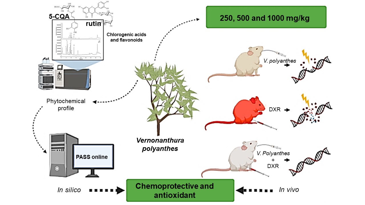
Phytochemical Composition and Protective Effect of Vernonanthura polyanthes Leaf against In Vivo Doxorubicin-Mediated Toxicity

by
Jamira Dias Rocha
1,
Marilia Elias Gallon
2,
Abel Vieira de Melo Bisneto
3,
Vanessa Cristiane Santana Amaral
1,
Luciane Madureira de Almeida
1,
Leonardo Luiz Borges
1,4,
Lee Chen-Chen
3,
Leonardo Gobbo-Neto
2 and
Elisa Flávia Luiz Cardoso Bailão
1,*
1
Laboratório de Biotecnologia, Campus Central, Universidade Estadual de Goiás, Anápolis 75132-903, GO, Brazil
2
Núcleo de Pesquisa em Produtos Naturais e Sintéticos de Ribeirão Preto, Universidade de São Paulo, Av. do Café s/n, Ribeirão Preto 14040-903, SP, Brazil
3
Laboratório de Radiobiologia e Mutagênese, Departamento de Genética, Instituto de Ciências Biológicas I, Universidade Federal de Goiás, Goiânia 74045-155, GO, Brazil
4
Escola de Ciências Médicas e da Vida, Pontifícia Universidade Católica de Goiás, Goiânia 74605-010, GO, Brazil
*
Author to whom correspondence should be addressed.
Molecules 2022, 27(8), 2553; https://doi.org/10.3390/molecules27082553
Submission received: 2 March 2022 / Revised: 22 March 2022 / Accepted: 24 March 2022 / Published: 15 April 2022
(This article belongs to the Special Issue Natural Compounds: A Lead for Drug Discovery and Development)

Abstract

:
Vernonanthura polyanthes (Spreng.) A.J. Vega & Dematt. (syn.: Vernonia polyanthes Less) is popularly known as “assa-peixe” and its leaves are used in folk medicine mainly to treat respiratory diseases. In this study, we evaluated the cytogenotoxic and anticytogenotoxic potential of the V. polyanthes leaf aqueous extract (VpLAE) and its n-butanol fraction (n-BF) in the presence or absence of doxorubicin (DXR) (pre-, co-, and post-treatments) on a murine model for 24 h or 120 h. The micronucleus test (MN) and the comet assay were used to assess the cytogenotoxic and anticytogenotoxic potential of VpLAE and n-BF (250, 500, and 1000 mg/kg) administered via gavage to Swiss Webster mice. The chemical profiles of VpLAE and n-BF were assessed by liquid chromatography coupled to mass spectrometry, and their metabolites were putatively identified. Lastly, the possible biological activities related to the (anti) cytogenotoxicity of the compounds were predicted using the PASS online webserver. The in vivo results showed that different doses of VpLAE and n-BF did not present cytotoxic activity; however, the MN test revealed a slight mutagenic activity for the 24 h treatments. Moderate genotoxic effects were demonstrated for all treatments in the comet assay. Regarding anticytotoxicity and antimutagenicity, VpLAE and n-BF presented a high cytoprotective potential against DXR toxic effects. In the co-treatment, VpLAE reduced the DXR genotoxicity by ~27%, and n-BF did not demonstrate antigenotoxic potential. In contrast, an antigenotoxic effect was observed for both VpLAE and n-BF in the pre- and post-treatments, reducing DXR genotoxicity by ~41% and ~47%, respectively. Chemical analysis of VpLAE and n-BF showed the presence of eight phenolic compounds, including seven chlorogenic acids and a flavonoid. The PASS online tool predicted antimutagenic, anticancer, antineoplastic, chemoprotective, antioxidant, and radical scavenging activities for all constituents identified in VpLAE and n-BF. V. polyanthes leaves presented a protective effect against DXR cytogenotoxicity. In general, VpLAE and n-BF showed a greater antigenotoxic potential in the pre- and post-treatments. The metabolites putatively identified in VpLAE and n-BF exhibited antioxidant and chemoprotective potential according to computational prediction analysis. Altogether, our results highlight the potential application of V. polyanthes to protect against toxic manifestations induced by DXR.

Graphical Abstract

1. Introduction

The search for plant metabolites is constantly increasing because they are more eco-friendly and usually less toxic to normal cells. According to the Food and Drug Administration agency, 40% of the approved molecules are natural compounds or inspired by them [1]. Moreover, many plant species are considered a good source of natural antioxidants and could be used as a functional food [2,3,4].
Vernonanthura polyanthes (Spreng.) A.J. Vega & Dematt. (syn.: Vernonia polyanthes Less), popularly known as “assa-peixe”, is an Asteraceae family species [5]. V. polyanthes leaves are used in folk medicine to treat respiratory disorders, pneumonia, bronchitis, coughs, flu, and colds, as well as kidney diseases, uterine infections, ulcers, hypertension, leishmaniasis, fever, hemorrhages, and gastric conditions [6,7,8,9,10,11,12]. Although there are other species popularly called “assa-peixe”, such as Vernonia brasiliana, Vernonia cognata Less., and Vernonia ferruginea Less., only V. polyanthes is regulated for phytotherapeutic use, registered in the Brazilian Pharmacopoeia of Herbal Medicines as an expectorant, in the form of tea obtained by infusion of the dry leaves [13]. The presence of flavonoids, saponins, tannins, coumarins, triterpenes, sesquiterpene lactones, chlorogenic acids, and phenolic acids was demonstrated in extracts from V. polyanthes leaves [14,15,16,17,18].
The V. polyanthes leaf aqueous extract (VpLAE) did not demonstrate toxicity, genotoxicity, or antigenotoxicity on Drosophila melanogaster. However, this extract potentiated the genotoxicity of doxorubicin (DXR) [19]. In contrast, VpLAE was cytotoxic to Allium cepa cells and Artemia salina [14]. Similarly, V. polyanthes leaf hydroalcoholic extract (2000 mg/kg) was cytogenotoxic when administrated to mice [20]. VpLAE and its three fractions (aqueous; n-butanol, n-BF; and ethyl acetate) showed cytogenotoxicity against human lymphocytes in vitro using a cell viability test and CometChip assay. However, when co-treated with DXR, V. polyanthes decreased DXR genotoxicity by ~15% [21].
DXR is a chemotherapeutic drug widely used in anticancer therapy; however, its use is limited by the increased oxidative stress caused by the drug [22,23]. It is already known that the redox balance disorder caused by DXR activates mechanisms that lead to cell damage, causing an increase in the production of free radicals and a decrease in endogenous antioxidants, inducing toxicity in various organs and tissues [22,23,24,25].
In this sense, it is important to study the effects of V. polyanthes and its interaction with DXR on a murine model. Initially, a micronucleus test (MN) and comet assay were performed, associated (pre-, co-, and post-treatments) or not with DXR on Swiss Webster mice, aiming to evaluate the cytogenotoxic and anticytogenotoxic potentials of VpLAE and n-BF. Then, the chemical profiles of both were determined, and the putatively identified metabolites were submitted to computational prediction analysis to predict their biological activities.

2. Methodology

2.1. Botanical Material

V. polyanthes leaves were collected at the Central Campus, in Anápolis, Goiás, Brazil (S 16°23′0.16″/W 48°56′37.8″, 1073 m) in November 2018. The botanical identification of this specimen was carried out by Dr. Aristônio Magalhães Teles from the Federal University of Goiás and an exsiccate (N° 10512) was deposited in the Herbarium of the State University of Goiás.

2.2. Infusion Preparation and Fractionation

The botanical material was subjected to drying at room temperature and pulverized in a knife mill E-625 (Tecnal Ltda, Piracicaba, SP, Brazil). Powdered material was stored sheltered from light and moisture for subsequent use. Initially, the powder from V. polyanthes leaves was subjected to quality parameters. The aqueous extract was prepared, according to Brazilian Pharmacopoeia (1st edition), by infusion of V. polyanthes (syn.: Vernonia polyanthes Less) leaves in the proportion of 0.02 g of dried and powdered plant material for each mL of water [13]. Samples were frozen at −18 °C and lyophilized. The solvent partitioning was obtained by percolation as previously described [21], resulting in three fractions (aqueous; n-BF; and ethyl acetate). The n-BF was evaporated under reduced pressure, and the dry extract and fraction were lyophilized and stored at −20 °C for further biological assays. The n-BF was selected to continue the experiments because it presented the highest total phenolic, flavonoids, and tannins contents, and the highest antioxidant activity, according to previous work [21].

2.3. Animals

Male Mus musculus (Swiss Webster) outbred mice, weighing between 20 to 30 g and aged from 7 to 12 weeks, were obtained from the Central Animal Facility of the Federal University of Goiás. This study was approved by the Animal Research Ethics Committee of the Federal University of Goiás (CEUA/UFG), protocol number 069/18 (Supplementary Materials).

2.4. In Vivo Experimental Procedures

This study used 140 mice (28 groups with 5 animals). Before the experiments were carried out, the animals remained acclimated for 7 days in the Laboratory of Radiobiology and Mutagenesis of the Federal University of Goiás. On these days, the animals were kept in polypropylene cages (Length: 40 cm, Width: 30 cm, and Height: 16 cm) with 5 animals in each cage lined with shavings changed every two days, and fed commercial feed and filtered water, both offered ad libitum. The animals were kept at room temperature, humidity 50% ± 20%, and a 12 h light/dark-light cycle. Doses of 250, 500, and 1000 mg/kg of VpLAE and n-BF were selected for the micronucleus test in mouse bone marrow based on previous work [20]. The animals in Group 1 (negative control) received mineral water orally in the same volume used to administer the aqueous extract of V. polyanthes. The animals in Group 2 (positive control) received DXR administered intraperitoneally (ip) at 50 mg/kg p.c. Animals from Groups 3 to 28 received different treatments with VpLAE or n-BF (Table 1). After 24 or120 h of treatment, the animals were euthanized by cervical dislocation. Bone marrow cells were obtained from the femurs of mice using 1 mL of fetal bovine serum and centrifuged at 300× g for 5 min to make slides for the MN test and the comet assay.

2.5. Micronucleus Test (MN)

Mouse bone marrow cells were used to make cell smears on glass slides. After drying, the smears were fixed in absolute methanol (CH4O) for 5 min, then stained in buffered Giemsa dye (dibasic sodium phosphate and monobasic sodium phosphate, pH 6.8). For each animal, four slides were prepared. As recommended [26], 4000 polychromatic erythrocytes (PCEs) were analyzed for each animal to determine the frequency of micronucleated polychromatic erythrocytes (MNPCE). Cytotoxicity and anticytotoxicity were evaluated by the ratio of PCE and normochromatic erythrocytes (NCE). Whereas genotoxicity and antigenotoxicity were assessed by the frequency of MNPCE concerning the total number of cells analyzed. The analysis of the slides was performed under an optical light microscope (Olympus BH-2, Tokyo, Japan, objective 100×).

2.6. Comet Assay

Microscope slides were previously coated with 1.5% agarose of normal melting point (1.5%). Subsequently, a solution containing 10 µL of bone marrow cells previously diluted in fetal bovine serum, and 120 µL of low melting point agarose at 0.5% and 37 °C, was placed on the agarose-precoated slides. The slides were then kept in the refrigerator, inside a foil-lined slide holder, containing lysis solution (2.5 M NaCl, 100 mM EDTA, 10 mM TRIS, 10% DMSO, 1% Triton X-100, and 1000 mL H2O, pH 10 at 4 °C overnight). After this period, the slides were transferred to a horizontal electrophoresis vat, containing electrophoresis buffer (30 mM NaOH, 1 mM EDTA, pH 13) at 5 ± 2 °C, where they remained for 30 min for DNA unwinding. Then, they were subjected to 300 mA and 1 V/cm for 30 min. After electrophoresis, the slides were immersed in a neutralizing buffer solution (0.4 M, Tris-HCl, pH 7.5) for 5 min. The slides were stained with 20 µL of Nucleic Acid Dye (DiamondTM, 10%) and covered with a coverslip. The capture of comet images was performed in a fluorescence microscope (Axio Imager® A2 and Zen 2.3 software Carl Zeiss AG, Germany, with 510–560 nm excitation filter and 590 nm barrier filter, in a 20× objective). The software TriTek CometScoreTM (version 1.5) was used to assess DNA damage. In this software, pixel intensity in nucleoid images provides values corresponding to the estimation of genomic damage, which is expressed as arbitrary units (AU). Nucleoids with fragmented heads (hedgehogs) were not included in the analyses. The parameter adopted for quantifying DNA damage was the percentage of DNA in the tail.

2.7. Statistical Analysis

For the parameters PCE/NCE and MNPCE, the mean ± standard deviations were calculated for each group. Data distribution was checked for normality using the Shapiro-Wilk test. The different groups were compared using the parametric (one-way ANOVA) or nonparametric Kruskal-Wallis test followed by the Dunn’s multiple comparison test. The comet assay results are presented as the mean ± standard deviations, and the analysis of variance (one-way ANOVA) was performed, followed by Tukey’s multiple comparison test. Analyzes were performed using the GraphPad Prism software version 8.0.1. p < 0.05 was considered significant.

2.8. Chemical Profiles

For the LC-MS analyses, extracts of VpLAE and n-BF were prepared using 10 mg of the dry powder of each extract or fraction and 1 mL of a MeOH:H2O (7:3, v:v) solution with hydrocortisone (10 mg/mL) as an internal standard. Extraction was performed in an ultrasonic bath for 10 min at room temperature. The extracts were filtered through a 0.20 mm PTFE membrane before analysis. Chemical profiles were obtained on an Accela UHPLC instrument (Thermo Scientific™, Waltham, MA, USA) with a diode array ultraviolet light detector (UV-DAD) coupled to an ExactiveTM Plus mass spectrometer (Thermo Scientific™, Waltham, MA, USA) with electrospray ionization source and orbitrap analyzer. Chromatograms were acquired in positive and negative ionization modes using a C18 Kinetex column (1.7 µm, XB-C18, 150 mm × 2.1 mm, Phenomenex) and an elution gradient of water and acetonitrile both with 0.1% of formic acid. All other chromatographic and spectroscopic parameters followed the methodology employed by Gallon et al. (2018b). Metabolites detected in VpLAE and n-BF were putatively identified by comparing the spectroscopic data of each chromatographic signal with the data available in the in-house database of secondary metabolites reported for Vernonieae species.

2.9. Prediction of Activity Spectra for Substances
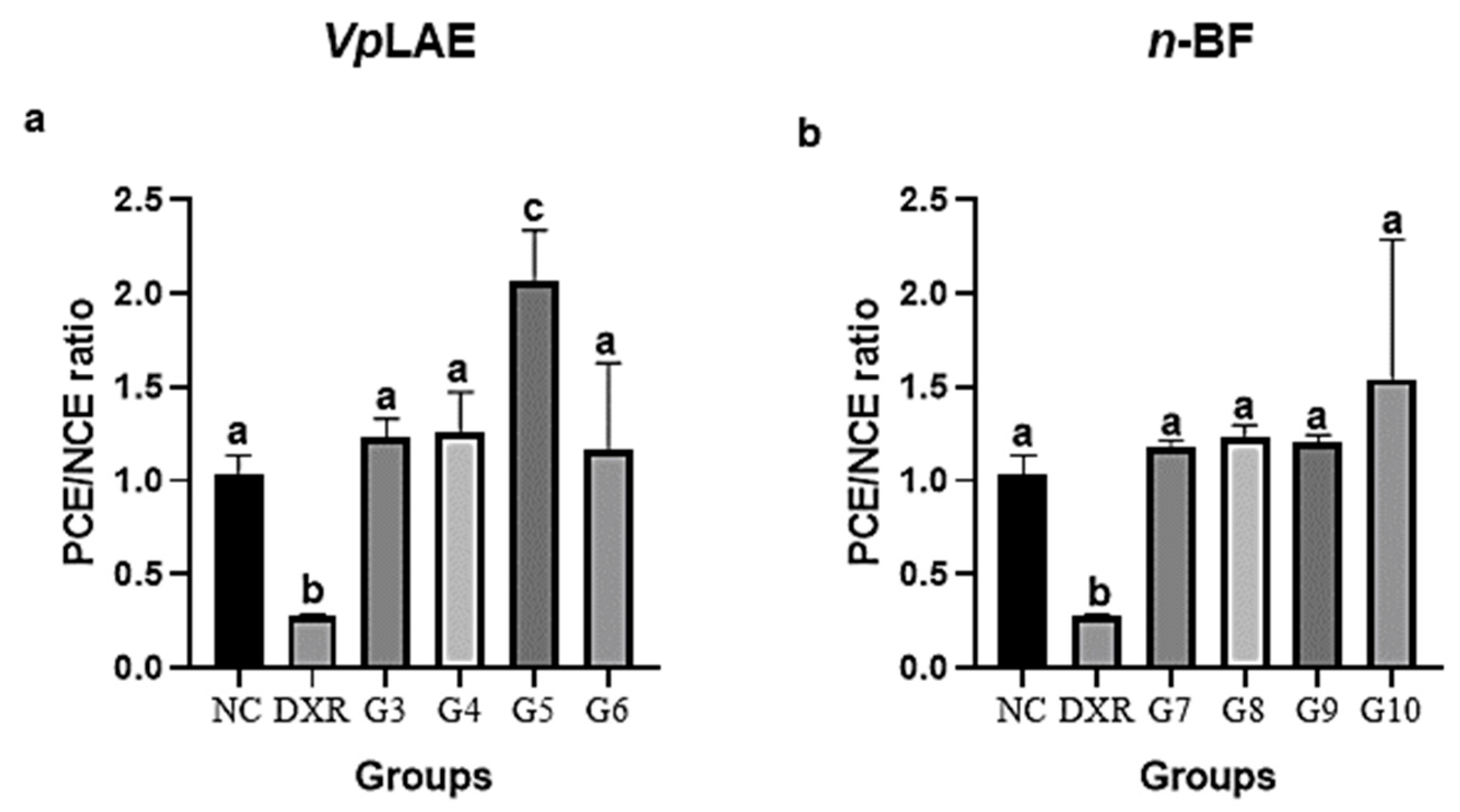
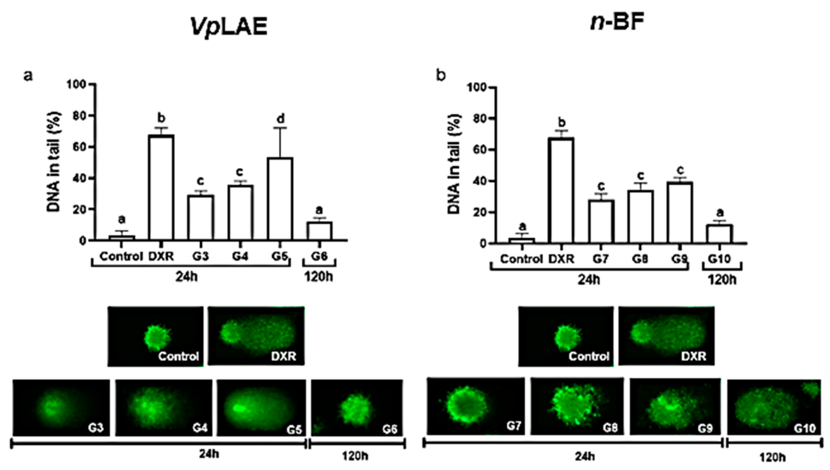
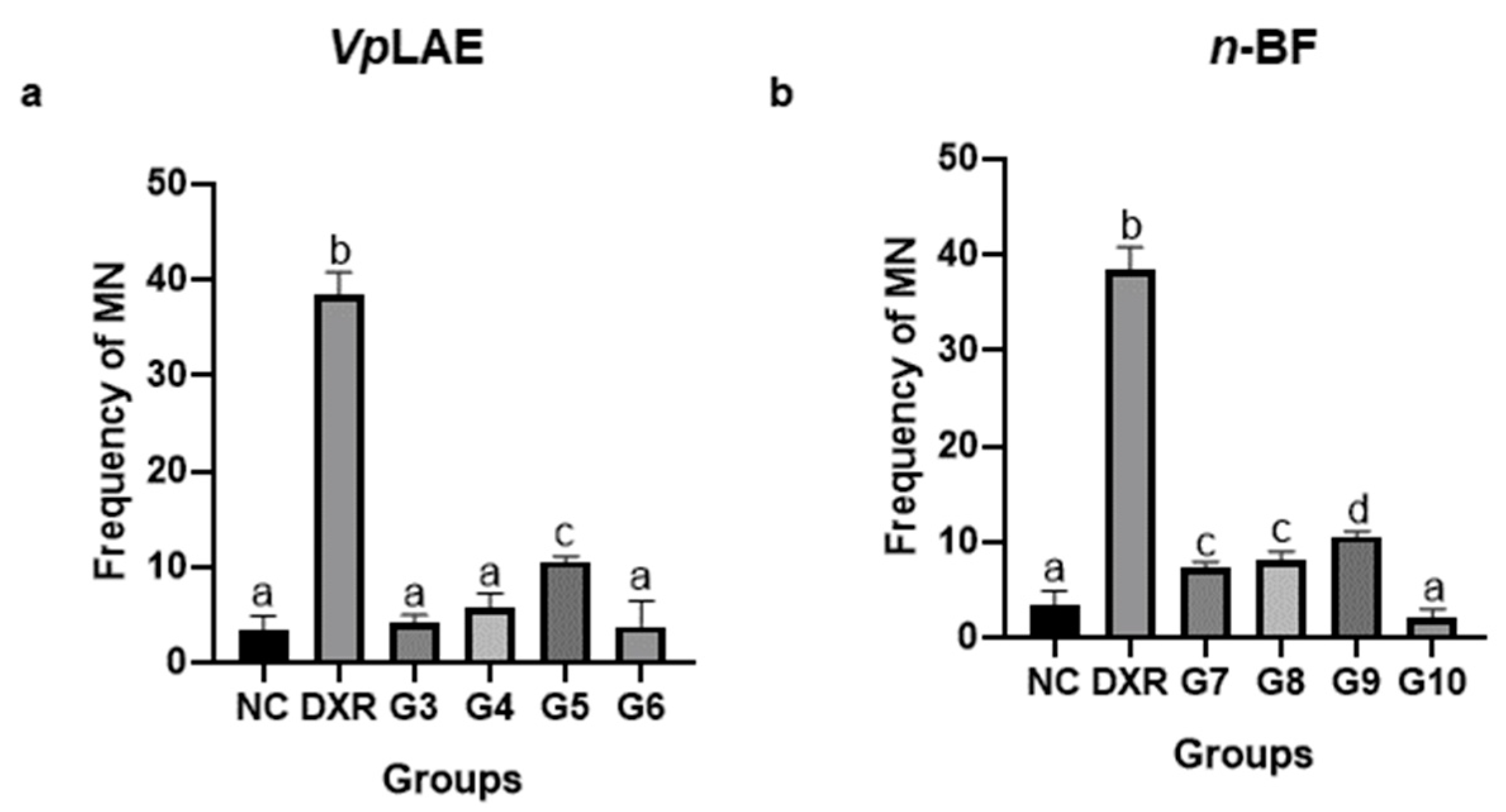
The bioactivity of the metabolites identified in the VpLAE and n-BF were predicted using the PASS online webserver (http://www.pharmaexpert.ru/passonline/, accessed on 20 May 2020). Pa and Pi estimate the probability of the substance to be active or inactive, respectively, for each type of activity from the database [27,28]. Therefore, the results presenting Pa > 0.7 and Pi < 0.05 were selected for the analysis.

3. Results

3.1. Cytogenotoxic Evaluation

VpLAE and n-BF did not present cytotoxicity against mice bone marrow cells (Figure 1) and presented genotoxicity after acute (24 h) exposition to all concentrations tested in this work (250, 500, and 1000 mg/kg) (Figure 2). The genotoxicity observed at the highest concentration of VpLAE (1000 mg/kg, G5) was similar to that observed for the positive control (DXR) (Figure 2a). Although VpLAE and n-BF presented a genotoxic potential on mice bone marrow cells, these materials did not present or presented a mild mutagenicity potential (Figure 3). As expected, animals treated with DXR (positive control) showed cytogenotoxic effects compared to the negative control (Figure 1, Figure 2, Figure 3, Figure 4, Figure 5 and Figure 6).

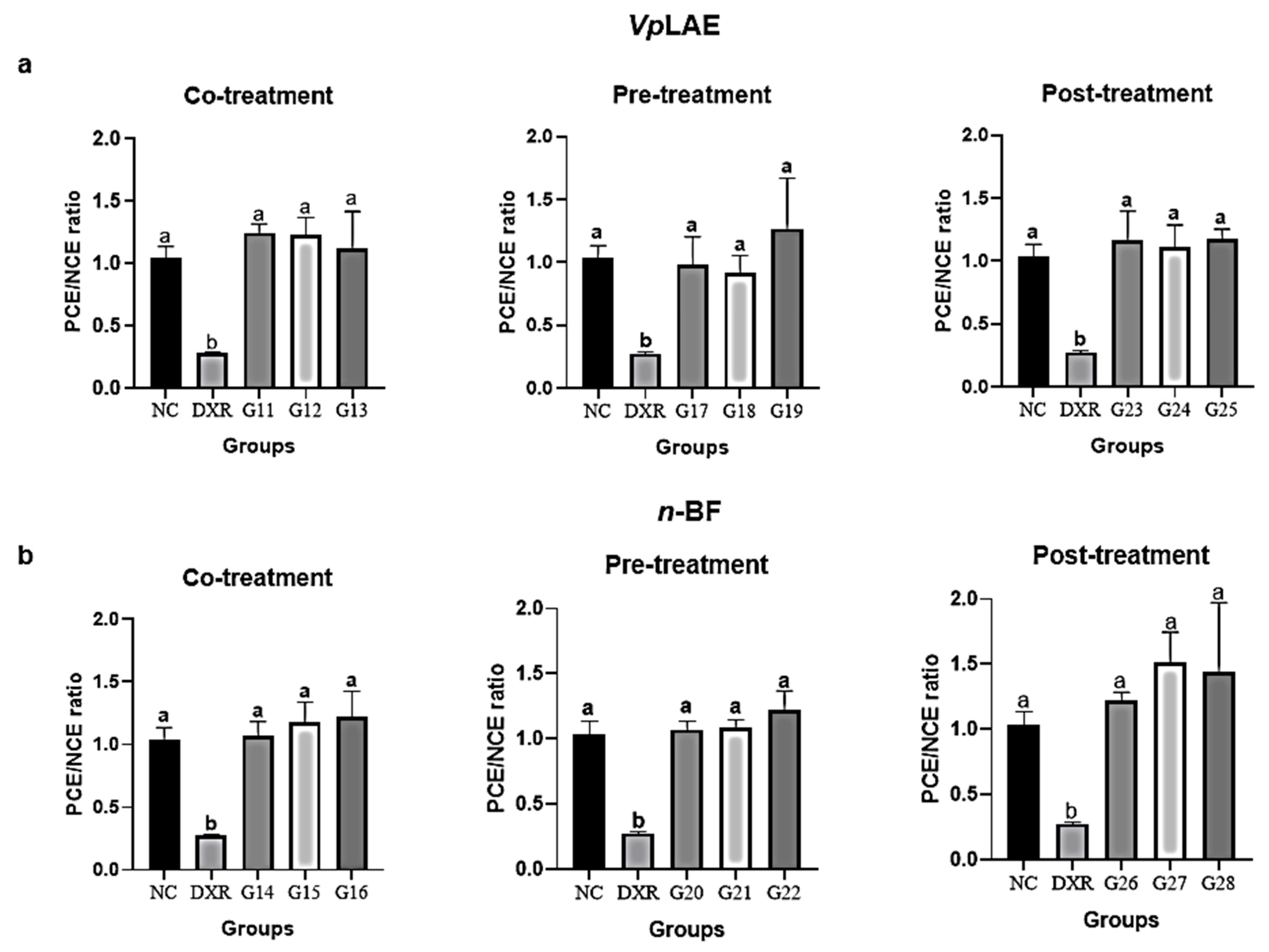
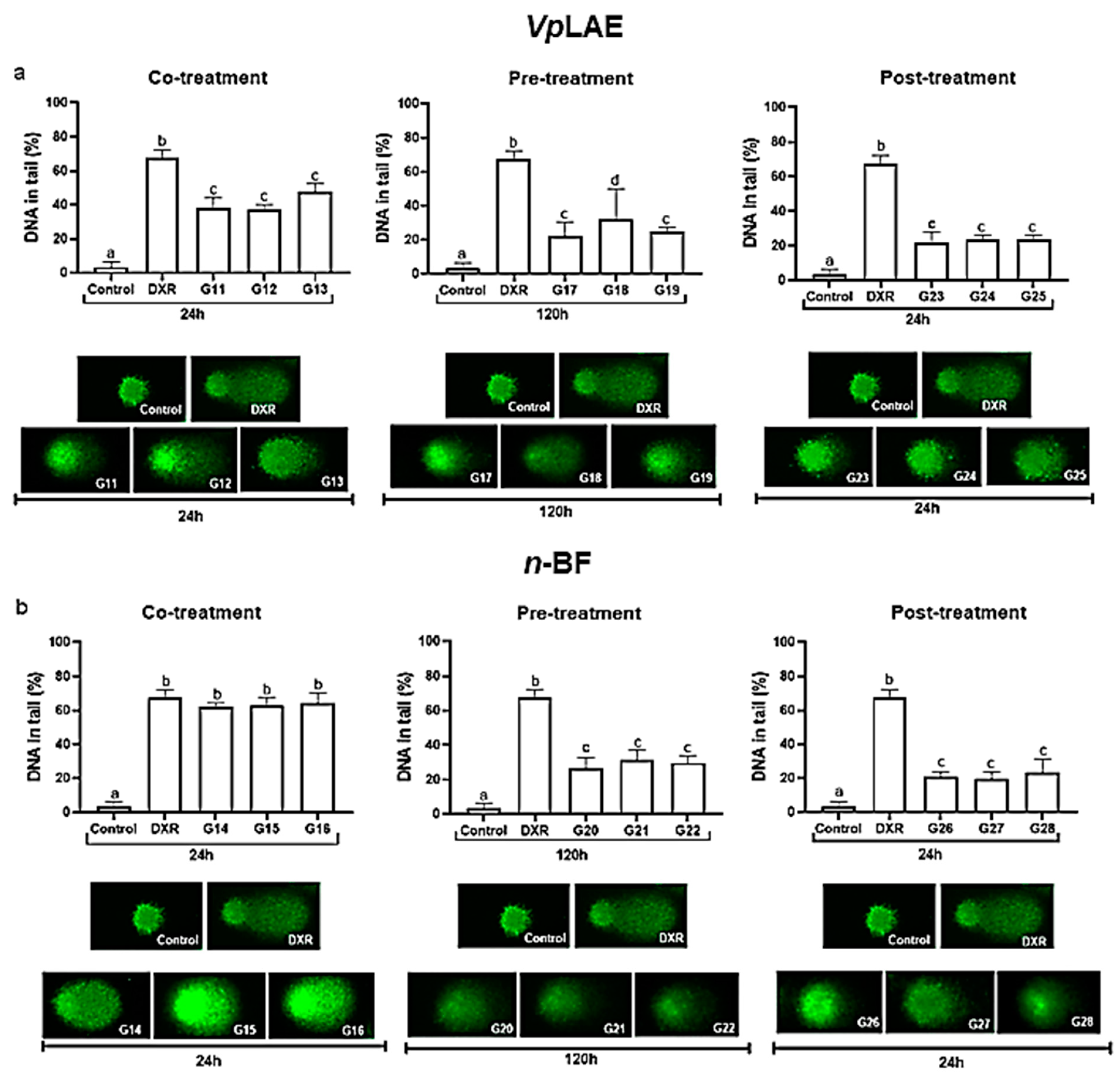
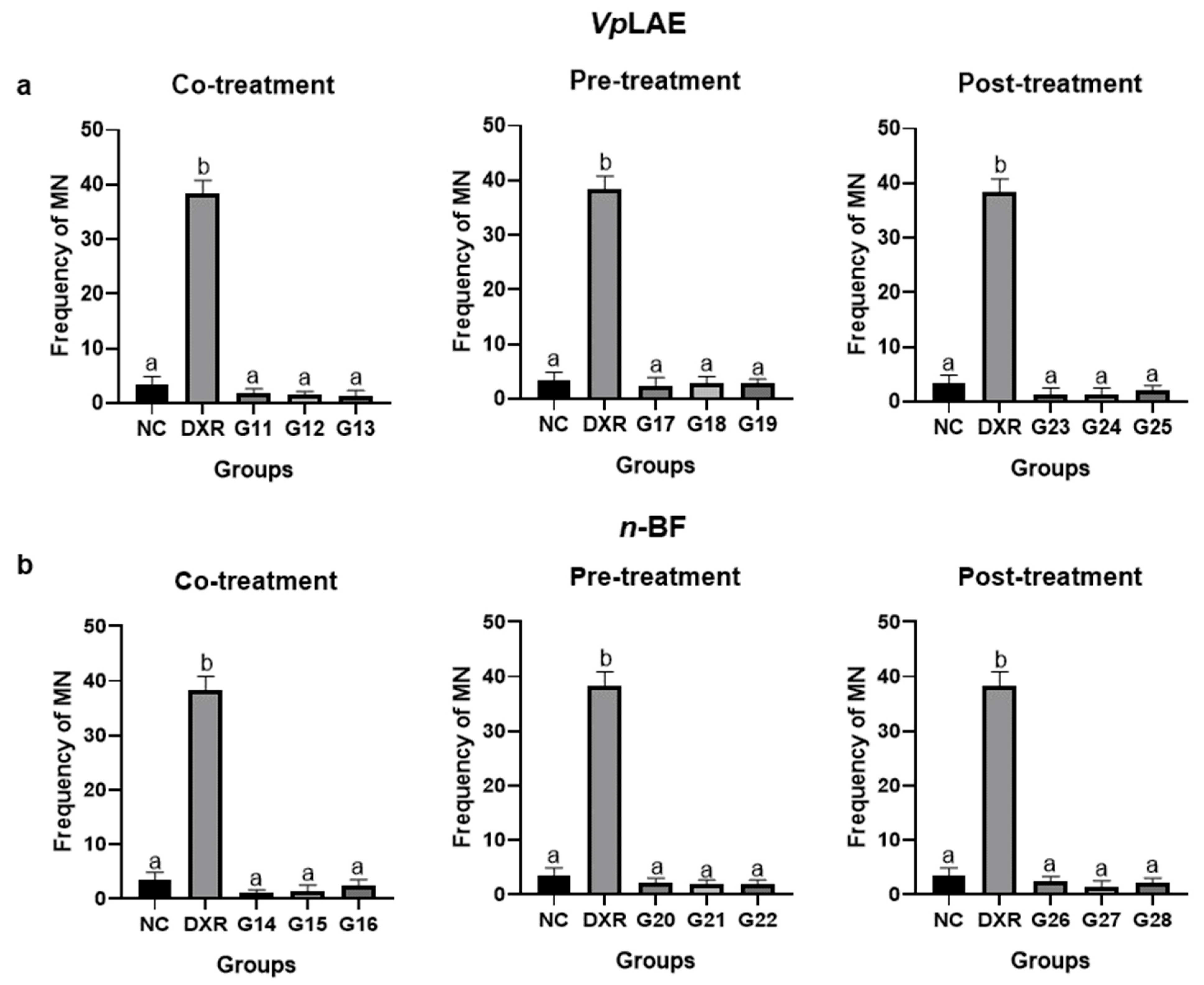
3.2. Anticytogenotoxic Evaluation

The evaluation of VpLAE and n-BF anti-cytogenotoxicity in the presence of the DXR positive control (co-, pre-, and post-treatments) was also performed. VpLAE and n-BF were anti-cytotoxic in all doses (250, 500, and 1000 mg/kg) and in different treatment schemes used in this work (Figure 4). The PCE/NCE ratios in treatments of DXR + VpLAE or n-BF were restored to the ratio obtained in the negative control (Figure 4). Regarding antigenotoxicity, VpLAE reduced the DXR genotoxicity by ~27% during co-treatment (Figure 5a). In contrast, n-BF did not demonstrate anti-genotoxic potential in the co-treatment with DXR (Figure 5b). Regarding pre-treatment, VpLAE and n-BF reduced the genotoxicity of DXR by ~41% (Figure 5). In the post-treatment, VpLAE and n-BF also reduced the genotoxicity of DXR by ~47% (Figure 5). Moreover, the association of VpLAE or n-BF with DXR in the co-, pre-, and post-treatments showed a significant reduction in the frequency of MN/PCE when compared to the positive control (Figure 6), revealing a relevant anti-genotoxic effect of V. polyanthes.

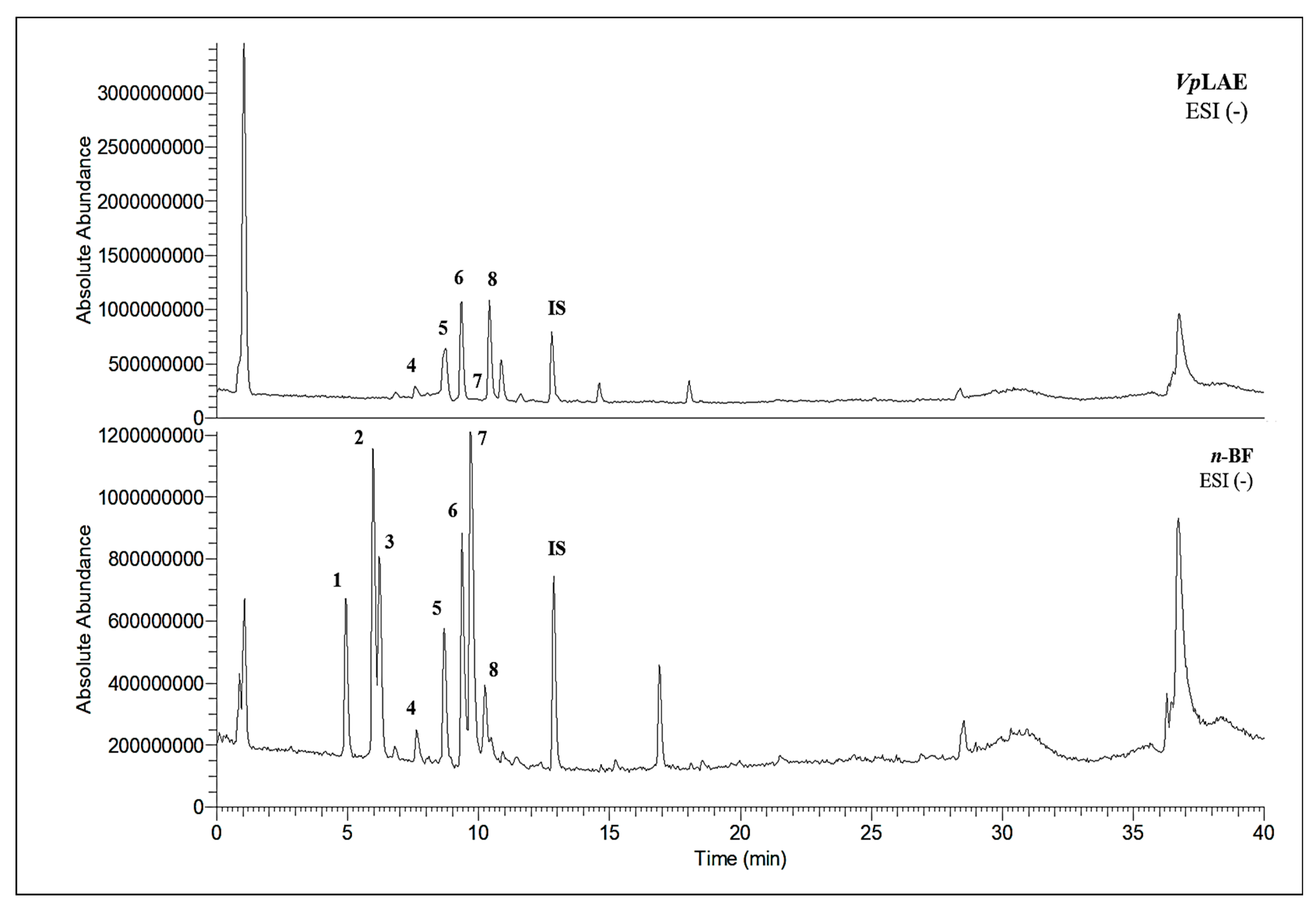
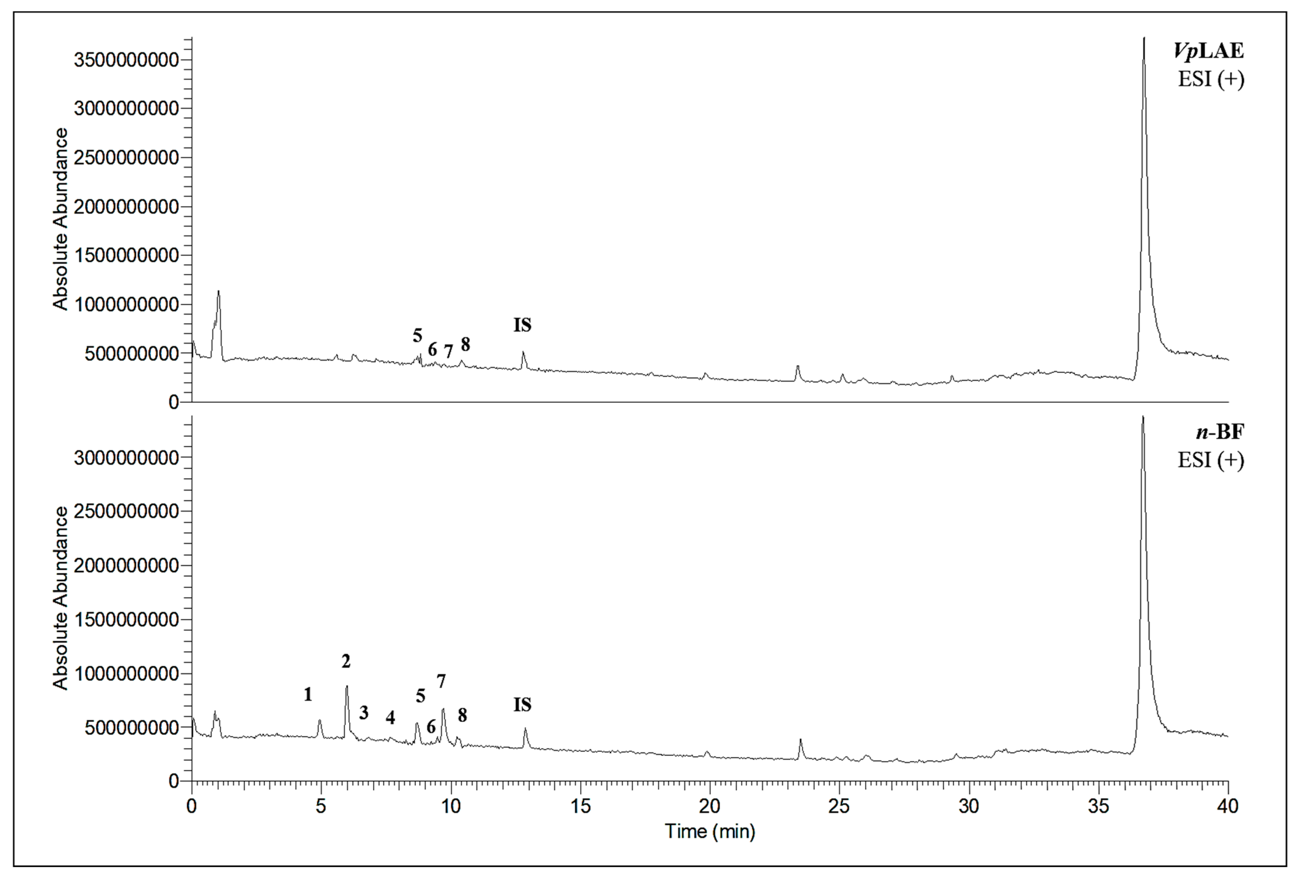
3.3. Chemical Profiles of VpLAE and n-BF

LC-MS analyses of VpLAE and n-BF confirmed the presence of phenolic compounds (flavonoids and chlorogenic acids) typically reported in V. polyanthes (Table 2 and Supplementary Table S1). The compounds 5-O-feruloylquinic acid, quercetin 3-O-rutinoside, 3,4-di-O-caffeoylquinic acid, 3,5-di-O-caffeoylquinic acid, and 4,5-di-O-caffeoylquinic acid were putatively identified in both the VpLAE and n-BF samples. Whereas 3-O-caffeoylquinic acid, 5-O-caffeoylquinic acid, and 4-O-caffeoylquinic acid were detected in higher relative abundance in the n-BF. Accordingly, all the detected O-caffeoylquinic acids and di-O-caffeoylquinic acids exhibited greater relative abundances in n-BF (Figure 7 and Figure 8).

3.4. Biological Activity Prediction of Identified Secondary Metabolites

Potential antimutagenic, anticancer, antineoplastic, chemoprotective, antioxidant, and radical scavenging effects were attributed to all metabolites putatively identified in VpLAE and n-BF by computational prediction analysis using the PASS online tool (Table 3). Moreover, the identified molecules are predicted modulators of enzymes involved in antioxidant processes such as HMOX1, lipid peroxidase inhibitors, aryl sulfotransferase, β-glucuronidase, α-glucosidase, UDP-glucuronosyl transferase, glutathione-disulfide reductase, and different P450 isoforms (CYP1A, CYP1A1, CYP3A4, CYP2C9, CYP3A) (Table 4). Among the metabolites detected in VpLAE and n-BF, quercetin-3-O-rutinoside (rutin) seems to be the most active considering the analyzed antioxidant and chemopreventive parameters provided by the PASS online tool (Table 3 and Table 4).

4. Discussion

In this study, the MN test and the comet assay in mouse bone marrow were performed to assess the cytotoxic, genotoxic, and mutagenic potential of VpLAE and n-BF. Moreover, the anticytogenotoxic potential of VpLAE and n-BF was evaluated in the presence of DXR, a widely used chemotherapy agent. The micronucleus test in mammals assesses the cytotoxic and mutagenic effects of chemical or physical agents, providing a system to detect cytogenetic damage resulting from clastogenic or aneugenic activity [29,30]. The comet assay (single-cell gel electrophoresis) is a methodology used to measure deoxyribonucleic acid (DNA) strand breaks in cells [31,32,33,34]. Both approaches can determine the antigenotoxic potential of a plant extract or isolated compound [35,36,37].
The animals treated with VpLAE and n-BF (250, 500, and 1000 mg/kg) did not promote a PCE/NCE ratio reduction compared to the negative control group, revealing the cytotoxic activity absence of V. polyanthes leaves. These results are consistent with our previous study using the wing recombination and somatic mutation test (SMART/wing), which revealed the absence of cytotoxic activity of the aqueous extract of V. polyanthes leaves (0.25–1 mg/mL) on Drosophila melanogaster [19]. In contrast, when the V. polyanthes leaf hydroalcoholic extract (1000, 1500, and 2000 mg/kg) was administrated to mice, the relationship between PCE and NCE was significantly reduced in all treatments, regardless the exposition time (24 and 48 h) or gender, indicating cytotoxicity of the V. polyanthes leaf hydroalcoholic extract [20]. V. polyanthes leaves were also cytotoxic to human lymphocytes, sarcoma-180 cells, Allium cepa cells, and Artemia salina [14,16,21]. The mode of action of plant extracts may vary depending on different model systems and chemical compositions [38,39,40]. So, these contrasting results could be explained by the model used to study cytotoxicity (if it is a metabolizing system or not), the type of solvent used to prepare the extracts, and the part of the plant used [38,40].
The comet assay has several applications in testing new chemicals, particularly genotoxicity or DNA repair damage evaluation [32,33]. VpLAE and n-BF showed genotoxicity for all concentrations used (250, 500, and 1000 mg/kg) after 24 h exposition. Likewise, VpLAE and its fractions (aqueous, n-butanol, and ethyl acetate) demonstrated genotoxic activity on human lymphocytes [21]. In contrast, when the hydroalcoholic extract of V. polyanthes leaves (1000, 1500, and 2000 mg/kg) was administrated to mice, just the highest dose tested was demonstrated to be genotoxic [20]. Similarly, the absence of genotoxic activity of V. polyanthes was verified on D. melanogaster and A. cepa at the studied conditions [14,19]. These discrepant results highlight the importance of using different models to study plant derivatives.
Slight mutagenicity was observed for some groups treated with VpLAE (G5) and n-BF (G7–G9). On the other hand, the aqueous extract of V. polyanthes did not show mutagenicity at the concentrations used (0.25–1 mg/mL) on D. melanogaster using the wing somatic mutation and recombination test (SMART-wing) [19]. V. polyanthes aqueous extract did not also show a mutagenic effect on A. cepa meristematic cells [14]. The hydroalcoholic extract of V. polyanthes leaves (ethanol/water, 70/30 v/v) was mutagenic for mice only at the highest dose used (2000 mg/kg). The low and intermediate doses (1000 and 1500 mg/kg) were not mutagenic [20]. This discrepancy between the results may be associated with different models tested and the different solvents used to prepare the V. polyanthes extract (water or ethanol). The polarity-dependent increase in antioxidant activity may indicate that polar solvents are more likely to extract antioxidant compounds [41]. One of the most suitable solvents for aqueous mixtures is ethanol because it maximizes polyphenols extraction and is safe for human consumption [21,42].
This study also evaluated the anticytotoxic and antigenotoxic potential of VpLAE and n-BF against DXR, a widely used chemotherapeutic, capable of performing single and double breaks in DNA. This drug is part of one of the most effective groups of antineoplastics used in current clinical practice. However, its use is limited by chronic and acute toxic side effects and susceptibility to numerous drug interactions [25,43,44,45]. The antitumor and toxic effects of DXR contribute to the production of free radicals and to the occurrence of oxidative stress, which can occur in four different ways: (i) semiquinone production; (ii) activation of NAD (P) H oxidases (NOXs); (iii) a non-enzymatic mechanism; and (iv) the generation of DXR metabolism products [24,46]. The generated oxidative stress confers non-specific cytotoxicity, leading to undesirable effects of chemotherapy with DXR [47]. DXR-mediated generation of excessive free radicals caused by increased free radical production and decreased endogenous antioxidants plays an important role in the pathogenesis of induced toxicity in various organs and tissues. DXR can also induce apoptosis and hyperlipidemia [24,25]. One of the oxidative stress reduction strategies has been the combination of the drug together with an antioxidant agent [25,44]. In this context, phytochemical compounds have been described as an alternative to mitigate the harmful effects caused by DXR since metabolites from natural products have shown to be promising in overcoming the limitations of DXR in pre-clinical models such as chemosensitizers, chemoresistance inhibitors, and protectors chemotherapeutics in different types of cancer [48].
The association of DXR with VpLAE or n-BF in the co-, pre-, and post-treatments demonstrated an anticytotoxic potential of V. polyanthes. In contrast, the cytotoxicity of DXR was potentiated by VpLAE and its aqueous, n-butanol, and ethyl acetate fractions (0.25–1 mg/mL) on human lymphocytes during co-treatment [21]. In vivo toxicity assessment involves important factors related to the dynamics of the accumulation of substances in cells that can occur through electron transport, absorption and diffusion, apoptosis process, production of reactive oxygen molecules, and biotransformation of molecules by intracellular/extracellular via activation of specific enzymes. The cell’s communication with its environment is a key factor in differentiating between in vivo and in vitro models [49].
The antigenotoxic potential of VpLAE and n-BF (in the presence of DXR) was also evaluated in co-, pre-, and post-treatment schemes by comet assay. The results revealed that VpLAE and n-BF reduced DXR genotoxicity by ~38%. The exception is n-BF co-treated with DXR, which could not protect the DNA. Similar results were observed for VpLAE and its fractions (aqueous, ethyl acetate, and n-butanol) co-treated with DXR in human lymphocytes. V. polyanthes decreased DXR genotoxicity by ~15% [21]. However, previous results with D. melanogaster showed that VpLAE potentiated DXR genotoxicity when both were administered in a co-treatment regimen [19].
The association of VpLAE and n-BF with DXR in co-, pre-, and post-treatments showed a significant reduction in the frequency of MN in erythrocytes from the bone marrow of mice, showing a protective effect against damage caused by DXR. Unlike this result, tests conducted to evaluate the genotoxic effects using the wing recombination and somatic mutation test (SMART-wing) showed that the association of the aqueous extract of V. polyanthes with DXR potentiated its mutagenic effect, increasing the number of mutations in D. melanogaster somatic cells [19]. The observed differences may be associated with the model used to conduct the studies (murine x insect), highlighting the importance of using different study models during the pre-clinical phase of drug development.
This study also investigated secondary metabolites in VpLAE and n-BF to infer their association with the anti-cytogenotoxic activity against the redox effect generated by DXR. Our LC-MS results of VpLAE and n-BF confirmed the presence of phenolic compounds abundantly found in plants [50,51,52,53]. We putatively identified eight phenolic compounds, including seven phenols identified as chlorogenic acids and a flavonoid identified as quercetin 3-O-rutinoside (rutin). The presence of these metabolites was observed for other species of the genus Vernonia [54,55]. Flavonoids are known for their antioxidant potential aiding in the capture and scavenging of radicals, as well as being one of the most widely found secondary metabolites in medicinal plants [23,50,51,53]. In this sense, due to their radical scavenging properties, flavonoids may act in the redox effect mediated by DXR in the generation of cellular oxidative stress. The phenolic compounds noted here, more specifically the chlorogenic acids, have been widely studied in recent years regarding their effects and redox effects in various cell types [45].
Recently, the antioxidant potential of V. polyanthes leaves aqueous extract and its fractions (aqueous, n-butanol, and ethyl acetate) was investigated by the DPPH method showing great antioxidant potential for the extract and the n-butanol fraction [21]. In this sense, the metabolites detected in VpLAE and n-BF may contribute to the prevention of DXR-induced overproduction of free radicals, protecting the bone marrow cells of mice. Phenolic compounds have hydroxyl groups, which can help in the process of scavenging free radicals by direct proton transfer. ROS elimination occurs in proportion to the number of functional hydroxyl groups present within each molecule [56,57]. There is growing evidence that compounds with antioxidant properties can remove ROS before these species react with DNA resulting in a mutation [56,58]. In this way, metabolites in plant extracts can increase the maintenance of DNA structure and modulation of DNA metabolism and repair, minimizing the redox effect of toxic compounds [59].
A high degree of correlation was found between the in vivo and in silico results. Among the metabolites detected in VpLAE and n-BF, rutin stands out as a potential antioxidant and chemopreventive molecule against DXR-mediated cytogenotoxicity. The result of the PASS online prediction tool showed that rutin might be able to enhance hmox1 expression. Hmox1 is an antioxidant enzyme produced to respond to oxidative stress [60,61]. Hmox1 activation inhibits lipid peroxidation, preventing cell damage caused by DXR [60,62]. It had been demonstrated that rutin attenuated the toxic effects caused by DXR in cardiac and renal tissues via improvement of the antioxidant state of cells [63]. Rutin administration also decreased DXR-induced heart failure, inhibiting excessive autophagy and apoptosis [64]. This suggests that using antioxidant agents, such as flavonoids, in combination with DXR may reduce or inhibit DXR-induced side effects.
Moreover, rutin is a predicted CYP inducer. The cytochrome P450 enzymes metabolize xenobiotics, participating mainly in the conversion of toxic substances into more polar and water-soluble metabolites to be rapidly excreted, preventing cytotoxic and genotoxic effects [65]. However, during the biotransformation process, various unstable and reactive intermediates that react with DNA can be formed, causing genotoxicity and cell damage [65,66,67]. In this sense, it can be suggested that the moderate genotoxicity presented for the VpLAE (G3, G4, G5, and G6) and the n-BF (G7, G8, G9, and G10), as well as the mild mutagenicity demonstrated for the G5 group (VpLAE) and the groups G7, G8 and G9 (n-BF), could be a result of the CYP family induction. In addition, since the cytochrome P450 enzymes catalyze many metabolic reactions involving xenobiotics [66], these enzymes may also have acted in the potent antigenotoxic and antimutagenic activity observed for VpLAE and n-BF.

5. Conclusions

VpLAE and n-BF in co-, pre-, and post-treatments significantly inhibited DXR toxicity, protecting the mouse bone marrow cells against the cytotoxic, genotoxic, and mutagenic effects of DXR. This cytoprotective activity may be correlated with the antioxidant potential of phenolic compounds present in VpLAE and n-BF. Furthermore, both VpLAE and n-BF did not demonstrate to be cytotoxic at the concentrations used. Thus, studies about the association of DXR with natural antioxidants are encouraged to reduce the level of oxidative stress generated by the drug; that is, its side effects.

Supplementary Materials

The following are available online at https://www.mdpi.com/article/10.3390/molecules27082553/s1, Table S1: Spectroscopic data of the putatively identified metabolites in V. polyanthes leaf aqueous extract (VpLAE) and its n-butanol fraction (n-BF), Figure S1: Letter of the Animal Research Ethics Committee of the Federal University of Goiás (CEUA/UFG), protocol number 069/18.

Author Contributions

Conceptualization, J.D.R. and E.F.L.C.B.; analysis, J.D.R., M.E.G., A.V.d.M.B. and V.C.S.A.; data curation, L.L.B., L.C.-C., L.G.-N., and E.F.L.C.B.; formal analysis, J.D.R., L.M.d.A., L.L.B., L.C.-C. and E.F.L.C.B.; Comet assay and MN test, J.D.R., A.V.d.M.B., V.C.S.A., L.M.d.A., L.C.-C. and E.F.L.C.B.; HPLC analysis, J.D.R., M.E.G. and L.G.-N.; in silico analysis, J.D.R., L.L.B. and E.F.L.C.B.; supervision, L.L.B., L.C.-C., L.G.-N., E.F.L.C.B.; writing—original draft, J.D.R., M.E.G. and E.F.L.C.B.; writing—review and editing, J.D.R., M.E.G., A.V.d.M.B., V.C.S.A., L.M.d.A., L.L.B., L.C.-C., L.G.-N. and E.F.L.C.B. All authors have read and agreed to the published version of the manuscript.

Funding

This research was funded by Fundação de Amparo à Pesquisa do Estado de Goiás (FAPEG), grant number PPP/201610267001019. Financiadora de Estudos e Projetos (Finep), grant number 01.14.0035.00. And Coordenação de Aperfeiçoamento de Pessoal de Nível Superior (CAPES), scholarships. The APC was funded by CAPES (Convênio UEG/CAPES N. 817164/2015-PROAP).

Acknowledgments

We thank Aristônio Magalhães Teles for botanical identification.

Conflicts of Interest

The authors declare no conflict of interest.

References

  1. Seca, A.M.L.; Pinto, D.C.G.A. Plant secondary metabolites as anticancer agents: Successes in clinical trials and therapeutic application. Int. J. Mol. Sci. 2018, 19, 263. [Google Scholar] [CrossRef] [PubMed] [Green Version]
  2. Rizwan, K.; Zubair, M.; Rasool, N.; Riaz, M.; Zia-Ul-Haq, M.; de Feo, V. Phytochemical and biological studies of Agave attenuata. Int. J. Mol. Sci. 2012, 13, 6440–6451. [Google Scholar] [CrossRef]
  3. Majeed, I.; Rizwan, K.; Ashar, A.; Rasheed, T.; Amarowicz, R.; Kausar, H.; Zia-ul-haq, M.; Marceanu, L.G. A comprehensive review of the ethnotraditional uses and biological and pharmacological potential of the genus mimosa. Int. J. Mol. Sci. 2021, 22, 7463. [Google Scholar] [CrossRef]
  4. Bailão, E.F.L.C.; Devilla, I.A.; da Conceição, E.C.; Borges, L.L. Bioactive compounds found in Brazilian cerrado fruits. Int. J. Mol. Sci. 2015, 16, 23760–23783. [Google Scholar] [CrossRef] [PubMed]
  5. Lorenzi, H.; Matos, F.J. Plantas Medicinais no Brasil. Nativas e Exóticas, 2nd ed.; Instituto, Plantarum: Nova Odessa, Brazil, 2008; ISBN 978-8586714283. [Google Scholar]
  6. Rodrigues, V.E.G.; Carvalho, D.A. De Levantamento Etnobotânico De Plantas Medicinais No Domíniodo Cerrado Na Região Do Alto Etnobotanical Survey of Medicinal Plants in the Dominion of Meadows in the Region of the Alto Rio Grande—Minas Gerais. Ciênc. Agratecnol. 2001, 25, 102–123. [Google Scholar]
  7. Silveira, R.R.; Foglio, M.A.; Gontijo, J.A.R. Effect of the crude extract of Vernonia polyanthes Less. on blood pressure and renal sodium excretion in unanesthetized rats. Phytomedicine 2003, 10, 127–131. [Google Scholar] [CrossRef] [PubMed]
  8. Braga, F.G.; Bouzada, M.L.M.; Fabri, R.L.; de O. Matos, M.; Moreira, F.O.; Scio, E.; Coimbra, E.S. Antileishmanial and antifungal activity of plants used in traditional medicine in Brazil. J. Ethnopharmacol. 2007, 111, 396–402. [Google Scholar] [CrossRef]
  9. Barbastefano, V.; Cola, M.; Luiz-Ferreira, A.; Farias-Silva, E.; Hiruma-Lima, C.A.; Rinaldo, D.; Vilegas, W.; Souza-Brito, A.R.M. Vernonia polyanthes as a new source of antiulcer drugs. Fitoterapia 2007, 78, 545–551. [Google Scholar] [CrossRef]
  10. dos Santos Temponi, V.; Da Silva, J.B.; Alves, M.S.; Ribeiro, A.; Pinho, J.D.; Yamamoto, C.H.; Pinto, M.A.; Del-Vechio-Vieira, G.; De Sousa, O.V. Antinociceptive and anti-inflammatory effects of ethanol extract from Vernonia polyanthes leaves in rodents. Int. J. Mol. Sci. 2012, 13, 3887–3899. [Google Scholar] [CrossRef] [Green Version]
  11. Oliveira, D.G.; Prince, K.A.; Higuchi, C.T.; Santos, A.C.B.; Lopes, L.M.X.; Simões, M.J.S.; Leite, C.Q.F. Antimycobacterial activity of some Brazilian indigenous medicinal drinks. Rev. Cienc. Farm. Basica E Aplicada 2007, 28, 165–169. [Google Scholar]
  12. Silva, N.C.C.; Barbosa, L.; Seito, L.N.; Junior, A.F. Antimicrobial activity and phytochemical analysis of crude extracts and essential oils from medicinal plants. Nat. Prod. Res. 2012, 26, 1510–1514. [Google Scholar] [CrossRef] [PubMed]
  13. Formulário de Fitoterápicos da Farmacopeia Brasileira; 2011 Brasília-DF, Brazil. pp. 1–126. Available online: https://www.gov.br/anvisa/pt-br/assuntos/farmacopeia/formulario-fitoterapico/arquivos/2021-fffb2-final-c-capa2.pdf (accessed on 1 March 2022).
  14. Almeida, L.M.; Prado, A.D.L.; Xavier-Silva, K.R.; Firmino, M.T.; Paula, M.I.M.; Gomes, P.N.; Paula, J.A.M.; Bailão, E.F.L.C. Cytotoxic effect of Vernonanthura polyanthes leaves aqueous extracts. Braz. J. Biol. 2021, 81, 575–583. [Google Scholar] [CrossRef] [PubMed]
  15. Martucci, M.E.P.; De Vos, R.C.H.; Carollo, C.A.; Gobbo-neto, L. Metabolomics as a Potential Chemotaxonomical Tool: Application in the Genus Vernonia Schreb. PLoS ONE 2014, 9, e93149. [Google Scholar] [CrossRef] [Green Version]
  16. Feleti, S.M.V.; Aleluia, R.L.; Gervásio, S.V.; Dutra, J.C.V.; Oliveira, J.R.P.; Gonçalves, R.C.R.; Jamal, C.M.; Kuster, R.M.; Brasileiro, B.G.; Battituci, M.C.R. Phytochemical screening, antioxidant, anti-cytotoxic and anticancer effects of Galinsoga parviflora and Vernonia polyanthes (asteraceae) extracts. Int. J. Res. Granthaalayah 2020, 8, 84–98. [Google Scholar] [CrossRef]
  17. Gallon, M.E.; Jaiyesimi, O.A.; Gobbo-Neto, L. LC-UV-HRMS dereplication of secondary metabolites from Brazilian Vernonieae (Asteraceae) species supported through in-house database. Biochem. Syst. Ecol. 2018, 78, 5–16. [Google Scholar] [CrossRef]
  18. Igual, M.O.; Martucci, M.E.P.; Da Costa, F.B.; Gobbo-Neto, L. Sesquiterpene lactones, chlorogenic acids and flavonoids from leaves of Vernonia polyanthes Less (Asteraceae). Biochem. Syst. Ecol. 2013, 51, 94–97. [Google Scholar] [CrossRef]
  19. Guerra-Santos, I.J.; Rocha, J.D.; Vale, C.R.; Sousa, W.C.; Teles, A.M.; Chen-Chen, L.; Carvalho, S.; Bailão, E.F.L.C. Vernonanthura polyanthes leaves aqueous extract enhances doxorubicin genotoxicity in somatic cells of Drosophila melanogaster and presents no antifungal activity against Candida spp. Braz. J. Biol. 2016, 76, 928–936. [Google Scholar] [CrossRef] [PubMed] [Green Version]
  20. Jorgetto, G.V.; Boriolo, M.F.; Silva, L.M.; Nogueira, D.A.; da Silva José, T.D.; Ribeiro, G.E.; Oliveira, N.D.; Fiorini, J.E. Ensaios de atividade antimicrobiana in vitro e mutagênica in vivo com extrato de Vernonia polyanthes Less (Assa-peixe). Rev. Do Inst. Adolfo Lutz 2011, 70, 53–61. [Google Scholar]
  21. Rocha, J.D.; da Silva Ferreira, J.; Vieira Silva, J.G.; Silva Fernandes, A.; Hollanda Véras, J.; Madureira de Almeida, L.; Magalhães Teles, A.; Luiz Borges, L.; Chen-Chen, L.; Luiz Cardoso Bailão, E.F. In vitro hematotoxicity of Vernonanthura polyanthes leaf aqueous extract and its fractions. Drug Chem. Toxicol. 2020, 1–9. [Google Scholar] [CrossRef]
  22. Xu, M.F.; Tang, P.L.; Qian, Z.M.; Ashraf, M. Effects by doxorubicin on the myocardium are mediated by oxygen free radicals. Life Sci. 2001, 68, 889–901. [Google Scholar] [CrossRef]
  23. Korga, A.; Józefczyk, A.; Zgórka, G.; Homa, M.; Ostrowska, M.; Burdan, F.; Dudka, J. Evaluation of the phytochemical composition and protective activities of methanolic extracts of Centaurea borysthenica and Centaurea daghestanica (Lipsky) wagenitz on cardiomyocytes treated with doxorubicin. Food Nutr. Res. 2017, 61. [Google Scholar] [CrossRef] [PubMed] [Green Version]
  24. Granados-Principal, S.; Quiles, J.L.; Ramirez-Tortosa, C.L.; Sanchez-Rovira, P.; Ramirez-Tortosa, M.C. New advances in molecular mechanisms and the prevention of adriamycin toxicity by antioxidant nutrients. Food Chem. Toxicol. 2010, 48, 1425–1438. [Google Scholar] [CrossRef] [PubMed]
  25. Singal, P.K.; Li, T.; Kumar, D.; Danelisen, I.; Iliskovic, N. Adriamycin-induced heart failure: Mechanism and modulation. Mol. Cell. Biochem. 2000, 207, 77–86. [Google Scholar] [CrossRef] [PubMed]
  26. OECD. Test No. 474: Mammalian Erythrocyte Micronucleus Test; OECD Guidelines for the Testing of Chemicals, Section 4; OECD: Paris, France, 2016; p. 21. [Google Scholar] [CrossRef] [Green Version]
  27. Filimonov, D.A.; Lagunin, A.A.; Gloriozova, T.A.; Rudik, A.V.; Druzhilovskii, D.S.; Pogodin, P.V.; Poroikov, V.V. Prediction of the biological activity spectra of organic compounds using the pass online web resource. Chem. Heterocycl. Compd. 2014, 50, 444–457. [Google Scholar] [CrossRef]
  28. Goel, R.K.; Singh, D.; Lagunin, A.; Poroikov, V. PASS-assisted exploration of new therapeutic potential of natural products. Med. Chem. Res. 2011, 20, 1509–1514. [Google Scholar] [CrossRef]
  29. Hayashi, M. The micronucleus test-most widely used in vivo genotoxicity test. Genes Environ. 2016, 38, 4–9. [Google Scholar] [CrossRef] [PubMed] [Green Version]
  30. Heddle, J.A. A rapid in vivo test for chromosomal damage. Mutat. Res. Fundam. Mol. Mech. Mutagenesis 1973, 18, 187–190. [Google Scholar] [CrossRef]
  31. Collins, A.R.; Dušinská, M.; Horská, A. Detection of alkylation damage in human lymphocyte DNA with the comet assay. Acta Biochim. Pol. 2001, 48, 611–614. [Google Scholar] [CrossRef] [PubMed]
  32. Collins, A.R. The comet assay for DNA damage and repair: Principles, applications, and limitations. Appl. Biochem. Biotechnol. Part B Mol. Biotechnol. 2004, 26, 249–261. [Google Scholar] [CrossRef]
  33. Collins, A.R. Investigating oxidative DNA damage and its repair using the comet assay. Mutat. Res. Rev. Mutat. Res. 2009, 681, 24–32. [Google Scholar] [CrossRef]
  34. Hartmann, A.; Agurell, E.; Beevers, C.; Brendler-Schwaab, S.; Burlinson, B.; Clay, P.; Collins, A.; Smith, A.; Speit, G.; Thybaud, V.; et al. Recommendations for conducting the in vivo alkaline Comet assay. Mutagenesis 2003, 18, 45–51. [Google Scholar] [CrossRef] [PubMed] [Green Version]
  35. Elgorashi, E.E.; Taylor, J.L.S.; Maes, A.; De Kimpe, N.; Van Staden, J.; Verschaeve, L. The use of plants in traditional medicine: Potential genotoxic risks. S. Afr. J. Bot. 2002, 68, 408–410. [Google Scholar] [CrossRef] [Green Version]
  36. Boeira, J.M.; Da Silva, J.; Erdtmann, B.; Henriques, J.A.P. Genotoxic effects of the alkaloids harman and harmine assessed by Comet assay and chromosome aberration test in mammalian cells in vitro. Pharmacol. Toxicol. 2001, 89, 287–294. [Google Scholar] [CrossRef] [PubMed]
  37. Medrano-Padial, C.; Puerto, M.; Prieto, A.I.; Ayala, N.; Beaumont, P.; Rouger, C.; Krisa, S.; Pichardo, S. In vivo genotoxicity evaluation of a stilbene extract prior to its use as a natural additive: A combination of the micronucleus test and the comet assay. Foods 2021, 10, 439. [Google Scholar] [CrossRef]
  38. Karkossa, I.; Bannuscher, A.; Hellack, B.; Wohlleben, W.; Laloy, J.; Stan, M.S.; Dinischiotu, A.; Wiemann, M.; Luch, A.; Haase, A.; et al. Nanomaterials induce different levels of oxidative stress, depending on the used model system: Comparison of in vitro and in vivo effects. Sci. Total Environ. 2021, 801, 149538. [Google Scholar] [CrossRef] [PubMed]
  39. Li, N. Use of a stratified oxidative stress model to study the biological effects of ambient concentrated and diesel exhaust particulate matter. Inhal. Toxicol. 2002, 14, 459–486. [Google Scholar] [CrossRef]
  40. Brandão, F.; Costa, C.; Bessa, M.J.; Dumortier, E.; Debacq-Chainiaux, F.; Hubaux, R.; Salmon, M.; Laloy, J.; Stan, M.S.; Hermenean, A.; et al. Genotoxicity and gene expression in the rat lung tissue following instillation and inhalation of different variants of amorphous silica nanomaterials (Asio2 nm). Nanomaterials 2021, 11, 1502. [Google Scholar] [CrossRef]
  41. Nawaz, H.; Shad, M.A.; Rehman, N.; Andaleeb, H.; Ullah, N. Effect of solvent polarity on extraction yield and antioxidant properties of phytochemicals from bean (Phaseolus vulgaris) seeds. Braz. J. Pharm. Sci. 2020, 56, 1–9. [Google Scholar] [CrossRef] [Green Version]
  42. Dai, J.; Mumper, R.J. Plant phenolics: Extraction, analysis and their antioxidant and anticancer properties. Molecules 2010, 15, 7313–7352. [Google Scholar] [CrossRef]
  43. Wang, B.; Ma, Y.; Kong, X.; Ding, X.; Gu, H.; Chu, T.; Ying, W. NAD+ administration decreases doxorubicin-induced liver damage of mice by enhancing antioxidation capacity and decreasing DNA damage. Chem. Biol. Interact. 2014, 212, 65–71. [Google Scholar] [CrossRef]
  44. Quiles, J.L.; Huertas, J.R.; Battino, M.; Mataix, J.; Ramírez-Tortosa, M.C. Antioxidant nutrients and adriamycin toxicity. Toxicology 2002, 180, 79–95. [Google Scholar] [CrossRef]
  45. Hall, S.; Anoopkumar-Dukie, S.; Grant, G.D.; Desbrow, B.; Lai, R.; Arora, D.; Hong, Y. Modulation of chemotherapy-induced cytotoxicity in SH-SY5Y neuroblastoma cells by caffeine and chlorogenic acid. Toxicol. Mech. Methods 2017, 27, 363–369. [Google Scholar] [CrossRef] [PubMed]
  46. Aslam, M.S.; Naveed, S.; Ahmed, A.; Abbas, Z.; Gull, I.; Athar, M.A. Side effects of chemotherapy in cancer patients and evaluation of patients opinion about starvation based differential chemotherapy. J. Cancer Ther. 2014, 05, 817–822. [Google Scholar] [CrossRef] [Green Version]
  47. Dewanjee, S.; Joardar, S.; Bhattacharjee, N.; Dua, T.K.; Das, S.; Kalita, J.; Manna, P. Edible leaf extract of Ipomoea aquatica Forssk. (Convolvulaceae) attenuates doxorubicin-induced liver injury via inhibiting oxidative impairment, MAPK activation and intrinsic pathway of apoptosis. Food Chem. Toxicol. 2017, 105, 322–336. [Google Scholar] [CrossRef]
  48. Salzillo, A.; Ragone, A.; Spina, A.; Naviglio, S.; Sapio, L. Chlorogenic acid enhances doxorubicin-mediated cytotoxic effect in osteosarcoma cells. Int. J. Mol. Sci. 2021, 22, 8586. [Google Scholar] [CrossRef]
  49. Astashkina, A.; Mann, B.; Grainger, D.W. A critical evaluation of in vitro cell culture models for high-throughput drug screening and toxicity. Pharmacol. Ther. 2012, 134, 82–106. [Google Scholar] [CrossRef] [PubMed]
  50. Chirinos, R.; Pedreschi, R.; Rogez, H.; Larondelle, Y.; Campos, D. Phenolic compound contents and antioxidant activity in plants with nutritional and/or medicinal properties from the Peruvian Andean region. Ind. Crops Prod. 2013, 47, 145–152. [Google Scholar] [CrossRef]
  51. Shah, S.R.; Ukaegbu, C.I.; Hamid, H.A.; Alara, O.R. Evaluation of antioxidant and antibacterial activities of the stems of Flammulina velutipes and Hypsizygus tessellatus (white and brown var.) extracted with different solvents. J. Food Meas. Charact. 2018, 12, 1947–1961. [Google Scholar] [CrossRef]
  52. Alara, O.R.; Abdurahman, N.H.; Abdul Mudalip, S.K.; Olalere, O.A. Effect of drying methods on the free radicals scavenging activity of Vernonia amygdalina growing in Malaysia. J. King Saud Univ. Sci. 2017, 31, 495–499. [Google Scholar] [CrossRef]
  53. Alara, O.R.; Abdurahman, N.H.; Ukaegbu, C.I.; Azhari, N.H. Vernonia cinerea leaves as the source of phenolic compounds, antioxidants, and anti-diabetic activity using microwave-assisted extraction technique. Ind. Crops Prod. 2018, 122, 533–544. [Google Scholar] [CrossRef]
  54. Arantes, A.A.; Falé, P.L.; Costa, L.C.B.; Pacheco, R.; Ascensão, L.; Serralheiro, M.L. Inhibition of HMG-CoA reductase activity and cholesterol permeation through Caco-2 cells by caffeoylquinic acids from Vernonia condensata leaves. Rev. Bras. Farmacogn. 2016, 26, 738–743. [Google Scholar] [CrossRef]
  55. Amuthan, A.; Devi, V.; Shreedhara, C.S.; Rao, V.; Jasphin, S.; Kumar, N. Vernonia cinerea regenerates tubular epithelial cells in cisplatin induced nephrotoxicity in cancer bearing mice without affecting antitumor activity. J. Tradit. Complement. Med. 2021, 11, 279–286. [Google Scholar] [CrossRef] [PubMed]
  56. Ayoub, M.; De Camargo, A.C.; Shahidi, F. Antioxidants and bioactivities of free, esterified and insoluble-bound phenolics from berry seed meals. Food Chem. 2016, 197, 221–232. [Google Scholar] [CrossRef] [PubMed]
  57. Shahidi, F.; Zhong, Y. Measurement of antioxidant activity. J. Funct. Foods 2015, 18, 757–781. [Google Scholar] [CrossRef]
  58. Shahidi, F.; Ambigaipalan, P. Phenolics and polyphenolics in foods, beverages and spices: Antioxidant activity and health effects—A review. J. Funct. Foods 2015, 18, 820–897. [Google Scholar] [CrossRef]
  59. Mara Serpeloni, J.; Mazzaron Barcelos, G.R.; Prates Mori, M.; Yanagui, K.; Vilegas, W.; Aparecida Varanda, E.; de Syllos Cólus, I.M. Cytotoxic and mutagenic evaluation of extracts from plant species of the Miconia genus and their influence on doxorubicin-induced mutagenicity: An in vitro analysis. Exp. Toxicol. Pathol. 2011, 63, 499–504. [Google Scholar] [CrossRef]
  60. Zhang, D.D. mechanistic studies of the NRF2-KEAP1 signaling pathway. Drug Metab. Rev. 2006, 38, 769–789. [Google Scholar] [CrossRef] [PubMed]
  61. Reichard, J.F.; Motz, G.T.; Puga, A. Heme oxygenase-1 induction by NRF2 requires inactivation of the transcriptional repressor BACH1. Nucleic Acids Res. 2007, 35, 7074–7086. [Google Scholar] [CrossRef] [PubMed] [Green Version]
  62. Ayala, A.; Muñoz, M.F.; Argüelles, S. Lipid Peroxidation: Production, Metabolism, and Signaling Mechanisms of Malondialdehyde and 4-Hydroxy-2-Nonenal. Oxidative Med. Cell. Longev. 2014, 1–31. [Google Scholar] [CrossRef] [PubMed]
  63. Mahmoud, H.U.; Ahmed, O.M.; Fahim, H.I.; Ahmed, N.A.; Ashour, M.B. Effects of Rutin and Quercetin on Doxorubicin-Induced Renocardiotoxicity in Male Wistar Rats. Adv. Anim. Vet. Sci. 2020, 8, 370–384. [Google Scholar] [CrossRef] [Green Version]
  64. Ma, Y.; Yang, L.; Ma, J.; Lu, L.; Wang, X.; Ren, J.; Yang, J. Rutin attenuates doxorubicin-induced cardiotoxicity via regulating autophagy and apoptosis. BBA Mol. Basis Dis. 2016, 17, 1904–1911. [Google Scholar] [CrossRef] [PubMed]
  65. Shimada, T. Review Xenobiotic-Metabolizing Enzymes Involved in Activation and Detoxication of Carcinogenic Polycyclic Aromatic Hydrocarbons. Drug Metab. Pharmacokinet. 2006, 21, 257–276. [Google Scholar] [CrossRef] [Green Version]
  66. Rendic, S.P.; Guengerich, F.P. Human Family 1–4 Cytochrome P450 Enzymes Involved in the Metabolic Activation of Xenobiotic and Physiological Chemicals: An Update; Springer: Berlin/Heidelberg, Germany, 2021; Volume 95, ISBN 0123456789. [Google Scholar]
  67. Chen, Z.; Xie, J.; Li, Q.; Hu, K.; Yang, Z.; Yu, H.; Liu, Y. Human CYP enzyme-activated clastogenicity of 2-ethylhexyl diphenyl phosphate (a flame retardant) in mammalian cells. Environ. Pollut. 2021, 285, 117527. [Google Scholar] [CrossRef] [PubMed]
Figure 1. Evaluation of the cytotoxic potential of Vernonanthura polyanthes leaves aqueous extract (VpLAE) and its n-butanol fraction (n-BF) on mouse bone marrow cells using the micronucleus test. The animals were treated with different concentrations of VpLAE or its n-BF fraction. (a) Polychromatic erythrocytes (PCE)/normochromatic erythrocytes (NCE) ratio of animals treated with VpLAE (G3: 250 mg/kg, G4: 500 mg/kg, G5: 1000 mg/kg for 24 h, and G6: 1000 mg/kg for 120 h). (b) PCE/NCE ratio of animals treated with n-BF (G7: 250 mg/kg, G8: 500 mg/kg, G9: 1000 mg/kg for 24 h, and G10: 1000 mg/kg for 120 h). NC: negative control (mineral water); DXR: positive control (doxorubicin: 50 mg/kg ip). Results are expressed as the mean ± standard deviation. The groups were compared by ANOVA (Vplae) or Kruskal–Wallis (n-BF) followed by the respective Tukey or Dunn’s tests. Different letters indicate statistically significant differences between groups (p < 0.05).
Molecules 27 02553 g001
Figure 2. Evaluation of the genotoxic potential of Vernonanthura polyanthes leaves aqueous extract (VpLAE) and its n-butanol fraction (n-BF) on mouse bone marrow cells using the comet assay. The animals were treated with different concentrations of VpLAE or its n-BF fraction. The parameter used to assess genetic damage was % DNA in the tail. (a) VpLAE (G3: 250 mg/kg, G4: 500 mg/kg, G5: 1000 mg/kg for 24 h, and G6: 1000 mg/kg for 120 h). (b) n-BF (G7: 250 mg/kg, G8: 500 mg/kg, G9: 1000 mg/kg for 24 h, and G10: 1000 mg/kg for 120 h). NC: negative control (mineral water); DXR: positive control (doxorubicin: 50 mg/kg ip). Results are expressed as the mean ± standard deviation. The groups were compared by ANOVA followed by the Tukey test. Different letters indicate statistically significant differences between groups (p < 0.05). Images below graphs are representative photomicrographs of nucleoids stained with DiamondTM Nucleic Acid Dye. The images were captured using a fluorescence microscope (Axioplan-ImagingVR) and the Lucia software, with an excitation filter of 510–560 nm and a barrier filter of 590 nm (10× objective).
Molecules 27 02553 g002
Figure 3. Evaluation of the mutagenic potential of Vernonanthura polyanthes leaves aqueous extract (VpLAE) and its n-butanol fraction (n-BF) on mouse bone marrow cells using the micronucleus test. The animals were treated with different concentrations of VpLAE or its n-BF fraction. (a) Micronucleated polychromatic erythrocyte frequency in animals treated with VpLAE (G3: 250 mg/kg, G4: 500 mg/kg, G5: 1000 mg/kg for 24 h, and G6: 1000 mg/kg for 120 h). (b) Micronucleated polychromatic erythrocyte frequency in animals treated with n-BF (G7: 250 mg/kg, G8: 500 mg/kg, G9: 1000 mg/kg for 24 h, and G10: 1000 mg/kg for 120 h). NC: negative control (mineral water); DXR: positive control (doxorubicin: 50 mg/kg ip). Results are expressed as the mean ± standard deviation. The groups were compared by ANOVA followed by the Tukey test. Different letters indicate statistically significant differences between groups (p < 0.05).
Molecules 27 02553 g003
Figure 4. Evaluation of the anticytotoxic potential of Vernonanthura polyanthes leaves aqueous extract (VpLAE) and its n-butanol fraction (n-BF) on mouse bone marrow cells using the micronucleus test. The animals were treated with different concentrations of VpLAE or its n-BF fraction associated with the positive control doxorubicin (DXR; 50 mg/kg ip). (a) Polychromatic erythrocytes (PCE)/normochromatic erythrocytes (NCE) ratio of animals treated with VpLAE (G3: 250 mg/kg, G4: 500 mg/kg, G5: 1000 mg/kg for 24 h, and G6: 1000 mg/kg for 120 h). (b) PCE/NCE ratio of animals treated with n-BF (G7: 250 mg/kg, G8: 500 mg/kg, G9: 1000 mg/kg for 24 h, and G10: 1000 mg/kg for 120 h). NC: negative control (mineral water). Results are expressed as the mean ± standard deviation. The groups were compared by ANOVA followed by the Tukey test. Different letters indicate statistically significant differences between groups (p < 0.05).
Molecules 27 02553 g004
Figure 5. Evaluation of the antigenotoxic potential of Vernonanthura polyanthes leaves aqueous extract (VpLAE) and its n-butanol fraction (n-BF) on mouse bone marrow cells using the comet assay. The animals were treated with different concentrations of VpLAE or its n-BF fraction associated with the positive control doxorubicin (DXR; 50 mg/kg ip). The parameter used to assess genetic damage was % DNA in the tail. (a) VpLAE (G3: 250 mg/kg, G4: 500 mg/kg, G5: 1000 mg/kg for 24 h, and G6: 1000 mg/kg for 120 h). (b) n-BF (G7: 250 mg/kg, G8: 500 mg/kg, G9: 1000 mg/kg for 24 h, and G10: 1000 mg/kg for 120 h). NC: negative control (mineral water). Results are expressed as the mean ± standard deviation. The groups were compared by ANOVA followed by the Tukey test. Different letters indicate statistically significant differences between groups (p < 0.05). Images below graphs are representative photomicrographs of nucleoids stained with DiamondTM Nucleic Acid Dye. The images were captured using a fluorescence microscope (Axioplan-ImagingVR) and Lucia software, with an excitation filter of 510–560 nm and a barrier filter of 590 nm (10× objective).
Molecules 27 02553 g005
Figure 6. Evaluation of the antimutagenic potential of Vernonanthura polyanthes leaves aqueous extract (VpLAE) and its n-butanol fraction (n-BF) on mouse bone marrow cells using the micronucleus test. The animals were treated with different concentrations of VpLAE or its n-BF fraction associated with the positive control doxorubicin (DXR; 50 mg/kg ip). (a) Micronucleated polychromatic erythrocyte frequency in animals treated with VpLAE (G3: 250 mg/kg, G4: 500 mg/kg, G5: 1000 mg/kg for 24 h, and G6: 1000 mg/kg for 120 h). (b) Micronucleated polychromatic erythrocyte frequency in of animals treated with n-BF (G7: 250 mg/kg, G8: 500 mg/kg, G9: 1000 mg/kg for 24 h, and G10: 1000 mg/kg for 120 h). NC: negative control (mineral water). Results are expressed as the mean ± standard deviation. The groups were compared by ANOVA followed by the Tukey test. Different letters indicate statistically significant differences between groups (p < 0.05).
Molecules 27 02553 g006
Figure 7. Total ion chromatograms (TIC) of Vernonanthura polyanthes leaf aqueous extract (VpLAE) and its n-butanol fraction (n-BF) analyzed by UHPLC-UV-MS (Orbitrap) in the negative ionization mode. 1, 3-O-caffeoylquinic acid; 2, 5-O-caffeoylquinic acid; 3, 4-O-caffeoylquinic acid; 4, 5-O-feruloylquinic acid; 5, rutin; 6, 3,4-di-O-caffeoylquinic acid; 7, 3,5-di-O-caffeoylquinic acid; 8, 4,5-di-O-caffeoylquinic acid; IS, internal standard (hydrocortisone 10 mg/mL).
Molecules 27 02553 g007
Figure 8. Total ion chromatograms (TIC) of V. polyanthes leaf aqueous extract (VpLAE) and its n-butanol fraction (n-BF) analyzed by UHPLC-UV-MS (Orbitrap) in the positive ionization mode. 1, 3-O-caffeoylquinic acid; 2, 5-O-caffeoylquinic acid; 3, 4-O-caffeoylquinic acid; 4, 5-O-feruloylquinic acid; 5, rutin; 6, 3,4-di-O-caffeoylquinic acid; 7, 3,5-di-O-caffeoylquinic acid; 8, 4,5-di-O-caffeoylquinic acid; IS, internal standard (hydrocortisone 10 mg/mL).
Molecules 27 02553 g008
Table 1. Experimental design of the micronucleus test and comet assay.
Animal GroupsDoseNumber of Animals (n)Exposure Time
Controls
G1NC (H2O)524 h
G2PC (DXR 50 mg/kg ip)524 h
Genotoxicity
VpLAE
G3250 mg/kg524 h
G4500 mg/kg524 h
G51000 mg/kg524 h
G61000 mg/kg5120 h
n-BF
G7250 mg/kg524 h
G8500 mg/kg524 h
G91000 mg/kg524 h
G101000 mg/kg5120 h
Co-treatment
VpLAE
G11DXR + 250 mg/kg524 h
G12DXR + 500 mg/kg524 h
G13DXR +1000 mg/kg524 h
n-BF
G14DXR + 250 mg/kg524 h
G15DXR + 500 mg/kg524 h
G16DXR + 1000 mg/kg524 h
Pre-treatment
VpLAE
G17250 mg/kg + DXR5120 h
G18500 mg/kg + DXR5120 h
G191000 mg/kg + DXR5120 h
n-BF
G20250 mg/kg + DXR5120 h
G21500 mg/kg + DXR5120 h
G22250 mg/kg + DXR5120 h
Post-treatment
VpLAE
G23DXR + 250 mg/kg 524 h
G24DXR + 500 mg/kg 524 h
G25DXR + 1000 mg/kg 524 h
n-BF
G26DXR + 250 mg/kg 524 h
G27DXR + 500 mg/kg 524 h
G28DXR + 1000 mg/kg 524 h
NC: Negative control; PC: positive control; DXR: doxorubicin; VpLAE: Vernonanthura polyanthes leaf aqueous extract; n-BF: n-butanol fraction of V. polyanthes leaf aqueous extract.
Table 2. Secondary metabolites putatively identified in Vernonanthura polyanthes leaf aqueous extract (VpLAE) and its n-butanol fraction (n-BF).
MetabolitesVpLAEn-BF
O-caffeoylquinic acids
Molecules 27 02553 i001 Molecules 27 02553 i002 Molecules 27 02553 i003 X
3-CQA4-CQA5-CQA
O-feruloylquinic acid
Molecules 27 02553 i004 XX
5-FQA
di-O-caffeoylquinic acids
Molecules 27 02553 i005 Molecules 27 02553 i006 Molecules 27 02553 i007XX
3,4-di-CQA3,5-di-CQA4,5-di-CQA
Flavonoid
Molecules 27 02553 i008 XX
rutin
The X symbol indicates the presence of the metabolite on VpLAE or n-BF. 3-CQA, 3-O-caffeoylquinic acid; 4-CQA, 4-O-caffeoylquinic acid; 5-CQA, O-caffeoylquinic acid; 5FQA, 5-O-feruloylquinic acid; 3,4-di-CQA, 3,4-di-O-caffeoylquinic acid; 3,5-di-CQA, 3,5- di-O-caffeoylquinic acid; 4,5-di-CQA, 4,5-di-O-caffeoylquinic acid.
Table 3. Biological activity prediction of putatively identified metabolites in Vernonanthura polyanthes leaf aqueous extract (VpLAE) and its n-butanol fraction (n-BF) by the PASS online webserver.
3-CQA5-CQA4-CQA5-FQARUTIN3,4-di-CQA3,5-di-CQA4,5-di-CQA
Biological ActivityPaPiPaPiPaPiPaPiPaPiPaPiPaPiPaPi
AntimutagenicN/AN/AN/AN/A0.9460.0010.9660.001N/AN/A0.9550.0010.9610.0010.9550.001
Anticarcinogenic0.8460.0040.8460.0040.8240.0040.8630.0030.9830.0010.8500.0040.8370.0040.8500.004
Antineoplastic0.7780.0140.7780.0140.7560.0180.7920.0130.8490.0070.7900.0130.7770.0150.7900.013
Chemopreventive0.8330.0030.8330.0030.8120.0040.8750.0030.9680.0010.8300.0030.8270.0030.8300.003
Antioxidant0.7850.0040.8330.0030.7710.0040.7270.0040.9230.0030.8060.0030.7800.0040.8060.003
Free radical scavenger0.8560.0020.8560.0020.8300.0020.9130.0020.9880.0010.8480.0020.8410.0020.8480.002
Pa: probability to be active (Pa > 0.7); Pi: probability to be inactive (Pi > 0.3); 3-CQA: 3-O-Caffeoylquinic acid; 4-CQA: 4-O-Caffeoylquinic acid; 5-CQA: 5-O-Caffeoylquinic acid; 5-FQA: 5-O-feruloylquinic; RUTIN: quercetin-3-O-rutinoside; 3,4-di-CQA: 3,4-di-O-caffeoylquinic acid; 3,5-di-CQA: 3,5-di-O-caffeoylquinic acid; 4,5-di-CQA: 4,5-di-O-caffeoylquinic acid. N/A: not active.
Table 4. Prediction of enzyme modulation by bioactive compounds putatively identified in Vernonanthura polyanthes leaf aqueous extract (VpLAE) and its n-butanol fraction (n-BF).
Enzymatic Activity3-CQA5-CQA4-CQA5-FQARUTIN3,4-di-CQA3,5-di-CQA4,5-di-CQA
CYP1A inducerN/AN/AN/AN/A0.980N/AN/AN/A
CYP1A1 inducerN/AN/AN/AN/A0.970N/AN/AN/A
CYP3A4 inducerN/AN/AN/AN/A0.850N/AN/AN/A
CYP2C9 inducerN/AN/AN/AN/A0.830N/AN/AN/A
CYP3A inducerN/AN/AN/AN/A0.820N/AN/AN/A
CYP2H substrateN/AN/AN/AN/A0.820N/AN/AN/A
UDP-glucuronosyltransferase substrate0.7100.7100.8200.8500.9700.8300.8400.830
Aryl sulfotransferase inhibitorN/AN/A0.9400.997N/A0.9500.9500.950
Glutathione-disulfide reductase inhibitorN/AN/AN/AN/A0.740N/AN/AN/A
Lipid peroxidase inhibitor0.8500.8500.8300.8800.9990.8500.8400.850
HMOX1 expression enhancerN/AN/AN/AN/A0.750N/AN/AN/A
NADPH oxidase inhibitorN/AN/AN/AN/A0.850N/AN/AN/A
β-glucuronidase inhibitorN/AN/AN/AN/A0.760N/AN/AN/A
α-glucosidase inhibitorN/AN/AN/AN/A0.860N/AN/AN/A
3-CQA:3-O-Caffeoilquinic acid; 4-CQA: 4-O-Caffeoilquinic acid; 5-CQA: 5-O-Caffeoilquinic acid; 5-FQA: 5-O-feruloylquinic; RUTIN: quercetin 3-O-rutinoside; 3,4-di-CQA: 3,4-di-O-caffeoylquinic acid; 3,5-di-CQA: 3,5-di-O-caffeoylquinic acid; 4,5-di-CQA: 4,5-di-O-caffeoylquinic acid; CYP: cytochrome P450. N/A: not active.
Publisher’s Note: MDPI stays neutral with regard to jurisdictional claims in published maps and institutional affiliations.

