
Phytoconstituents and Pharmacological Activities of Indian Camphorweed (Pluchea indica): A Multi-Potential Medicinal Plant of Nutritional and Ethnomedicinal Importance

 ,
, 
Abstract
:1. Introduction
2. Methodology
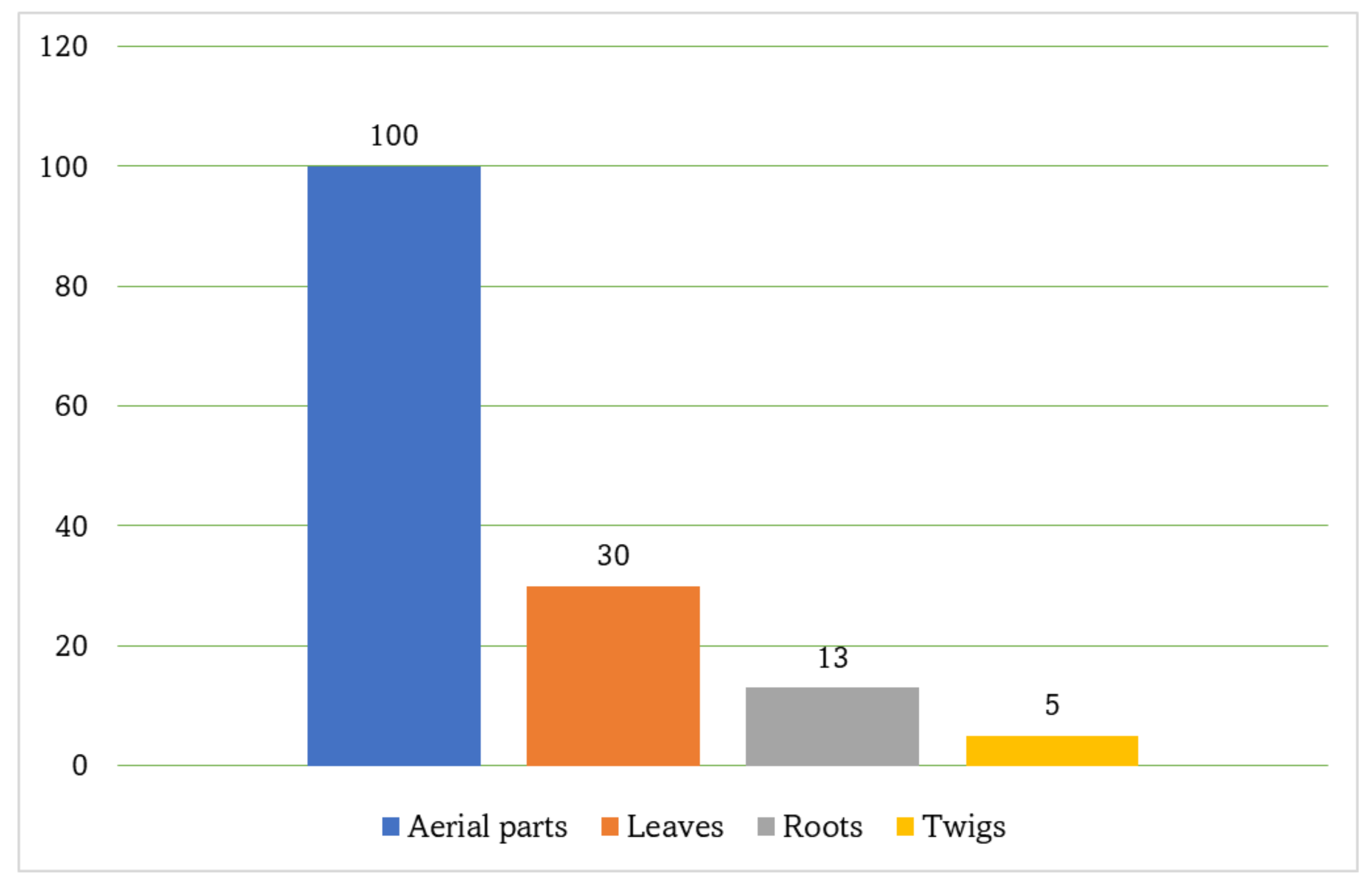
3. Morphology
4. Nutritional Value
5. Ethnomedicinal Uses
6. Phytochemistry
7. Biological Activities
7.1. Anti-Inflammatory Activity

| Plant Organ/Tested Extract/Fraction | Activity | Model | Results | Ref. | |
| Fraction/Extract (Tested Parameter, Dose) | Control | ||||
| Root/MeOH fraction of the defatted chloroform extract | Anti-inflammatory | Carrageenin-induced paw edema/Charles Foster rats | 0.103 mL (Paw volume, 100 mg/kg) | Phenylbutazone 0.204 mL (Paw volume) | [36] |
| 0.046 mL (Paw volume, 300 mg/kg) | |||||
| 75.2 (%Inhibition, 100 mg/kg) | Phenylbutazone 50.9 (%Inhibition) | [36] | |||
| 88.9 (%Inhibition, 300 mg/kg) | |||||
| Carrageenin-induced paw edema/adrenalectomized rats | 0.113 mL (Paw volume, 300 mg/kg) | Phenylbutazone 0.31 mL (Paw volume) | [36] | ||
| 80.7 (%Inhibition, 300 mg/kg) | Phenylbutazone 45.4 (%Inhibition) | [36] | |||
| Mediator-induced edema/Histamine | 0.030 mL (Paw volume, 300 mg/kg) | Control* 0.313 mL (Paw volume) | [36] | ||
| Mediator-induced edema/Histamine | 0.180 mL (Paw volume, 300 mg/kg) | Control* 0.403 mL (Paw volume) | [36] | ||
| Mediator-induced edema/Histamine | 0.030 mL (Paw volume, 300 mg/kg) | Control* 0.353 mL (Paw volume) | [36] | ||
| Carrageenin-induced pleurisy/Charles Foster rats | 46.9 mg (Rear paw weight, 100 mg/kg) | Phenylbutazone 24.7 mg (Rear paw weight) | [36] | ||
| 27.4 mg (Rear paw weight, 300 mg/kg) | [36] | ||||
| Cotton pellet-induced granuloma/Charles Foster rats | 26.2 g (Granuloma weight, 100 mg/kg) | Phenylbutazone 15.4 g (Granuloma weight) | [36] | ||
| 21.8 g (Granuloma weight, 300 mg/kg) | [36] | ||||
| 41.9 (%Inhibition, 100 mg/kg) | Phenylbutazone 65.7 (%Inhibition) | [36] | |||
| 51.8 (%Inhibition, 300 mg/kg) | [36] | ||||
| Carrageenin-induced granuloma/Charles Foster rats | 1.22 g (Granuloma weight, 100 mg/kg) | Phenylbutazone 2.65 g (Granuloma weight) | [36] | ||
| 1.00 g (Granuloma weight, 300 mg/kg) | [36] | ||||
| 63.1 (%Inhibition, 100 mg/kg) | Phenylbutazone 19.9 (%Inhibition) | [36] | |||
| 69.8 (%Inhibition, 100 mg/kg) | [36] | ||||
| Sodium urate-inducededema/Charles Foster rats | 0.40 (Rear paw weight, 100 mg/kg) | Control* 0.68 (Rear paw weight) | [36] | ||
| 0.22 (Rear paw weight, 300 mg/kg) | [36] | ||||
| Root/MeOH extract | Antiulcer | Indomethacin-induced gastric ulcer/Charles Foster rats | 2.50 mm (Ulcer lesion index, 100 mg/kg) | Control* 4.7 1 (Ulcer lesion index) | [32] |
| 0.83 (Ulcer lesion index, 300 mg/kg) | [32] | ||||
| 46.92 (%Inhibition, 100 mg/kg) | [32] | ||||
| 82.37 (%Inhibition, 300 mg/kg) | |||||
| Alcohol-induced gastric ulcer/Charles Foster rats | 1.33 (Ulcer lesion index, 100 mg/kg) | Control* 4.1 (Ulcer lesion index) | [32] | ||
| 1.16 (Ulcer lesion index, 300 mg/kg) | [32] | ||||
| 67.56 (%Inhibition, 100 mg/kg) | [32] | ||||
| 71.70 (%Inhibition, 300 mg/kg) | [32] | ||||
| Alcohol-indomethacin-induced gastric ulcer/Charles Foster rats | 5.72 (Ulcer lesion index, 100 mg/kg) | Control* 19.73 (Ulcer lesion index) | [32] | ||
| 4.36 (Ulcer lesion index, 300 mg/kg) | [32] | ||||
| 71.0 (%Inhibition, 100 mg/kg) | [32] | ||||
| 78.0 (%Inhibition, 300 mg/kg) | [32] | ||||
| Gastric secretion following pyloric ligation/Charles Foster rats | 13.0 mEq acid (HCl)/1/h (Total acidity, 100 mg/kg) | *Control 16.25 mEq acid (HCl)/1/h | [32] | ||
| 7.5 mEq acid (HCl)/1/h (Total acidity, 300 mg/kg) | [32] | ||||
| 9.00 mEq acid (HCl)/1/h (Total acidity, 100 mg/kg) | *Control 9.5 mEq acid (HCl)/1/h | [32] | |||
| 5.16 mEq acid (HCl)/1/h (Total acidity, 300 mg/kg) | [32] | ||||
| 10.3 mg Tyrosin/mL (Peptic activity, 100 mg/kg) | Control* 11.03 Tyrosin/mL (Peptic activity) | [32] | |||
| 9.6 Tyrosin/mL (Peptic activity, 300 mg/kg) | [32] | ||||
| PAF-induced gastric ulcer/Charles Foster rats | 15.0 mm (Ulcer lesion index, 100 mg/kg) | BW 755C 8.33 mm (Ulcer lesion index) | [64] | ||
| 11.66 mm (Ulcer lesion index, 300 mg/kg) | BW 755C 8.33 mm (Ulcer lesion index) | [64] | |||
| 35.6 (%Inhibition, 100 mg/kg) | BW 755C 64.2 (%Inhibition) | [64] | |||
| 50.2 (%Inhibition, 300 mg/kg) | BW 755C 64.2 (%Inhibition) | [64] | |||
| PAF-induced hematological change/Charles Foster rats | 41.2 (Haematocrit % change, 300 mg/kg) | BW 755C 37.6 (Haematocrit% change) | [64] | ||
| 20.4 (RBC % change) | BW 755C 14.3 (RBC % change) | [64] | |||
| 25.2 (Hemoglobin % change) | BW 755C 21.2 (Hemoglobin %change) | [64] | |||
| 15.8 (WBC % change) | BW 755C 16.6 (WBC % change) | [64] | |||
| Croton oil-induced ear edema/Swiss A mice | 3.3 mg (Weight of 5 mm ear-punch, 100 mg/kg) | *Control 19.6 mg (Weight of 5 mm ear-punch | [65] | ||
| 6.8 mg (Weight of 5 mm ear-punch, 300 mg/kg) | - | [65] | |||
| 7.2 mg (Weight of 5 mm ear-punch, 250 µg/ear) | - | [65] | |||
| 15.4 mg (Weight of 5 mm ear-punch, 500 µg/ear) | - | [65] | |||
| Root/EtOH extract | Antioxidant | Hydroxyl (OH) radical-scavenging | 10.77 µg/mL (OH radicals IC50) | Vitamin E 33.2 µg/mL (OH radicals, IC50) | [63] |
| CCl4-induced lipid peroxidation/Swiss albino mice | 54.5 (%Inhibition, 300 µg/mL) | vitamin E 46.28 (%Inhibition) | [63] | ||
| Hydrogen peroxide (H2O2)-scavenging/Charles Foster rats | 65.2 (% Lysis of erythrocytes, 10 µg/mL) | BW 755C 86.9 (% Lysis of erythrocytes) Phenidone 76.8 (% Lysis of erythrocytes) | [63] | ||
| Root/EtOH extract | Cytotoxic | NPC-TW 01 cells/WST-1 colorimetric assay | 14.07 (%Migration, 80 µg/mL) | - | [66] |
| 5.65 (%Relative migration rate, 80 µg/mL) | - | [66] | |||
| 24.57 (%Colony forming efficiency, 50 µg/mL) | - | [66] | |||
| 108.5 µg/mL (24 h) (Growth inhibition 50%) | - | [66] | |||
| NPC-TW04/WST-1 colorimetric assay | 7.76 (%Migration, 60 µg/mL) | - | [66] | ||
| 93.2 (24 h) (Growth inhibition 50%) | - | [66] | |||
| 3.47 (%Relative migration rate, 60 µg/mL) | - | [66] | |||
| Leaves/EtOAc fraction | Anti-inflammatory | EPP-induced ear edema/Male Sprague Dawley rats | 8.30 µm (15 min) (ED, 3 mg/ear) | PB 3.3 µm (15 min) | [67] |
| 18.30 µm (30 min) (ED, 3 mg/ear) | PB 25.0 µm (30 min) | ||||
| 45 µm (60 min) (ED, 3 mg/ear) | PB 43.3 µm (60 min) | ||||
| 65 µm (120 min) (ED, 3 mg/ear) | PB 48.3 µm (120 min) | ||||
| 93.24 (15 min) (%Inhibition) | PB 97.3 (15 min) (%Inhibition) | [67] | |||
| 89 (30 min) (%Inhibition) | PB 85 (30 min) (%Inhibition) | ||||
| 73 (60 min) (%Inhibition) | PB 74 (60 min) (%Inhibition) | ||||
| 54.65 (120 min) (%Inhibition) | PB 66.28 (120 min) (%Inhibition) | ||||
| Carrageenan-induced hind paw edema/Male Sprague Dawley rats | 0.12 mL (1 h) (EV, 600 mg/kg) | DC 0.16 (1 h) (EV) | [67] | ||
| 0.20 mL (3 h) (EV, 600 mg/kg) | DC 0.21 (3 h) (EV) | ||||
| 66.2 (1 h) (%Inhibition, 600 mg/kg) | DC 55.09 (1 h) (%Inhibition) | [67] | |||
| 56.74 (3 h) (%Inhibition, 600 mg/kg) | DC 54.61 (3 h) (%Inhibition) | ||||
| Leaves/EtOH extract | Anti-inflammatory | Carrageenan-induced hind paw edema/Male Sprague Dawley rats | 5.75 (180 min) (%Oedema, 300 mg/kg) | Indo 3.46 (180 min) (%Oedema) | [37] |
| 87.9 (%Inhibition, 300 mg/kg) | 92.7 Indo (180 min) (%Inhibition) | [37] | |||
| Analgesic | Acetic acid-induced writhing/Adult Balb/calbino mice | 20.3 (15 min) (Mean of writhings) | 19.0 (15 min) (Mean of writhings) | [37] | |
| 60.7 (15 min) (%Inhibition) | 63.2 (15 min) (%Inhibition) | [37] | |||
| Cytotoxicity | EA.hy926 cells/MTT assay | 54.9 (%Cell viability, 100 µg/mL) | - | [68] | |
| 27.5 (%Cell viability, 200 µg/mL) | - | [68] | |||
| 26.4 (%Cell viability, 400 µg/mL) | - | [68] | |||
| Lipase inhibitory | 3T3-L1 adipocytes/Pancreatic lipase assay | 1708.35 µg/mL (IC50, 250–1000 µg/mL) | Orlistat 68.23 µg/mL (IC50) | [69] | |
| Antihyperlipidemic | 3T3-L1 adipocytes/Lipid accumulation | 76.87 (%Inhibition, 750 µg/mL) | - | [69] | |
| 71.93 (%Inhibition, 1000 µg/mL) | - | [69] | |||
| Leaves/H2O extract | Anti-dyslipidemia | Prediabetic patients/Clinical trials | 109.22 mg/dL (TG, 1.5 g/once/12 weeks) | Placebo 145.56 mg/dL (TG) | [70] |
| 122.20 mg/dL (LDL-C, 1.5 g/once/12 weeks) | Placebo 142.07 mg/dL (LDL-C) | [70] | |||
| 57.56 mg/dL (HDL-C, 1.5 g/once/12 weeks) | Placebo 46.44 mg/dL (HDL-C) | [70] | |||
| Fibroblast hyperproliferation inhibition | Flowcytometry analysis/ Fibroblast cultures | 1.3 (%Fibroblast density, 20 µmol/L) | Control 1.5 (%Fibroblast density) | [5] | |
| 1.0 (%Fibroblast density, 40 µmol/L) | - | [5] | |||
| 0.7 (%Fibroblast density, 80 µmol/L) | - | [5] | |||
| Leaves/MeOH extract | Antidiabetic | Normoglycemic/Wistar albino rats | 72.67 mg/dL (24 h) (Plasma glucose, 200 mg/kg) | Glibenclamide 73.67 mg/dL (24 h) (Plasma glucose) | [71] |
| 71.67 mg/dL (24 h) (Plasma glucose, 400 mg/kg) | [71] | ||||
| STZ-induced hyperglycemia/Wistar albino rats | 190.0 mg/dL (24 h) (Plasma glucose, 200 mg/kg) | Glibenclamide 168.0 mg/dL (24 h) (Plasma glucose) | [71] | ||
| 172.83 mg/dL (24 h) (Plasma glucose, 400 mg/kg) | - | [71] | |||
| Milk production | Total milk yield/lactating rats | 6.82 g/L/day (23 h) (Milk yield, 750 mg/kg) | Domperidone 7.17 g/L/day (23 h) (Milk yield) | [72] | |
| Growth hormone promotion | 1963.25 pg/µL (Serum growth hormone level, 750 mg/kg) | Domperidone 409.46 pg/µL (Serum growth hormone level) | [72] | ||
| Weight gain | Bodyweight gain/Wistar rat dams | 8.44 (%Dams body weight gain) | Domperidone 0.66 (%Dams body weight gain) | [72] | |
| Leaves/Et OH extract | Antioxidant | DPPH scavenging/DPPH assay | 96.4 µmol TE/g fw (%DPPH scavenging, 100 mg/mL) | - Quercetin 6.70 µg/mL (IC50, DPPH scavenging) | [28] |
| 24.45 µg/mL (IC50, DPPH scavenging) | [41] | ||||
| DPPH scavenging/DPPH assay | 42.24 µg/mL (IC50, DPPH scavenging) | α-Tocopherol 35.57 µg/mL (IC50, DPPH scavenging) | [73] | ||
| ABTS scavenging/TEAC method | 3.75 µmol TE/g fw, ABTS scavenging | - | [28] | ||
| Ferric-reducing power/FRAP assay | 81.1 (µmol TE/g fw, Ferric-reducing | - | [28] | ||
| Inhibition of lipid oxidation/TBA method | 98.5 (%Inhibition) | - | [28] | ||
| β-Carotene-linoleic scavenging activity/β-carotene bleaching method | 59.8 (%Antioxidant activity) | BHA 93.5 (% Antioxidant activity) | [73] | ||
7.2. Anti-Obesity and Anti-Hyperlipidemic Activities
7.3. Antidiabetic Activity
7.4. Insecticidal and Herbicidal Activity
7.5. Cytotoxicity Activity
7.6. Venom Neutralizing Activity
7.7. Hepatoprotective and Neurological Activities
7.8. Antifertility Activity
7.9. Wound-Healing Activity
7.10. Anti-Hemorrhoidal Activity
7.11. Antimicrobial Activity and Pharmaceutical Preparations
7.12. Antioxidant Activity
7.13. Other Activities
8. Toxicity and Safety of P. indica
9. Clinical Traits
10. Conclusion and Recommendations
Author Contributions
Funding
Institutional Review Board Statement
Informed Consent Statement
Data Availability Statement
Conflicts of Interest
Abbreviations
References
- Kesharwani, R.K.; Misra, K.; Singh, D.B. Perspectives and challenges of tropical medicinal herbs and modern drug discovery in the current scenario. Asian Pac. J. Trop. Med. 2019, 12, 1. [Google Scholar] [CrossRef]
- Abdallah, H.M.; Mohamed, G.A.; Ibrahim, S.R.M. Lansium domesticum—A fruit with multi-benefits: Traditional uses, phytochemicals, nutritional value, and bioactivities. Nutrients 2022, 14, 1531. [Google Scholar]
- Lautié, E.; Russo, O.; Ducrot, P.; Boutin, J.A. Unraveling Plant Natural Chemical Diversity for Drug Discovery Purposes. Front. Pharmacol. 2020, 11, 397. [Google Scholar] [CrossRef] [PubMed]
- Katiyar, C.; Kanjilal, S.; Gupta, A.; Katiyar, S. Drug discovery from plant sources: An integrated approach. Ayu 2012, 33, 10. [Google Scholar] [CrossRef] [PubMed]
- Maharani, S.C.; Julianto, I.; Widhiati, S. The role of beluntas (Pluchea indica L.) leaf extract in preventing the occurrence of fibroblasts hyperproliferation: An in vitro preliminary study. Dermatol. Rep. 2019, 11, 8019. [Google Scholar] [CrossRef] [Green Version]
- Ibrahim, S.R.; Mohamed, G.A. Litchi chinensis: Medicinal uses, phytochemistry, and pharmacology. J. Ethnopharmacol. 2015, 174, 492–513. [Google Scholar] [CrossRef]
- Ibrahim, S.R.M.; Mohamed, G.A.; Khedr, A.; Zayed, M.; El-Kholy, A.A.-E.S. Genus Hylocereus: Beneficial phytochemicals, nutritional importance, and biological relevance-A review. J. Food Biochem. 2018, 42, e12491. [Google Scholar] [CrossRef]
- Riaz, U.; Iqbal, S.; Sohail, M.I.; Samreen, T.; Ashraf, M.; Akmal, F.; Siddiqui, A.; Ahmad, I.; Naveed, M.; Khan, N.I.; et al. A Comprehensive Review on Emerging Importance and Economical Potential of Medicinal and Aromatic Plants (MAPs) in Current Scenario. Pak. J. Agric. Res. 2021, 34, 381–392. [Google Scholar] [CrossRef]
- Ekor, M. The growing use of herbal medicines: Issues relating to adverse reactions and challenges in monitoring safety. Front. Pharmacol. 2014, 4, 177. [Google Scholar] [CrossRef] [Green Version]
- Taylor, J.; Rabe, T.; McGaw, L.; Jäger, A.; Van Staden, J. Towards the scientific validation of traditional medicinal plants. Plant Growth Regul. 2001, 34, 23–37. [Google Scholar] [CrossRef]
- Setorki, M. Medicinal herbs with anti-depressant effects. J. Herbmed Pharmacol. 2020, 9, 309–317. [Google Scholar] [CrossRef]
- Abraham, J.; Thomas, T.D. Recent Advances in Asteraceae Tissue Culture. In Plant Tissue Culture: Propagation, Conservation and Crop Improvement; Anis, M., Ahmad, N., Eds.; Springer: Singapore, 2016. [Google Scholar] [CrossRef]
- Rolnik, A.; Olas, B. The Plants of the Asteraceae Family as Agents in the Protection of Human Health. Int. J. Mol. Sci. 2021, 22, 3009. [Google Scholar] [CrossRef] [PubMed]
- Nikolic, M.; Stevovic, S. Family Asteraceae as a sustainable planning tool in phytoremediation and its relevance in urban areas. Urban For. Urban Green. 2015, 14, 782–789. [Google Scholar] [CrossRef]
- Anderberg, A.A. Asteraceae. In Cladistics & Classification; Bremer, K., Ed.; Timber Press: Portland, OR, USA, 1994; pp. 292–303. [Google Scholar]
- Aggarwal, P. A review on phytochemical and biological investigation of plant genus Pluchea. Indo Am. J. Pharm. Res. 2013, 3, 3373–3392. [Google Scholar]
- Wang, J.; Pei, Y.H.; Lin, W.H.; Deng, Z.W.; Qiao, L. Chemical constituents from the stems and leaves of marine mangrove plant Pluchea indica (L.) Less. Shenyang Yaoke Daxue Xuebao 2008, 25, 960–963. [Google Scholar]
- Rodsom, T. A Study on the Diuretic Effects of Pluchea Indica in Healthy Subjects and Patients; Mahidol University: Bangkok, Thailand, 1993. [Google Scholar]
- Susetyarini, E.; Wahyono, P.; Latifa, R.; Nurrohman, E. The Identification of Morphological and Anatomical Structures of Pluchea indica. J. Phys Conf. Ser. 2020, 1539, 012001. [Google Scholar] [CrossRef]
- Susetyarini, E. Jumlah sel spermiogenesis tikus putih yang diberi tanin daun Beluntas (Pluchea indica) sebagai sumber belajar. Proc. Biol. Educ. Conf. 2015, 12, 462–466. [Google Scholar]
- Susetyarini, E. The level of glutamic acid in the semen of male white rat (Ratus norwegicus) after being treated with tannin of Pluchea indica. Procedia Chem. 2015, 14, 152–156. [Google Scholar] [CrossRef] [Green Version]
- Polsiri, K. Antioxidant Activity of Pluchea Indica Less. Extract After In Vitro Digestion and Absorption by Caco–2 Cell Line. Ph.D. Thesis, Burapha University, Chon Buri, Thailand, 2015. [Google Scholar]
- Sudjaroen, Y. Evaluation of ethnobotanical vegetables and herbs in Samut Songkram province. Procedia Eng. 2012, 32, 160–165. [Google Scholar] [CrossRef] [Green Version]
- US Department of Agriculture. Handbooks 8–1 to 8–21: Composition of Food Raw, Processed, Prepared; Government Printing Office: Washington, DC, USA, 1972–1991. [Google Scholar]
- Neamsuwan, A.; Sengnon, N.; Yingcharoen, K. Ethnobotany of edible plants from mangrove and beach forest in Sating Phra Peninsula, Songkhla province. KKU Sci. J. 2012, 40, 981–991. [Google Scholar]
- Pramanik, K.C.; Biswas, R.; Mitra, A.; Bandyopadhyay, D.; Mishra, M.; Chatterjee, T.K. Tissue culture of the plant Pluchea indica (L.) and evaluation of diuretic potential of its leaves. Adv. Tradit. Med. 2007, 7, 197–204. [Google Scholar] [CrossRef]
- Shannon, M.; Grieve, C. Tolerance of vegetable crops to salinity. Sci. Hortic. 1998, 78, 5–38. [Google Scholar] [CrossRef]
- Andarwulan, N.; Batari, R.; Sandrasari, D.A.; Bolling, B.; Wijaya, C.H. Flavonoid content and antioxidant activity of vegetables from Indonesia. Food Chem. 2010, 121, 1231–1235. [Google Scholar] [CrossRef] [PubMed] [Green Version]
- Andarwulan, N.; Kurniasih, D.; Apriady, R.A.; Rahmat, H.; Roto, A.V.; Bolling, B. Polyphenols, carotenoids, and ascorbic acid in underutilized medicinal vegetables. J. Funct. Foods 2012, 4, 339–347. [Google Scholar] [CrossRef]
- Kirtikar, K.R.; Basu, B.D. Indian Medicinal Plants; International Book Distributors: Dehradun, India, 1999; pp. 1344–1345. [Google Scholar]
- Ahem, S.A.; Kamel, E.M. Phenolic constituents and biological activity of the genus Pluchea. Pharma Chem. 2014, 5, 109–114. [Google Scholar]
- Sen, T.; Basu, A.; Ray, R.N.; Chaudhuri, A.K.N. Hepatoprotective effects of Pluchea indica (L.) extract in experimental acute liver damage in rodents. Phytother. Res. 1993, 7, 352–355. [Google Scholar] [CrossRef]
- Sen, T.; Ghosh, T.K.; Chaudhuri, A.K. Studies on the mechanism of anti-inflammatory and anti-ulcer activity of Pluchea indica–probable involvement of 5–lipooxygenase pathway. Life Sci. 1993, 52, 737–743. [Google Scholar] [CrossRef]
- Thongpraditchote, S.; Matsumoto, K.; Temsiririrkkul, R.; Tohda, M.; Murakami, Y.; Watanabe, H. Neuropharmacological Actions of Pluchea indica L. Root Extract in Socially Isolated Mice. Biol. Pharm. Bull. 1996, 19, 379–383. [Google Scholar] [CrossRef] [Green Version]
- Locher, C.; Burch, M.; Mower, H.; Berestecky, J.; Davis, H.; Van Poel, B.; Lasure, A.; Berghe, D.; Vlietinck, A. Anti-microbial activity and anti-complement activity of extracts obtained from selected Hawaiian medicinal plants. J. Ethnopharmacol. 1995, 49, 23–32. [Google Scholar] [CrossRef]
- Sen, T.; Chaudhuri, A. Antiinflammatory evaluation of a Pluchea indica root extract. J. Ethnopharmacol. 1991, 33, 135–141. [Google Scholar] [CrossRef]
- Roslida, A.H.; Erazuliana, A.K.; Zuraini, A. Anti-inflammatory and antinociceptive activities of the ethanolic extract of Pluchea indica (L.) Less leaf. Pharmacologyonline 2008, 2, 349–360. [Google Scholar]
- Bandaranayake, W. Bioactivities, bioactive compounds and chemical constituents of mangrove plants. Wetl. Ecol. Manag. 2002, 10, 421–452. [Google Scholar] [CrossRef]
- Widyawati, P.S.; Budianta, T.D.W.; Gunawan, D.I.; Wongso, R.S. Evaluation antidiabetic activity of various leaf extracts of Pluchea indica L. Int. J. Pharmacog. Phytochem. Res. 2015, 7, 597–603. [Google Scholar]
- Sumanth, M.; Narasimharaju, K. Evaluation of galactagogue activity of lactovedic: A polyherbal formulation. Int. J. Green Pharm. 2011, 5, 61–66. [Google Scholar] [CrossRef]
- Noridayu, A.R.; Hii, Y.F.; Faridah, A.; Khozirah, S.; Lajis, N. Antioxidant and antiacetylcholinesterase activities of Pluchea indica Less. Int. Food Res. J. 2011, 18, 925–929. [Google Scholar]
- Due, R.; Symaswisna, M.R. Ethnobotany of dayak pesaguan medicinal plant and its implementation in the making of biodiversity flash cards. Biol. Study Fac. Teach. Train. Educ. Untan Pontianak 2013, 3, 1–15. [Google Scholar]
- Widyawati, P.S.; Wijaya, C.H.; Hardjosworo, P.S.; Sajuthi, D. Volatile compounds of Pluchea indica less and Ocimum basillicum linn essential oil and potency as antioxidant. HAYATI J. Biosci. 2013, 20, 117–126. [Google Scholar] [CrossRef] [Green Version]
- Ruan, J.; Yan, J.; Zheng, D.; Sun, F.; Wang, J.; Han, L.; Zhang, Y.; Wang, T. Comprehensive Chemical Profiling in the Ethanol Extract of Pluchea indica Aerial Parts by Liquid Chromatography/Mass Spectrometry Analysis of Its Silica Gel Column Chromatography Fractions. Molecules 2019, 24, 2784. [Google Scholar] [CrossRef] [Green Version]
- Nopparat, J.; Nualla–Ong, A.; Phongdara, A. Ethanolic extracts of Pluchea indica (L.) leaf pretreatment attenuates cytokine–induced β–cell apoptosis in multiple low–dose streptozotocin–induced diabetic mice. PLoS ONE 2019, 14, e0212133. [Google Scholar] [CrossRef]
- Chewchida, S.; Vongsak, B. Simultaneous HPTLC quantification of three caffeoylquinic acids in Pluchea indica leaves and their commercial products in Thailand. Rev. Bras. Farmacogn. 2019, 29, 179–181. [Google Scholar] [CrossRef]
- Uchiyama, T.; Miyase, T.; Ueno, A.; Usmanghani, K. Terpenic glycosides from Pluchea indica. Phytochemistry 1989, 28, 3369–3372. [Google Scholar] [CrossRef]
- Uchiyama, T.; Miyase, T.; Ueno, A.; Usmanghani, K. Terpene and lignan glycosides from Pluchea indica. Phytochemistry 1991, 30, 655–657. [Google Scholar] [CrossRef]
- Mukhopadhyay, S.; Cordel, G.A. Traditional medicinal plants of Thailand. IV. 3-(2’,3’-diacetoxy-2’-methyl butyryl)-cuauhtemone from Pluchea indica. J. Nat. Prod. 1983, 46, 671–674. [Google Scholar] [CrossRef]
- Wang, H.M.; Kao, C.L.; Liu, C.M.; Li, W.J.; Yeh, H.C.; Li, H.T.; Kuo, C.N.; Chen, C.Y. Chemical Constituents of the Roots of Pluchea indica. Chem. Nat. Compd. 2017, 53, 736–737. [Google Scholar] [CrossRef]
- Ruan, J.; Li, Z.; Yan, J.; Huang, P.; Yu, H.; Han, L.; Zhang, Y.; Wang, T. Bioactive Constituents from the Aerial Parts of Pluchea indica L. Molecules 2018, 23, 2104. [Google Scholar] [CrossRef] [Green Version]
- Qiu, Y.Q.; Qi, S.H.; Zhang, C.; Li, Q.X. Chemical constituents of Pluchea indica (II). Zhongcaoyao 2010, 41, 24–27. [Google Scholar]
- Huong, D.T.V.; Giang, P.M.; Sim, H.T.; Chinh, T.T.T. Triterpenoids and Phytosterols Isolated from Pluchea indica L. Leaves. VNU J. Sci. Nat. Sci. Technol. 2019, 35, 106–111. [Google Scholar] [CrossRef]
- Huong, D.T.V.; Giang, P.M. Sterol, glycerol ester, and thiophene constituents from the twigs of Pluchea indica L. of Vietnam. VNU J. Sci. Nat. Sci. Technol. 2018, 34, 78–82. [Google Scholar] [CrossRef]
- Shukri, M.A.M.; Alan, C.; Noorzuraini, A.R.S. Polyphenols and antioxidant activities of selected traditional vegetables. J. Trop. Agric. Food Sci. 2011, 39, 69–83. [Google Scholar]
- Kongkiatpaiboon, S.; Chewchinda, S.; Vongsak, B. Optimization of extraction method and HPLC analysis of six caffeoylquinic acids in Pluchea indica leaves from different provenances in Thailand. Rev. Bras. Farm. 2018, 28, 145–150. [Google Scholar] [CrossRef]
- Arsiningtyas, I.S.; Gunawan–Puteri, M.D.; Kato, E.; Kawabata, J. Identification of α–glucosidase inhibitors from the leaves of Pluchea indica (L.) Less., a traditional Indonesian herb: Promotion of natural product use. Nat. Prod. Res. 2014, 28, 1350–1353. [Google Scholar] [CrossRef] [PubMed]
- Ohtsuki, T.; Yokosawa, E.; Koyano, T.; Preeprame, S.; Kowithayakorn, T.; Sakai, S.; Toida, T.; Ishibashi, M. Quinic acid esters from Pluchea indica with collagenase, MMP-2 and MMP-9 inhibitory activities. Phytother. Res. 2008, 22, 264–266. [Google Scholar] [CrossRef] [PubMed]
- Boonruang, S.; Prakobsri, K.; Pouyfung, P.; Srisook, E.; Prasopthum, A.; Rongnoparut, P.; Sarapusit, S. Inhibition of human cytochromes P450 2A6 and 2A13 by flavonoids, acetylenic thiophenes and sesquiterpene lactones from Pluchea indica and Vernonia cinerea. J. Enzym. Inhib. Med. Chem. 2017, 32, 1136–1142. [Google Scholar] [CrossRef] [PubMed] [Green Version]
- Biswas, R.; Dutta, P.; Achari, B.; Bandyopadhyay, D.; Mishra, M.; Pramanik, K.; Chatterjee, T. Isolation of pure compound R/J/3 from Pluchea indica (L.) Less. and its anti-amoebic activities against Entamoeba histolytica. Phytomedicine 2007, 14, 534–537. [Google Scholar] [CrossRef]
- Goswami, S.; Debnath, S.; Karan, S.; Chatterjee, T.K. In vivo antitumor activity of phytochemical pitc-2 obtained from tissue cultured plant Pluchea indica on sarcoma-180 solid tumor mice model. Asian J. Pharm. Clin. Res. 2018, 11, 211–218. [Google Scholar] [CrossRef] [Green Version]
- Qiu, Y.-Q.; Qi, S.-H.; Zhang, S.; Tian, X.-P.; Xiao, Z.-H.; Li, M.-Y.; Li, Q.-X. Thiophene derivatives from the aerial part of Pluchea indica. Heterocycles 2008, 75, 1757–1764. [Google Scholar]
- Sen, T.; Dhara, A.K.; Bhattacharjee, S.; Pal, S.; Chaudhuri, A.K.N. Antioxidant activity of the methanol fraction of Pluchea indica root extract. Phytother. Res. 2002, 16, 331–335. [Google Scholar] [CrossRef]
- Sen, T.; Ghosh, T.K.; Bhattacharjee, S.; Nag Chaudhuri, A.K. Action of Pluchea indica methanol extract as a dual inhibitor on PAF–induced paw oedema and gastric damage. Phytother. Res. 1996, 10, 74–76. [Google Scholar] [CrossRef]
- Sen, T.; Pal, S.; Izzo, A.A.; Capasso, F.; Nag Chaudhuri, A.K. Studies on the methanolic fraction of Pluchea indica on croton oil–induced mouse ear oedema and lipid peroxidation. Pharm. Sci. 1996, 2, 433–435. [Google Scholar]
- Kao, C.-L.; Cho, J.; Lee, Y.-Z.; Cheng, Y.-B.; Chien, C.-Y.; Hwang, C.-F.; Hong, Y.-R.; Tseng, C.-N.; Cho, C.-L. Ethanolic Extracts of Pluchea indica Induce Apoptosis and Antiproliferation Effects in Human Nasopharyngeal Carcinoma Cells. Molecules 2015, 20, 11508–11523. [Google Scholar] [CrossRef] [Green Version]
- Buapool, D.; Mongkol, N.; Chantimal, J.; Roytrakul, S.; Srisook, E.; Srisook, K. Molecular mechanism of anti-inflammatory activity of Pluchea indica leaves in macrophages RAW 264.7 and its action in animal models ofinflammation. J. Ethnopharmacol. 2013, 146, 495–504. [Google Scholar] [CrossRef] [PubMed]
- Srisook, K.; Jinda, S.; Srisook, E. Anti-inflammatory and Antioxidant Effects of Pluchea indica Leaf Extract in TNF-α-Induced Human Endothelial Cells. Walailak J. Sci. Technol. 2021, 18, 10271. [Google Scholar] [CrossRef]
- Sirichaiwetchakoon, K.; Lowe, G.M.; Thumanu, K.; Eumkeb, G. The Effect of Pluchea indica (L.). Tea on Adipogenesis in 3T3-L1 Adipocytes and Lipase Activity. Evid. Based Complement. Altern. Med. 2018, 2018, 4108787–4108813. [Google Scholar] [CrossRef] [PubMed]
- Sirichaiwetchakoon, K.; Churproong, S.; Kupittayanant, S.; Eumkeb, G. The Effect of Pluchea indica (L.). Tea on Blood Glucose and Lipid Profile in People with Prediabetes: A Randomized Clinical Trial. J. Altern. Complement. Med. 2021, 27, 669–677. [Google Scholar] [CrossRef] [PubMed]
- Pramanik, K.C.; Bhattacharya, P.; Biswas, R.; Bandyopadhyay, D.; Mishra, M.; Chatterjee, T. Hypoglycemic and antihyperglycemic activity of leaf extract of Pluchea indica L. Orient. Pharm. Exp. Med. 2006, 6, 232–236. [Google Scholar]
- Syarif, R.A.; Anggorowati, N.; Yuniyanti, M.M.; Wahyuningsih, M.S.H. Ethanolic extract of Pluchea indica L. leaf increases serum growth hormone in lactating rats. Trad. Med. J. 2021, 26, 111–116. [Google Scholar] [CrossRef]
- Abdul Rahman, H.; Saari, N.; Abas, F.; Ismail, A.; Mumtaz, M.W.; Abdul Hamid, A. Anti-obesity and antioxidant activities of selected medicinal plants and phytochemical profiling of bioactive compounds. Int. J. Food Prop. 2017, 20, 2616–2629. [Google Scholar] [CrossRef]
- Chaudhuri, A.K.N.; Mahapatra, P.K. Preliminary studies on anti inflammatory actions of Pluchea indica less roots. Med. Sci. Res. 1987, 15, 487–488. [Google Scholar]
- Mohamed, G.A.; Ibrahim, S.R.; Elkhayat, E.S.; El Dine, R.S. Natural anti-obesity agents. Bull. Facu. Pharm. Cairo Univ. 2014, 52, 269–284. [Google Scholar] [CrossRef] [Green Version]
- Filippatos, T.D.; Derdemezis, C.S.; Gazi, I.F.; Nakou, E.S.; Mikhailidis, D.P.; Elisaf, M.S. Orlistat–associated adverse effects and drug interactions: A critical review. Drug Safety 2008, 31, 53–65. [Google Scholar] [CrossRef]
- Payne, C.; Wiffen, P.J.; Martin, S. Interventions for fatigue and weight loss in adults with advanced progressive illness. Cochrane Database Syst. Rev. 2012, 1, CD008427. [Google Scholar] [PubMed]
- Tomita, T. Apoptosis in pancreatic beta–islet cells in type 2 diabetes. Bosn. J. Basic Med. Sci. 2016, 16, 162–179. [Google Scholar] [CrossRef] [PubMed] [Green Version]
- Thomas, H.E.; Kay, T.W. Intracellular pathways of pancreatic beta–cell apoptosis in type 1 diabetes. Diabetes Metab. Res. Rev. 2011, 27, 790–796. [Google Scholar] [CrossRef] [PubMed]
- Ibrahim, S.R.; Mohamed, G.A.; Khayat, M.T.; Ahmed, S.; Abo-Haded, H.; Alshali, K.Z. Mangostanaxanthone VIIII, a new xanthone from Garcinia mangostana pericarps, α-amylase inhibitory activity, and molecular docking studies. Rev. Bras. Farm. 2019, 29, 206–212. [Google Scholar] [CrossRef]
- Ibrahim, S.R.M.; Mohamed, G.A.A.; Khayat, M.T.A.; Ahmed, S.; Abo-Haded, H. Garcixanthone D, a New Xanthone, and Other Xanthone Derivatives from Garcinia mangostana Pericarps: Their α-Amylase Inhibitory Potential and Molecular Docking Studies. Starch-Stärke 2019, 71, 1800354. [Google Scholar] [CrossRef]
- Ibrahim, S.R.M.; Mohamed, G.A.; Khayat, M.T.; Ahmed, S.; Abo–Haded, H. α–Amylase inhibitors xanthones from Garcinia mangostana pericarps and its possible use for the treatment of diabetes with their molecular docking studies. J. Food Biochem. 2019, 43, e12844. [Google Scholar] [CrossRef]
- Ibrahim, S.R.; Mohamed, G.A.; Zayed, M.; Ross, S.A. 8-Hydroxyirilone 5-methyl ether and 8-hydroxyirilone, new antioxidant and α-amylase inhibitors iso flavonoids from Iris germanica rhizomes. Bioorg. Chem. 2017, 70, 192–198. [Google Scholar] [CrossRef]
- Vongsak, B.; Kongkiatpaiboon, S.; Jaisamut, S.; Konsap, K. Comparison of active constituents, antioxidant capacity, and α-glucosidase inhibition in Pluchea indica leaf extracts at different maturity stages. Food Biosci. 2018, 25, 68–73. [Google Scholar] [CrossRef]
- Suriyah, W.H.; Ichwan, S.J.A.; Kasmuri, A.R.; Taher, M. In vitro Evaluation of the Effect of Pluchea indica Extracts in Promoting Glucose Consumption Activity on A Liver Cell Line. Makara J. Health Res. 2019, 23, 48–52. [Google Scholar] [CrossRef]
- Reddy, P.P. Crop Residue Management and Organic Amendments; Springer Science and Business Media LLC: Berlin/Heidelberg, Germany, 2017; pp. 29–41. [Google Scholar]
- Metcalf, R.L. The Ecology of Insecticides and the Chemical Control of Insects Ecological Theory an Integrated Pest Management Practice; Kogan, M., Ed.; Wiley: New York, NY, USA, 1986. [Google Scholar]
- Rahayu, Y.S. The Using of Fenolic Compounds of Pluchea indica (L.). Leaves Extracts as A Bioinsecticide and Bioherbicide; In Proceedings of the Journal of Physics: Conference Series; IOP Publishing: Bristol, UK, 2018; Volume 953, p. 012206. [Google Scholar]
- Zhang, S.; Xu, H.; Zhang, L.; Qiao, Y. Cervical cancer: Epidemiology, risk factors and screening. Chin. J. Cancer Res. 2020, 32, 720–728. [Google Scholar] [CrossRef]
- Baskar, R.; Lee, K.A.; Yeo, R.; Yeoh, K.-W. Cancer and Radiation Therapy: Current Advances and Future Directions. Int. J. Med. Sci. 2012, 9, 193–199. [Google Scholar] [CrossRef] [Green Version]
- Cho, J.; Cho, C.-L.; Kao, C.-L.; Chen, C.-M.; Tseng, C.-N.; Lee, Y.-Z.; Liao, L.-J.; Hong, Y.-R. Crude aqueous extracts of Pluchea indica (L.) inhibit proliferation and migration of cancer cells through induction of p53-dependent cell death. BMC Complement. Altern. Med. 2012, 12, 265. [Google Scholar] [CrossRef] [PubMed] [Green Version]
- Kao, C. Antiproliferation and Apoptosis Induction of Ethanolic Extracts of Pluchea indica Root on Human Nasopharyngeal Carcinoma Cell Lines. Ph.D. Thesis, National Sun Yat–Sen University, Kaohsiung City, Taiwan, 2015. [Google Scholar]
- Ko, H.J. Effect of Crude Ethanol Extracts of Pluchea Indica on Human Leukemia K562 Cells. Master’s Thesis, National Sun Yat–Sen University, Kaohsiung City, Taiwan, 2015. [Google Scholar]
- Cho, C.-L.; Lee, Y.-Z.; Tseng, C.-N.; Cho, J.; Cheng, Y.-B.; Wang, K.; Chen, H.-J.; Chiou, S.-J.; Chou, C.-H.; Hong, Y.-R. Hexane fraction of Pluchea indica root extract inhibits proliferation and induces autophagy in human glioblastoma cells. Biomed. Rep. 2017, 7, 416–422. [Google Scholar] [CrossRef] [PubMed] [Green Version]
- Goswami, S.; Chakraborty, S.; Das, P.; Karan, S.; Naskar, D.; Debnath, S.; Chatterjee, T.K. PITC–2 loaded solid lipid nanoparticle: Design, preparation, characterization, and therapeutic comparison with free phytochemical PITC–2 isolated from tissue cultured plant Pluchea indica. Int. J. Pharm. Biol. Sci. 2019, 9, 23–36. [Google Scholar]
- Gomes, A.; Saha, A.; Chatterjee, I.; Chakravarty, A.K. Viper and cobra venom neutralization by beta–sitosterol and stigmasterol isolated from the root extract of Pluchea indica L. (Asteraceae). Phytomedicine 2007, 14, 637–643. [Google Scholar] [CrossRef] [PubMed]
- Alam, M.I.; Auddy, B.; Gomes, A. Viper venom neutralization by Indian medicinal plant (Hemidesmus indicus and Pluchea indica) root extracts. Phytother. Res. 1996, 10, 58–61. [Google Scholar] [CrossRef]
- Sutherland, S.K. Antivenom use in Australia: Premedication, adverse reactions and the use of venom detection kits. Med. J. Aust. 1992, 157, 734–739. [Google Scholar] [CrossRef] [PubMed]
- Stahel, E.; Wellauer, R.; Freyvogel, T.A. Vergiftungem durch einheimische (Vipera vipera berrus and Vipera aspis) Eine retrospective studies on 113 patient. Schweiz. Med. Wochenschr. 1985, 115, 890–896. [Google Scholar] [PubMed]
- Kadali, V.N.; Kameswara, R.K.; Sandeep, B.V. Medicinal plants with anti-Snake Venom property–A review. Pharm. Innov. J. 2015, 4, 11–15. [Google Scholar]
- Nopparat, J.; Nualla–Ong, A.; Phongdara, A. Treatment with Pluchea indica (L.) leaf ethanol extract alleviates liver injury in multiple low-dose streptozotocin-induced diabetic BALB/c mice. Exp. Therap. Med. 2020, 20, 1385–1396. [Google Scholar] [CrossRef]
- Mahapatra, P.K.; Chaudhuri, A.K.N. Neuropharmacological Studies on Pluchea indica. Planta Med. 1986, 6, 546–547. [Google Scholar] [CrossRef] [PubMed]
- Campbell, C.D.; Lee, J.Z. Fertility control in historical China revisited: New Methods for an Old Debate. Hist. Fam. 2010, 15, 370–385. [Google Scholar] [CrossRef] [PubMed] [Green Version]
- Payne, C.; Goldberg, E. Male contraception: Past, present and future. Curr. Mol. Pharmacol. 2014, 7, 175–181. [Google Scholar] [CrossRef] [PubMed]
- Dias, T.; Alves, M.; Oliveira, P.F.; Silva, B.M. Natural Products as Modulators of Spermatogenesis: The Search for a Male Contraceptive. Curr. Mol. Pharmacol. 2014, 7, 154–166. [Google Scholar] [CrossRef] [PubMed]
- Amalina, N.; Suyatmi, S.; Suparyanti, E.L. Effect of Beluntas (Pluchea indica) leaf extract on mice spermatogenesis. Biofarmasi 2010, 8, 47–51. [Google Scholar] [CrossRef] [Green Version]
- O’Donnell, L. Mechanisms of spermiogenesis and spermiation and how they are disturbed. Spermatogenesis 2015, 4, e979623. [Google Scholar] [CrossRef] [PubMed] [Green Version]
- Susetyarini, E.; Latifa, R.; Miharja, F.J. DNA profile of white male rats spermatozoa after treatment with tannins Beluntas (Pluchea indica). Int. J. Eng. Technol. 2019, 8, 302–305. [Google Scholar]
- Sivastana, S.; Desai, P.; Coutinno, E.; Govil, G. Mechanism of action of L–arginine on the vitality of spermatozoa is primarily through increase biosynthesis of nitric oxide. Biol. Reprod. 2006, 74, 954–958. [Google Scholar]
- Gurtner, G.C.; Werner, S.; Barrandon, Y.; Longaker, M.T. Wound repair and regeneration. Nature 2008, 453, 314–321. [Google Scholar] [CrossRef]
- Guo, S.; DiPietro, L.A. Factors Affecting Wound Healing. J. Dent. Res. 2010, 89, 219–229. [Google Scholar] [CrossRef]
- Sugiaman, V.K.; Nisyah, N.Q.; Anisa, N.; Pranata, N. Pluchea indica extract as a potential source of nutrition for accelerate wound healing. Sys. Rev. Pharm. 2021, 12, 570–573. [Google Scholar]
- Buranasukhon, W.; Athikomkulchai, S.; Tadtong, S.; Chittasupho, C. Wound healing activity of Pluchea indica leaf extract in oral mucosal cell line and oral spray formulation containing nanoparticles of the extract. Pharm. Biol. 2017, 55, 1767–1774. [Google Scholar] [CrossRef] [PubMed] [Green Version]
- Reinke, J.; Sorg, H. Wound Repair and Regeneration. Eur. Surg. Res. 2012, 49, 35–43. [Google Scholar] [CrossRef]
- Landén, N.X.; Li, D.; Ståhle, M. Transition from inflammation to proliferation: A critical step during wound healing. Cell. Mol. Life Sci. 2016, 73, 3861–3885. [Google Scholar] [CrossRef] [PubMed] [Green Version]
- Ichim, T.E.; O’Heeron, P.; Kesari, S. Fibroblasts as a practical alternative to mesenchymal stem cells. J. Transl. Med. 2018, 16, 212. [Google Scholar] [CrossRef] [Green Version]
- Stipcevic, T.; Piljac, J.; Berghe, D.V. Effect of Different Flavonoids on Collagen Synthesis in Human Fibroblasts. Mater. Veg. 2006, 61, 29–34. [Google Scholar] [CrossRef]
- Caliskan, U.K.; Aka, C.; Oz, M.G. Plants used in Anatolian traditional medicine for the treatment of hemorrhoid. Rec. Nat. Prod. 2017, 11, 235–250. [Google Scholar]
- Senvorasinh, K.; Phunikhom, K.; Sattayasai, J. Anti-Hemorrhoidal activity of Pluchea indica leaves aqueous extract in croton oil–induced hemorrhoids in experimental animals. Srinagarind Med. J. 2019, 34, 590–594. [Google Scholar]
- Nurrohman, E.; Pantiwati, Y.; Susetyarini, E.; Umami, E.K. Extract of Beluntas (Pluchea indica) as an Antibacterial Towards Streptococcus mutans ATCC 25175 Causes of Dental Carries. BIO-EDU J. Pendidik. Biol. 2021, 6, 9–17. [Google Scholar] [CrossRef]
- Fatimatuzzahr, N.; Rahayu, F.; Ningsih, N.S.; Darsono, A.; Salasia, S.I.O. Anticariogenic effect of Beluntas (Pluchea indica) Leaces extract as growth inhibitor of Streptococcus mutans which caused dental caries. J. Sain Vet. 2016, 34, 182–193. [Google Scholar]
- Fatimatuzzahr, N.; Rahayu, F.; Ningsih, N.S.; Darsono, A.; Salasia, S.I.O. Rontgen Result of Caries Molar in White Rat (Rattus novergicus) with Treatment of Herbal Toothpaste (Pluchea Indica Leaves Extract). In Proceedings of the 4th Asian Academic Society International Conference (AASIC), Globalizing Asia: Integrating Science, Technology and Humanities for Future Growth and Development, Nakhon Pathom, Thailand, 12–13 May 2016; pp. 183–187. [Google Scholar]
- Demolsky, W.L.; Sugiaman, V.K.; Pranata, N. Antifungal activity of Beluntas “Indian Camphorweed” (Pluchea indica) ethanol extract on Candida albicans in vitro using different solvent concentrations. Eur. J. Dent. 2021. [Google Scholar] [CrossRef] [PubMed]
- Sulistyani, N.; Alvionida, F.; Sugihartini, N. Composition of carbopol 940 and HPMC affects antibacterial activity of beluntas (Pluchea indica (L.)) leaves extract gel. Pharmaciana 2021, 11, 427–438. [Google Scholar] [CrossRef]
- Komala, O.; Wiendarlina, I.Y.; Rizqiyana, N. Antibacterial activity roll on deodorant with Pluchea indica (L.) leaf extract against Staphylococcus epidermidis (Evans 1916) in vitro. IOP Conf. Ser. Earth Environ. Sci. 2019, 293, 012031. [Google Scholar] [CrossRef]
- Sylvana, D.; Amir, M.; Purnamasari, C.B.; Iskandar, A.; Asfirizal, V. Antibacterial activity of ethanol extract of Beluntas leaves on Streptococcus mutans, Porphyromonas gingivalis, and Enterococcus faecalis. Padjadjaran J. Dent. 2021, 33, 191–198. [Google Scholar] [CrossRef]
- Pargaputri, A.F.; Munadziroh, E.; Indrawati, R. The Effect of Pluchea indica L. Leaves Extract Againts Biofilm of Enterococcus faecalis and Fusobacterium nucleatum In Vitro. Denta 2017, 11, 51. [Google Scholar] [CrossRef]
- Simanjuntak, N.; Yuniarni, U.; Prayugo, D. Antibacterial activity of Pluchea indica and Piper beetle ethanol extract on Staphylococcus epidermidis and Pseudomonas aeruginosa. Pharmacol. Clin. Pharm. Res. 2016, 1, 62–68. [Google Scholar]
- Sittiwet, C. In vitro Antimicrobial Activity of Pluchea indica Aqueous Extract: The Potential for Urinary Tract Infection Treatment. J. Pharmacol. Toxicol. 2009, 4, 87–90. [Google Scholar] [CrossRef] [Green Version]
- Pramanik, K.C.; Chatterjee, T.K. In vitro and in vivo antibacterial activities of root extract of tissue cultured Pluchea indica (L.) Less. Orient. Pharm. Exp. Med. 2008, 8, 295–301. [Google Scholar] [CrossRef] [Green Version]
- Srimoon, R.; Ngiewthaisong, S. Antioxidant and antibacterial activities of Indian marsh fleabane (Pluchea indica (L.). KKU Res J. 2015, 20, 144–154. [Google Scholar]
- Farhamzah, H.A.; Mursal, I.L. Formulation and antibacterial activity test of foot spray with Beluntas leaf ethanol extract (Pluchea indica L.). IOP Conf. Ser. Mater. Sci. Eng. 2021, 1071, 12013. [Google Scholar] [CrossRef]
- Phaniendra, A.; Jestadi, D.B.; Periyasamy, L. Free Radicals: Properties, Sources, Targets, and Their Implication in Various Diseases. Indian J. Clin. Biochem. 2015, 30, 11–26. [Google Scholar] [CrossRef] [PubMed] [Green Version]
- Halliwell, B.; Gutteridge, J.M.C. Free Radicals in Biology and Medicine, 5th ed.; Oxford University Press: New York, NY, USA, 2015. [Google Scholar]
- Srisook, K.; Buapool, D.; Boonbai, R.; Simmasut, P.; Charoensuk, Y.; Srisook, E. Antioxidant and anti-inflammatory activities of hot water extract from Pluchea indica L. herbal tea. J. Med. Plant Res. 2012, 6, 4077–4408. [Google Scholar]
- Widyawati, P.S.; Budianta, T.D.W.; Werdani, Y.D.W.; Halim, M.O. Antioxidant Activity of Pluchea Leaves–Black Tea Drink (Pluchea indica L.–Camelia sinensis). Agritech 2018, 38, 200–207. [Google Scholar] [CrossRef] [Green Version]
- Widyawati, P.S.; Budianta, T.D.W.; Kusuma, F.A.; Wijaya, E.L.; Yaunatan, D.I.; Wongso, R.S. Potency of Beluntas (Pluchea indica L.) Leaves to Extract as Antioxidant and Anti-Warmed–Over Flavor (WOF) of Duck Meat. In Proceedings of the International Congress Challenges of Biotechnological Research in Food and Health, Surakarta, Indonesia, 15 November 2014; pp. 81–89. [Google Scholar]
- Sirichaiwetchakoon, K.; Lowe, G.M.; Eumkeb, G. The Free Radical Scavenging and Anti-Isolated Human LDL Oxidation Activities of Pluchea indica (L.) Tea Compared to Green Tea (Camellia sinensis). BioMed Res. Int. 2020, 2020, 4183643. [Google Scholar] [CrossRef]
- Sirichaiwetchakoon, K.; Lowe, G.M.; Kupittayanant, S.; Churproong, S.; Eumkeb, G. Pluchea indica (L.) tea ameliorates hyperglycemia, dyslipidemia, and obesity in high fat diet–fed mice. Evid. Based Complemen. Altern. Med. 2020, 2020, 8746137. [Google Scholar] [CrossRef] [PubMed]
- Werdani, Y.D.W.; Widyawati, P.S. Antidiabetic Effect on Tea of Pluchea indica Less as Functional Beverage in Diabetic Patients. In Proceedings of the 1st International Conference Postgraduate School Universitas Airlangga: “Implementation of Climate Change Agreement to Meet Sustainable Development Goals” (ICPSUAS 2017), Jawa Timur, Indonesia, 1–2 August 2017; Atlantis Press: Paris, France, 2018; Volume 98, pp. 164–167. [Google Scholar]


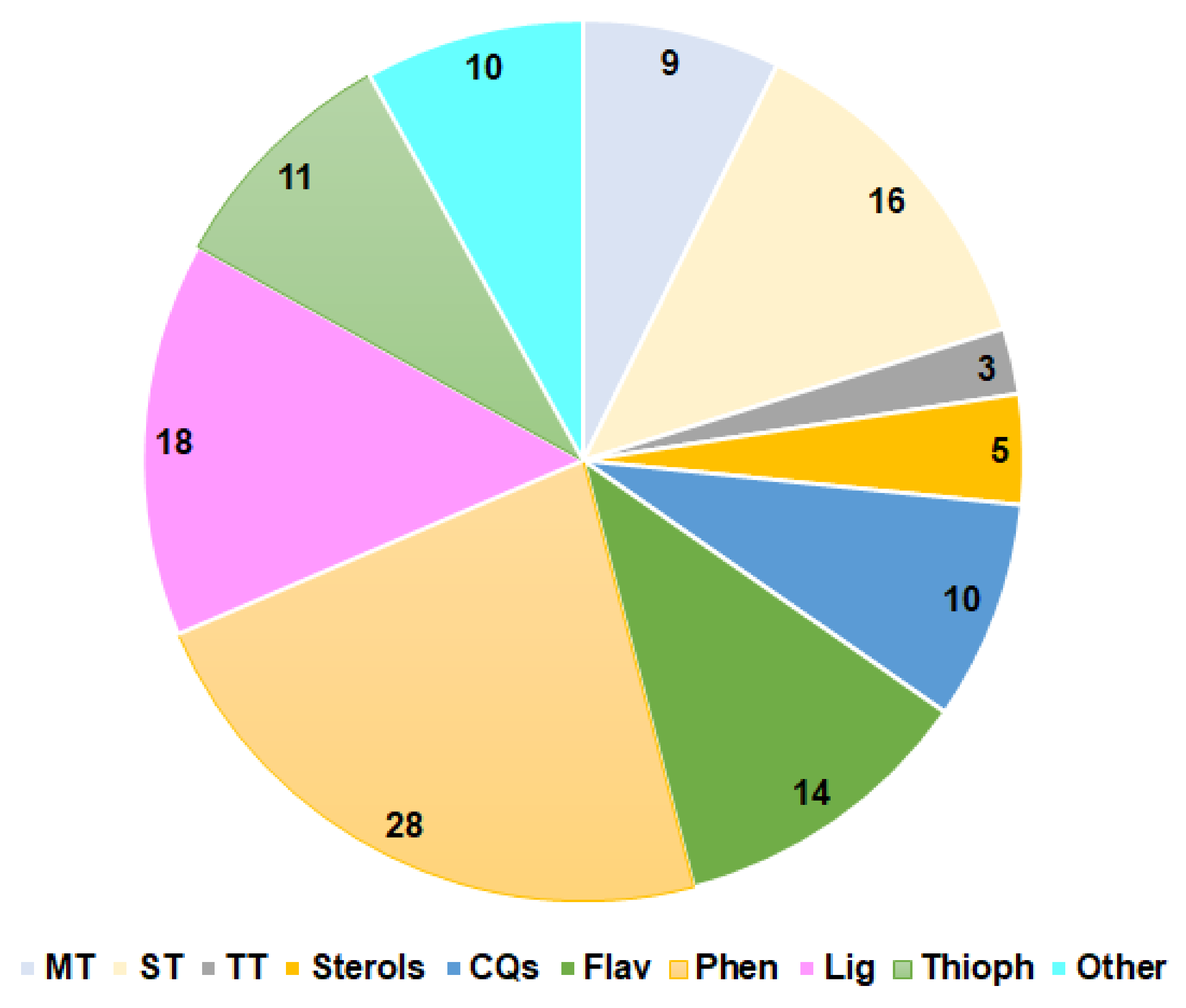
| Compound Name | Plant Part | Extract/Fraction | Mol. Wt. | Mol. Formula | City, Country | Ref. |
|---|---|---|---|---|---|---|
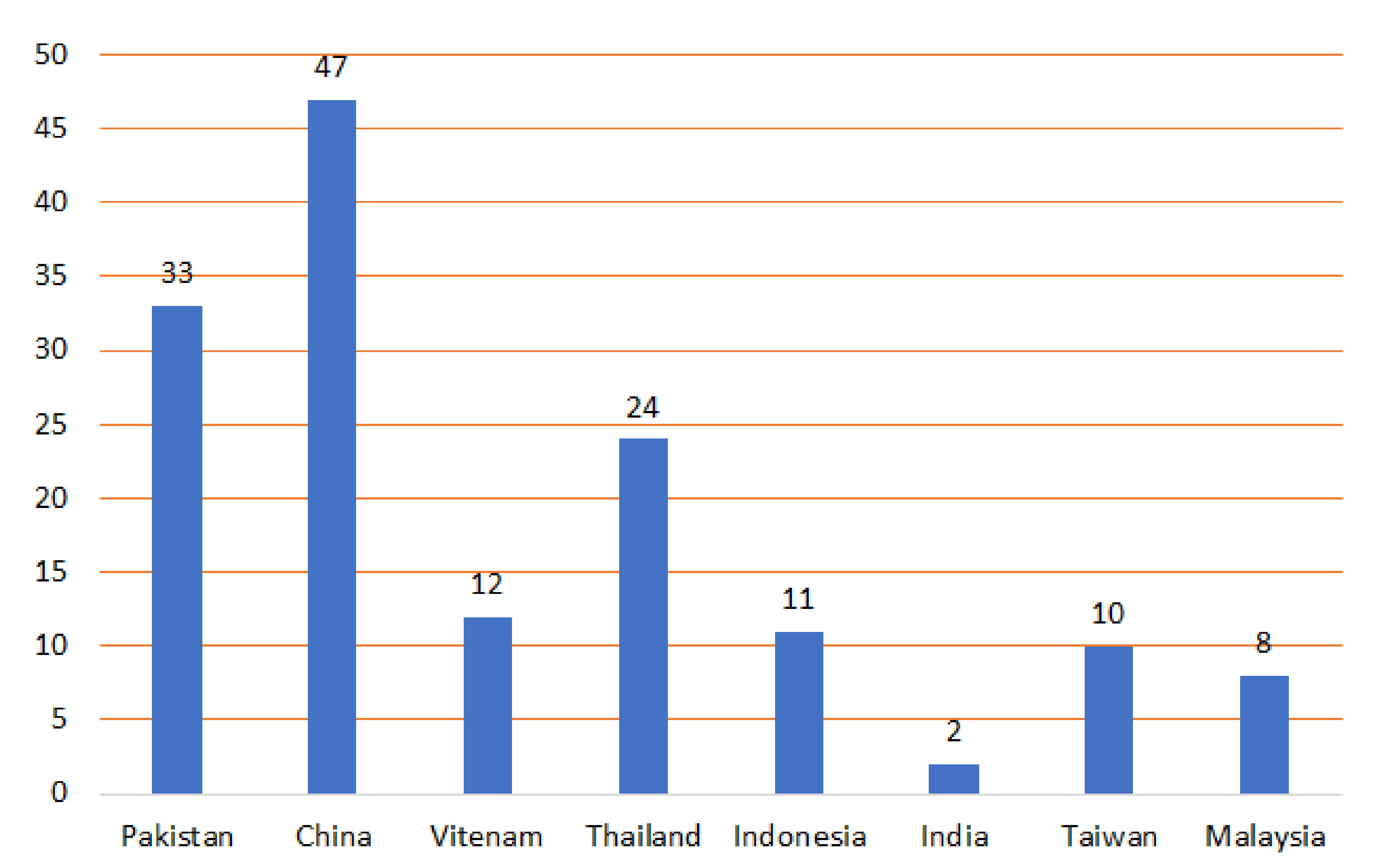
| Monoterpenes | ||||||
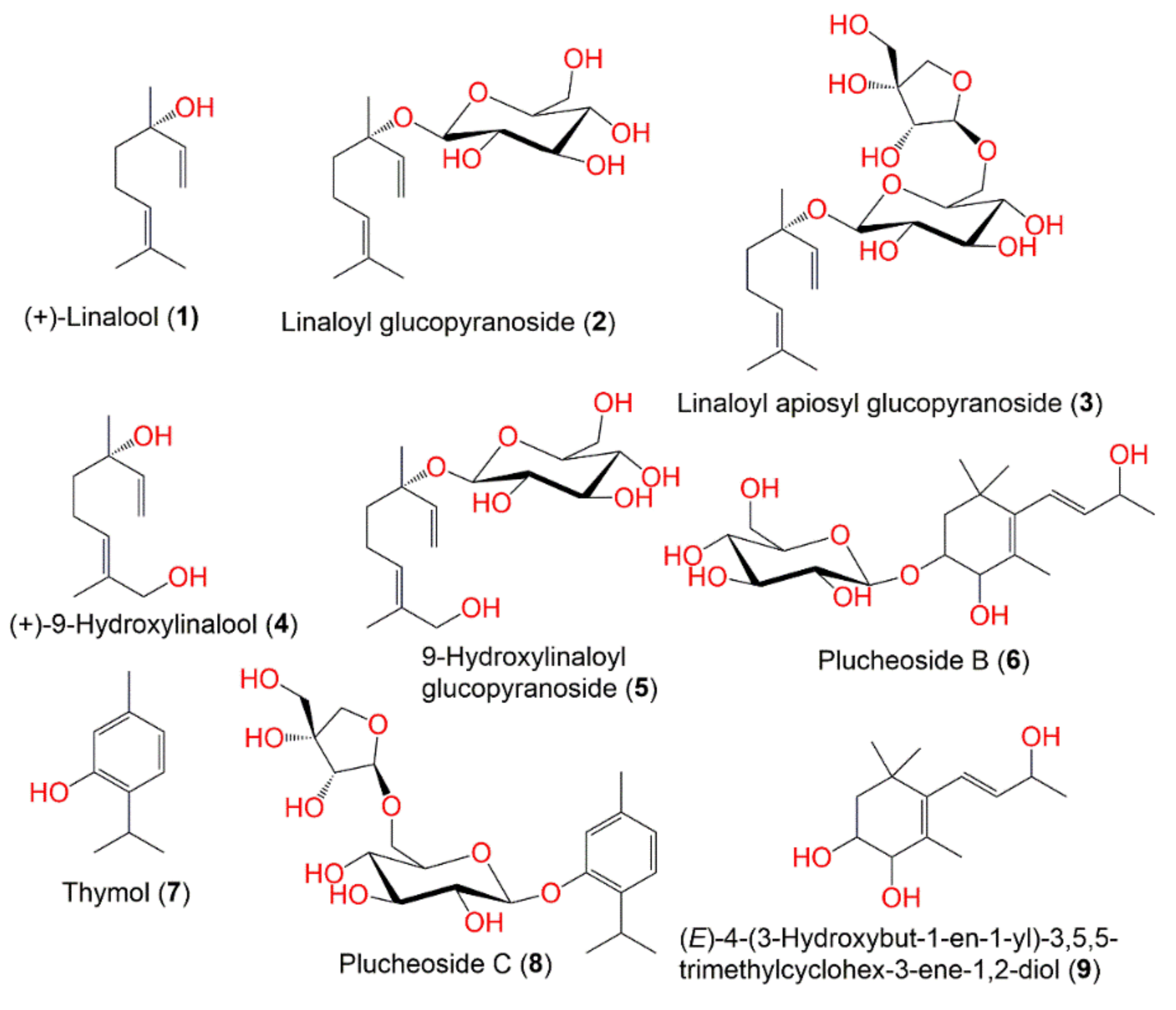
| (+)-Linalool (1) | Aerial parts | Polar fraction of MeOH extract | 154 | C10H18O | Drigh Road, Karachi, Pakistan | [47] |
| Linaloyl glucopyranoside (2) | Aerial parts | Polar fraction of MeOH extract | 316 | C16H28O6 | Drigh Road, Karachi, Pakistan | [47] |
| Linaloyl apiosyl glucopyranoside (3) | Aerial parts | Polar fraction of MeOH extract | 448 | C21H36O10 | Drigh Road, Karachi, Pakistan | [47] |
| (+)-9-Hydroxylinalool (4) | Aerial parts | Polar fraction of MeOH extract | 170 | C10H18O2 | Drigh Road, Karachi, Pakistan | [47] |
| 9-Hydroxylinaloyl glucopyranoside (5) | Aerial parts | Polar fraction of MeOH extract | 332 | C16H28O7 | Drigh Road, Karachi, Pakistan | [47] |
| Plucheoside B (6) | Aerial parts | Polar fraction of MeOH extract | 388 | C19H32O8 | Drigh Road, Karachi, Pakistan | [47] |
| Thymol (7) | Aerial parts | Polar fraction of MeOH extract | 150 | C10H14O | Drigh Road, Karachi, Pakistan | [48] |
| Plucheoside C (8) | Aerial parts | Polar fraction of MeOH extract | 444 | C21H32O10 | Drigh Road, Karachi, Pakistan | [48] |
| (E)-4-(3-Hydroxybut-1-en-1-yl)-3,5,5-trimethylcyclohex-3-ene-1,2-diol (9) | Aerial parts | Polar fraction of MeOH extract | 226 | C13H22O3 | Drigh Road, Karachi, Pakistan | [47] |
| Sesquiterpenes | ||||||
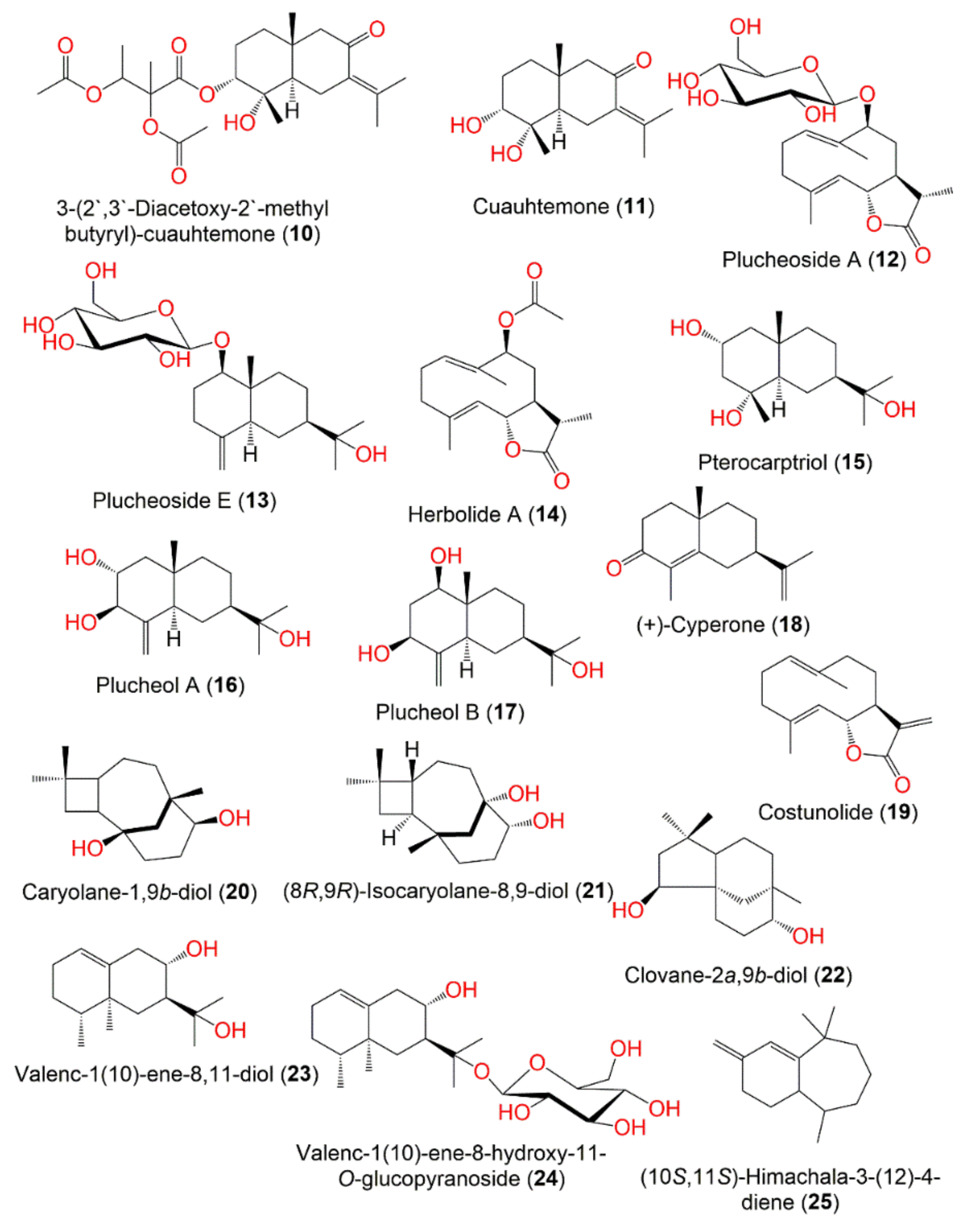
| 3-(2′,3′-Diacetoxy-2′-methyl butyryl)-cuauhtemone (10) | Leaves | CHCl3 fraction of EtOH extract | 452 | C24H36O8 | Nakornpathom, Thailand | [49] |
| Cuauhtemone (11) | Leaves | CHCl3 fraction of EtOH extract | 252 | C15H24O3 | Nakornpathom, Thailand | [49] |
| Plucheoside A (12) | Aerial parts | Polar fraction of MeOH extract | 412 | C21H32O8 | Drigh Road, Karachi, Pakistan | [47] |
| Plucheoside E (13) | Aerial parts | Polar fraction of MeOH extract | 400 | C21H36O7 | Drigh Road, Karachi, Pakistan | [48] |
| Herbolide A (14) | Aerial parts | Polar fraction of MeOH extract | 292 | C17H24O4 | Drigh Road, Karachi, Pakistan | [47] |
| Pterocarptriol (15) | Aerial parts | Polar fraction of MeOH extract | 256 | C15H28O3 | Drigh Road, Karachi, Pakistan | [48] |
| Plucheol A (16) | Aerial parts | Polar fraction of MeOH extract | 254 | C15H26O3 | Drigh Road, Karachi, Pakistan | [48] |
| Plucheol B (17) | Aerial parts | Polar fraction of MeOH extract | 254 | C15H26O3 | Drigh Road, Karachi, Pakistan | [48] |
| (+)-Cyperone (18) | Roots | CH2Cl2 fraction of MeOH extract | 218 | C15H22O | Kaohsiung, Taiwan | [50] |
| Costunolide (19) | Roots | CH2Cl2 fraction of MeOH extract | 232 | C15H20O2 | Kaohsiung, Taiwan | [50] |
| Caryolane-1,9β-diol (20) | Aerial parts | n-Hexane fraction of EtOH extract | 238 | C15H26O2 | Hepu, Guangxi, China | [51] |
| (8R,9R)-Isocaryolane-8,9-diol (21) | Aerial parts | n-Hexane fraction of EtOH extract | 238 | C15H26O2 | Hepu, Guangxi, China | [51] |
| Clovane-2α,9β-diol (22) | Aerial parts | n-Hexane fraction of EtOH extract | 238 | C15H26O2 | Hepu, Guangxi, China | [51] |
| Valenc-1(10)-ene-8,11-diol (23) | Aerial parts | n-Hexane fraction of EtOH extract | 238 | C15H26O2 | Hepu, Guangxi, China | [51] |
| Valenc-1(10)-ene-8-hydroxy-11-O-glucopyranoside (24) | Aerial parts | EtOAc fraction of MeOH extract | 400 | C21H37O7 | China | [52] |
| (10S,11S)-Himachala-3-(12)-4-diene (25) | Leaves | Essential oil | 204 | C15H24 | Sidoarjo and Surabaya, Indonesia | [43] |
| Triterpenes | ||||||
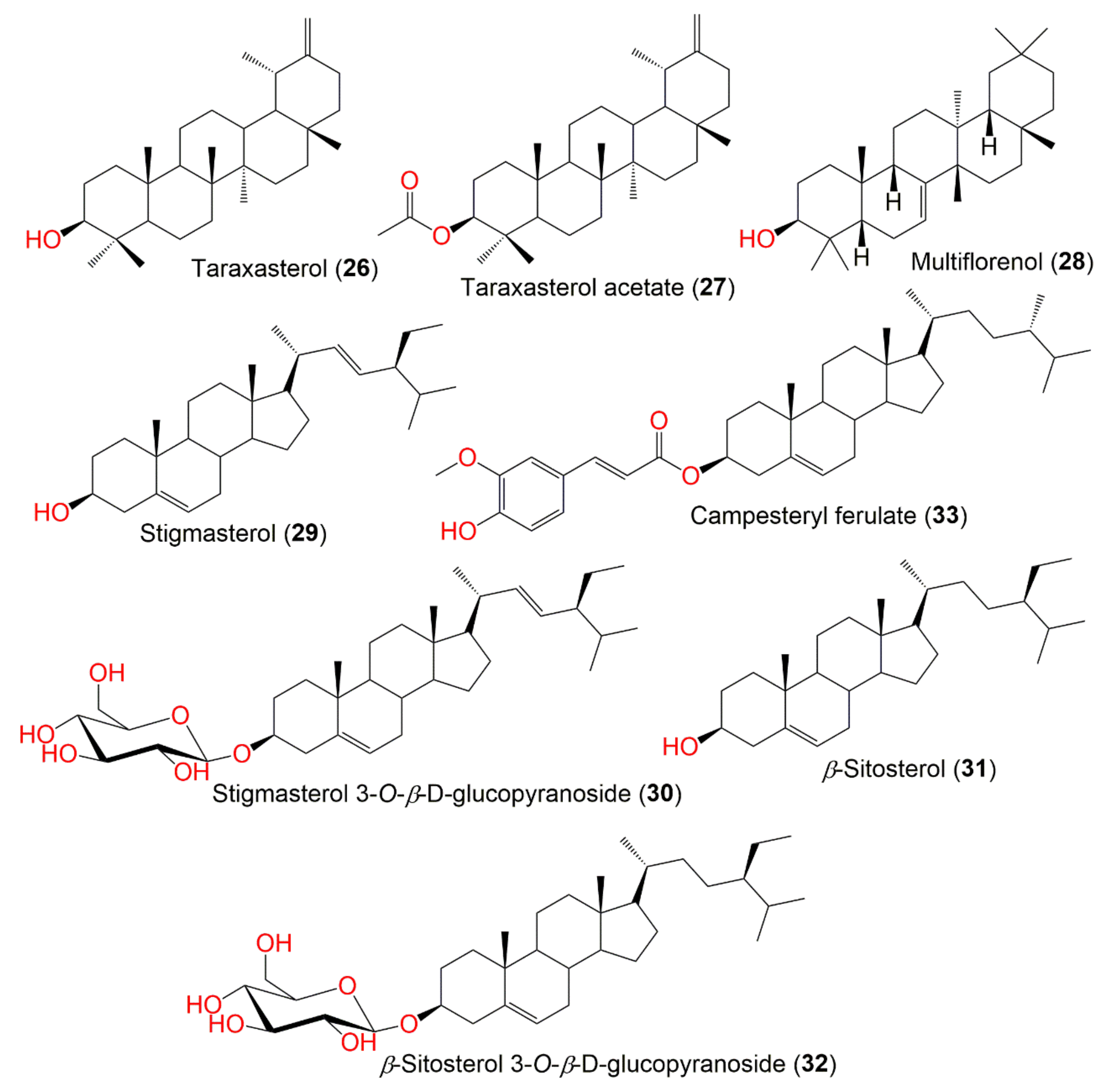
| Taraxasterol (26) | Leaves | n-Hexane fraction of MeOH extract | 426 | C30H50O | Gia Lam, Hanoi, Vietnam | [53] |
| Taraxasterol acetate (27) | Leaves | n-Hexane fraction of MeOH extract | 468 | C32H52O2 | Gia Lam, Hanoi, Vietnam | [53] |
| Multiflorenol (28) | Aerial parts | n-Hexane fraction of MeOH extract | 426 | C30H50O | China | [52] |
| Sterols | ||||||
| Stigmasterol (29) | Aerial parts | n-Hexane fraction of EtOH extract | 412 | C29H48O | Hepu, Guangxi, China | [51] |
| Twigs | n-Hexane fraction of MeOH extract | - | - | Vietnam | [54] | |
| Leaves | n-Hexane fraction of MeOH extract | 412 | C29H48O | Gia Lam, Hanoi, Vietnam | [53] | |
| Stigmasterol 3-O-β-D-glucopyranoside (30) | Aerial parts | Polar fraction of MeOH extract | 574 | C35H58O6 | Drigh Road, Karachi, Pakistan | [47] |
| Twigs | EtOAc fraction of MeOH extract | - | - | Vietnam | [54] | |
| Leaves | EtOAc fraction of MeOH extract | - | - | Gia Lam, Hanoi, Vietnam | [53] | |
| β-Sitosterol (31) | Twigs | n-Hexane fraction of MeOH extract | 414 | C29H50O | Vietnam | [54] |
| β-Sitosterol 3-O-β-D-glucopyranoside (32) | Leaves | EtOAc fraction of MeOH extract | 576 | C35H60O6 | Gia Lam, Hanoi, Vietnam | [53] |
| Campesteryl ferulate (33) | Leaves | EtOH extract | 576 | C38H56O4 | Hat Yai, Songkhla, Thailand | [45] |
| Caffeoylquinic acid derivatives | ||||||
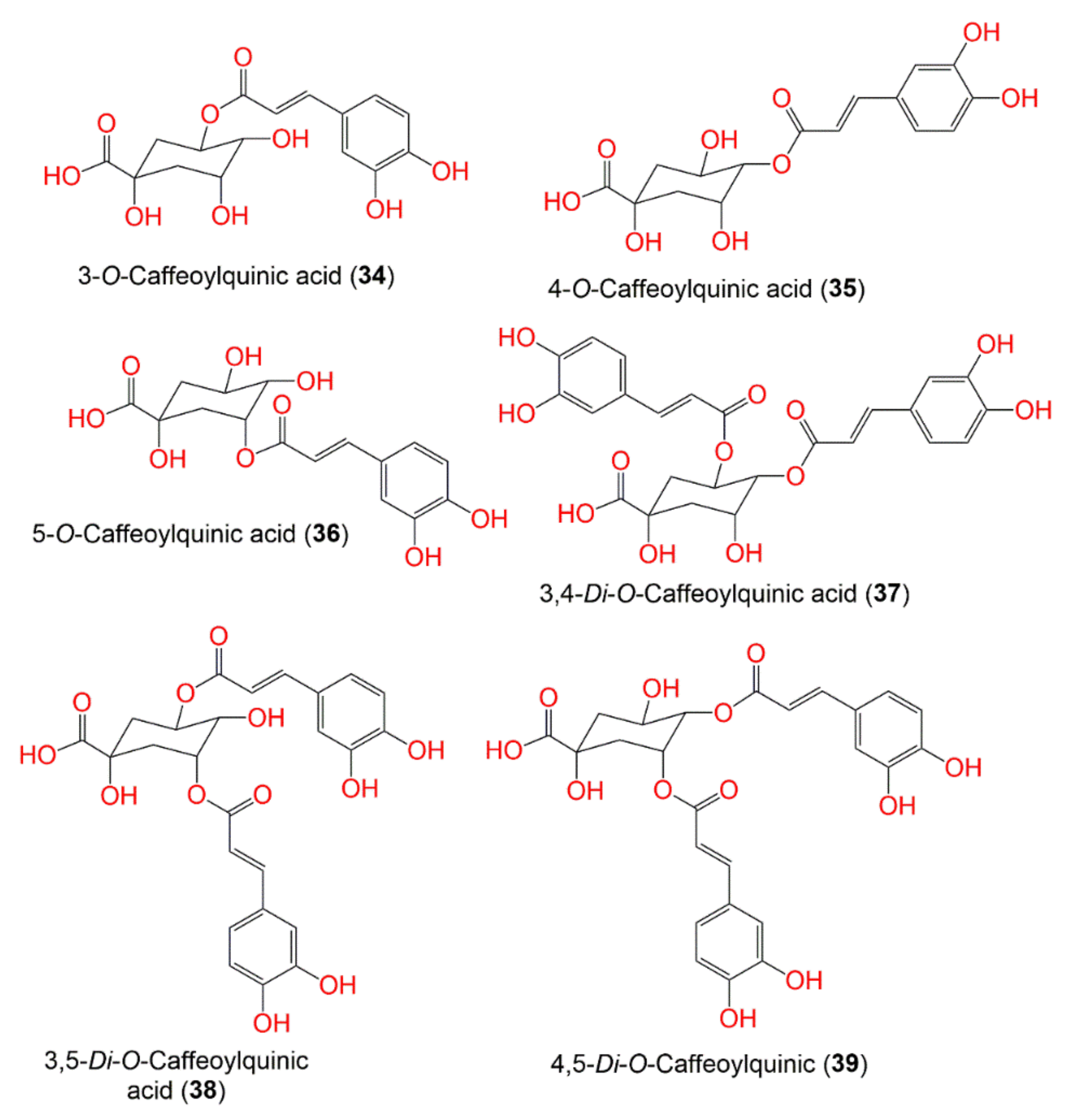
| 3-O-Caffeoylquinic acid (34) | Aerial parts | Acidified MeOH | 354 | C16H18O9 | Seberang Perai, Malaysia | [55] |
| Leaves | 50% EtOH extract | - | - | Different provinces in Thailand | [56] | |
| 4-O-Caffeoylquinic acid (35) | Leaves | 50% EtOH extract | 354 | C16H18O9 | Different provinces in Thailand | [56] |
| 5-O-Caffeoylquinic acid (36) | Aerial parts | Acidified MeOH | 354 | C16H18O9 | Seberang Perai, Malaysia | [55] |
| Leaves | 50% EtOH extract | - | - | Different provinces in Thailand | [56] | |
| 3,4-Di-O-Caffeoylquinic acid (37) | Aerial parts | Acidified MeOH | - | - | Seberang Perai, Malaysia | [55] |
| Leaves | 50% EtOH extract | 530 | C26H26O12 | Different provinces in Thailand | [56] | |
| Leaves | 50% EtOH extract | - | - | Different provinces in Thailand | [46] | |
| 3,5-Di-O-Caffeoylquinic acid (38) | Aerial parts | Acidified MeOH | 516 | C25H24O12 | Seberang Perai, Malaysia | [55] |
| Leaves | EtOAc fraction of MeOH extract | - | - | Yogyakarta, Indonesia | [57] | |
| Leaves | 50% EtOH extract | - | - | Different provinces in Thailand | [56] | |
| Leaves | 50% EtOH extract | - | - | Different provinces in Thailand | [46] | |
| 4,5-Di-O-Caffeoylquinic acid (39) | Aerial parts | Acidified MeOH | 516 | C25H24O12 | Seberang Perai, Malaysia | [55] |
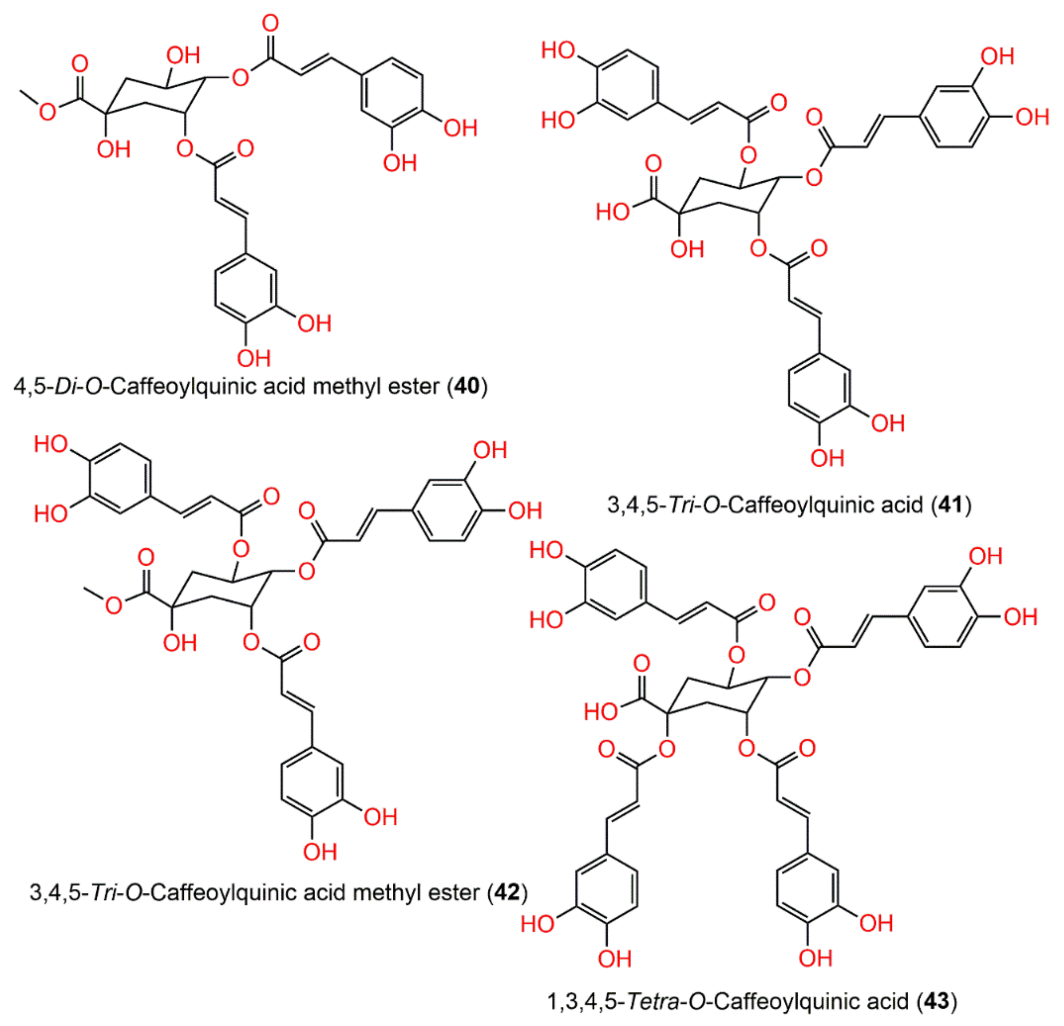
| 4,5-Di-O-caffeoylquinic acid methyl ester (40) | Leaves | EtOAc fraction of MeOH extract | 530 | C26H26O12 | Yogyakarta, Indonesia | [57] |
| Leaves | 50% EtOH extract | - | - | Different provinces in Thailand | [56] | |
| 3,4,5-Tri-O-Caffeoylquinic acid (41) | Leaves | EtOAc fraction of MeOH extract | 678 | C34H30O15 | Khon Kaen, Thailand | [58] |
| Leaves | EtOAc fraction of MeOH extract | - | - | Yogyakarta, Indonesia | [57] | |
| 3,4,5-Tri-O-Caffeoylquinic acid methyl ester (42) | Leaves | EtOAc fraction of MeOH extract | 692 | C35H32O15 | Yogyakarta, Indonesia | [57] |
| 1,3,4,5-Tetra-O-Caffeoylquinic acid (43) | Leaves | EtOAc fraction of MeOH extract | 840 | C43H36O18 | Khon Kaen, Thailand | [58] |
| Leaves | EtOAc fraction of MeOH extract | - | - | Yogyakarta, Indonesia | [57] | |
| Flavonoids | ||||||
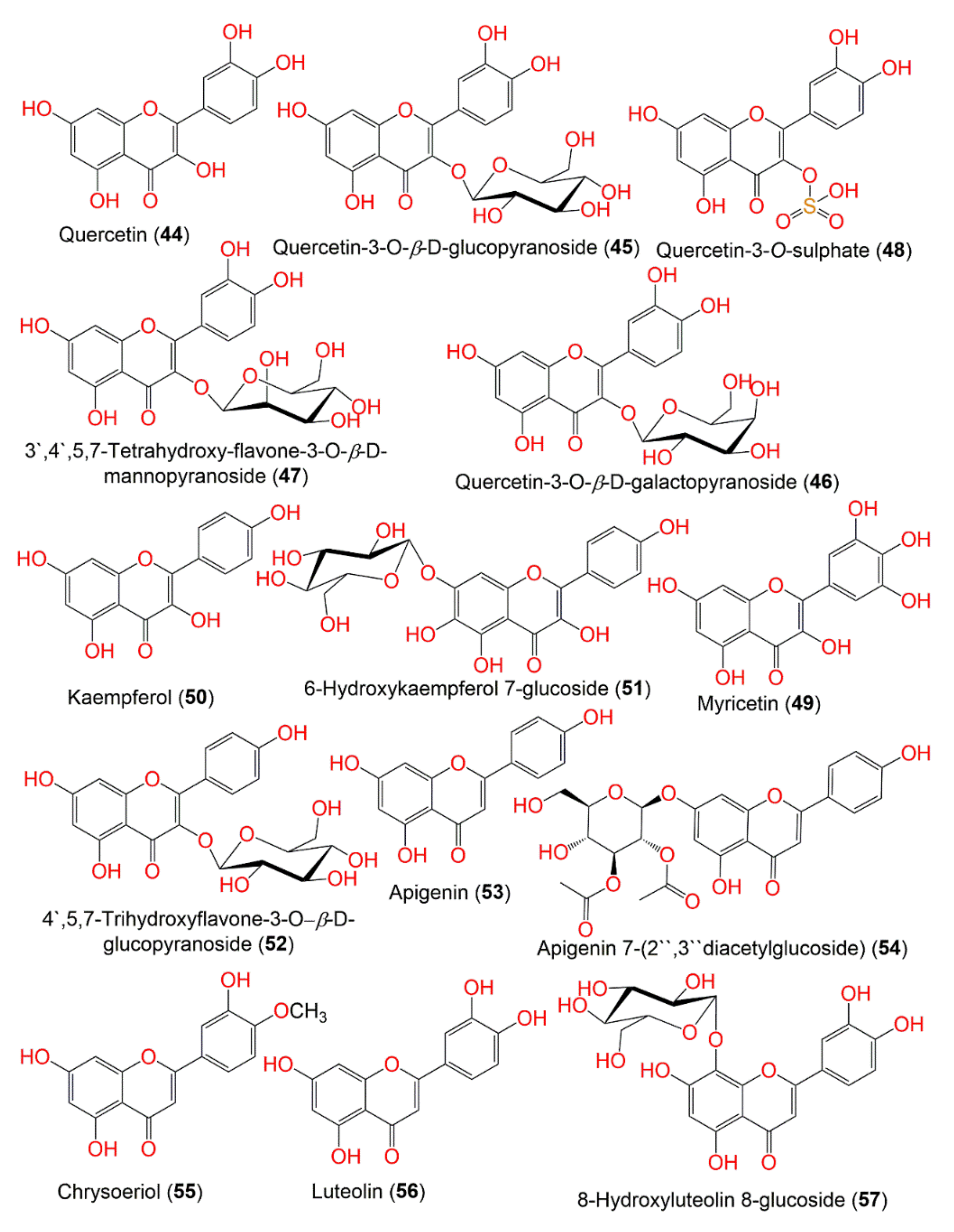
| Quercetin (44) | Leaves | EtOAc fraction of MeOH extract | 302 | C15H10O7 | Khon Kaen, Thailand | [58] |
| Aerial parts | Acidified 50% MeOH | - | - | Bogor, west Java, Indonesia | [29] | |
| Aerial parts | EtOAc fraction of MeOH extract | - | - | Chantaburi, Thailand | [59] | |
| Quercetin-3-O-β-D-glucopyranoside (45) | Aerial parts | EtOAc fraction of MeOH extract | 464 | C21H20O12 | China | [52] |
| Aerial parts | Acidified MeOH | - | - | Seberang Perai, Malaysia | [55] | |
| Quercetin-3-O-β-D-galactopyranoside (46) | Aerial parts | Acidified MeOH | 464 | C21H20O12 | Seberang Perai, Malaysia | [55] |
| 3′,4′,5,7-Tetrahydroxy-flavone-3-O-β-D-mannopyranoside (47) | Aerial parts | EtOAc fraction of MeOH extract | 464 | C21H20O12 | China | [52] |
| Quercetin-3-O-sulphate (48) | Aerial parts | Acidified MeOH | 382 | C15H10O10S | Seberang Perai, Malaysia | [55] |
| Myricetin (49) | Aerial parts | Acidified 50% MeOH | 318 | C15H10O8 | Bogor, west Java, Indonesia | [29] |
| Kaempferol (50) | Aerial parts | Acidified 50% MeOH | 286 | C15H10O6 | Bogor, west Java, Indonesia | [29] |
| 6-Hydroxykaempferol 7-glucoside (51) | 464 | C20H20O12 | Hat Yai, Songkhla, Thailand | [45] | ||
| 4′,5,7-Trihydroxyflavone-3-O-β-D-glucoside (52) | Aerial parts | EtOAc fraction of MeOH extract | 448 | C21H20O11 | China | [52] |
| Apigenin (53) | Aerial parts | Acidified 50% MeOH | 270 | C15H10O5 | Bogor, west Java, Indonesia | [29] |
| Aerial parts | EtOAc fraction of MeOH extract | - | - | Chantaburi, Thailand | [59] | |
| Apigenin 7-(2″,3″diacetylglucoside (54) | [45] | |||||
| Chrysoeriol (55) | Aerial parts | EtOAc fraction of MeOH extract | 300 | C16H12O6 | Chantaburi, Thailand | [59] |
| Luteolin (56) | Aerial parts | Acidified 50% MeOH | 286 | C15H10O6 | Bogor, west Java, Indonesia | [29] |
| Aerial parts | EtOAc fraction of MeOH extract | - | - | Chantaburi, Thailand | [59] | |
| 8-Hydroxyluteolin 8-glucoside (57) | Leaves | EtOH extract | 464 | C20H20O12 | Hat Yai, Songkhla, Thailand | [45] |
| Phenolic acids, aldehydes, esters, and ketones | ||||||
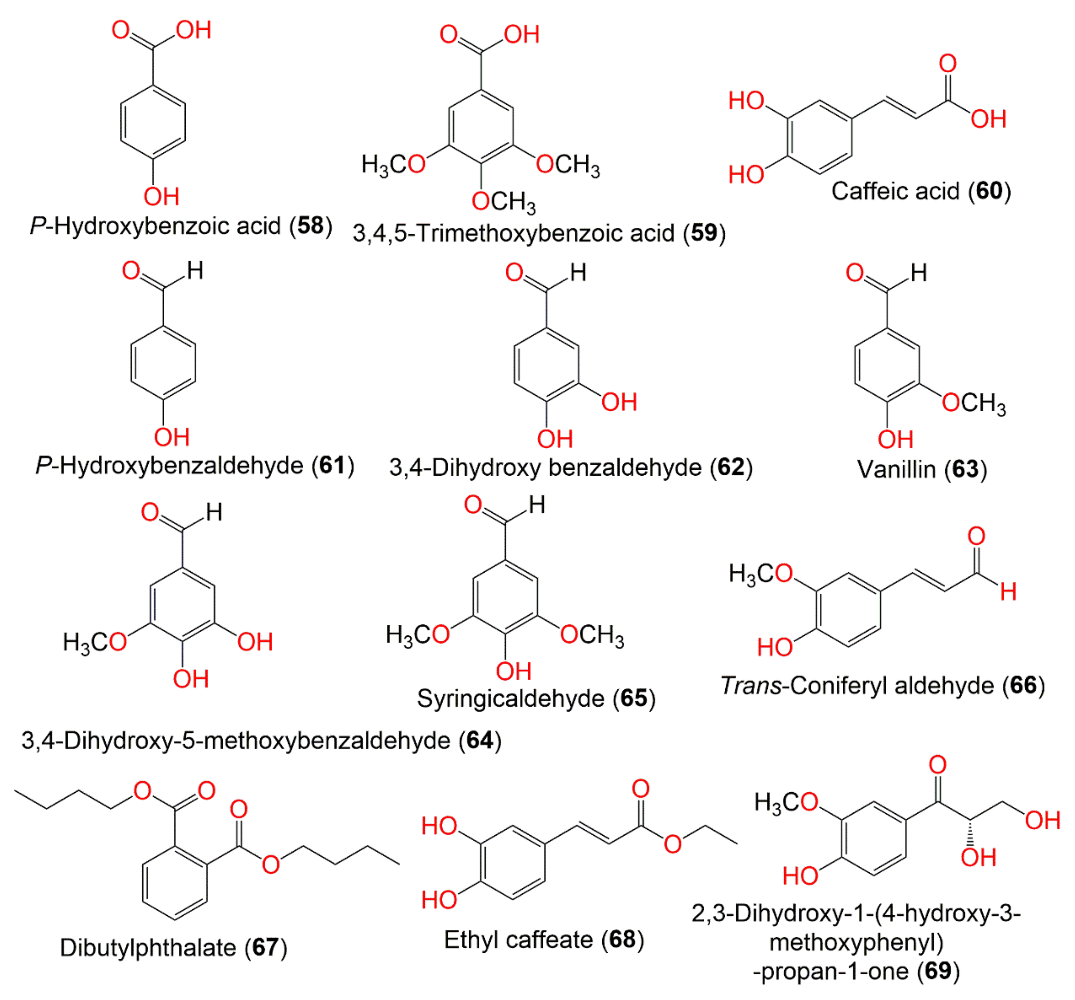
| P-Hydroxybenzoic acid (58) | Aerial parts | CHCl3 fraction of EtOH extract | 138 | C7H6O3 | Hepu, Guangxi, China | [51] |
| 3,4,5-Trimethoxybenzoic acid (59) | Roots | CH2Cl2 fraction of MeOH extract | 212 | C10H12O5 | Kaohsiung, Taiwan | [50] |
| Caffeic acid (60) | Aerial parts | EtOAc fraction of MeOH extract | 180 | C9H8O4 | China | [52] |
| P-Hydroxybenzaldehyde (61) | Roots | CH2Cl2 fraction of MeOH extract | 122 | C7H6O2 | Kaohsiung, Taiwan | [50] |
| 3,4-Dihydroxy benzaldehyde (62) | Aerial parts | CHCl3 fraction of EtOH extract | 138 | C7H6O3 | Hepu, Guangxi, China | [51] |
| Vanillin (63) | Aerial parts | CHCl3 fraction of EtOH extract | 152 | C8H8O3 | Hepu, Guangxi, China | [51] |
| 3,4-Dihydroxy-5-methoxybenzaldehyde (64) | Aerial parts | EtOAc fraction of MeOH extract | 168 | C8H8O4 | China | [52] |
| Aerial parts | CHCl3 fraction of EtOH extract | - | - | Hepu, Guangxi, China | [51] | |
| Syringicaldehyde (65) | Aerial parts | CHCl3 fraction of EtOH extract | 182 | C9H10O4 | Hepu, Guangxi, China | [51] |
| Trans-Coniferyl aldehyde (66) | Aerial parts | CHCl3 fraction of EtOH extract | 178 | C10H10O3 | Hepu, Guangxi, China | [51] |
| Dibutylphthalate (67) | Aerial parts | CHCl3 fraction of EtOH extract | 278 | C16H22O4 | Hepu, Guangxi, China | [51] |
| Ethyl caffeate (68) | Aerial parts | CHCl3 fraction of EtOH extract | 208 | C11H12O4 | Hepu, Guangxi, China | [51] |
| 2,3-Dihydroxy-1-(4-hydroxy-3-methoxyphenyl)-propan-1-one (69) | Aerial parts | CHCl3 fraction of EtOH extract | 212 | C10H12O5 | Hepu, Guangxi, China | [51] |
| Phenolics and phenolic glucosides | ||||||
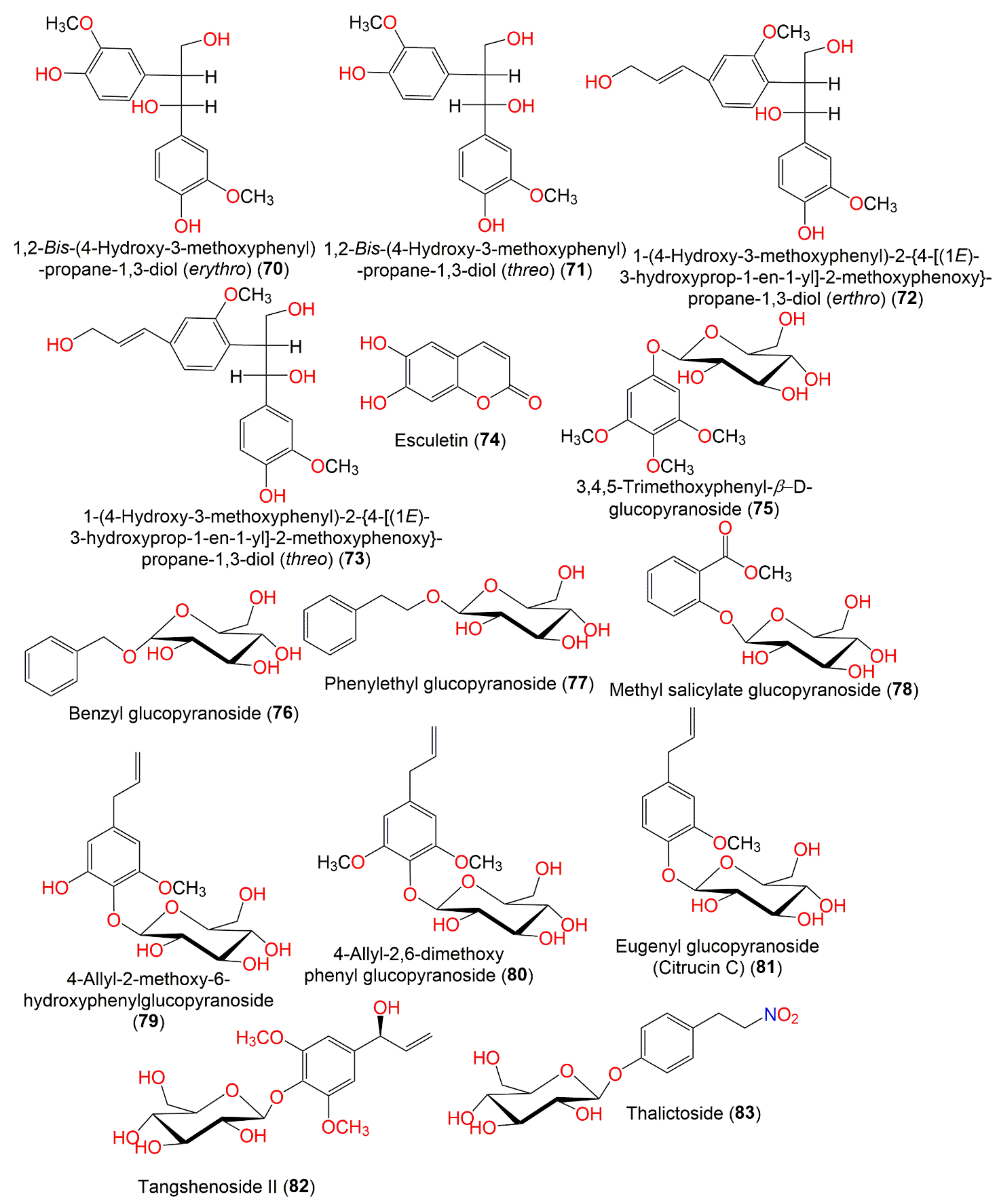
| 1,2-Bis-(4-Hydroxy-3-methoxyphenyl)-propane-1,3-diol (erythro) (70) | Aerial parts | Polar fraction of MeOH extract | 320 | C17H20O6 | Drigh Road, Karachi, Pakistan | [47] |
| 1,2-Bis-(4-Hydroxy-3-methoxyphenyl)-propane-1,3-diol (threo) (71) | Aerial parts | Polar fraction of MeOH extract | 320 | C17H20O6 | Drigh Road, Karachi, Pakistan | [47] |
| 1-(4-Hydroxy-3-methoxyphenyl)-2-{4-[(1E)-3-hydroxyprop-1-en-1-yl]-2-methoxyphenoxy}-propane-1,3-diol (erythro) (72) | Aerial parts | Polar fraction of MeOH extract | 360 | C20H24O6 | Drigh Road, Karachi, Pakistan | [47] |
| 1-(4-Hydroxy-3-methoxyphenyl)-2-{4-[(1E)-3-hydroxyprop-1-en-1-yl]-2-methoxyphenoxy}-propane-1,3-diol (threo) (73) | Aerial parts | Polar fraction of MeOH extract | 360 | C20H24O6 | Drigh Road, Karachi, Pakistan | [47] |
| Esculetin (74) | Aerial parts | CHCl3 fraction of EtOH extract | 178 | C9H6O4 | Hepu, Guangxi, China | [51] |
| 3,4,5-Trimethoxyphenyl-β-D-glucopyranoside (75) | Roots | CH2Cl2 fraction of MeOH extract | 346 | C15H22O9 | Kaohsiung, Taiwan | [50] |
| Benzyl glucopyranoside (76) | Aerial parts | Polar fraction of MeOH extract | 270 | C13H18O6 | Drigh Road, Karachi, Pakistan | [47] |
| Phenylethyl glucopyranoside (77) | Aerial parts | Polar fraction of MeOH extract | 284 | C14H20O6 | Drigh Road, Karachi, Pakistan | [47] |
| Methyl salicylate glucoside (78) | Aerial parts | Polar fraction of MeOH extract | 314 | C14H18O8 | Drigh Road, Karachi, Pakistan | [47] |
| 4-Allyl-2-methoxy-6-hydroxyphenylglucoside (79) | Aerial parts | EtOAc fraction of MeOH extract | 342 | C16H22O8 | China | [52] |
| 4-Allyl-2,6-dimethoxy phenyl glucopyranoside (80) | Aerial parts | Polar fraction of MeOH extract | 356 | C17H24O8 | Drigh Road, Karachi, Pakistan | [47] |
| Eugenyl glucoside (Citrucin C) (81) | Aerial parts | Polar fraction of MeOH extract | 326 | C16H22O7 | Drigh Road, Karachi, Pakistan | [47] |
| Tangshenoside Ⅱ (82) | Aerial parts | EtOAc fraction of MeOH extract | 372 | C17H24O9 | China | [52] |
| Thalictoside (83) | Roots | CH2Cl2 fraction of MeOH extract | 329 | C14H19NO8 | Kaohsiung, Taiwan | [50] |
| Lignans and their derivatives | ||||||
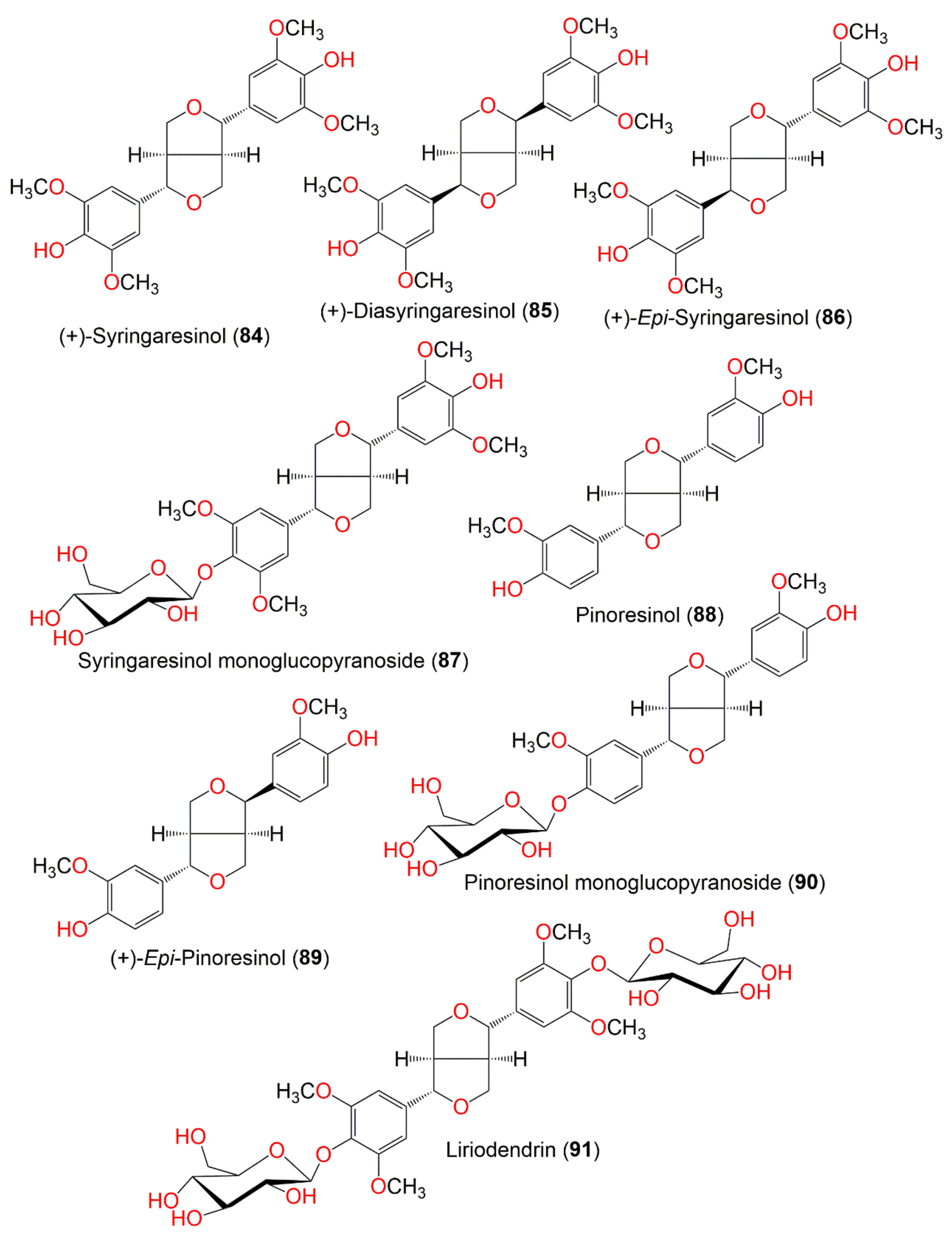
| (+)-Syringaresinol (84) | Roots | CH2Cl2 fraction of MeOH extract | 418 | C22H26O8 | Kaohsiung, Taiwan | [50] |
| (+)-Diasyringaresinol (85) | Roots | CH2Cl2 fraction of MeOH extract | 418 | C22H26O8 | Kaohsiung, Taiwan | [50] |
| Aerial parts | CHCl3fraction of EtOH extract | - | - | Hepu, Guangxi, China | [51] | |
| (+)-Epi-Syringaresinol (86) | Roots | CH2Cl2 fraction of MeOH extract | 418 | C22H26O8 | Kaohsiung, Taiwan | [50] |
| Syringaresinol monoglucopyranoside (87) | Aerial parts | Polar fraction of MeOH extract | 580 | C28H36O13 | Drigh Road, Karachi, Pakistan | [47] |
| Pinoresinol (88) | Aerial parts | EtOAc fraction of MeOH extract | 358 | C20H22O6 | China | [52] |
| (+)-Epi-Pinoresinol (89) | Aerial parts | EtOAc fraction of MeOH extract | 358 | C20H22O6 | China | [52] |
| Pinoresinol monoglucopyranoside (90) | Aerial parts | Polar fraction of MeOH extract | 520 | C26H32O11 | Drigh Road, Karachi, Pakistan | [47] |
| Liriodendrin (91) | Roots | CH2Cl2 fraction of MeOH extract | 742 | C34H46O18 | Kaohsiung, Taiwan | [50] |
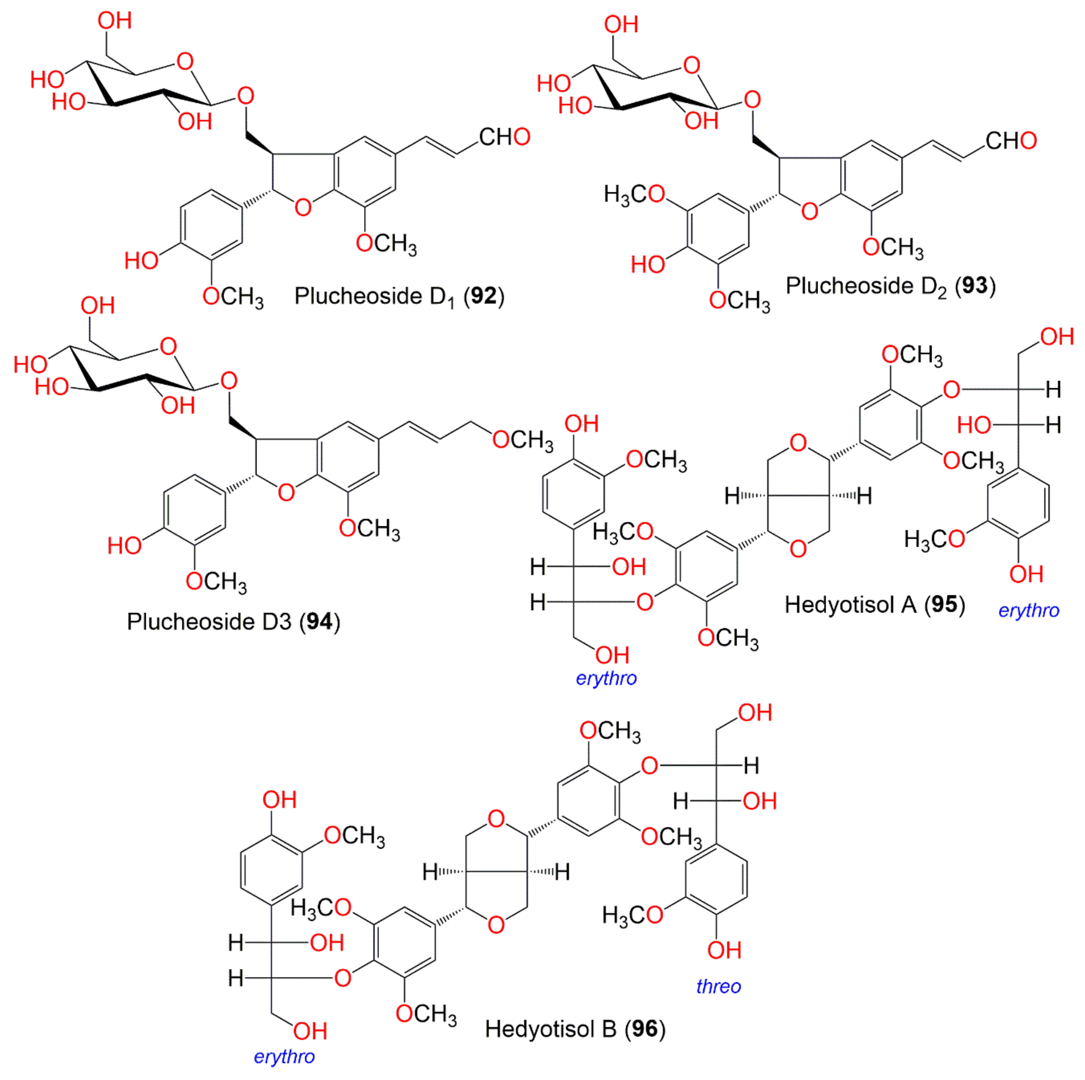
| Plucheoside D1 (92) | Aerial parts | Polar fraction of MeOH extract | 518 | C26H30O11 | Drigh Road, Karachi, Pakistan | [48] |
| Plucheoside D2 (93) | Aerial parts | Polar fraction of MeOH extract | 548 | C27H32O12 | Drigh Road, Karachi, Pakistan | [48] |
| Plucheoside D3 (94) | Aerial parts | Polar fraction of MeOH extract | 534 | C27H34O11 | Drigh Road, Karachi, Pakistan | [48] |
| Hedyotisol A (95) | Aerial parts | Polar fraction of MeOH extract | 810 | C42H50O16 | Drigh Road, Karachi, Pakistan | [47] |
| Hedyotisol B (96) | Aerial parts | Polar fraction of MeOH extract | 810 | C42H50O16 | Drigh Road, Karachi, Pakistan | [47] |
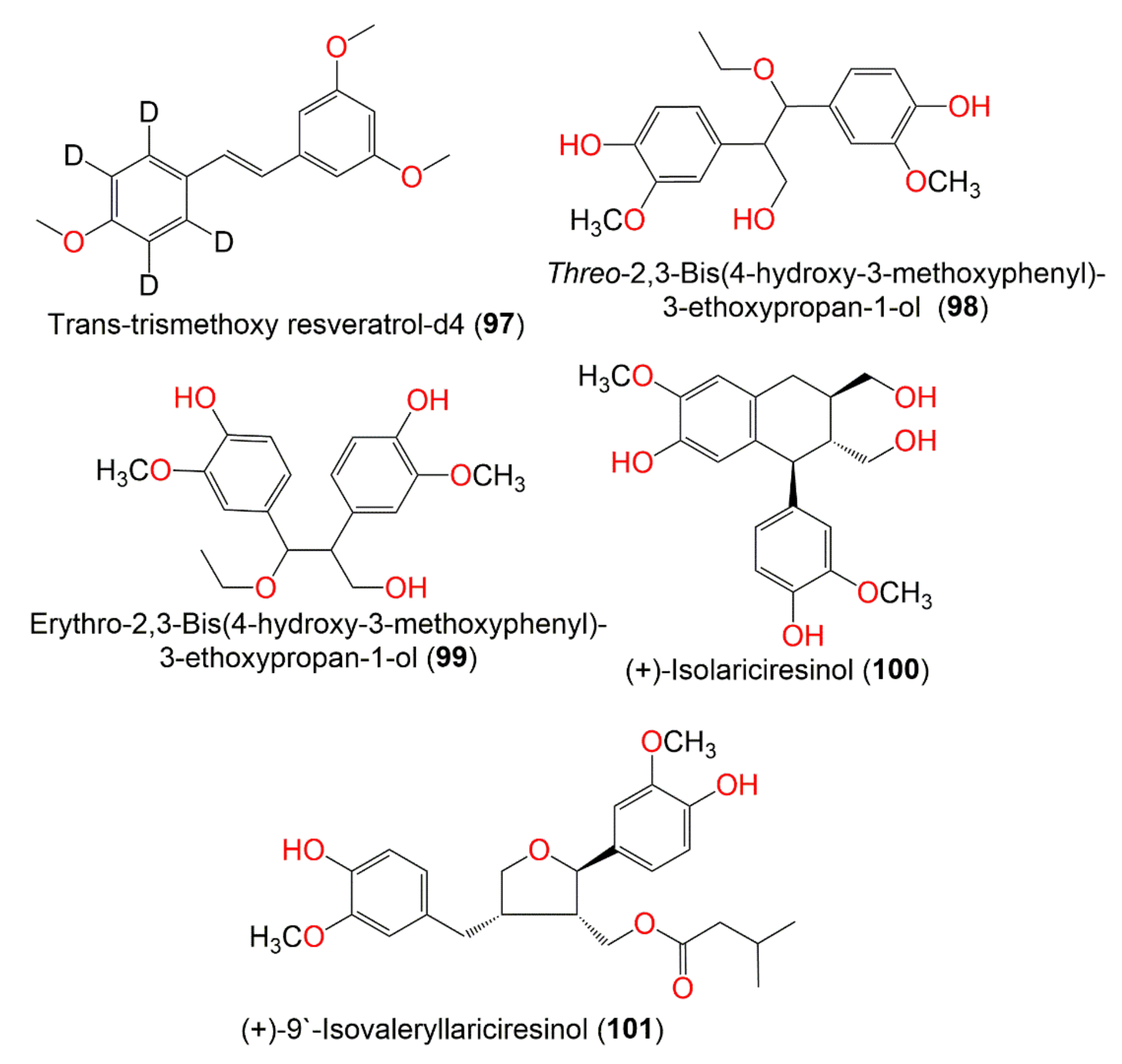
| Trans-Trismethoxy resveratrol-d4 (97) | Leaves | EtOH extract | 274 | C17H14D4O3 | Hat Yai, Songkhla, Thailand | [45] |
| Threo-2,3-Bis(4-hydroxy-3-methoxyphenyl)-3-ethoxypropan-1-ol (98) | Aerial parts | EtOAc fraction of EtOH extract | 348 | C19H24O6 | Hepu, Guangxi, China | [51] |
| Erythro-2,3-Bis(4-hydroxy-3-methoxyphenyl)-3-ethoxypropan-1-ol (99) | Aerial parts | EtOAc fraction of EtOH extract | 348 | C19H24O6 | Hepu, Guangxi, China | [51] |
| (+)-Isolariciresinol (100) | Aerial parts | EtOAc fraction of EtOH extract | 360 | C20H24O6 | Hepu, Guangxi, China | [51] |
| (+)-9′-Isovaleryllariciresinol (101) | Aerial parts | EtOAc fraction of EtOH extract | 444 | C25H32O7 | Hepu, Guangxi, China | [51] |
| Thiophenes and their derivatives | ||||||
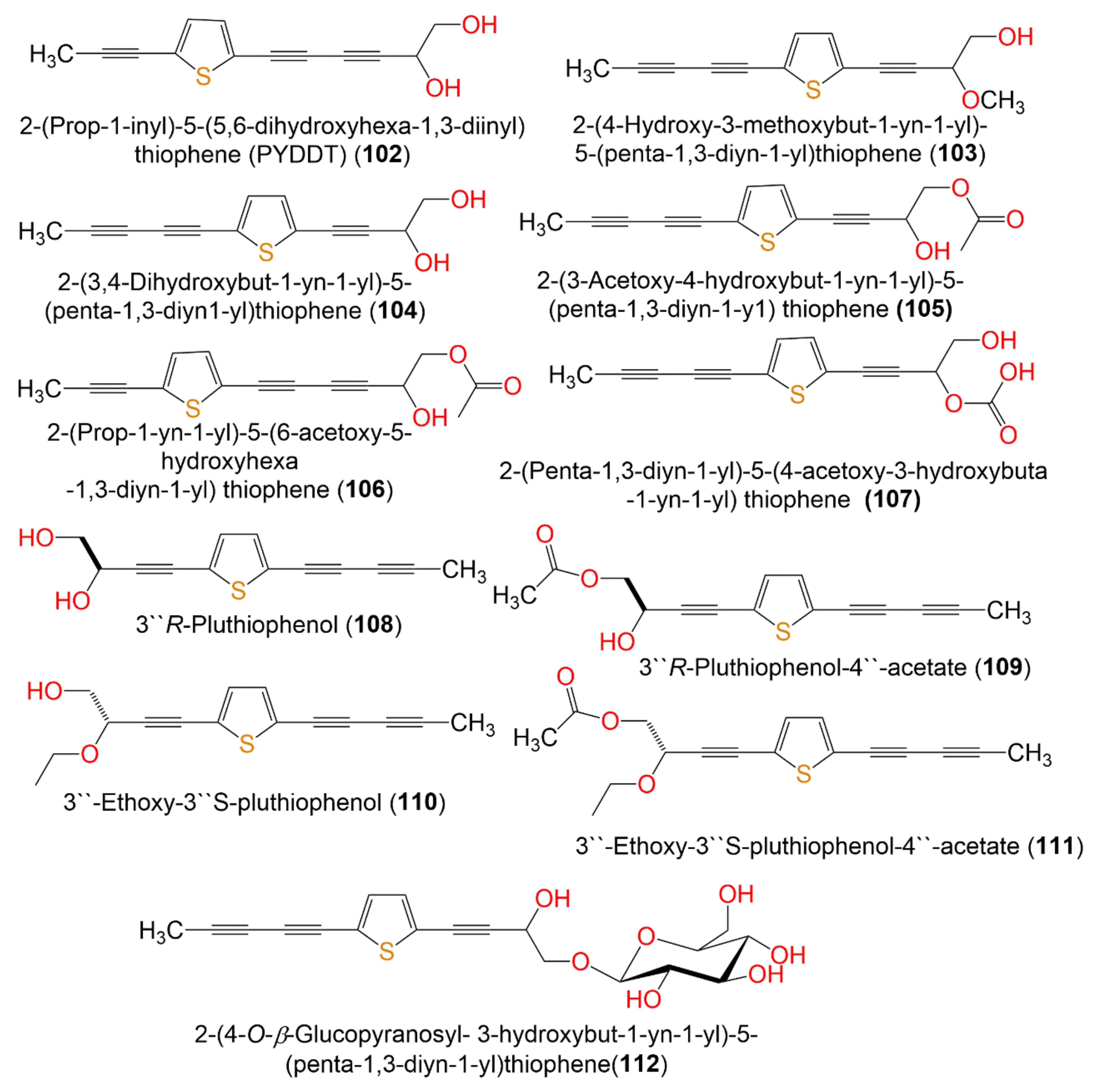
| 2-(Pro-1-ynyl)-5-(5,6-dihydroxypenta-1,3-diynyl) thiophene (PYDDT) = 2-(Prop-1-ynyl)-5(5,6-dihydroxyhexa-1, 3-diynyl)-thiophene = PITC-2 = R/J/3 (102) | Roots | EtOAc fraction of MeOH extract | 230 | C13H10O2S | West Bengal, India | [60] |
| Aerial parts | n-Hexane fraction of MeOH extract | - | - | Chantaburi, Thailand | [59] | |
| Leaves | n-Hexane fraction of MeOH extract | - | - | Gia Lam, Hanoi, Vietnam | [53] | |
| Roots | EtOAc fraction of MeOH extract | - | - | West Bengal, India | [61] | |
| Twigs | n-Hexane fraction of MeOH extract | - | - | Vietnam | [54] | |
| 2-(4-Hydroxy-3-methoxybut-1-yn-1-yl)-5-(penta-1,3-diyn-1-yl)thiophene (103) | Aerial parts | EtOAc fraction of MeOH extract | 244 | C14H12O2S | Guangzhou, China | [62] |
| 2-(3,4-Dihydroxybut-1-yn-1-yl)-5-(penta-1,3-diyn1-yl)thiophene (104) | Aerial parts | EtOAc fraction of MeOH extract | 230 | C13H10O2S | Guangzhou, China | [62] |
| 2-(3-Acetoxy-4-hydroxybut-1-yn-1-yl)-5-(penta-1,3-diyn-1-y1) thiophene (105) | Aerial parts | EtOAc fraction of MeOH extract | 272 | C15H12O3S | Guangzhou, China | [62] |
| 2-(Prop-1-yn-1-yl)-5-(6-acetoxy-5-hydroxyhexa-1,3-diyn-1-yl) thiophene = 2-(Prop-1-inyl)-5-(6-acetoxy-5-hydroxyhexa-1,3-diinyl) thiophene (106) | Aerial parts | EtOAc fraction of MeOH extract | 272 | C15H12O3S | Guangzhou, China | [62] |
| Aerial parts | n-Hexane fraction of MeOH extract | - | - | Chantaburi, Thailand | [59] | |
| 2-(Penta-1,3-diyn-1-yl)-5-(4-acetoxy-3-hydroxybuta-1-yn-1-yl) thiophene (107) | Aerial parts | n-Hexane fraction of MeOH extract | 272 | C15H12O3S | Chantaburi, Thailand | [59] |
| 3″R-Pluthiophenol (108) | Aerial parts | n-Hexane fraction of EtOH extract | 230 | C13H10O2S | Hepu, Guangxi, China | [51] |
| 3″R-Pluthiophenol-4″-acetate (109) | Aerial parts | n-Hexane fraction of EtOH extract | 272 | C15H12O3S | Hepu, Guangxi, China | [51] |
| 3″-Ethoxy-3″S-pluthiophenol (110) | Aerial parts | n-Hexane fraction of EtOH extract | 258 | C15H14O2S | Hepu, Guangxi, China | [51] |
| 3″-Ethoxy-3″S-pluthiophenol-4″-acetate (111) | Aerial parts | n-Hexane fraction of EtOH extract | 300 | C17H16O3S | Hepu, Guangxi, China | [51] |
| 2-(4-O-β-Glucopyranosyl-3-hydroxybut-1-yn-1-yl)-5-(penta-1,3-diyn-1-yl)thiophene (112) | Aerial parts | EtOAc fraction of MeOH extract | 392 | C19H20O7S | Guangzhou, China | [62] |
| Other metabolites | ||||||
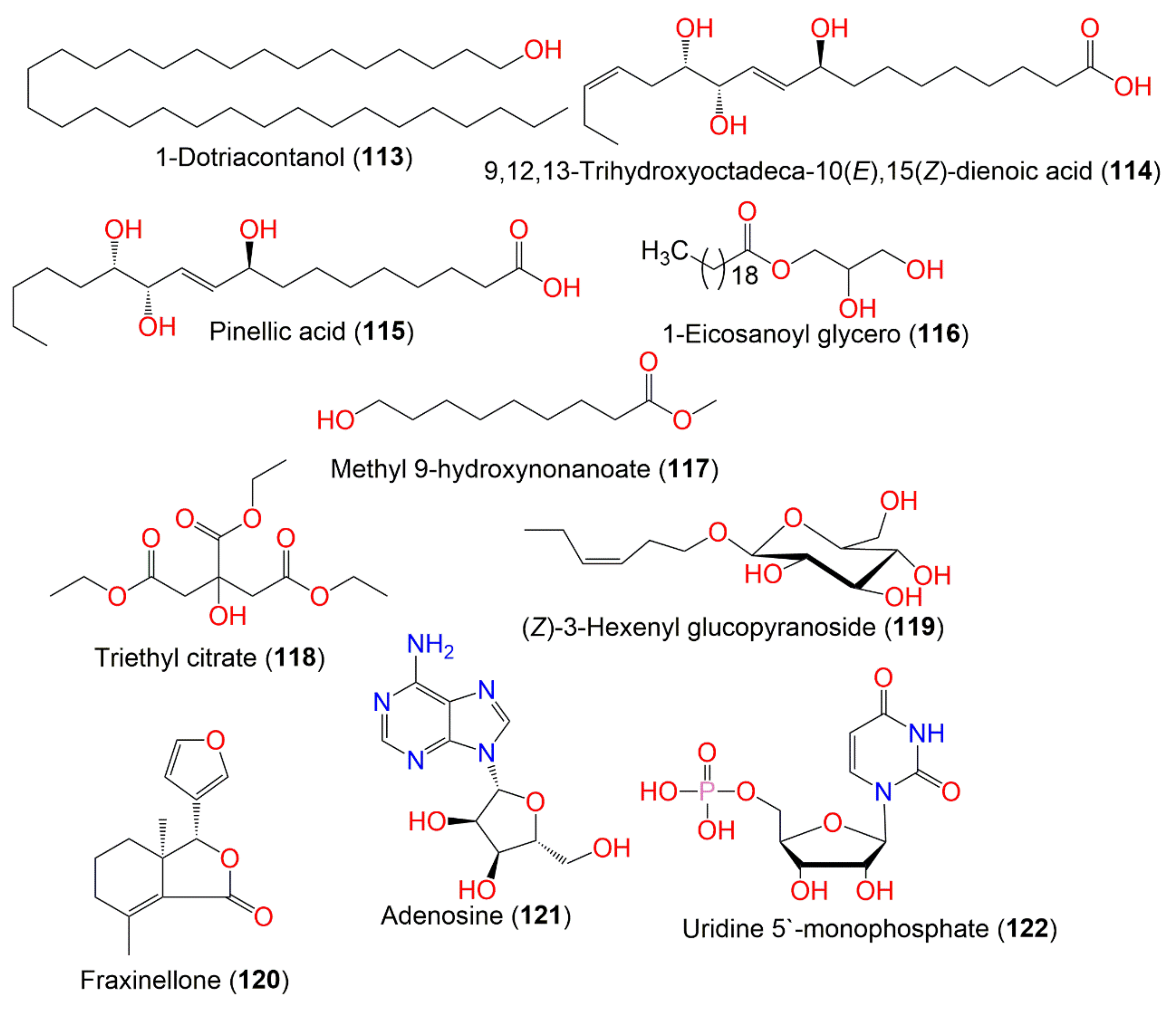
| 1-Dotriacontanol (113) | Leaves | n-Hexane fraction of MeOH extract | 466 | C32H66O | Gia Lam, Hanoi, Vietnam | [53] |
| 9,12,13-Trihydroxyoctadeca-10(E),15(Z)-dienoic acid (114) | Aerial parts | n-Hexane fraction of EtOH extract | 328 | C18H32O5 | Hepu, Guangxi, China | [51] |
| Pinellic acid (115) | Aerial parts | n-Hexane fraction of EtOH extract | 330 | C18H34O5 | Hepu, Guangxi, China | [51] |
| 1-Eicosanoyl glycerol (116) | Twigs | n-Hexane fraction of MeOH extract | 386 | C23H46O4 | Vietnam | [54] |
| Methyl 9-hydroxynonanoate (117) | Aerial parts | n-Hexane fraction of EtOH extract | 188 | C10H20O3 | Hepu, Guangxi, China | [51] |
| Triethyl citrate (118) | Aerial parts | n-Hexane fraction of EtOH extract | 276 | C12H20O7 | Hepu, Guangxi, China | [51] |
| (Z)-3-Hexenyl glucopyranoside (119) | Aerial parts | Polar fraction of MeOH extract | 262 | C12H22O6 | Drigh Road, Karachi, Pakistan | [47] |
| Fraxinellone (120) | Aerial parts | n-Hexane fraction of EtOH extract | 232 | C14H16O3 | Hepu, Guangxi, China | [51] |
| Adenosine (121) | Aerial parts | EtOAc fraction of EtOH extract | 267 | C10H13N5O4 | Hepu, Guangxi, China | [51] |
| Uridine 5′-monophosphate (122) | Aerial parts | EtOAc fraction of MeOH extract | 324 | C9H13N2O9P | China | [52] |
| Compound Name | Biological Activity | Assay, Organism, or Cell Line | Biological Results | Ref. | |
|---|---|---|---|---|---|
| Compound | Positive Control | ||||
| Caryolane-1,9β-diol (20) | Anti-inflammatory/inhibition of NO production | LPS-stimulated production in RAW 264.7 macrophages cells | 104.8 (NRC % inhibition) | Dexamethasone 62.2 (NRC % inhibition) | [51] |
| (8R,9R)-Isocaryolane-8,9-diol (21) | Anti-inflammatory/inhibition of NO production | LPS-stimulated production in RAW 264.7 macrophages cells | 95.1 (NRC % inhibition) | Dexamethasone 62.2 (NRC % inhibition) | [51] |
| Clovane-2α,9β-diol (22) | Anti-inflammatory/inhibition of NO production | LPS-stimulated production in RAW 264.7 macrophages cells | 101.6 (NRC % inhibition) | Dexamethasone 62.2 (NRC % inhibition) | [51] |
| Valenc-1(10)-ene-8,11-diol (23) | Anti-inflammatory/inhibition of NO production | LPS-stimulated production in RAW 264.7 macrophages cells | 103.8 (NRC % inhibition) | Dexamethasone 62.2 (NRC % inhibition) | [51] |
| Stigmasterol (29) | Anti-inflammatory/inhibition of NO production | LPS-stimulated production in RAW 264.7 macrophages cells | 92.5 (NRC % inhibition) | Dexamethasone 62.2 (NRC % inhibition) | [51] |
| 3,5-Di-O-caffeoylquinic acid (38) | α-Glucosidase inhibition | Colorimetric/Rat intestinal maltase | 1166 µM (IC50) | Acarbose 0.5 µM (IC50) | [57] |
| 4,5-Di-O-caffeoylquinic acid methyl ester (40) | α-Glucosidase inhibition | Colorimetric/Rat intestinal maltase | 208.0 µM (IC50) | Acarbose 0.5 µM (IC50) | [57] |
| 3,4,5-Tri-O-caffeoylquinic acid (41) | α-Glucosidase inhibition | Colorimetric/Rat intestinal maltase | 13.0 µM (IC50) | Acarbose 0.5 µM (IC50) | [57] |
| Collagenase inhibition | Fluorometric/Collagenase type IV | 1.5 µM (IC50) | Phosphramidon 7.4 µM (IC50) | [58] | |
| MMP-2 inhibition | Fluorometric/MMP-2 proenzyme | 2.5 µM (IC50) | Chlorhexidine 7.3 µM (IC50) | [58] | |
| MMP-9 inhibition | Fluorometric/MMP-9 monomer | 6.4 µM (IC50) | Chlorhexidine 25.2 µM (IC50) | [58] | |
| 3,4,5-tri-O-caffeoylquinic acid methyl ester (42) | α-Glucosidase inhibition | Colorimetric/Rat intestinal maltase | 2.0 µM (IC50) | Acarbose 0.5 µM (IC50) | [57] |
| 1,3,4,5-Tetra-O-Caffeoylquinic acid (43) | α-Glucosidase inhibition | Colorimetric/Rat intestinal maltase | 11.0 µM (IC50) | Acarbose 0.5 µM (IC50) | [57] |
| Collagenase inhibition | Fluorometric/Collagenase type IV | 6.3 µM (IC50) | Phosphramidon 7.4 µM (IC50) | [58] | |
| MMP-2 inhibition | Fluorometric/MMP-2 proenzyme | 18.4 µM (IC50) | Chlorhexidine 7.3 µM (IC50) | [58] | |
| MMP-9 inhibition | Fluorometric/MMP-9 monomer | 16.8 µM (IC50) | Chlorhexidine 25.2 µM (IC50) | [58] | |
| Quercetin (44) | Collagenase inhibition | Fluorometric/Collagenase type IV | 16.9 µM (IC50) | Phosphramidon 7.4 µM (IC50) | [58] |
| CYP2A6 inhibition | Enzymatic reconstitution | 2.66 µM (IC50) | Methoxsalen 0.19 µM (IC50) | [59] | |
| CYP2A13 inhibition | 0.80 µM (IC50) | Methoxsalen 0.43 µM (IC50) | [59] | ||
| Apigenin (53) | CYP2A6 inhibition | Enzymatic reconstitution | 0.9 µM (IC50) | Methoxsalen 0.19 µM (IC50) | [59] |
| CYP2A13 inhibition | 0.05 µM (IC50) | Methoxsalen 0.43 µM (IC50) | [59] | ||
| Luteolin (56) | CYP2A6 inhibition | Enzymatic reconstitution | 1.38 µM (IC50) | Methoxsalen 0.19 µM (IC50) | [59] |
| CYP2A13 inhibition | 0.18 µM (IC50) | Methoxsalen 0.43 µM (IC50) | [59] | ||
| Chrysoeriol (55) | CYP2A6 inhibition | Enzymatic reconstitution | 1.14 µM (IC50) | Methoxsalen 0.19 µM (IC50) | [59] |
| CYP2A13 inhibition | 0.82 µM (IC50) | Methoxsalen 0.43 µM (IC50) | [59] | ||
| 3,4-Dihydroxy benzaldehyde (62) | Anti-inflammatory/inhibition of NO production | LPS-stimulated production in RAW 264.7 macrophages cells | 92.9 (NRC % inhibition) | Dexamethasone 62.2 (NRC % inhibition) | [51] |
| Vanillin (63) | Anti-inflammatory/inhibition of NO production | LPS-stimulated production in RAW 264.7 macrophages cells | 99.6 (NRC % inhibition) | Dexamethasone 62.2 (NRC % inhibition) | [51] |
| 3,4-Dihydroxy-5-methoxybenzaldehyde (64) | Anti-inflammatory/inhibition of NO production | LPS-stimulated production in RAW 264.7 macrophages cells | 103.9 (NRC % inhibition) | Dexamethasone 62.2 (NRC % inhibition) | [51] |
| Syringicaldehyde (65) | Anti-inflammatory/inhibition of NO production | LPS-stimulated production in RAW 264.7 macrophages cells | 92.6 (NRC % inhibition) | Dexamethasone 62.2 (NRC % inhibition) | [51] |
| Trans-Coniferyl aldehyde (66) | Anti-inflammatory/inhibition of NO production | LPS-stimulated production in RAW 264.7 macrophages cells | 94.2 (NRC % inhibition) | Dexamethasone 62.2 (NRC % inhibition) | [51] |
| Dibutylphthalate (67) | Anti-inflammatory/inhibition of NO production | LPS-stimulated production in RAW 264.7 macrophages cells | 101.1 (NRC % inhibition) | Dexamethasone 62.2 (NRC % inhibition) | [51] |
| Ethyl caffeate (68) | Anti-inflammatory/inhibition of NO production | LPS-stimulated production in RAW 264.7 macrophages cells | 77.9 (NRC % inhibition) | Dexamethasone 62.2 (NRC % inhibition) | [51] |
| 2,3-Dihydroxy-1-(4-hydroxy-3-methoxyphenyl)-propan-1-one (69) | Anti-inflammatory/inhibition of NO production | LPS-stimulated production in RAW 264.7 macrophages cells | 100.9 (NRC % inhibition) | Dexamethasone 62.2 (NRC % inhibition) | [51] |
| Esculetin (74) | Anti-inflammatory/inhibition of NO production | LPS-stimulated production in RAW 264.7 macrophages cells | 88.5 (NRC % inhibition) | Dexamethasone 62.2 (NRC % inhibition) | [51] |
| (+)-Diasyringaresinol (85) | Anti-inflammatory/inhibition of NO production | LPS-stimulated production in RAW 264.7 macrophages cells | 101.7 (NRC % inhibition) | Dexamethasone 62.2 (NRC % inhibition) | [51] |
| Threo-2,3-Bis(4-hydroxy-3-methoxyphenyl)-3-ethoxypropan-1-ol (98) | Anti-inflammatory/inhibition of NO production | LPS-stimulated production in RAW 264.7 macrophages cells | 101.7 (NRC % inhibition) | Dexamethasone 62.2 (NRC % inhibition) | [51] |
| Erythro-2,3-Bis(4-hydroxy-3-methoxyphenyl)-3-ethoxypropan-1-ol (99) | Anti-inflammatory/inhibition of NO production | LPS-stimulated production in RAW 264.7 macrophages cells | 99.7 (NRC % inhibition) | Dexamethasone 62.2 (NRC % inhibition) | [51] |
| (+)-Isolariciresinol (100) | Anti-inflammatory/inhibition of NO production | LPS-stimulated production in RAW 264.7 macrophages cells | 101.9 (NRC % inhibition) | Dexamethasone 62.2 (NRC % inhibition) | [51] |
| (+)-9′-Isovaleryllariciresinol (101) | Anti-inflammatory/inhibition of NO production | LPS-stimulated production in RAW 264.7 macrophages cells | 77.6 (NRC % inhibition) | Dexamethasone 62.2 (NRC % inhibition) | [51] |
| 2-(Prop-1-inyl)-5-(5,6-dihydroxyhexa-1,3-diinyl) thiophene (102) | CYP2A6 inhibition | Enzymatic reconstitution | 3.90 µM (IC50) | Methoxsalen 0.19 µM (IC50) | [59] |
| CYP2A13 inhibition | 2.40 µM (IC50) | Methoxsalen 0.43 µM (IC50) | [59] | ||
| Anti-amoebic | Cell count/Entamoeba histolytica (HM1) | 50 µg/mL (MIC) | Metronidazole 5 µg/mL (MIC) | [60] | |
| 2-(Prop-1-inyl)-5-(6-acetoxy-5-hydroxyhexa-1, 3-diinyl) thiophene (106) | CYP2A6 inhibition | Enzymatic reconstitution | 4.44 µM (IC50) | Methoxsalen 0.19 µM (IC50) | [59] |
| CYP2A13 inhibition | 2.94 µM (IC50) | Methoxsalen 0.43 µM (IC50) | [59] | ||
| 2-(Penta-1,3-diyn-1-yl)-5-(4-acetoxy-3-hydroxybuta-1-yn-1-yl) thiophene (107) | CYP2A6 inhibition | Enzymatic reconstitution | 6.43 µM (IC50) | Methoxsalen 0.19 µM (IC50) | [59] |
| CYP2A13 inhibition | 6.18 µM (IC50) | Methoxsalen 0.43 µM (IC50) | [59] | ||
| 3″R-Pluthiophenol (108) | Anti-inflammatory/inhibition of NO production | LPS-stimulated production in RAW 264.7 macrophages cells | 84.5 (NRC % inhibition) | Dexamethasone 62.2 (NRC % inhibition) | [51] |
| 3″R-Pluthiophenol-4″-acetate (109) | Anti-inflammatory/inhibition of NO production | LPS-stimulated production in RAW 264.7 macrophages cells | 83.4 (NRC % inhibition) | Dexamethasone 62.2 (NRC % inhibition) | [51] |
| 3″-Ethoxy-3″S-pluthiophenol (110) | Anti-inflammatory/inhibition of NO production | LPS-stimulated production in RAW 264.7 macrophages cells | 86.9 (NRC % inhibition) | Dexamethasone 62.2 (NRC % inhibition) | [51] |
| 3″-Ethoxy-3″S-pluthiophenol-4″-acetate (111) | Anti-inflammatory/inhibition of NO production | LPS-stimulated production in RAW 264.7 macrophages cells | 90.1 (NRC % inhibition) | Dexamethasone 62.2 (NRC % inhibition) | [51] |
| 9,12,13-Trihydroxyoctadeca-10(E),15(Z)-dienoic acid (114) | Anti-inflammatory/inhibition of NO production | LPS-stimulated production in RAW 264.7 macrophages cells | 90.3 (NRC % inhibition) | Dexamethasone 62.2 (NRC % inhibition) | [51] |
| Pinellic acid (115) | Anti-inflammatory/inhibition of NO production | LPS-stimulated production in RAW 264.7 macrophages cells | 89.5 (NRC % inhibition) | Dexamethasone 62.2 (NRC % inhibition) | [51] |
| Methyl 9-hydroxynonanoate (117) | Anti-inflammatory/inhibition of NO production | LPS-stimulated production in RAW 264.7 macrophages cells | 93.6 (NRC % inhibition) | Dexamethasone 62.2 (NRC % inhibition) | [51] |
| Triethyl citrate (118) | Anti-inflammatory/inhibition of NO production | LPS-stimulated production in RAW 264.7 macrophages cells | 91.1 (NRC % inhibition) | Dexamethasone 62.2 (NRC % inhibition) | [51] |
| Fraxinellone (120) | Anti-inflammatory/inhibition of NO production | LPS-stimulated production in RAW 264.7 macrophages cells | 52.1 (NRC % inhibition) | Dexamethasone 62.2 (NRC % inhibition) | [51] |
| Adenosine (121) | Anti-inflammatory/inhibition of NO production | LPS-stimulated production in RAW 264.7 macrophages cells | 88.7 (NRC % inhibition) | Dexamethasone 62.2 (NRC % inhibition) | [51] |
Publisher’s Note: MDPI stays neutral with regard to jurisdictional claims in published maps and institutional affiliations. |
© 2022 by the authors. Licensee MDPI, Basel, Switzerland. This article is an open access article distributed under the terms and conditions of the Creative Commons Attribution (CC BY) license (https://creativecommons.org/licenses/by/4.0/).
Share and Cite
Ibrahim, S.R.M.; Bagalagel, A.A.; Diri, R.M.; Noor, A.O.; Bakhsh, H.T.; Mohamed, G.A. Phytoconstituents and Pharmacological Activities of Indian Camphorweed (Pluchea indica): A Multi-Potential Medicinal Plant of Nutritional and Ethnomedicinal Importance. Molecules 2022, 27, 2383. https://doi.org/10.3390/molecules27082383
Ibrahim SRM, Bagalagel AA, Diri RM, Noor AO, Bakhsh HT, Mohamed GA. Phytoconstituents and Pharmacological Activities of Indian Camphorweed (Pluchea indica): A Multi-Potential Medicinal Plant of Nutritional and Ethnomedicinal Importance. Molecules. 2022; 27(8):2383. https://doi.org/10.3390/molecules27082383
Chicago/Turabian StyleIbrahim, Sabrin R. M., Alaa A. Bagalagel, Reem M. Diri, Ahmad O. Noor, Hussain T. Bakhsh, and Gamal A. Mohamed. 2022. "Phytoconstituents and Pharmacological Activities of Indian Camphorweed (Pluchea indica): A Multi-Potential Medicinal Plant of Nutritional and Ethnomedicinal Importance" Molecules 27, no. 8: 2383. https://doi.org/10.3390/molecules27082383