
Synthesis, Characterization, and In Vivo Study of Some Novel 3,4,5-Trimethoxybenzylidene-hydrazinecarbothioamides and Thiadiazoles as Anti-Apoptotic Caspase-3 Inhibitors

 , and
, and
Abstract
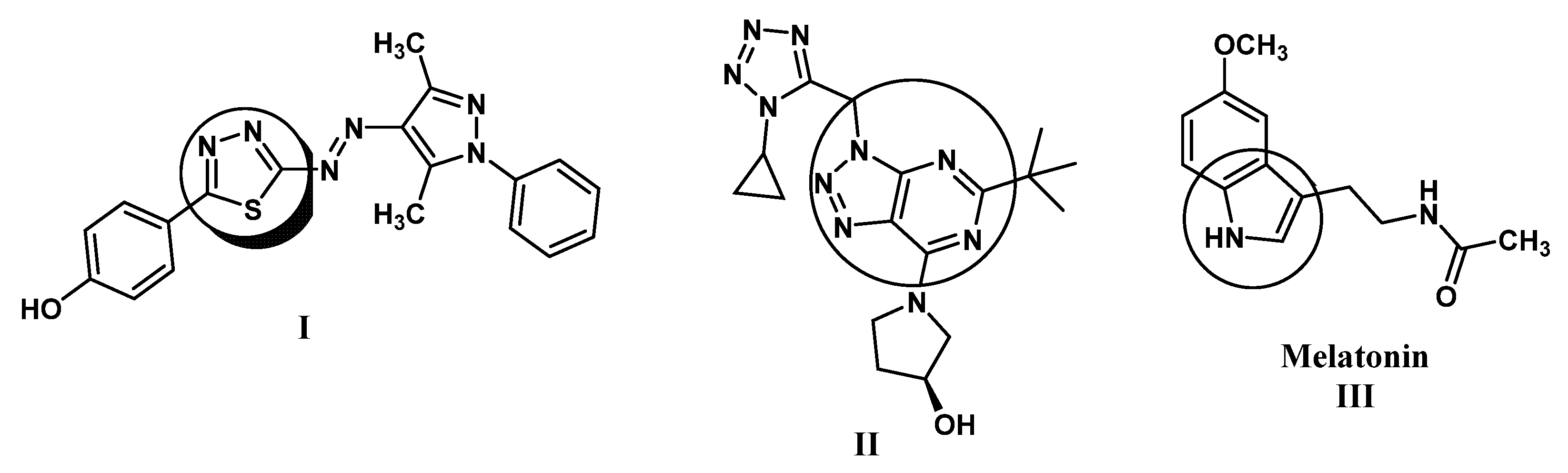
:1. Introduction
2. Results and Discussion
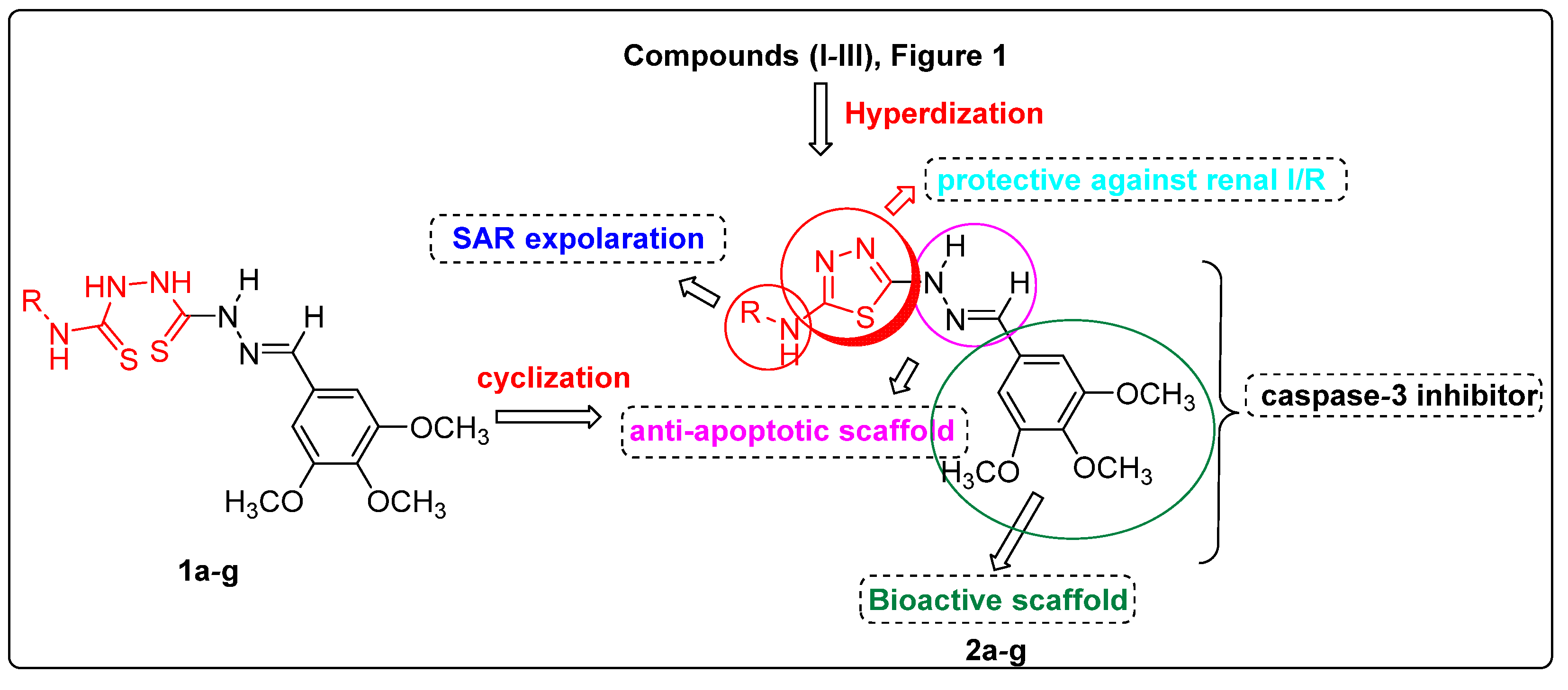
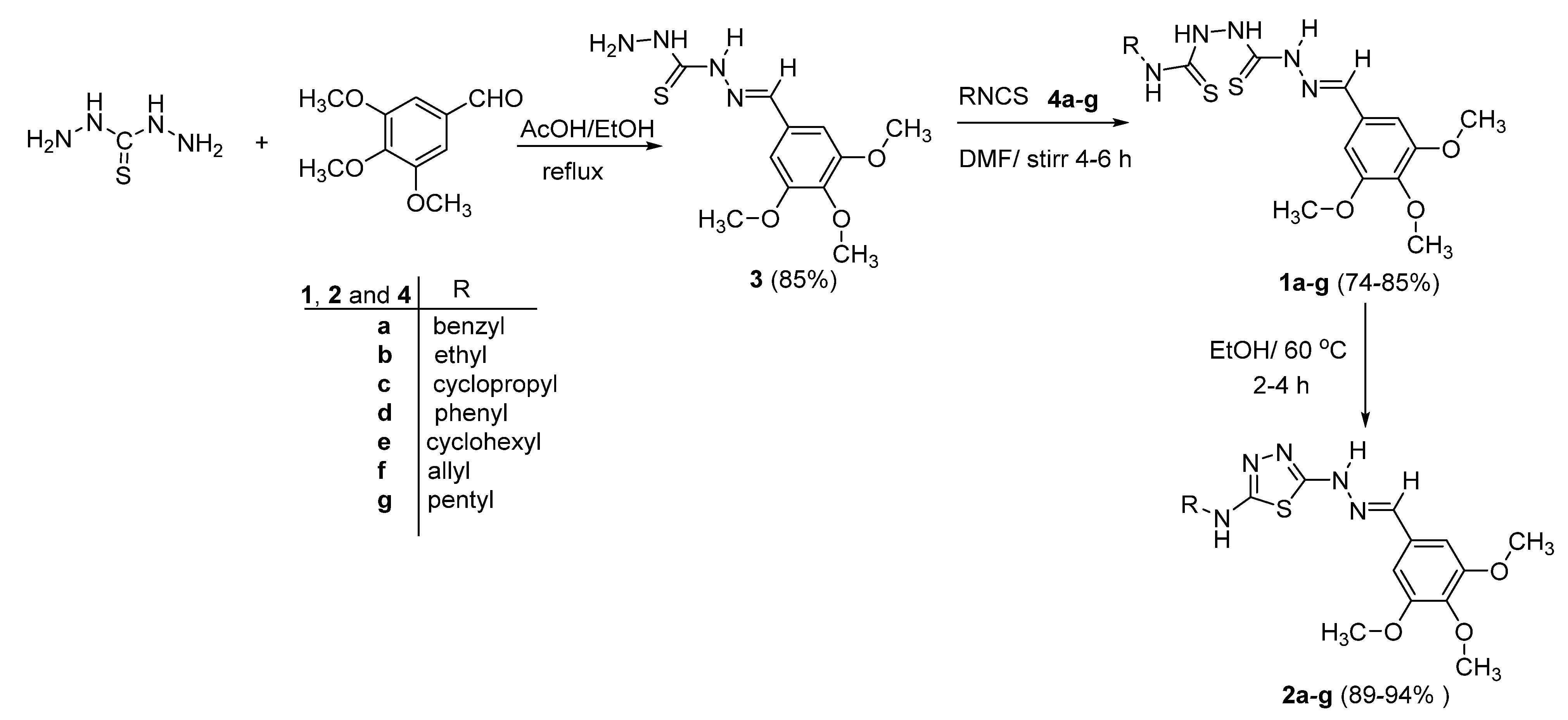
2.1. Chemistry
2.2. Evaluation of Biological Activity
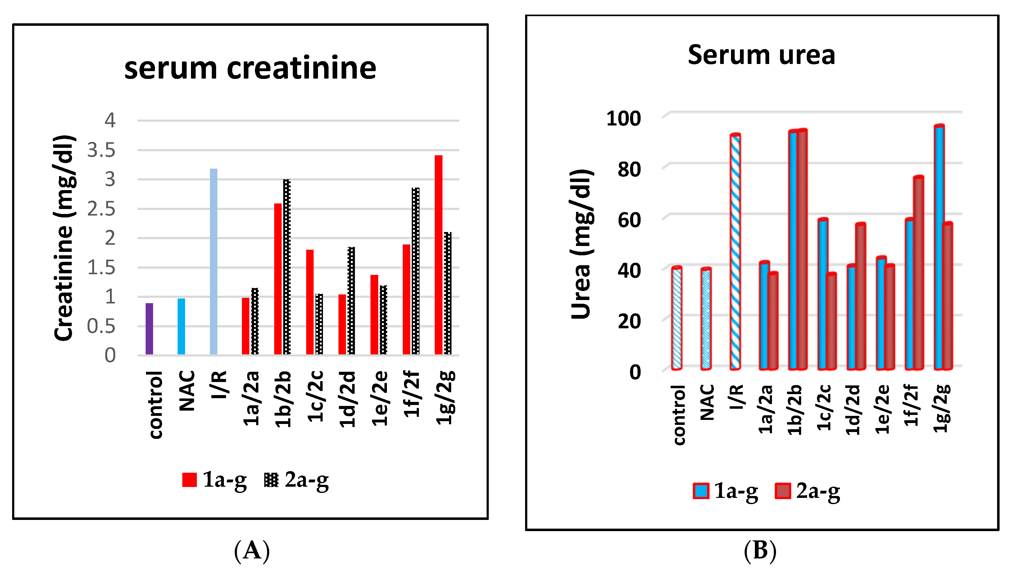
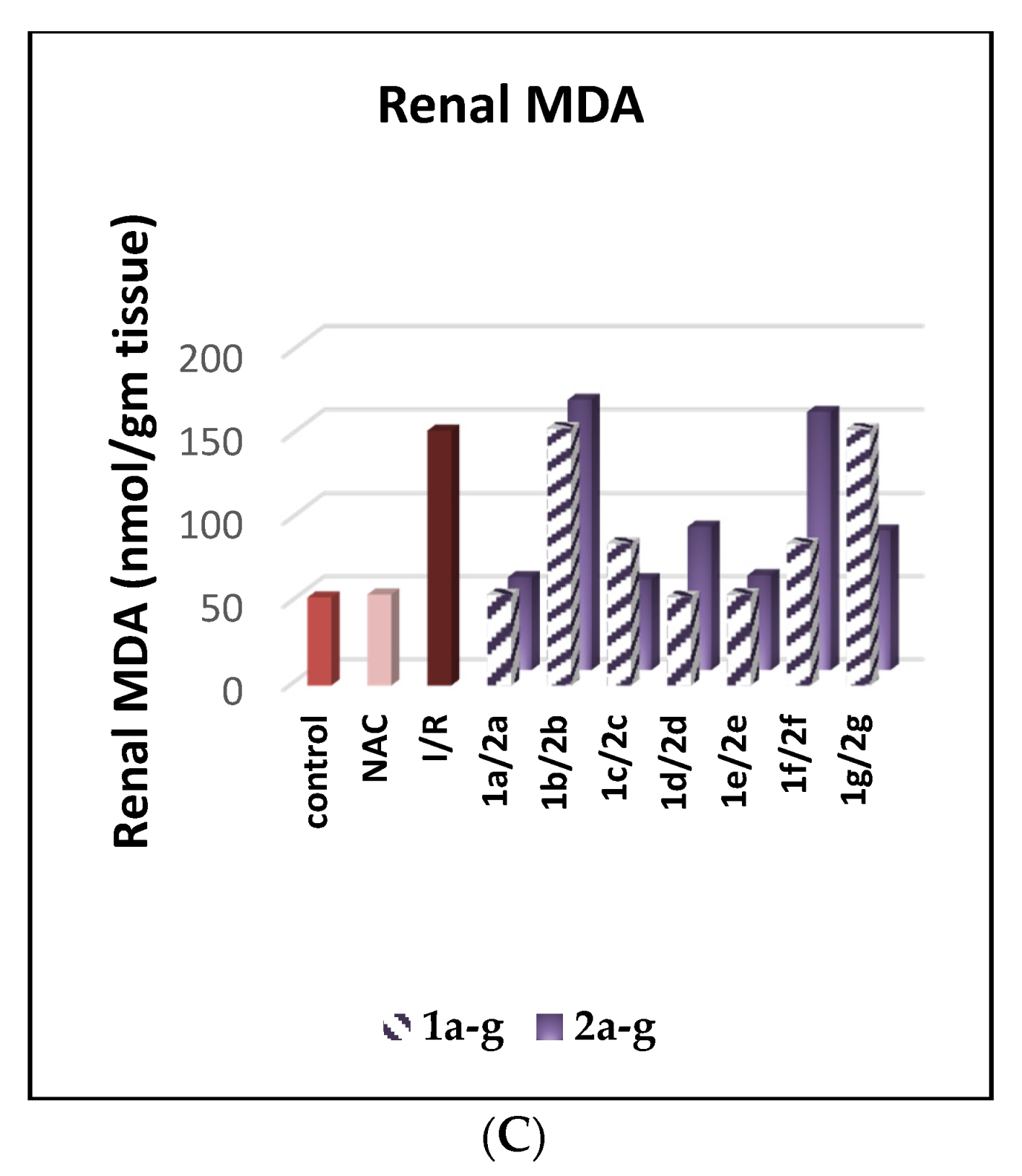
2.2.1. Inhibition Effect on Serum Creatinine, Urea, and Renal MDA
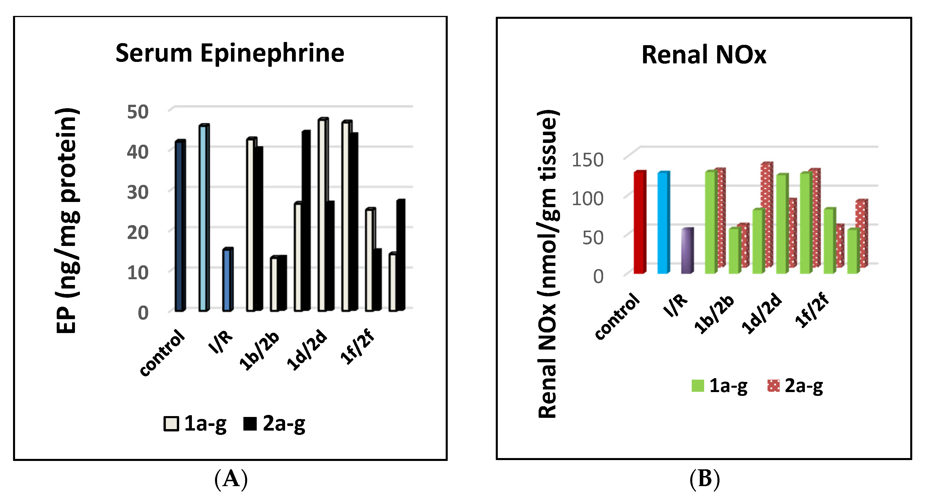
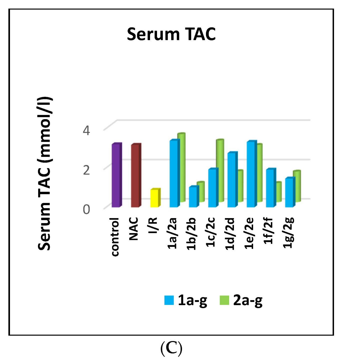
2.2.2. Activation Effect on Serum Epinephrine, Renal NOx, and Serum TAC
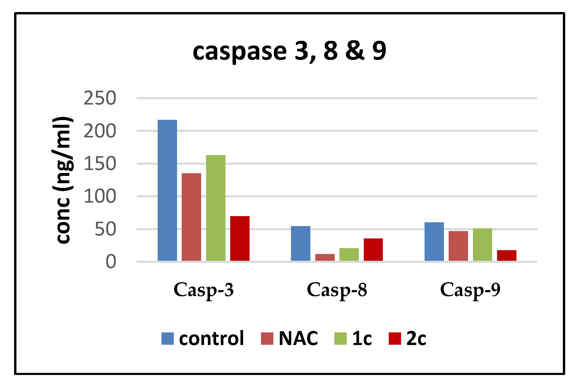
2.2.3. Caspases-3, 8, and 9 Inhibition and Selectivity
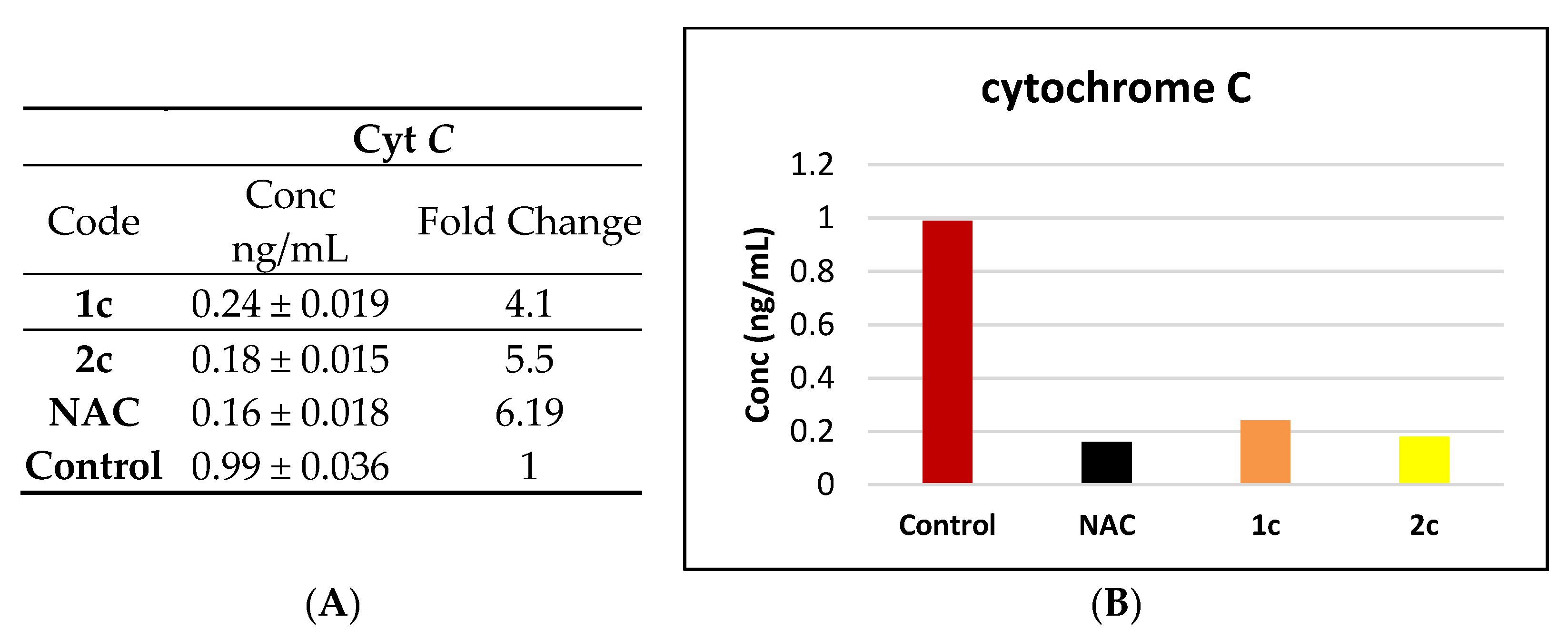
2.2.4. Assay of Cytochrome C Inactivation
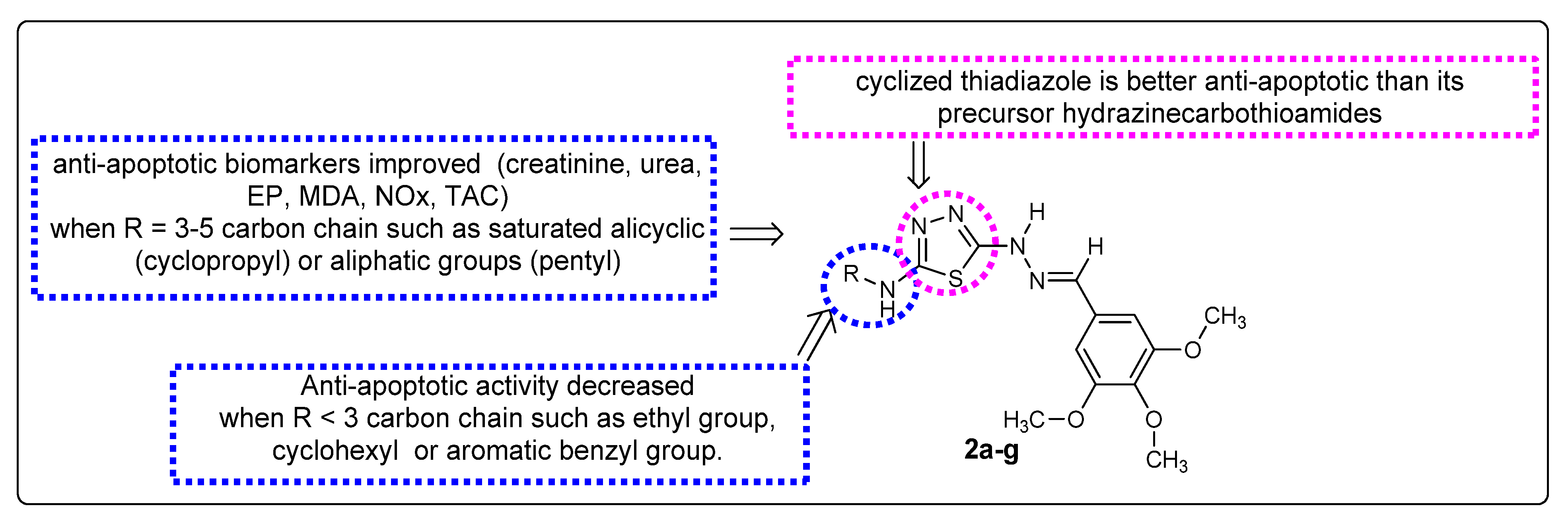
Structure Activity Relationship
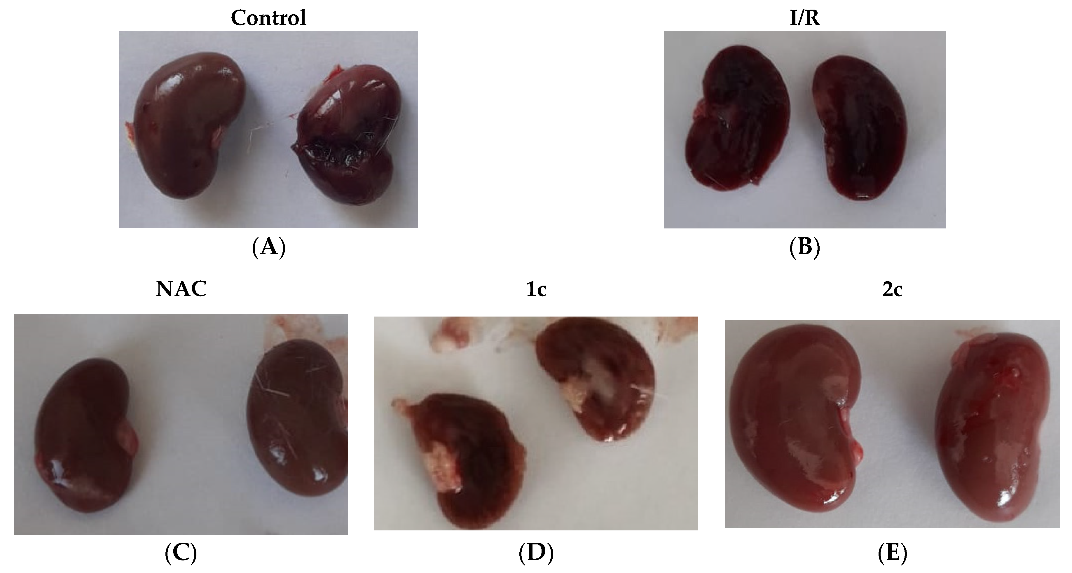
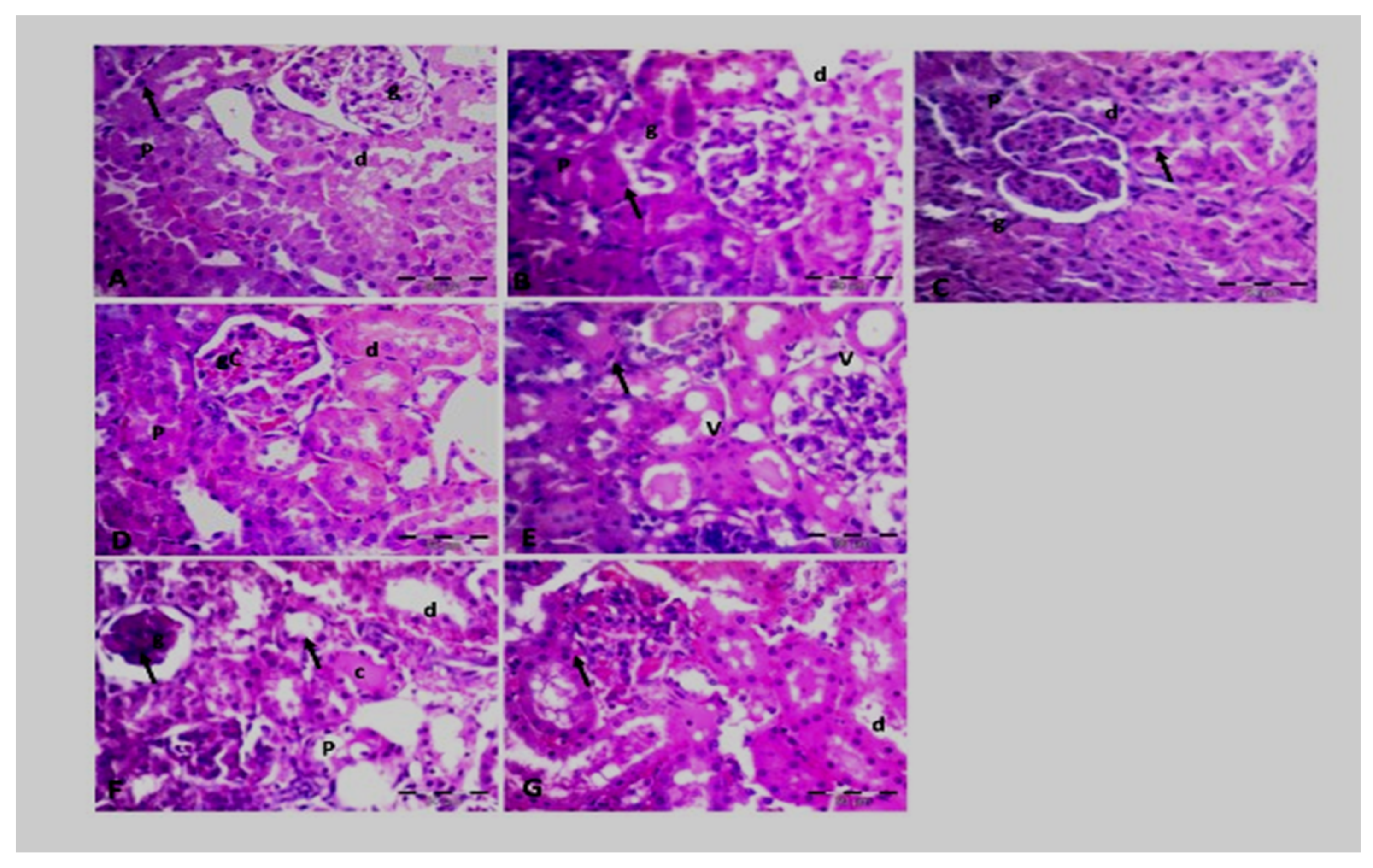
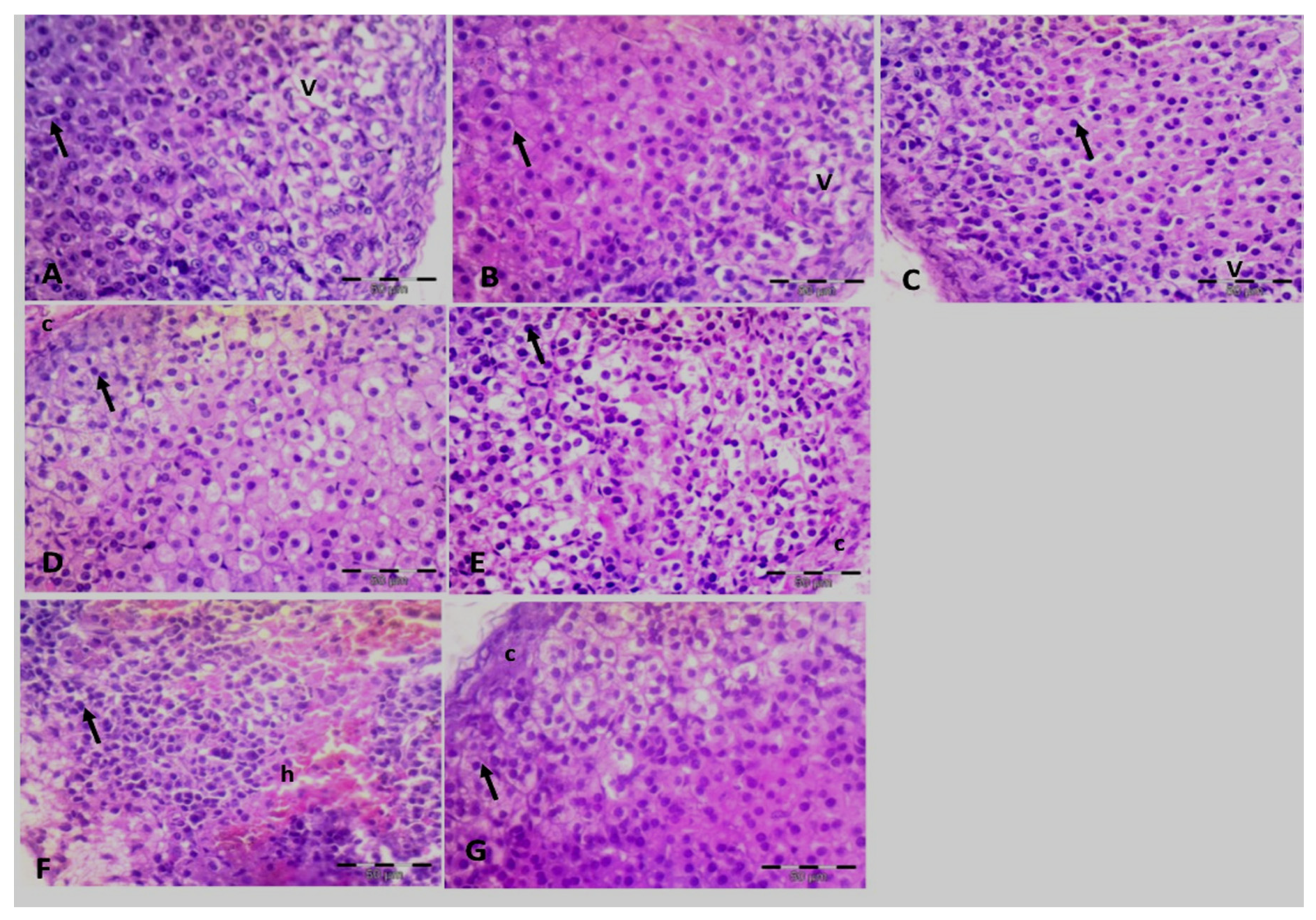
2.2.5. Histopathological Investigation
Renal Cortex
Supra Renal Cortex
Renal Tissue
Suprarenal Tissue
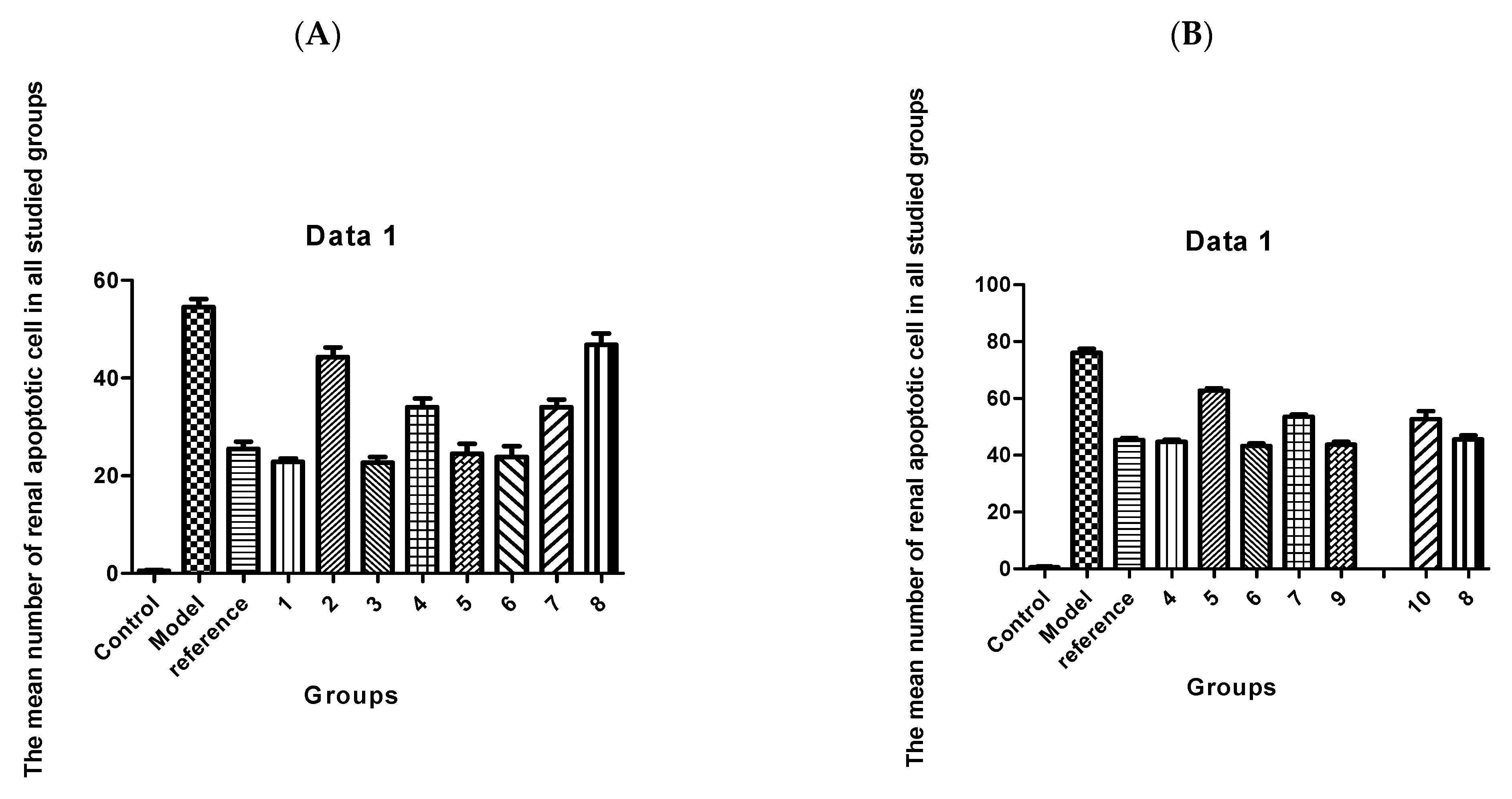
Morphometric Study
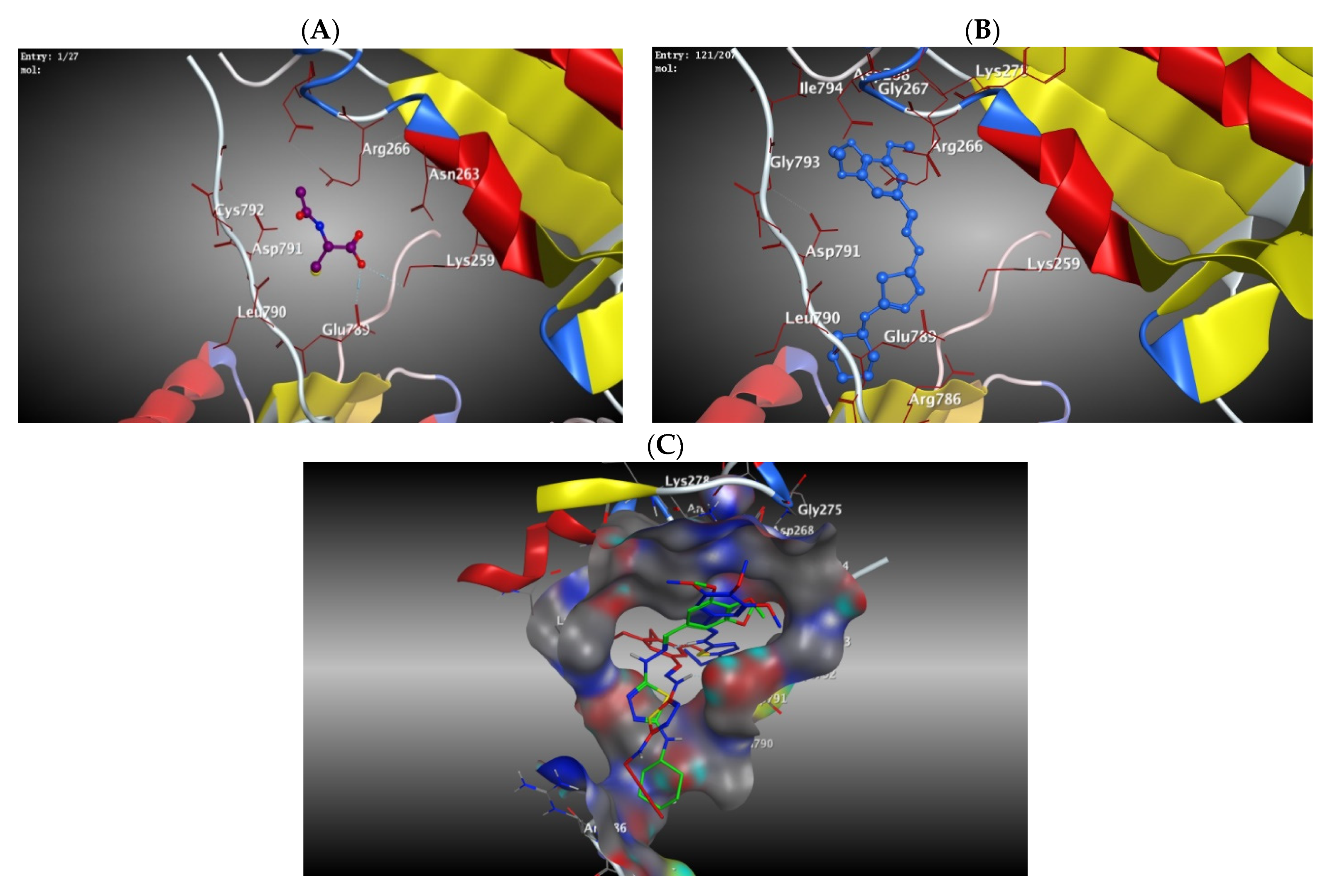
2.2.6. Molecular Docking Studies Using MOE® Program
3. Conclusions
4. Experimental Section
4.1. Chemistry
4.1.1. General Method for the Synthesis of Compounds 1a–g
4.1.2. General Method for the Synthesis of Compounds 2a–g
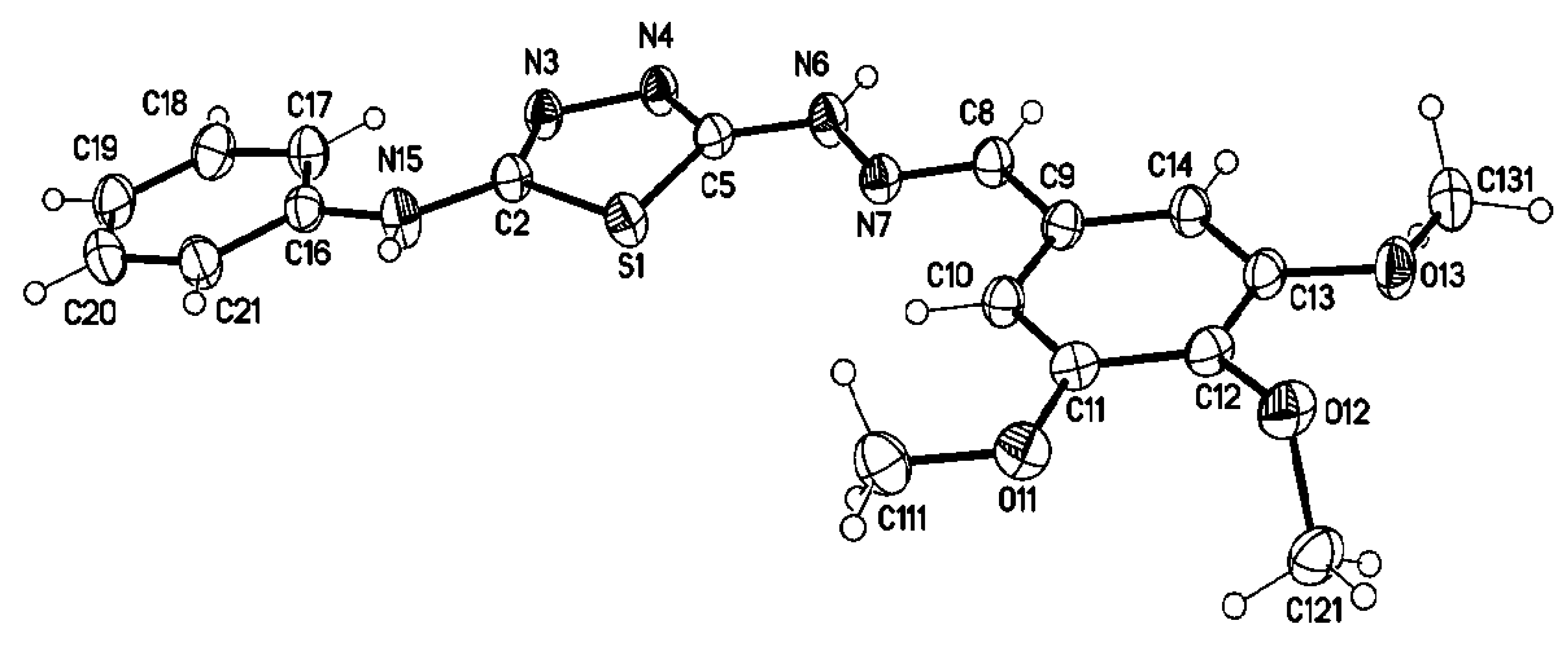
4.2. Single Crystal X-ray Structure Determination of 2d
4.3. Biological Evaluation
4.3.1. Materials and Methods of Biomarkers (Creatinine, Urea, Ep, MDA, TAC, NOx)
4.3.2. Assay of Caspase-3, 8, and 9 Inhibition
4.3.3. Assay of Cytochrome C
4.3.4. Histopathological Investigation
4.3.5. Molecular Docking Study
Supplementary Materials
Author Contributions
Funding
Institutional Review Board Statement
Informed Consent Statement
Data Availability Statement
Acknowledgments
Conflicts of Interest
References
- Lobana, T.S.; Sharma, R.; Bawa, G.; Khanna, S. Bonding and structure trends of thiosemi-carbazone derivatives of metals—An overview. Coord. Chem. Rev. 2009, 253, 977–1055. [Google Scholar] [CrossRef]
- Padhyé, S.; Kayffman, G.B. Transition metal complexes of semicarbazones and thiosemicarbazones. Coord. Chem. Rev. 1985, 63, 127–160. [Google Scholar] [CrossRef]
- Garoufis, A.; Hadjikakou, S.K.; Hadjiliadis, N. Palladium coordination compounds as anti-viral, anti-fungal, anti-microbial and anti-tumor agents. Coord. Chem. Rev. 2009, 253, 1384–1397. [Google Scholar] [CrossRef]
- Singh, D.P.; Kumar, P.; Malik, V.; Tyagi, P. Template synthesis, spectroscopic studies and biological activities of macrocyclic complexes derived from thiocarbohydrazide and glyoxal. J. Enzym. Inhib. Med. Chem. 2007, 22, 177–182. [Google Scholar] [CrossRef] [PubMed]
- Mendes, I.C.; Moreira, J.P.; Speziali, N.L.; Mangrich, A.S.; Takahashi, J.A.; Beraldo, H. N(4)-tolyl-2-benzoylpyridine thiosemicarbazones and their copper(II) complexes with significant antifungal activity. Crystal structure of N(4)-para-tolyl-2-benzoylpyridine thiosemi-carbazone. J. Brazil. Chem. Soc. 2006, 17, 1571–1577. [Google Scholar] [CrossRef] [Green Version]
- Kolocouris, A.; Dimas, K.; Pannecouque, C.; Witvrouw, M.; Foscolos, G.B.; Stamatiou, G.; Fytas, G.; Zoidis, G.; Kolocouris, N.; Andrei, G.; et al. New 2-(1-adamantylcarbonyl)pyridine and 1-acetyladamantane thiosemicarbazonesthiocarbonohydrazones: Cell growth inhibitory, antiviral and antimicrobial activity evaluation. Bioorg. Med. Chem. Lett. 2002, 12, 723–727. [Google Scholar] [CrossRef]
- Ferrari, M.B.; Capacchi, S.; Pelosi, G.; Reffo, G.; Tarasconi, P.; Albertini, R.; Pinelli, S.; Lunghi, P. Synthesis, structural characterization and biological activity of helicin thiosemicarbazone monohydrate and a copper(II) complex of salicylaldehyde thiosemicarbazone. Inorg. Chim. Acta 1999, 276, 134–141. [Google Scholar] [CrossRef]
- Quiroga, A.G.; Pérez, J.M.; López-Solera, I.; Masaguer, J.R.; Luque, A.; Román, P.; Edwards, A.; Alonso, C.; Navarro-Ranninger, C. Novel tetranuclear orthometalated complexes of Pd(II) and Pt(II) derived from p-isopropylbenzaldehyde thiosemicarbazone with cytotoxic activity in cis-DDP resistant tumor cell lines. Interaction of these complexes with DNA. J. Med. Chem. 1998, 41, 1399–1408. [Google Scholar] [CrossRef]
- Glotova, T.E.; Dvorko, M.Y.; Chipanina, N.N.; Albanov, A.I.; Sherstyannikova, L.V.; Kazheva, O.N.; Chekhlov, A.N.; D’yachenko, O.A. Synthesis and steric structure of N″-(arylmethylidene)-N″′-(1-methyl-3-phenylprop-2-yn-1-ylidene) thiocarbonohydrazides. Russ. J. Org. Chem. 2008, 44, 114–119. [Google Scholar] [CrossRef]
- Karegoudar, P.; Prasad, D.J.; Ashok, M.; Mahalinga, M.; Poojary, B.; Holla, B.S. Synthesis, antimicrobial and antiinflammatory activities of some 1,2,4-triazolo[3,4-b][1,3,4]thiadiazoles and 1,2,4-triazolo[3,4-b][1,3,4]thiadiazines bearing trichlorophenyl moiety. Eur. J. Med. Chem. 2008, 43, 808–815. [Google Scholar] [CrossRef] [PubMed]
- Prasad, D.J.; Ashok, M.; Karegoudar, P.; Poojary, B.; Shivarama, P.H.; Holla, B.S.; Kumari, N.S. Synthesis and antimicrobial activities of some new triazolothiadiazoles bearing 4-methylthio-benzyl moiety. Eur. J. Med. Chem. 2009, 44, 551–557. [Google Scholar] [CrossRef] [PubMed]
- Kaplancikli, Z.A.; Turan-Zitouni, G.; Özdemir, A.; Revial, G. New triazole and triazolothiadiazine derivatives as possible antimicrobial agents. Eur. J. Med. Chem. 2008, 43, 155–159. [Google Scholar] [CrossRef] [PubMed]
- Aly, A.A.; Hassan, A.A.; Ibrahim, Y.R. Synthesis of 1,3-thiazin-2-ylidene-substituted hydrazides via reaction of N-substitut edhydrazinocarbothioamides with 1,4-diphenylbut-2-yne-1,4-dione. J. Chem. Res. 2008, 2008, 699–701. [Google Scholar] [CrossRef]
- Ashok, M.; Holla, B.S. Synthesis and antimicrobial evaluation of some new thiadiazinotriazinones carrying 4-methylthiobenzyl moiety. Phosphorus Sulfur Silicon Relat. Elem. 2007, 182, 1599–1608. [Google Scholar] [CrossRef]
- Amin, A.H.; Bakherad, M. Synthesis of a novel heterocyclic ring system: 4-substituted-1-thioxo[1,2,4,5]tetraazino[1,2-b]- phtalazine-6,11-dione. Heterocycl. Commun. 2007, 13, 311. [Google Scholar] [CrossRef]
- Aly, A.A.; Hassan, A.A.; El-Sheref, E.M.; Mohamed, M.A.; Brown, A.B. Conventional and microwave irradiation assisted synthesis of new 1,2,4-triazepine-3-thiones. J. Heterocycl. Chem. 2008, 45, 521–526. [Google Scholar] [CrossRef]
- Aly, A.A.; El-Shaieb, K.M. Reaction of N-imidoylthioureas with dimethyl acetylenedicarboxylate: Synthesis of new 1,3,5-thiadiazepines. J. Chem. Res. 2007, 2007, 563–565. [Google Scholar] [CrossRef]
- Aly, A.A.; Hassan, A.A.; Ameen, M.A.; Brown, A.B. Chemistry of cyclopropenones: Synthesis of new pyrrolo[2,1-b]-1,3,4-oxadiazoles. Tetrahedron Lett. 2008, 49, 4060–4062. [Google Scholar] [CrossRef]
- Aly, A.A.; Hassan, A.A.; Gomaa, M.A.-M.; El-Sheref, E.M. Unusual reactivity of thiosemicarbazides towards 2,3-diphenyl cyclopropenone: Synthesis of new pyridazinethiones and 1,2,4-triazolo[4,3-b]pyridazinethiones. ARKIVOC 2007, 2007, 1–11. [Google Scholar] [CrossRef] [Green Version]
- Singh, D.P.; Kumar, R.; Tyagi, P. Template synthesis, spectroscopic studies and biological screening of macrocyclic complexes derived from thiocarbohydrazide and benzyl. Transit. Met. Chem. 2006, 31, 970–973. [Google Scholar] [CrossRef]
- Suni, M.M.; Nair, V.A.; Joshua, C.P. Heterocyclization of 1-aryl/alkyl-2-thiobiureas to 4-aryl/alkyl-3-substituted-Δ2-1,2,4-triazolin-5-ones. Tetrahedron 2001, 57, 2003. [Google Scholar] [CrossRef]
- Turner, S.; Myers, M.; Gadie, B.; Nelson, A.J.; Pape, R.; Saville, J.F.; Doxey, J.C.; Berridge, T.L. Antihypertensive thiadiazoles, 1: Synthesis of some 2-aryl-5-hydrazino-1,3,4-thiadiazoles with vasodilator activity. J. Med. Chem. 1988, 31, 902–906. [Google Scholar] [CrossRef] [PubMed]
- Mullican, M.D.; Wilson, M.W.; Connor, D.T.; Kostlan, C.R.; Schrier, D.J.; Dyer, R.D. Design of 5-(3,5-di-tert-butyl-4- hydroxy- phenyl)-l,3,4-thiadiazoles, -1,3,4-oxadiazoles, and -1,2,4-triazoles as orally active, nonulcerogenic anti-inflammatory agents. J. Med. Chem. 1993, 36, 1090–1099. [Google Scholar] [CrossRef] [PubMed]
- Farshori, N.N.; Banday, M.R.; Ahmad, A.; Khan, A.U.; Rauf, A. Synthesis, characterization, and in vitro antimicrobial activities of 5-alkenylhydroxy-alkenyl-2- phenylamine-1,3,4-oxadiazoles and thiadiazoles. Bioorg. Med. Chem. Lett. 2010, 20, 1933–1938. [Google Scholar] [CrossRef] [PubMed]
- Oruc, E.E.; Rollas, S.; Kandemirli, F.; Shvets, N.; Dimoglo, A.S. 1,3,4-Thiadiazole derivatives: Synthesis, structure elucidation, and structure antituberculosis activity relationship investigation. J. Med. Chem. 2004, 47, 6760–6767. [Google Scholar] [CrossRef] [PubMed]
- Chen, H.; Li, Z.; Han, Y. Synthesis and fungicidal activity against Rhizoctonia solani of 2-alkyl (alkylthio)-5-pyrazolyl-1,3,4-oxadiazoles (thiadiazoles). J. Agric. Food Chem. 2000, 48, 5312–5315. [Google Scholar] [CrossRef]
- Stillings, M.R.; Welbourn, A.P.; Walter, D.S. Substituted 1,3,4-thiadiazoles with anticonvul-sant activity, 2: Aminoalkyl derivatives. J. Med. Chem. 1986, 29, 2280–2284. [Google Scholar] [CrossRef]
- Rzeski, W.; Matysiak, J.; Kandefer-Szerszeń, M. Anticancer, neuroprotective activities and computational studies of 2-amino- 1,3,4-thiadiazole-based compound. Bioorg. Med. Chem. 2007, 15, 3201–3207. [Google Scholar] [CrossRef]
- Fujiwara, M.; Ijichi, K.; Hanasaki, Y.; Ide, T.; Katsuura, K.; Takayama, H.; Aimi, N.; Shigeta, S.; Konno, K.; Yokota, T.; et al. Thiadiazole derivatives were highly potent inhibitors of human immunodeficiency virus type 1 (HIV-1) replications in vitro. Microbiol. Immunol. 1997, 41, 301–308. [Google Scholar] [CrossRef] [Green Version]
- Poorrajab, F.; Ardestani, S.K.; Emani, S.; Behrouzi-Fardmoghadam, M.; Shafiee, A.; Foroumadi, A. Nitroimidazolyl-1,3,4-thiadiazole-based anti-leishmanial agents: Synthesis and in vitro biological evaluation. Eur. J. Med. Chem. 2009, 44, 1758–1762. [Google Scholar] [CrossRef]
- Yusuf, M.; Khan, R.; Ahmed, A.B. Syntheses and anti-depressant activity of 5-amino-1, 3, 4-thiadiazole-2-thiol imines and thio benzyl derivatives. Bioorg. Med. Chem. 2008, 16, 8029–8034. [Google Scholar] [CrossRef]
- Kaur, H.; Kumar, S.; Vishwakarma, P.; Sharma, M.; Saxena, K.K.; Kumar, A. Synthesis and antipsychotic and anticonvulsant activity of some new substituted oxa/thiadiazolylaze-tidinonyl/thiazolidinonylcarbazoles. Eur. J. Med. Chem. 2010, 45, 2777–2783. [Google Scholar] [CrossRef] [PubMed]
- Kumar, D.; Maruthi Kumar, N.; Chang, K.H.; Shah, K. Synthesis and anticancer activity of 5-(3-indolyl)-1,3,4-thiadiazoles. Eur. J. Med. Chem. 2010, 45, 4664–4668. [Google Scholar] [CrossRef] [PubMed]
- Asbury, R.F.; Blessing, J.A.; Smith, D.M.; Carson, L.F. Aminothiadiazole in the treatment of advanced leiomyosarcoma of the uterine corpus. A Gynecologic Oncology Group study. Am. J. Clin. Oncol. 1995, 18, 397–399. [Google Scholar] [CrossRef] [PubMed]
- Puthiyapurayil, P.; Poojary, B.; Chikkanna, C.; Buridipad, S.K. Design, synthesis and biological evaluation of a novel series of 1,3,4-oxadiazole bearing N-methyl-4- (trifluoromethyl)-phenyl pyrazole moiety as cytotoxic agents. Eur. J. Med. Chem. 2012, 53, 203–210. [Google Scholar] [CrossRef] [PubMed]
- Zhang, X.M.; Qui, M.; Sun, J.; Zhang, Y.-B.; Yang, Y.-S.; Wang, X.-L.; Tang, J.-F.; Zhu, H.-L. Synthesis, biological evaluation, and molecular docking studies of 1,3,4-oxadiazole derivatives possessing 1,4-benzodioxan moiety as potential anticancer agents. Bioorg. Med. Chem. 2011, 19, 6518–6524. [Google Scholar] [CrossRef] [PubMed]
- Aoyama, T.; Kabeya, M.; Fukushima, A.; Shioiri, T. New method and reagents in organic synthesis. Lithium trimethylsilyl diazomethane: A new synthon for the preparation of 2-amino-1,3,4-thiadiazoles from isothiocyanates. Heterocycles 1985, 23, 2367–2369. [Google Scholar] [CrossRef]
- Armstrong, D.R.; Davies, R.P.; Haigh, R.; Hendy, M.A.; Raithby, P.R.; Snaith, R.; Wheatley, A.E.H. A solid-state, solution, and theoretical structural study of kinetic and thermodynamic lithiated derivatives of a simple diazomethane and their reactivities towards aryl isothiocyanates. Eur. J. Inorg. Chem. 2003, 2003, 3363–3375. [Google Scholar] [CrossRef]
- Danial, N.N.; Korsmeyer, S.J. Cell death: Critical control points. Cell 2004, 116, 205–219. [Google Scholar] [CrossRef] [Green Version]
- Sagulenko, V.; Lawlor, K.E.; Vince, J.E. New insights into the regulation of innate immunity by caspase-8. Arthritis Res. Ther. 2016, 18, 4. [Google Scholar] [CrossRef] [PubMed] [Green Version]
- Bulut, S.; Özdemir, B.H. Apoptosis and expression of caspase-3 in cyclosporine-induced gingival overgrowth. J. Periodontal. 2007, 78, 2364–2368. [Google Scholar] [CrossRef]
- Pu, X.; Storr, S.J.; Zhang, Y.; Rakha, E.A.; Green, A.R.; Ellis, I.O.; Martin, S.G. Caspase-3 and caspase-8 expression in breast cancer: Caspase-3 is associated with survival. Apoptosis 2017, 22, 357–368. [Google Scholar] [CrossRef] [PubMed]
- Wang, C.-C.; Li, H.; Zhang, M.; Li, X.-L.; Yue, L.-T.; Zhang, P.; Zhao, Y.; Wang, S.; Duan, R.-N.; Li, Y.-B. Caspase-1 inhibitor ameliorates experimental autoimmune myasthenia gravis by innate dendric cell IL-1-IL-17 pathway. J. Neuroinflamm. 2015, 12, 118. [Google Scholar] [CrossRef] [PubMed] [Green Version]
- Akpan, N.E. The Intrinsic Caspase Death Pathway in Stroke [Dissertation]. Neurodegeneration; Columbia University: New York, NY, USA, 2013. [Google Scholar]
- Wang, X.-J.; Cao, Q.; Zhang, Y.; Su, X.-D. Activation and regulation of caspase-6 and its role in neurodegenerative diseases. Ann. Rev. Pharmacool. Toxicol. 2015, 55, 553–572. [Google Scholar] [CrossRef] [Green Version]
- Singh, P.K.; Kumar, A. Mitochondria mediates caspase-dependent and independent retinal cell death in Staphylococcus aureus endophthalmitis. Cell Death Discov. 2016, 2, 16034. [Google Scholar] [CrossRef] [Green Version]
- Malysheva, I.E.; Topchieva, L.V.; Barysheva, O.Y.; Kurbatova, I.V.; Vasykova, O.A.; Vezikova, N.N.; Marusenko, I.M.; Nemova, N.N. The level of cytokines and expression of caspase genes in rheumatoid arthritis. Dokl. Biochem. Biophys. 2016, 468, 226–228. [Google Scholar] [CrossRef]
- Woolbright, B.L.; Ding, W.-X.; Jaeschke, H. Caspase inhibitors for the treatment of liver disease: Friend or foe? Expert Rev. Gastroenterol. Hepatol. 2017, 11, 397–399. [Google Scholar] [CrossRef] [Green Version]
- Sun, C.; Liu, H.; Guo, J.; Yu, Y.; Yang, D.; He, F.; Du, Z. MicroRNA-98 negatively regulates myocardial infarction-induced apoptosis by down-regulating Fas and caspase-3. Sci. Rep. 2017, 7, 7460. [Google Scholar] [CrossRef] [Green Version]
- Aziz, M.; Jacob, A.; Wang, P. Revisiting caspases in sepsis. Cell Death Dis. 2014, 5, 1526. [Google Scholar] [CrossRef] [PubMed]
- Hwang, H.S.; Kim, H.A. Chondrocyte Apoptosis in the Pathogenesis of Osteoarthritis. Int. J. Mol. Sci. 2015, 16, 26035–26054. [Google Scholar] [CrossRef]
- Qi, X.; Gurung, P.; Malireddi, R.K.S.; Karmaus, P.W.F.; Sharma, D.; Vogel, P.; Chi, H.; Green, D.R.; Kanneganti, T.D. Critical role of caspase-8-mediated IL-1 signaling in promoting Th2 responses during asthma pathogenesis. Mucosal. Immunol. 2017, 10, 128–138. [Google Scholar] [CrossRef] [PubMed] [Green Version]
- Walsh, J.G.; Cullen, S.P.; Sheridan, C.; Luthi, A.U.; Gerner, C.; Martin, S.J. Executioner caspase-3 and caspase-7 are functionally distinct proteases. Proc. Natl. Acad. Sci. USA 2008, 105, 12815–12819. [Google Scholar] [CrossRef] [PubMed] [Green Version]
- Donnahoo, K.K.; Meldrum, D.R.; Shenkar, R.; Chung, C.-S.; Abrham, E.; Harken, A.H. Early renal ischemia, with or without reperfusion, activates NFκB and increases TNF-α bioactivity in the kidney. J. Urol. 2000, 163, 1328–1332. [Google Scholar] [CrossRef]
- Sivarajah, A.; Chatterjee, P.K.; Patel, N.S.A.; Todorovic, Z.; Hattori, Y.; Brown, P.A.J.; Stewart, K.N.; Mota-Filipe, H.; Cuzzocrea, S.; Thiemermann, C. Agonists of peroxisome-proliferator activated receptor-gamma reduce renal ischemia/reperfusion injury. Am. J. Nephrol. 2003, 23, 267–276. [Google Scholar] [CrossRef]
- O’Donnell, M.P.; Burne, M.; Daniels, F.; Rabb, H. Utility and limitations of serum creatinine as a measure of renal function in experimental renal ischemia-reperfusion injury1. Transplantation 2002, 73, 1841–1844. [Google Scholar] [CrossRef] [PubMed] [Green Version]
- Sancaktutar, A.A.; Bodakci, M.N.; Hatipoglu, N.K.; Soylemez, H.; Basarılı, K.; Turkcu, G. The protective effects of pomegranate extracts against renal ischemia-reperfusion injury in male rats. Urol. Ann. 2014, 6, 46–50. [Google Scholar] [CrossRef] [PubMed]
- Wang, J.; Guo, Y.; Wu, X.; Zhang, Y. Design and development of novel pyrazole-thiadiazole derivatives as NF-ĸB Inhibitor and cardioprotective effect against isoproterenol induced myocardial infarction in Sprague-Dawley rats. Pharmacology 2020, 105, 260–271. [Google Scholar] [CrossRef] [PubMed]
- Nettekoven, M.; Adam, J.M.; Bendels, S.; Bissantz, C.; Fingerle, J.; Grether, U.; Grüner, S.; Guba, W.; Kimbara, A.; Ottaviani, G.; et al. Novel triazolopyrimidine-derived cannabinoid receptor 2 agonists as potential treatment for inflammatory kidney diseases. Chem. Med. Chem. 2016, 11, 179–189. [Google Scholar] [CrossRef]
- Sener, G.; Sehirli, A.Ö.; Keyer-Uysal, M.; Arbak, S.; Ersoy, Y.; Yeğen, B.Ç. The protective effect of melatonin on renal ischemia–reperfusion injury in the rat. J. Pineal. Res. 2002, 32, 120–126. [Google Scholar] [CrossRef] [PubMed]
- Karakus, S.; Rollas, S. Synthesis and antituberculosis activity of new N-phenyl-N′-[4-(5-alkyl/arylamino-1,3,4-thiadiazole-2-yl)phenyl]thioureas. Farmaco 2002, 57, 577–581. [Google Scholar] [CrossRef]
- D’Amico, F.; Vitale, A.; Piovan, D.; Bertacco, A.; Morales, R.R.; Frigo, A.C.; Bassi, D.; Bonsignore, P.; Gringeri, E.; Valmasoni, M.; et al. Use of N-acetylcysteine during liver procure-ment: A prospective randomized controlled study. Liver Transpl. 2013, 19, 135–144. [Google Scholar] [CrossRef]
- Zhang, F.-X.; Chen, M.-L.; Yang, B.; Chen, H.-W.; Ju, W.-Z.; Wang, J.; Cao, K.-J. The antiapoptotic effects of N-acetylcysteine in neonatal rat cardiomyocytes underwent hypoxia-reoxygenation injury. Zhonghua Xin Xue Guan Bing Za Zhi 2010, 38, 445–449. [Google Scholar] [PubMed]
- Aly, A.A.; Sayed, S.M.; Abdelhafez, E.-S.M.; Abdelhafez, S.M.N.; Abdelzaher, W.Y.; Raslan, M.A.; Ahmed, A.E.; Thabet, K.; El-Reedy, A.A.; Brown, A.B. New quinoline-2-one/pyrazole derivatives; design, synthesis, molecular docking, antiapoptotic evaluation, and caspase-3 inhibition assay. Bioorg. Chem. 2020, 94, 103348. [Google Scholar] [CrossRef] [PubMed]
- Brito, M.; de Moreira, R.J.; Tavares, M.; Carballo, M.; Carneiro, T.X.; dos Santos Ade, A. Copaiba oil effect on urea and creatinine serum levels in rats submitted to kidney ischemia and reperfusion syndrome. Acta Cir. Bras. 2005, 20, 243–246. [Google Scholar] [CrossRef] [PubMed] [Green Version]
- Yamaki, V.N.; Gonçalves, T.B.; Coelho, J.V.B.; Pontes, R.V.S.; Costa, F.L.D.S.; Brito, M.V.H. Protective effect of remote ischemic per-conditioning in the ischemia and reperfusion-induce renal injury in rats. Rev Col Bras Cir. 2012, 39, 529–533. [Google Scholar] [CrossRef] [Green Version]
- Abdelbaset, M.S.; Abdel-Aziz, M.; Abuo-Rahma, G.E.D.A.; Abdelrahman, M.H.; Ramadan, M.; Youssif, B.G. Novel quinoline derivatives carrying nitrones/oximes nitric oxide donors: Design, synthesis, antiproliferative and caspase-3 activation activities. Arch. Pharm. 2019, 352, e1800270. [Google Scholar] [CrossRef] [Green Version]
- Giaccia, A.J.; Kastan, M.B. The complexity of p53 modulation: Emerging patterns from divergent signals. Genes Dev. 1998, 12, 2973–2983. [Google Scholar] [CrossRef] [Green Version]
- Hassan, A.A.; Abdel-Latif, F.F.; Abdel Aziz, M.; Mostafa, S.M.; Bräse, S.; Nieger, M. Heterocyclisation of substituted ylidene thiocarbonohydrazides using dimethyl acetylenedicarboxylate. Chem. Pap. 2015, 69, 973–982. [Google Scholar] [CrossRef]
- Mostafa, S.M.; Aly, A.A.; Brase, S.; Mohamed, A.H. An efficient approach for the synthesis of novel series of 1,3-dihydro- spiro[indene-2,6′-[1,3]thiazine] derivatives. Monatsh. Chem. 2022, 153, 87–94. [Google Scholar] [CrossRef]
- Sheldrick, G.M. SHELXT–Integrated space-group and crystal-structure determination. Acta Cryst. 2015, A71, 3–8. [Google Scholar] [CrossRef] [PubMed] [Green Version]
- Sheldrick, G.M. Crystal structure refinement with SHELX. Acta Cryst. 2015, C71, 3–8. [Google Scholar]
- Aziz, N.M.; Ragy, M.M.; Gayyed, M.F. Effect of acute immobilization stress with or without a heme oxygenase inducer on testicular structure and function in male albino rats. J. Basic Clin. Physiol. Pharmacol. 2013, 24, 255–262. [Google Scholar] [CrossRef] [PubMed]
- Tabacco, A.; Meiattini, F.; Moda, E.; Tarli, P. Simplified enzymic/colorimetric serum urea nitrogen determination. Clin. Chem. 1979, 25, 336–337. [Google Scholar] [CrossRef]
- Söğüt, S.; Zoroğlu, S.S.; Özyurt, H.; Yılmaz, H.R.; Özuğurlu, F.; Sivaslı, E.; Yetkin, Ö.; Yanık, M.; Tutkun, H.; Savaş, H.A.; et al. Changes in nitric oxide levels and antioxidant enzyme activities may have a role in the pathophysiological mechanisms involved in autism. Clin. Chim. Acta 2003, 331, 111–117. [Google Scholar] [CrossRef]
- Abdel-Aziz, A.M.; Hafez, S.M.N.A. Sitagliptin protects male albino rats with testicular ischaemia/reperfusion damage: Modulation of VCAM-1 and VEGF-A. Andrologia 2019, 52, e13472. [Google Scholar] [CrossRef]


| * Biomarker | Effect during Anti-Apoptosis in Kidney |
|---|---|
| EP [65], Renal NOx [63], TAC [57] | Increase |
| Creatinine [56], Urea [66], MDA [60] | Decrease |
Publisher’s Note: MDPI stays neutral with regard to jurisdictional claims in published maps and institutional affiliations. |
© 2022 by the authors. Licensee MDPI, Basel, Switzerland. This article is an open access article distributed under the terms and conditions of the Creative Commons Attribution (CC BY) license (https://creativecommons.org/licenses/by/4.0/).
Share and Cite
Mostafa, S.M.; Aly, A.A.; Bräse, S.; Nieger, M.; Abdelhafez, S.M.N.; Abdelzaher, W.Y.; Abdelhafez, E.-S.M.N. Synthesis, Characterization, and In Vivo Study of Some Novel 3,4,5-Trimethoxybenzylidene-hydrazinecarbothioamides and Thiadiazoles as Anti-Apoptotic Caspase-3 Inhibitors. Molecules 2022, 27, 2266. https://doi.org/10.3390/molecules27072266
Mostafa SM, Aly AA, Bräse S, Nieger M, Abdelhafez SMN, Abdelzaher WY, Abdelhafez E-SMN. Synthesis, Characterization, and In Vivo Study of Some Novel 3,4,5-Trimethoxybenzylidene-hydrazinecarbothioamides and Thiadiazoles as Anti-Apoptotic Caspase-3 Inhibitors. Molecules. 2022; 27(7):2266. https://doi.org/10.3390/molecules27072266
Chicago/Turabian StyleMostafa, Sara M., Ashraf A. Aly, Stefan Bräse, Martin Nieger, Sara Mohamed Naguib Abdelhafez, Walaa Yehia Abdelzaher, and El-Shimaa M. N. Abdelhafez. 2022. "Synthesis, Characterization, and In Vivo Study of Some Novel 3,4,5-Trimethoxybenzylidene-hydrazinecarbothioamides and Thiadiazoles as Anti-Apoptotic Caspase-3 Inhibitors" Molecules 27, no. 7: 2266. https://doi.org/10.3390/molecules27072266