
Microwave-Assisted Solution Synthesis of Metastable Intergrowth of AgInS2 Polymorphs


1
Department of Chemistry, Iowa State University, Ames, IA 50011, USA
2
Laboratoire Crismat, ENSICAEN, CNRS UMR 6508, 14050 Caen, France
*
Author to whom correspondence should be addressed.
Molecules 2022, 27(6), 1815; https://doi.org/10.3390/molecules27061815
Submission received: 1 February 2022
/
Revised: 1 March 2022
/
Accepted: 1 March 2022
/
Published: 10 March 2022
(This article belongs to the Special Issue Emerging Frontiers in Metastable Crystalline Solids)
Abstract
:The intergrowth of stable and metastable AgInS2 polymorphs was synthesized using a microwave-assisted synthesis. The samples were synthesized in water and in a deep eutectic solvent (DES) consisting of choline chloride and thiourea. An increase in the metal precursor concentration improved the crystallinity of the synthesized samples and affected the particle size. AgInS2 cannot be synthesized from crystalline binary Ag2S or In2S3 via this route. The solution synthesis reported here results in the intergrowth of the thermodynamically stable polymorph (space group I2d, chalcopyrite structure) and the high-temperature polymorph (space group Pna21, wurtzite-like structure) that is metastable at room temperature. A scanning transmission microscopy (STEM) study revealed the intergrowth of tetragonal and orthorhombic polymorphs in a single particle and unambiguously established that the long-thought hexagonal wurtzite polymorph has pseudo-hexagonal symmetry and is best described with the orthorhombic unit cell. The solution-synthesized AgInS2 polymorphs intergrowth has slightly lower bandgap values in the range of 1.73 eV–1.91 eV compared to the previously reported values for tetragonal I2d (1.86 eV) and orthorhombic Pna21 (1.98 eV) polymorphs.
1. Introduction
Deep eutectic solvents (DESs) are emerging solvents that are analogs of ionic liquids [1]. DESs are made by mixing a hydrogen bond donor and a hydrogen bond acceptor (usually a quaternary ammonium salt) in a specific molar ratio [1,2]. The eutectic mixture of the two DES constituents has a significantly lower melting point compared to its individual components because of the hydrogen bonding occurring in the mixture; hence a viscous liquid is formed [1,3]. DESs have high solubility rates for metal precursors, such as salts and binary oxides [4]; therefore, DESs have been used as the reaction medium in the synthesis of functional oxides [5,6,7,8], including a metastable oxide [9]. Due to the various possible combinations of hydrogen bond donors and acceptors, DESs allow for synthetic tunability and have also been employed in the syntheses of sulfides [10,11,12].
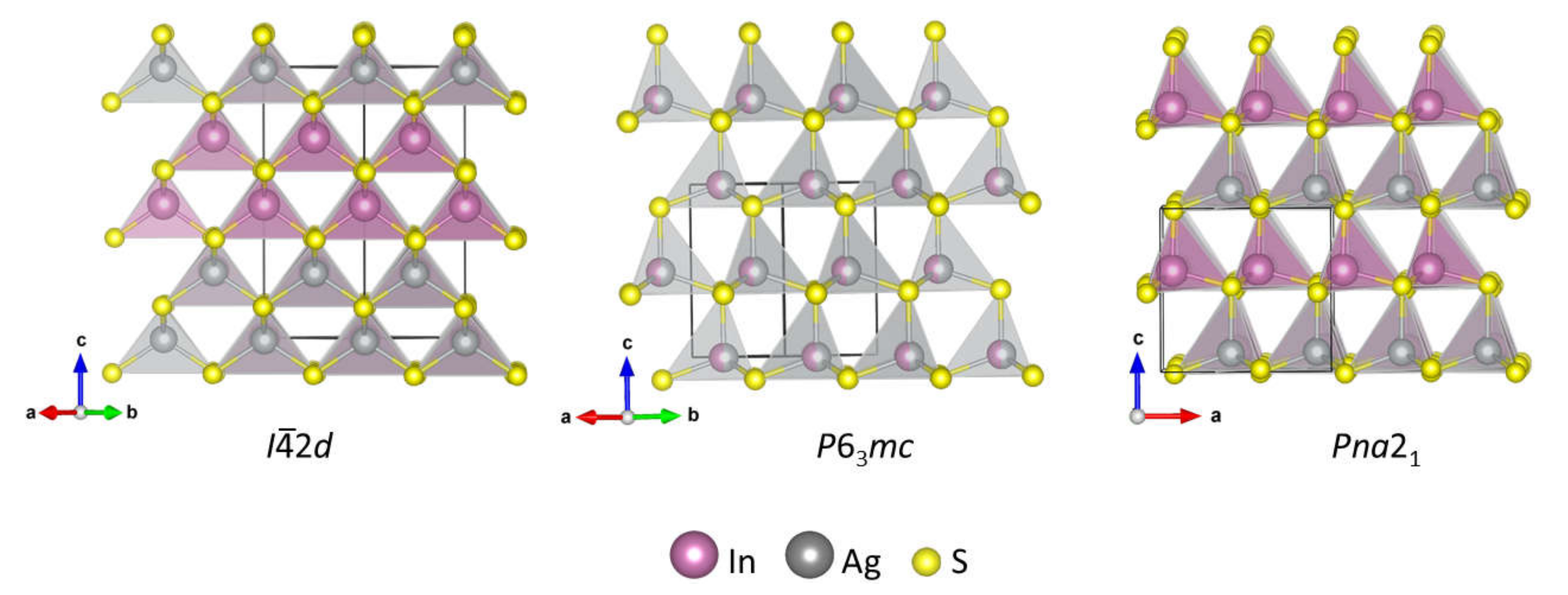
AgInS2 is one of the two ternary sulfides reported in the pseudo-binary Ag2S-In2S3 system. AgInS2 is reported to have four polymorphic modifications. The most common room-temperature polymorph has a tetragonal chalcopyrite structure (space group I2d) [13], which transforms into the high-temperature orthorhombic wurtzite-like structure (space group Pna21) [14] upon heating to ~620 °C [15]. The less common polymorphs are the hexagonal (space group P63mc) [13] wurtzite structure and the trigonal structure (space group Rm), the latter one was synthesized under high-pressure and high-temperature conditions [16]. The structures of the I2d, Pna21, and P63mc polymorphs comprise of the Ag- and In- centered AgS4 and InS4 tetrahedra that share corners to give different types of packing (Figure 1). The I2d polymorph is made of double layers of AgS4 tetrahedra alternating with double layers of InS4 tetrahedra. The I2d chalcopyrite structure is an ordered variant of a cubic sphalerite. The P63mc polymorph has a wurtzite structure, where Ag and In cations share the same crystallographic site. The high-temperature Pna21 polymorph has a pseudo-wurtzite structure, where Ag and In are ordered in different crystallographic sites, resulting in the similar alternating layers of InS4 and AgS4 tetrahedra as in the wurtzite structure, but with the lowering of the symmetry to orthorhombic. The unit cell parameters of the Pna21 and the P63mc structures are related as follows: aortho = √3ahex, bortho = 2ahex, and cortho ~ chex; where ortho represents Pna21 and hex represents P63mc [17].
According to the Materials Project Database [18], the I2d polymorph is thermodynamically stable, the high-temperature Pna21 polymorph is metastable by ~0.008 eV/atom, and the high-pressure Rm structure is metastable by 0.090 eV/atom. No information was found for the P63mc wurtzite polymorph in the Materials Project Database; however, a trigonal P3m1 structure with similar unit cell parameters as the P63mc polymorph and ordered arrangement of Ag and In cations is calculated to be metastable by ~0.97 eV/atom. A closer inspection of the publication reporting synthesis of wurtzite polymorph reveals that the structure assignment was done based on powder X-ray diffraction data collected for a multiphase sample that also contained a tetragonal polymorph [13]. Thus, the question regarding the validity of these data arises.
AgInS2 are currently being explored as non-toxic alternatives for photoluminescent compounds, such as the commercialized CdSe, because the size-dependent bandgap of AgInS2 falls within the visible light region [19,20,21]. However, the broad photoluminescent spectra, which is a result of the lack of monochromaticity, is a major challenge [22,23].
Here, we studied the microwave-assisted solution synthesis of AgInS2 using two solvents—DI water and a DES consisting of choline chloride and thiourea. We investigated the effect of the heating profile, solvents (DI water vs. DES), and metal precursor concentration on the synthesis of the AgInS2 polymorphs. The synthetic route resulted in the intergrowth of two polymorphs of AgInS2: the I2d and high-temperature Pna21 structures. We also investigated the thermal stability of the metastable intergrowth of two AgInS2 polymorphs using in-situ high-temperature powder X-ray diffraction. We further used scanning transmission electron microscopy to address a controversy regarding the hexagonal P63mc polymorph with a wurtzite structure.
2. Results and Discussion
A deep eutectic solvent utilized in this work consists of choline chloride as a hydrogen bond acceptor (melting point of 302 °C) and thiourea as both the hydrogen bond donor and the sulfur source (melting point of 182 °C). The eutectic mixture of choline chloride and thiourea in a 1:2 molar ratio exhibits a lower melting point of 69 °C compared to its components [2]. AgNO3 and InCl3·xH2O are utilized as the metal precursors in this work since they have appreciable solubility in the chosen DES [2]. The DES-assisted synthesis utilized in this work is similar to a previously reported solvothermal synthesis of binary sulfide using a DES consisting of choline chloride and thioacetamide, where thioacetamide is the hydrogen bond donor and also a sulfur source [11].
For the microwave solution synthesis of AgInS2, we studied two temperature profiles: 1-step and 2-step heating profiles (see Experimental for details). For both profiles, the highest synthesis temperature was set to 180 °C above which thiourea will decompose, while the 2-step profile included additional heating at 80 °C to ensure the dissolution of the metal precursors before ramping to 180 °C. We compared the syntheses utilizing DES, or deionized water, as a solvent. Furthermore, we varied concentrations of the metal precursors and observed that the variation in the amount of the metal precursors affected the crystallinity, particle size, and shape. Separately, we studied the reaction mechanism for the synthesis of AgInS2 and ruled out a partial cation exchange between a crystalline binary sulfide (Ag2S or In2S3) and a cation in the solution (In+3 and Ag+, respectively) as a possible mechanism. The reaction between the crystalline pre-synthesized Ag2S with the aqueous solution of InCl3·xH2O did not proceed, while the metathesis reaction of the crystalline pre-synthesized In2S3 with an aqueous solution of AgNO3 resulted in the formation of Ag2S. Further details of this study can be found in the Supplementary Materials.
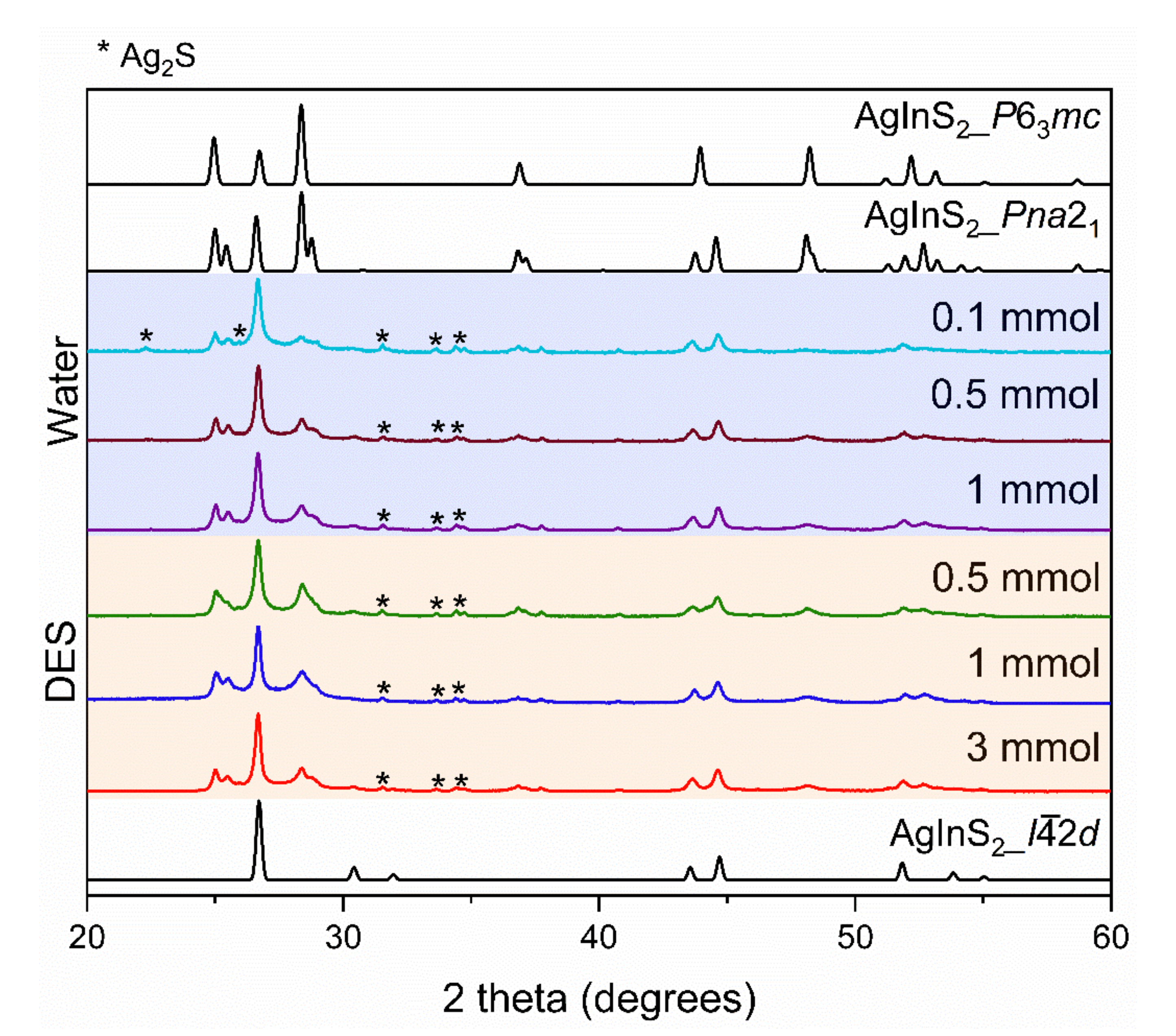
1-step synthesis. Samples of AgInS2 synthesized using the 1-step synthesis contained two polymorphs, regardless of the used solvent: the high-temperature orthorhombic Pna21 and the tetragonal I2d polymorphs. Varying molar concentrations of the metal precursors in the solution were employed in both the water and DES. However, we observed very similar powder X-ray diffraction (PXRD) patterns in all the 1-step syntheses, as shown in Figure 2. This suggests that the two polymorphs are present in similar ratios in all the 1-step-synthesized samples, irrespective of the solvent or metal precursor concentration employed. The dominant polymorph is the tetragonal I2d polymorph.
The crystallinity of the phases was qualitatively evaluated by tracking the full width at half maximum (FWHM) of the most intense diffraction peak at ~26.6°. Narrower peaks indicate a higher crystallinity. According to the PXRD data, the peaks of the orthorhombic Pna21 polymorph are better resolved in the samples made with water than in the samples made in the DES, indicating improved crystallinity. Comparing the FWHM of the 0.5 mmol sample made in the DES (0.273) and that made in water (0.235), we see that the sample made in water has a slightly better crystallinity. The crystallinity of the synthesized samples also increases with an increase in the metal precursor concentration in both water and DES. This is more significant for the DES-made samples, as seen in Figure 2, where the 3 mmol sample (FWHM of 0.231) is more crystalline than the 0.5 mmol sample (FWHM of 0.273).
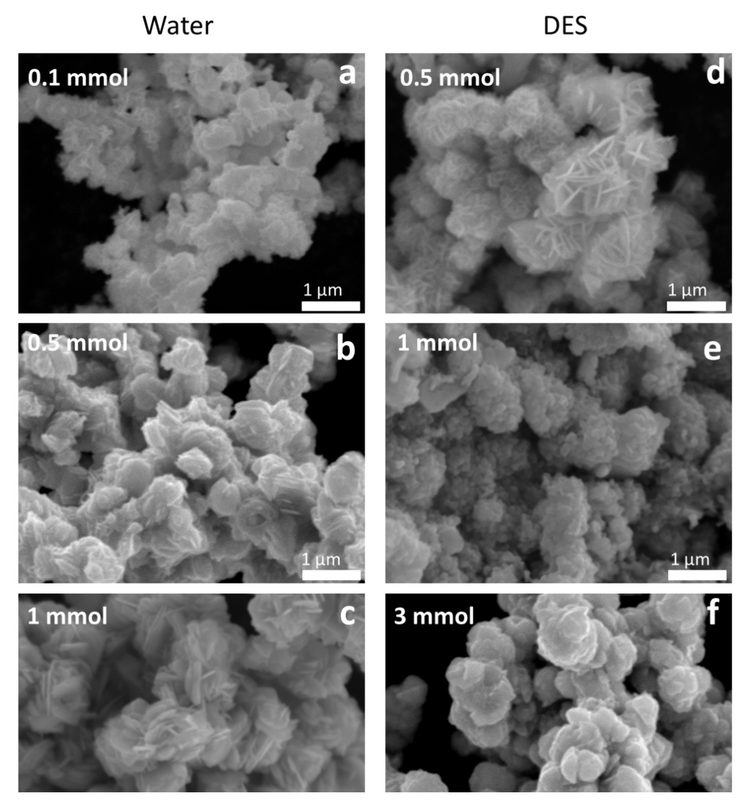
The morphology and particle size were also analyzed by scanning electron microscopy (SEM). As seen in the SEM images (Figure 3), the 1-step water-synthesized AgInS2 have plate-like morphologies arranged like flower petals, and the morphology is uniform throughout the sample. The uniform morphology may also suggest a metastable intergrowth of the orthorhombic Pna21 and the tetragonal I2d polymorphs in a single particle. Notably, an increase in the metal precursor concentration of the 1-step water-synthesized AgInS2 leads to an increase in the particle size. The particle size in the 0.1 mmol sample is evidently smaller than in the 0.5 mmol sample. Further increase in concentration to 1 mmol leads to even larger particles, consistent with the PXRD data. The samples with larger particle sizes (0.5 and 1 mmol 1-step water-synthesized AgInS2) have peaks in PXRD patterns that are more narrow and better-resolved than the samples with the smaller particle size synthesized from the less-concentrated solution (0.1 mmol 1-step water-synthesized AgInS2).
The SEM images of the 1-step DES -synthesized AgInS2 show that the particles also have plate-like morphologies, with varying arrangements depending on the metal precursor concentration. The most dilute concentration in the 1-step DES-synthesized AgInS2 (0.5 mmol sample) shows a flower, petal-like arrangement (Figure 3d), which is identical to the arrangement exhibited by the 1-step water-synthesized AgInS2. As we increased the metal precursor concentration to 1 mmol, the plate-like particles of the 1-step DES-synthesized AgInS2 exhibited an arrangement of small, stacked plates, as shown in Figure 3e. Increasing the metal precursor concentration further to 3 mmol led to larger stacked plates, as shown in Figure 3f.
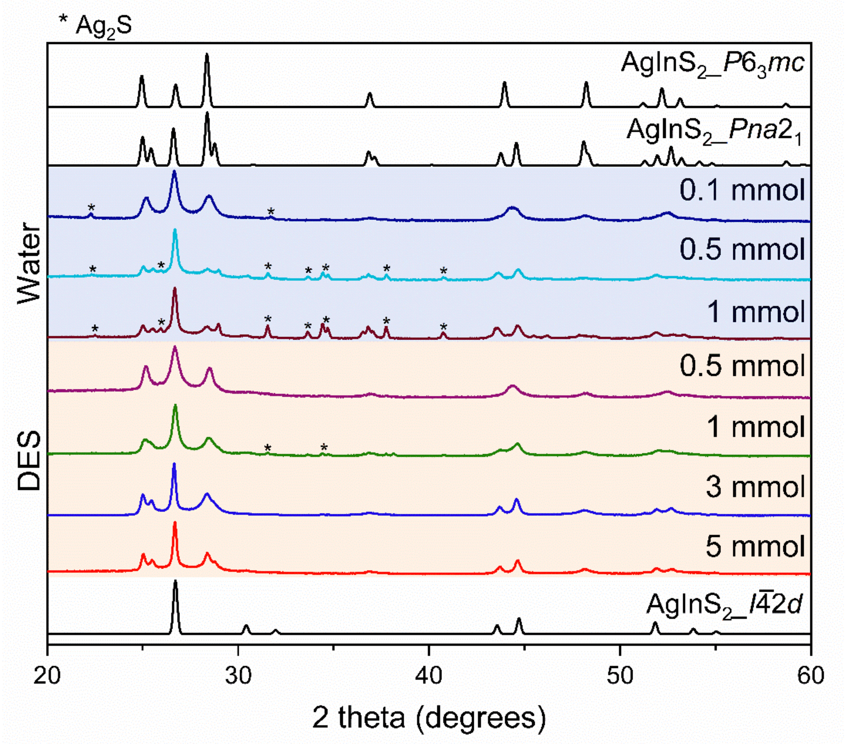
2-step synthesis. We further explored a 2-step synthesis of AgInS2, where reagents were dwelled at 80 °C to allow for the proper mixing and complete dissolution of the precursors before ramping to 180 °C. We discovered that the effect of the solvent used and the metal precursor concentration was more significant in the 2-step synthesis of AgInS2. We observed changes in the amount of impurities present and the ratios between the polymorphs in the 2-step-synthesized samples as we varied the solvent and the metal precursor concentration (Figure 4). From the distinct PXRD pattern, we suspect that the 2-step-synthesized AgInS2 at a dilute metal precursor concentration of 0.1 mmol for water and 0.5 mmol for DES syntheses are a mixture of the hexagonal P63mc polymorph and the tetragonal I2d polymorph. However, given that the observed peaks are broad, indicating a low crystallinity of the sample, and that the PXRD patterns of the wurtzite P63mc and Pna21 polymorphs are similar, the formation of wurtzite P63mc polymorph, based on PXRD data only, cannot be ascertained. The further increase of the metal precursor concentration led to a mixture of the orthorhombic Pna21 and the tetragonal I2d polymorphs, similar to that observed in the 1-step synthesis. An increase in the metal precursor concentration did not only lead to the prevalence of the tetragonal I2d polymorph but also resulted in an increased amount of Ag2S impurity.
Similar to the 2-step water-synthesized AgInS2, the 2-step DES-synthesized AgInS2 samples are also suspected to be a mixture of the hexagonal P63mc polymorph and the tetragonal I2d polymorph at the low metal precursor concentration of 0.5 and 1 mmol. It should be noted that while the PXRD patterns for Pna21 and P63mc are very similar, there are distinct differences: the P63mc PXRD pattern has single peaks at ~25, ~29, and ~45° 2θ, while the Pna21 PXRD pattern has double (split) peaks at the same diffraction angles (2θ). Yet again, the low crystallinity, thus broader diffraction peaks, does not allow to conclude the formation of the wurtzite P63mc polymorph unambiguously.
The PXRD pattern of the 0.1 mmol 2-step water-synthesized AgInS2 is almost identical to that of 0.5 mmol 2-step DES-synthesized AgInS2. A further increase in the metal precursor concentration first led to an increase in the fraction of tetragonal I2d polymorph in the 1 mmol 2-step DES-synthesized sample; then, the intergrowth of the Pna21 and I2d polymorphs appeared at 3 mmol and 5 mmol, with the 5 mmol sample (FWHM of 0.185) being more crystalline than the 3 mmol sample (FWHM of 0.191) of the 2-step DES-synthesis. The observed trend in the crystallinity of the 2-step synthesis is similar to that observed in the 1-step synthesis, i.e., the 5 mmol sample (FWHM of 0.185) is more crystalline than the 0.5 mmol sample (FWHM of 0.468) for the DES synthesis, and the 1 mmol sample (FWHM of 0.221) is more crystalline than the 0.1 mmol sample (FWHM of 0.412) for the water synthesis. The crystallinity also improved with an increase in the molar concentration in the 2-step synthesis.
In contrast to the 2-step water-synthesized AgInS2, the 2-step DES-synthesized AgInS2 samples have no impurities, except the 1 mmol 2-step DES-synthesized AgInS2, which has a very minor Ag2S impurity. Based on the PXRD data in Figure 4, we believe that the 2-step synthesis is better suited for a DES rather than water.
The SEM images obtained for the 2-step water synthesis samples are more diverse in particles arrangement and morphology than the 1-step water synthesis samples. The 0.1 mmol 2-step water-synthesized AgInS2 has a plate-like morphology with a flower, petal-like arrangement. The 0.5 mmol 2-step water-synthesized AgInS2 has a stacked, plate-like morphology, while the 1 mmol 2-step water-synthesized AgInS2 has a morphology that is clustered or rock-like. In contrast, the 2-step DES-synthesized AgInS2 samples have a stacked, plate-like morphology, as shown in Figure 5. There is an overall significant increase of particle size with an increase in the metal precursor concentration in the DES synthesis, but not in the water synthesis.
2.1. Scanning Transmission Electron Microscopy Study
The solution synthesis of AgInS2 reported here always results in two polymorphs: the tetragonal I2d chalcopyrite structure and the orthorhombic Pna21 wurtzite-like structure. Additionally, the PXRD patterns of AgInS2 samples (Figure 4) prepared using a 2-step heating profile and a low concentration of metal precursors (0.1 mmol for water and 0.5 mmol for DES) could be interpreted as a mixture of the tetragonal I2d and hexagonal P63mc wurtzite structure. We have utilized a high-resolution high-angle annular dark-field scanning transmission electron microscopy (HAADF–STEM) to address the following questions:
- Do tetragonal I2d and orthorhombic Pna21 wurtzite-like polymorphs form as an intergrowth, or do they precipitate as separate particles?
- Does the AgInS2 polymorph with P63mc wurtzite structure and disorder in Ag/In cations form at lower concentrations of metal precursors and 2-step heating profile?
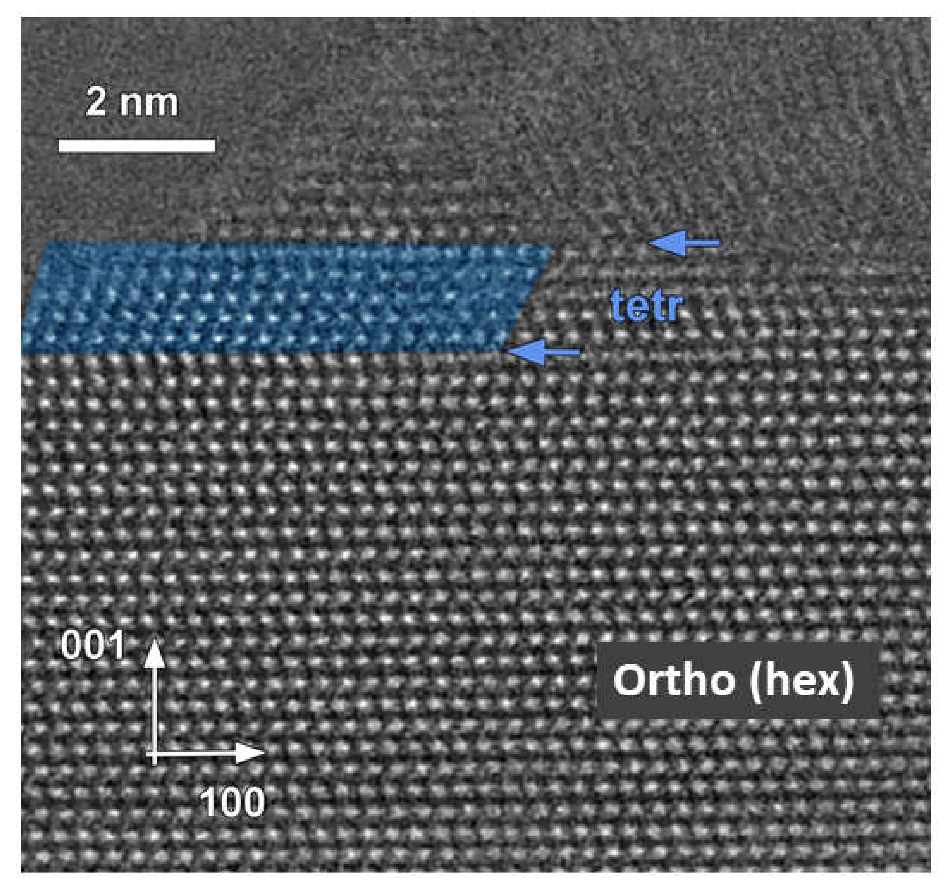
The HAADF–STEM image of AgInS2 prepared via the 2-step heating profile, DES solvent route, and 0.5 mmol metal precursors concentration (see Figure 4) suggests the intergrowth of two polymorphs: tetragonal I2d and orthorhombic Pna21 (Figure 6) with the clear phase boundary along (001)orth plane, indicated by arrows in Figure 6.
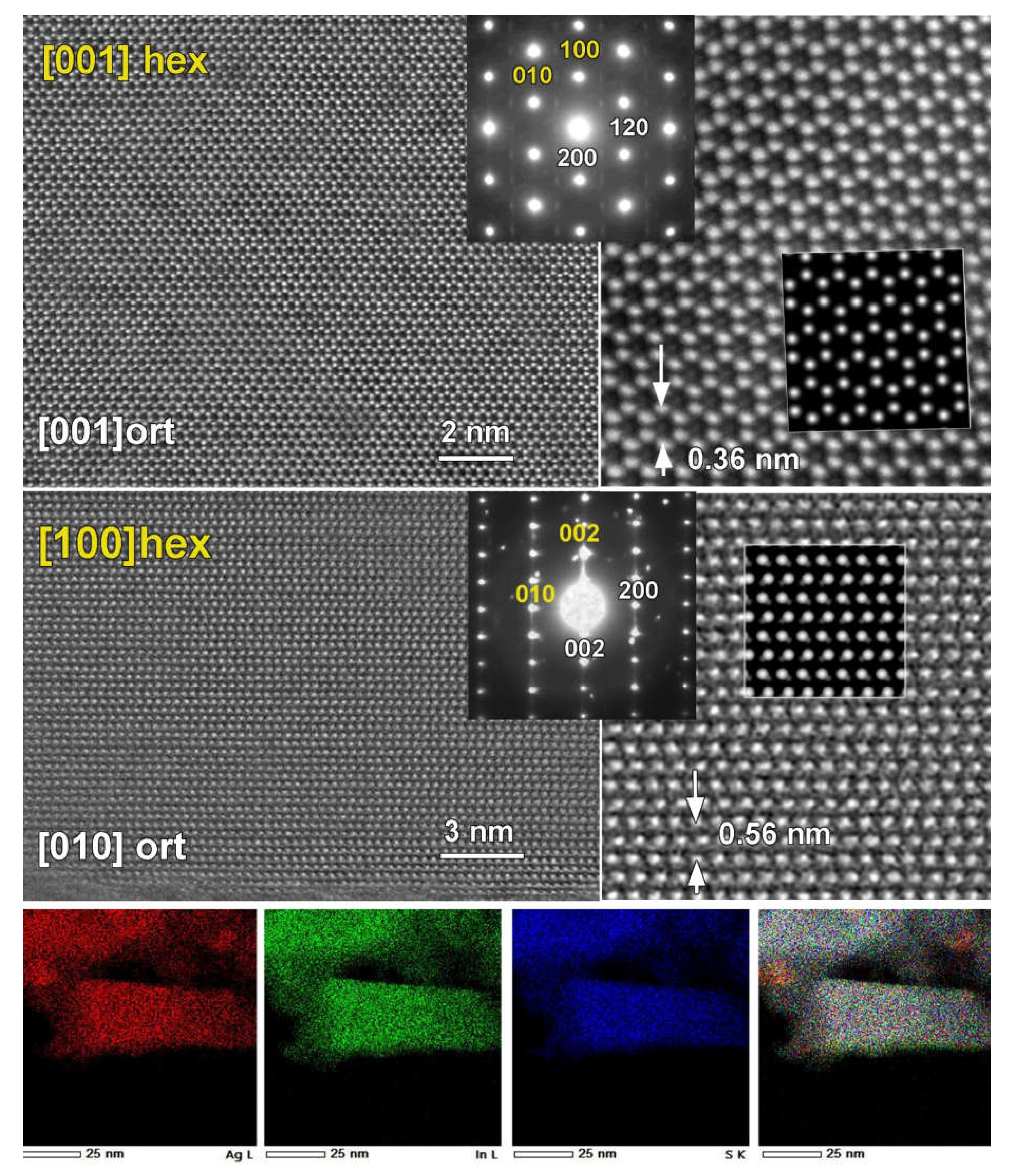
We further investigated the structure of the presumed P63mc wurtzite polymorph. The high-intensity diffraction spots in the electron diffraction (ED) patterns collected in the ab-plane and along the c-axis of the wurtzite structure can be indexed in a hexagonal unit cell (Figure 7: top, yellow), corresponding to the wurtzite structure. However, there are plenty of low-intensity reflections that cannot be indexed using the wurtzite cell but can be indexed in the P-orthorhombic cell with quadrupled volume. Therefore, the structure is better described as pseudo-wurtzite since the deviation from ideal hexagonal symmetry is observed. The HAADF–STEM images and simulated images of the structure further support this conclusion. The HAADF–STEM image in the ab-plane (Figure 7: middle right) supports the hexagonal in-plane arrangement of the Ag and In cations, while a variation of the contrast, seen in the lower magnification HAADF–STEM image, suggests the non-uniform distribution of Ag and In cations in the atomic columns, e.g., clustering. The Fast Fourier Transform (FFT) pattern (not shown here) includes the weak diffraction spots that are forbidden in the simple hexagonal wurtzite cell, further supporting the pseudo-wurtzite structure. All weak diffraction spots presented in the [001]orth ED pattern can be indexed based on the orthorhombic Pna21 structure. The simulated image of the structure performed along [001]ort and [010]ort directions (see insert Figure 7) strongly supports the orthorhombic structure with the clear pseudo-hexagonal symmetry. Importantly, the performed EDX–STEM elemental mapping (Figure 7 bottom panel) demonstrates the homogeneous distribution of all elements with a nominal composition close to AgInS2. The appearance of nanoparticles that are free from In (showing as more red in the bottom panel of Figure 7) is in agreement with the presence of the small Ag2S impurity.
Therefore, our study by ED and HAADF–STEM confirms the formation of an intergrowth of two AgInS2 polymorphs, tetragonal I2d and orthorhombic Pna21, within the same particle. Additionally, STEM clearly reveals that the previously reported wurtzite structure is best described as orthorhombic with pseudo-hexagonal symmetry. Thus, the synthesis using the 2-step profile and low concentrations of metal precursors results in the intergrowth of two polymorphs, tetragonal and pseudo-hexagonal wurtzite-like structures, with the latter best described as an orthorhombic structure.
2.2. High-Temperature PXRD
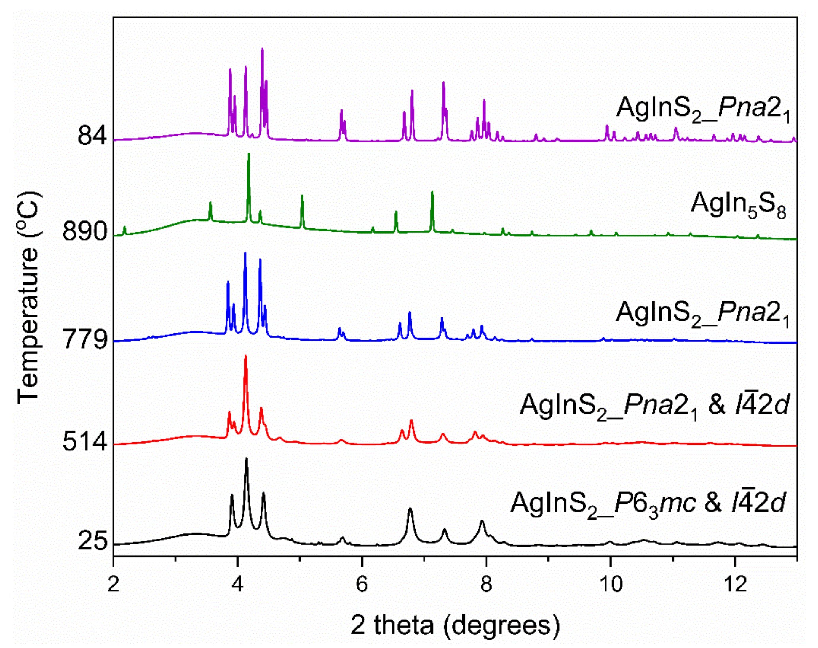
The 0.5 mmol 2-step DES-synthesized AgInS2 represents a metastable intergrowth of the two polymorphs–tetragonal and pseudo-hexagonal (best described as orthorhombic). We further investigated the thermal transformation of this metastable intergrowth by collecting the high-temperature synchrotron in situ PXRD data (HT–PXRD) on the 0.5 mmol 2-step DES-synthesized AgInS2. The data was collected on the powdered sample upon heating and cooling, as shown in Figure 8. The HT–PXRD data shows an intergrowth of the pseudo-hexagonal wurtzite-like polymorph and the I2d polymorph at room temperature. While the fraction of the I2d polymorph increased upon heating, a transformation of the pseudo-hexagonal polymorph to orthorhombic Pna21 happened between 355 and 380 °C. The combination of Pna21 and I2d polymorphs persisted until 700 °C, where the peaks belonging to the Pna21 polymorph began to increase in intensity and at 790 °C the orthorhombic phase was the only polymorph. The Pna21 AgInS2 remained the only crystalline phase upon heating until 863 °C, when the peaks belonging to the AgIn5S8 began to appear in the HT–PXRD pattern. At 890 °C AgIn5S8 was the only crystalline phase, suggesting that Ag2S and In2S3 were molten:
Upon cooling, AgIn5S8 reacted with the molten Ag2S and In2S3 and gave the Pna21 polymorph of AgInS2. It is important to note the high-temperature orthorhombic polymorph formed, albeit as a minor phase, at synthesis temperatures, well below the transition between tetragonal I2d and orthorhombic Pna21 polymorphs.
2.3. Diffuse Reflectance
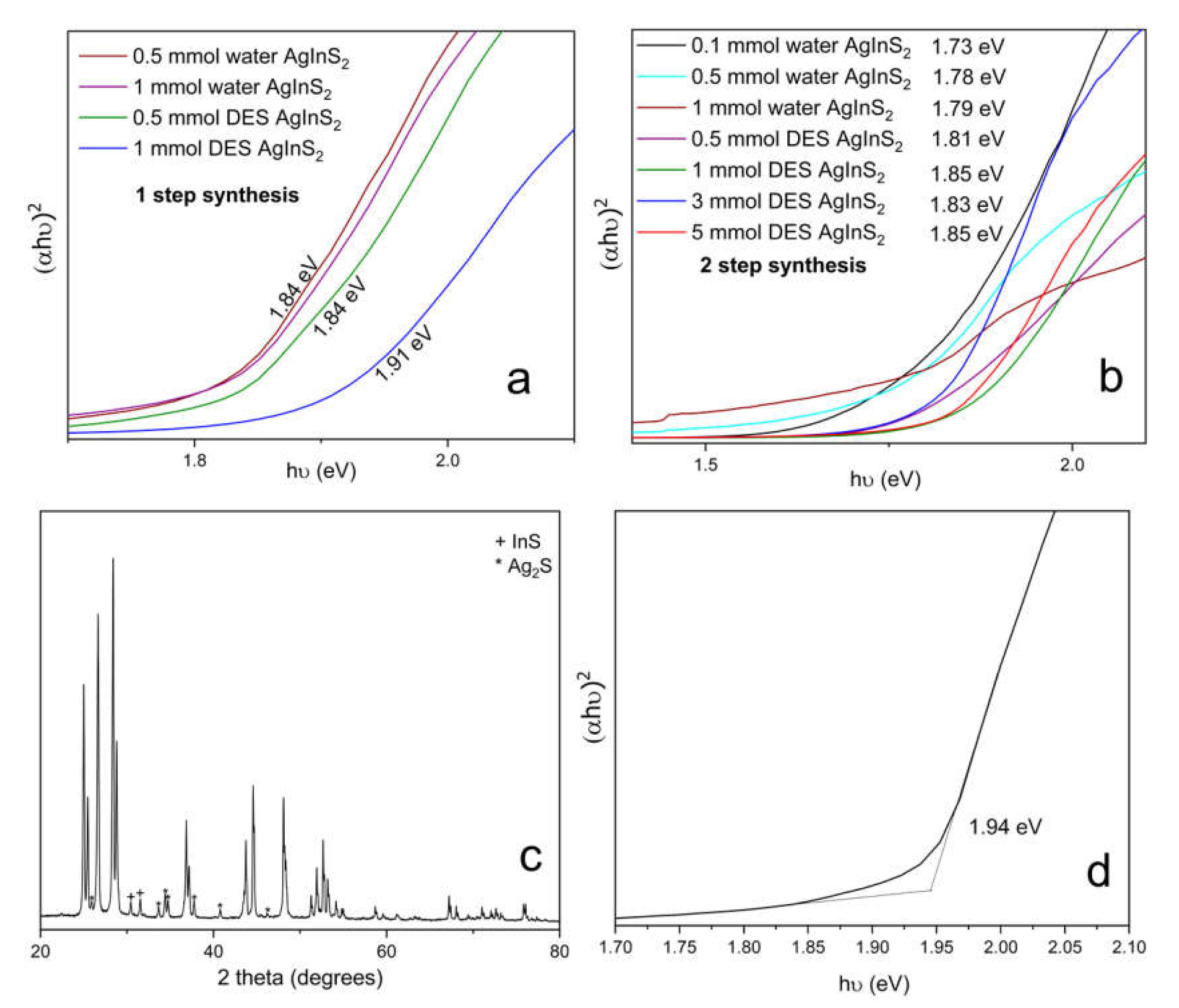
The effect of the synthesis conditions on the optical properties of the synthesized samples was investigated by diffuse reflectance using a UV-Vis-NIR spectrophotometer. In order to obtain the values of the bandgap, data was converted into Tauc plot, e.g. (a × hν)r vs. hν, where a is proportional to the absorption coefficient, hν is the excitation energy in eV, and r = 2 is for the direct allowed transitions. The bandgap was determined as the intercept of a tangent line and a baseline, as shown in Figure 9d. The additional absorption edge at ~1.0 eV was attributed to the Ag2S impurity. Three trends became apparent; firstly, the 2-step synthesis resulted in AgInS2 with slightly smaller bandgaps for the same precursor concentration and solvent used; secondly, the increase in the metal precursor concentrations led to the slight increase in the bandgap values; and, lastly, the bandgap values for the AgInS2 synthesized using the DES were overall slightly higher than that for the water synthesis.
The reported values of the bandgaps span the range of 1.86–1.87 eV for the tetragonal I2d polymorph (chalcopyrite structure) and 1.96–1.98 eV for the Pna21 orthorhombic (wurtzite-like structure) polymorph [17,20,21], while larger values of the bandgap were measured for the AgInS2 nanoparticles due to the quantum confinement effect and Ag/In non-stoichiometry [24,25]. The solution synthesis in either the water or the DES results in AgInS2 with the bandgap on the slightly lower side compared to the literature, which we tentatively attribute to the formation of intergrowth between the tetragonal and orthorhombic polymorphs.
We also compared the bandgap of the polymorph mixture obtained using the solution synthesis with the bandgap of the orthorhombic Pna21 polymorph, which was synthesized by high-temperature annealing (at 800 °C at 10 °C/min for 30 min) of the 0.1 mmol 2-step water-synthesized AgInS2 (Figure 9c,d). The high-temperature-synthesized Pna21 orthorhombic polymorph is highly crystalline and has a bandgap of 1.94 eV, which is comparable to the previously reported values [17,20,21].
3. Conclusions
We have studied the solution microwave-assisted synthesis of AgInS2 from two solvents—water and an eutectic mixture of choline chloride and thiourea (a deep eutectic solvent). AgInS2 was previously reported to have four polymorphic modifications. According to the literature data, the tetragonal I2d (chalcopyrite structure) is the thermodynamically stable polymorph at room-temperature., which transforms into a high-temperature orthorhombic (wurtzite-like) polymorph at ~620 °C. The microwave-assisted solution synthesis from either the water or the DES resulted in a metastable mixture of tetragonal and orthorhombic polymorphs of AgInS2, with the tetragonal I2d being prevalent. We also investigated how the fraction and crystallinity of the polymorphs formed can be controlled by varying the heating profile, solvent type, and metal precursor concentration. Overall, the increase in the metal precursor concentrations improved the crystallinity of the polymorphic intergrowth, while the 2-step microwave heating profile was better suited for the DES synthesis because the binary sulfides impurities were eliminated. We further studied the possible mechanism of the AgInS2 formation from the solution via the microwave-assisted synthesis and ruled out the partial cation exchange between the crystalline binary sulfide and a cation in the solution as a possible mechanism.
The PXRD data of the AgInS2 samples prepared via the 2-step heating profile and low concentrations of metal precursors have initially suggested the possible formation of hexagonal polymorph with wurtzite structure and Ag/In mixed occupied cation positions. The state-of-the-art study by scanning transmission electron microscopy (STEM) revealed that the long thought wurtzite polymorph has, indeed, pseudo-hexagonal symmetry, and its structure is best described as an orthorhombic structure alike to the high-temperature Pna21 polymorph. The STEM study further revealed that the solution synthesis from the DES resulted in the intergrowth of tetragonal and orthorhombic polymorphs within the same particle, thus resulting in the metastable product.
The bandgaps of the solution synthesized AgInS2 are similar, albeit a bit lower compared to the previously reported values for both the tetragonal and orthorhombic polymorphs.
4. Materials and Methods
Silver nitrate, AgNO3 (Alfa Aesar, 99.9+%); indium chloride, InCl3·yH2O (Alfa Aesar, 99.9+%); thiourea, CH4N2S (Acros 99+%); choline chloride, (CH3)3N(Cl)CH2CH2OH (Sigma-Aldrich, 98%), and deionized (DI) water were used as received.
AgInS2 was synthesized in either DI water or Deep Eutectic Solvent (DES). For the DI water synthesis, powders of AgNO3, InCl3·yH2O, and CH4N2S were weighted in x:x:5 molar ratio (where x = 0.1, 0.5 and 1 mmols), respectively, loaded into a microwave tube with 6 mL of DI water, and heated in a microwave reactor with a 1-step or 2-step heating profile. For the DES synthesis, powders of AgNO3 and InCl3·yH2O were weighted in x:x molar ratio (where x = 0.5, 1, 3 and 5 mmol). After that, approximately 4 g of thiourea and 3.59 g of choline chloride were added to the metal precursors in a microwave tube and heated in a Monowave 400 microwave reactor (Anton Paar) with a 1-step or 2-step heating profile. The resulting mixture of AgInS2 and solvent from either the water or DES synthesis was then washed with water by sonication and centrifuged several times. Then, it was further washed with ethanol and centrifuged several times until the supernatant is clear. The AgInS2 was then dried in a static vacuum overnight.
1-step heating profile: The loaded microwave tube was placed in a Monowave 400 microwave reactor and heated to 180 °C for 8 min. It then dwelled for 2 h. The resulting mixture was then cooled to room temperature.
2-step heating profile: The loaded microwave tube was placed in a Monowave 400 microwave reactor and heated to 80 °C for 5 min and dwelled for 20 min. The reaction mixture was then quickly (~1 min) heated to 180 °C and dwelled for 1 h. The resulting mixture was cooled to room temperature.
5. Characterization
5.1. Powder X-ray Diffraction
Samples were analyzed by powder X-ray diffraction (PXRD) using a Rigaku Miniflex 600 diffractometer with Cu Kα radiation (λ = 1.54051 Å). Measurement was carried out on a silicon, zero-background holder at room temperature in air. The powder pattern was collected in 0.02° steps increment at a rate of 10°/minute. Phase identification was performed using the Match! 2.0 software [26]. The Full width at half maximum (FWHM) was determined using the PDXL software [27].
5.2. Diffuse Reflectance UV–Vis Spectroscopy
The diffuse reflectance of the powdered samples was collected on a Perkin Elmer Lambda 1050+ UV/Vis/NIR spectrophotometer in reflectance mode. The instrument was equipped with a 150 mm Spectralon-coated integrating sphere. We loaded finely ground samples into a powder holder, which had a lens. The samples were pressed against the lens and held in place by a press and a spring within the holder. We then placed the holder at the reflectance port, while the specular port was left open. The Iris aperture was adjusted so that the sample beam was focused only on the samples. A sample holder containing a lens and Spectralon reference standard was used as a blank.
5.3. Scanning Electron Microscopy and Energy Dispersive Spectroscopy
Elemental analysis was performed using a JEOL JSM-IT200 scanning electron microscope (SEM) equipped with a JEOL energy-dispersive X-ray (EDX) analysis system with a silicon drift detector. Powder samples were adhered to aluminum stubs using carbon tape and then placed into an aluminum holder for insertion into the SEM. Samples were oriented perpendicular to the beam and analyzed using a 15 kV accelerating voltage with an accumulation time of 60 s.
5.4. High-Temperature Synchrotron Powder X-ray Diffraction (HT–PXRD)
High temperature in situ synchrotron powder diffraction data were collected for AgInS2 at beamline 17-BM Advanced Photon Source (APS) at Argonne National Laboratory, with a wavelength of 0.24158 Å. The powdered sample of AgInS2 was loaded into a 0.7 mm outer diameter silica capillary, which was sealed under vacuum. The sealed tube was then inserted into a second silica tube of 0.9 mm inner diameter and 1.1 mm outer diameter, and this second tube was mounted on a sample stage featuring two micro-heaters and a thermocouple, which was placed close to the area being measured. Details of the experimental setup can be found elsewhere [28]. Data were collected from room temperature to 895 °C at the rate of 15 °C/min, and also upon cooling. Diffraction patterns were analyzed using Match! 2.0 software [26].
5.5. Differential Scanning Calorimetry
The DSC/TGA experiment was performed using a Netzsch STA449 F1 Jupiter. 5 mg of the powdered sample (1-step water-synthesized In2S3) was loaded into a pan-type alumina (Al2O3) crucible with an alumina cover. The sample was heated from 40 to 200, or 500 °C, and, subsequently, cooled to 50 °C, with a rate of 10 °C/min under a constant argon gas flow.
5.6. Scanning Transmission Electron Microscopy (STEM) and Electron Diffraction
HAADF–STEM imaging and ED studies were performed using JEM ARM200F cold FEG double aberration corrected microscope operated at 200 kV, equipped with a large angle CENTURIO EDX detector, Orius CCD camera, and Quantum GIF. Samples for STEM were prepared by mechanical grinding of material in an agate mortar, adding ethanol and depositing obtained suspension on the Cu holey carbon grid. HAADF–STEM image simulation was done using JEMS Software developed by P. Stadelmann. The 0.5 mmol 2-step DES-synthesized AgInS2 was used for STEM analysis.
Supplementary Materials
The following supporting information can be downloaded at: https://www.mdpi.com/article/10.3390/molecules27061815/s1, Figure S1: PXRD pattern of 1-step and 2-step synthesized In2S3 in both water and DES; Figure S2: (a) DSC/TGA data of water synthesized In2S3 at 500 °C (b) PXRD pattern of water synthesized In2S3 before and after DSC at 200 °C and 500 °C; Figure S3: PXRD pattern of (a) 1-step water synthesized In2S3; (b) the instantaneously formed product of a reaction of pre-made In2S3 with aqueous AgNO3 solution; (c) the Ag2S product obtained from the metathesis reaction of In2S3 with a twice concentrated aqueous AgNO3 solution at room temperature which was left undisturbed for 2 days.
Author Contributions
Conceptualization, project supervision, resources, J.V.Z.; methodology, formal analysis, writing—original draft preparation, visualization, A.N.A.; investigation, data collection, curation, and validation, formal analysis, A.N.A., R.A.E., T.C., O.I.L. All authors have read and agreed to the published version of the manuscript.
Funding
This research was funded by J.V.Z. startup funds from Iowa State University. Use of the Advanced Photon Source at Argonne National Laboratory was supported by the U. S. Department of Energy, Office of Science, and Office of Basic Energy Sciences, under contract no. DE-AC02-06CH11357.
Data Availability Statement
The data presented in this study are available in article and Supplementary Materials, and are also available on request from the corresponding author.
Acknowledgments
We thank Kirill Kovnir (Department of Chemistry, Iowa State University, and Ames Laboratory) for the access to the PXRD diffractometer; Brett Boote (Chemical Instrumentation facility, Iowa State University) for the help with the DSC/TGA instrument and the SEM.
Conflicts of Interest
The authors declare no conflict of interest.
References
- Abbott, A.P.; Boothby, D.; Capper, G.; Davies, D.L.; Rasheed, R.K. Deep Eutectic Solvents Formed between Choline Chloride and Carboxylic Acids: Versatile Alternatives to Ionic Liquids. J. Am. Chem. Soc. 2004, 126, 9142–9147. [Google Scholar] [CrossRef] [PubMed]
- Abbott, A.P.; Capper, G.; Davies, D.L.; Rasheed, R.K.; Tambyrajah, V. Novel solvent properties of choline chloride/urea mixtures. Chem. Commun. 2003, 70–71. [Google Scholar] [CrossRef] [PubMed] [Green Version]
- Zhang, Q.H.; Vigier, K.D.; Royer, S.; Jerome, F. Deep eutectic solvents: Syntheses, properties and applications. Chem. Soc. Rev. 2012, 41, 7108–7146. [Google Scholar] [CrossRef]
- Abbott, A.P.; Capper, G.; Davies, D.L.; McKenzie, K.J.; Obi, S.U. Solubility of Metal Oxides in Deep Eutectic Solvents Based on Choline Chloride. J. Chem. Eng. Data 2006, 51, 1280–1282. [Google Scholar] [CrossRef]
- Hong, S.K.; Doughty, R.M.; Osterloh, F.E.; Zaikina, J.V. Deep eutectic solvent route synthesis of zinc and copper vanadate n-type semiconductors-mapping oxygen vacancies and their effect on photovoltage. J. Mater. Chem. A 2019, 7, 12303–12316. [Google Scholar] [CrossRef] [Green Version]
- Hong, S.; Burkhow, S.J.; Doughty, R.M.; Cheng, Y.; Ryan, B.J.; Mantravadi, A.; Roling, L.T.; Panthani, M.G.; Osterloh, F.E.; Smith, E.A.; et al. Local Structural Disorder in Metavanadates MV2O6 (M = Zn and Cu) Synthesized by the Deep Eutectic Solvent Route: Photoactive Oxides with Oxygen Vacancies. Chem. Mater. 2021, 33, 1667–1682. [Google Scholar] [CrossRef]
- Boston, R.; Foeller, P.Y.; Sinclair, D.C.; Reaney, I.M. Synthesis of Barium Titanate Using Deep Eutectic Solvents. Inorg. Chem. 2017, 56, 542–547. [Google Scholar] [CrossRef]
- Thorat, G.M.; Jadhav, H.S.; Roy, A.; Chung, W.J.; Seo, J.G. Dual Role of Deep Eutectic Solvent as a Solvent and Template for the Synthesis of Octahedral Cobalt Vanadate for an Oxygen Evolution Reaction. ACS Sustain. Chem. Eng. 2018, 6, 16255–16266. [Google Scholar] [CrossRef]
- Hong, S.; Cheng, Y.; Hariyani, S.; Li, J.Z.; Doughty, R.M.; Mantravadi, A.; Adeyemi, A.N.; Smith, E.A.; Brgoch, J.; Osterloh, F.E.; et al. The Deep Eutectic Solvent Precipitation Synthesis of Metastable Zn4V2O9. Inorg. Chem. 2022, 66, 154–169. [Google Scholar] [CrossRef]
- Zeng, J.R.; Chen, L.; Siwal, S.S.; Zhang, Q.B. Solvothermal sulfurization in a deep eutectic solvent; a novel route to synthesize Co-doped Ni3S2 nanosheets supported on Ni foam as active materials for ultrahigh-performance pseudocapacitors. Sustain. Energy Fuels 2019, 3, 1957–1965. [Google Scholar] [CrossRef]
- Zhang, T.; Doert, T.; Ruck, M. Synthesis of Metal Sulfides from a Deep Eutectic Solent Precursor (DESP). Z. Anorg. Allg. Chem. 2017, 643, 1913–1919. [Google Scholar] [CrossRef] [Green Version]
- Jiang, J.Y.; Chang, L.Y.; Zhao, W.C.; Tian, Q.Y.; Xu, Q. An advanced FeCoNi nitro-sulfide hierarchical structure from deep eutectic solvents for enhanced oxygen evolution reaction. Chem. Commun. 2019, 55, 10174–10177. [Google Scholar] [CrossRef] [PubMed]
- Hahn, H.; Frank, G.; Klinger, W.; Meyer, A.D.; Stöerger, G.; Anorg, Z. Untersuchungen über ternäre Chalkogenide. V. Über einige ternäre Chalkogenide mit Chalkopyritstruktur. Z. Anorg. Allg. Chem. 1953, 271, 153–170. [Google Scholar] [CrossRef]
- Delgado, G.; Mora, A.J.; Pineda, C.; Tinoco, T. Simultaneous Rietveld refinement of three phases in the Ag-In-S semiconducting system from X-ray powder diffraction. Mater. Res. Bull. 2001, 36, 2507–2517. [Google Scholar] [CrossRef]
- Roth, R.S.; Parker, H.S.; Brower, W.S. Comments on the system Ag2S·In2S3. Mater. Res. Bull. 1973, 8, 333–338. [Google Scholar] [CrossRef]
- Range, K.J.; Keubler, M.; Weiss, A. Eine Hochdruckmodifikation des AgInS2 mit α-NaFeO2-Structure. Z. Naturforschg. 1969, 24, 1060–1061. [Google Scholar] [CrossRef] [Green Version]
- Shay, J.L.; Tell, B.; Schiavon, L.M.; Kasper, H.M.; Thiel, F. Energy bands of AgInS2 in the chalcopyrite and orthorhombic structures. Phys. Rev. B 1974, 9, 1719–1723. [Google Scholar] [CrossRef]
- Jain, A.; Shyue Ping, O.; Hautier, G.; Chen, W.; Richards, W.D.; Dacek, S.; Cholia, S.; Gunter, D.; Skinner, D.; Ceder, G.; et al. Commentary: The Materials Project: A materials genome approach to accelerating materials innovation. APL Mater. 2013, 1, 011002. [Google Scholar] [CrossRef] [Green Version]
- Torimoto, T.; Adaichi, T.; Okazaki, K.-i.; Sakuraoka, M.; Shibayama, T.; Ohtani, B.; Kudo, A.; Kuwabata, S. Facile Synthesis of ZnS-AnInS2 Solid Solution Nanoparticles for a Color-Adjustable Luminophore. J. Am. Chem. Soc. 2007, 129, 12388. [Google Scholar] [CrossRef]
- Kowalik, P.; Penkala, M.; Bujak, P.; Kmita, A.; Gajewska, M.; Ostowski, A.; Slodek, A.; Pron, A. From Ag2S to luminescent Ag-In-S nanocrystals via an ultrasonic method—In situ synthesis study in an NMR tube. J. Mater. Chem. C 2020, 8, 8942–8952. [Google Scholar] [CrossRef]
- Chhikara, N.; Gupta, P.; Gupta, B.K.; Jain, K.; Chand, S. Synthesis of AgInS2 nanoparticles Directly in Poly (3-hexyl thiophene) (P3HT) Matrix: Photoluminescence quenching studies. In Physics of Semiconductor Devices; Environmental Engineering; Springer: Cham, Switzerland, 2014; pp. 335–338. [Google Scholar]
- Hamanaka, Y.; Ogawa, T.; Tsuzuski, M.; Kuzuya, T. Photoluminescence Properties and Its Origin of AgInS2 Quantum Dots with Chalcopyrite Structure. J. Phys. Chem. C 2011, 115, 1786–1792. [Google Scholar] [CrossRef]
- Uematsu, T.; Wajima, K.; Sharma, D.K.; Hirata, S.; Yamamoto, T.; Kameyama, T.; Vacha, M.; Torimoto, T.; Kuwabata, S. Narrow band-edge photoluminescence from AgInS2 semiconductor nanoparticles by the formation of amorphous III-VI semiconductor shells. NPG Asia Mater. 2018, 10, 713–726. [Google Scholar] [CrossRef]
- Park, Y.J.; Oh, J.H.; Han, N.H.; Yoon, H.C.; Park, S.M.; Do, Y.R.; Song, J.K. Phoyoluminescence of Band Gap States in AgInS2 Nanoparticles. J. Phys. Chem. C 2014, 118, 25677–25683. [Google Scholar] [CrossRef]
- Hong, S.P.; Park, H.K.; Oh, J.H.; Yang, H.; Do, Y.R. Comparisons of the structural and optical properties of o-AgInS2, t-AgInS2, and c-AgIn5S8 nanocrystals and their solid-solution nanocrystals with ZnS. J. Mater. Chem. 2012, 22, 18939–18949. [Google Scholar] [CrossRef]
- Putz, H.; Brandenburg, K. Match!-Phase Analysis Using Powder Diffraction, Crystal Impact. Available online: https://www.crystalimpact.com/contact.htm (accessed on 20 December 2021).
- PDXL: Integrated X-ray Powder Diffraction Software; Version 2.8.1.1; Rigaku: The Woodlands, TX, USA, 2018.
- Chupas, P.J.; Chapman, K.W.; Kurtz, C.; Hanson, J.C.; Lee, P.L.; Grey, C.P. A versatile sample-environment cell for non-ambient X-ray scattering experiments. J. Appl. Crystallogr. 2008, 41, 822–824. [Google Scholar] [CrossRef]
Figure 1.
The crystal structures of the tetragonal, orthorhombic, and hexagonal polymorphs of AgInS2. Atoms are color-coded: In—purple, Ag—gray, S—yellow, and the site with mixed In/Ag occupancy is denoted as a half-purple and half-grey sphere. The unit cell is shown with a black line. The orientation is chosen to highlight similarities in the structures of polymorphs.

Figure 2.
PXRD patterns of the 1-step synthesis of water (blue shaded region) and DES-synthesized (orange shaded region) AgInS2 with varying concentrations of metal precursors. Diffraction peaks for Ag2S impurity are denoted with an asterisk (*). The theoretical PXRD patterns include ICSD 51617, representing the I2d; ICSD 76276, representing the P63mc; and ICSD 605408, representing the Pna21 polymorph.

Figure 3.
SEM images of the 1-step-synthesized AgInS2 made with varying metal precursor concentrations in water: (a) 0.1 mmol, (b) 0.5 mmol, and (c) 1 mmol; and in DES: (d) 0.5 mmol, (e) 1 mmol, and (f) 3 mmol.
Figure 3.
SEM images of the 1-step-synthesized AgInS2 made with varying metal precursor concentrations in water: (a) 0.1 mmol, (b) 0.5 mmol, and (c) 1 mmol; and in DES: (d) 0.5 mmol, (e) 1 mmol, and (f) 3 mmol.

Figure 4.
PXRD patterns of the 2-step synthesis of water (blue shaded region) and DES-synthesized AgInS2 (orange shaded region), with varying concentrations of metal precursors. Diffraction peaks for the Ag2S impurity are denoted with an asterisk (*). The theoretical PXRD patterns include ICSD 51617, representing the I2d; ICSD 76276, representing the P63mc; and ICSD 605408, representing the Pna21 polymorph.

Figure 5.
SEM images of the 2-step-synthesized AgInS2 made with varying metal precursor concentrations in water: (a) 0.1 mmol, (b) 0.5 mmol, and (c) 1 mmol; and in DES: (d) 1 mmol, (e) 3 mmol, and (f) 5 mmol.
Figure 5.
SEM images of the 2-step-synthesized AgInS2 made with varying metal precursor concentrations in water: (a) 0.1 mmol, (b) 0.5 mmol, and (c) 1 mmol; and in DES: (d) 1 mmol, (e) 3 mmol, and (f) 5 mmol.

Figure 6.
High-resolution HAADF–STEM image of AgInS2 crystallite prepared via 2-step heating profile, 0.5 mmol metal concentration, and DES. The arrows indicate the phase boundary between orthorhombic and tetragonal (in blue) polymorphs.
Figure 6.
High-resolution HAADF–STEM image of AgInS2 crystallite prepared via 2-step heating profile, 0.5 mmol metal concentration, and DES. The arrows indicate the phase boundary between orthorhombic and tetragonal (in blue) polymorphs.

Figure 7.
(Top and Middle, Left) High-resolution HAADF–STEM images and corresponding electron diffraction (ED) patterns of the two main zones of the (pseudo)-wurtzite AgInS2 structure: top panel, [001] and middle panel, [010]ort/[100]hex. High-intensity diffraction spots can be indexed using hexagonal wurtzite structure (P63mc, yellow). All of the diffraction spots, including those with lower intensity can be indexed using the orthorhombic wurtzite-like structure (Pna21 space group). Top and Middle, Right: Magnified high-resolution HAADF–STEM images of the two main zones and simulated images based on orthorhombic wurtzite-like structure (Pna21 space group) as inserts. Bottom: Images of the EDX-STEM elemental mapping for Ag L(red), In L (green), S K (blue), and an overlapping color image.

Figure 8.
HT–PXRD data of 0.5 mmol the 2-step DES-synthesized AgInS2 upon heating and cooling.

Figure 9.
Tauc plots for AgInS2 obtained via (a) 1-step and (b) 2-step synthesis; (c) the PXRD pattern of high-temperature Pna21 orthorhombic AgInS2 polymorph, synthesized via a high-temperature route; and (d) Tauc plot of high-temperature Pna21 orthorhombic AgInS2 polymorph, synthesized via high-temperature route.

Publisher’s Note: MDPI stays neutral with regard to jurisdictional claims in published maps and institutional affiliations. |
© 2022 by the authors. Licensee MDPI, Basel, Switzerland. This article is an open access article distributed under the terms and conditions of the Creative Commons Attribution (CC BY) license (https://creativecommons.org/licenses/by/4.0/).
Share and Cite
MDPI and ACS Style
Adeyemi, A.N.; Earnest, R.A.; Cox, T.; Lebedev, O.I.; Zaikina, J.V. Microwave-Assisted Solution Synthesis of Metastable Intergrowth of AgInS2 Polymorphs. Molecules 2022, 27, 1815. https://doi.org/10.3390/molecules27061815
AMA Style
Adeyemi AN, Earnest RA, Cox T, Lebedev OI, Zaikina JV. Microwave-Assisted Solution Synthesis of Metastable Intergrowth of AgInS2 Polymorphs. Molecules. 2022; 27(6):1815. https://doi.org/10.3390/molecules27061815
Chicago/Turabian StyleAdeyemi, Adedoyin N., Rae Ann Earnest, Tori Cox, Oleg I. Lebedev, and Julia V. Zaikina. 2022. "Microwave-Assisted Solution Synthesis of Metastable Intergrowth of AgInS2 Polymorphs" Molecules 27, no. 6: 1815. https://doi.org/10.3390/molecules27061815