
Jasminum sambac: A Potential Candidate for Drug Development to Cure Cardiovascular Ailments

 , , ,
, , ,
Abstract
:1. Introduction
2. Results
2.1. Priliminary Phytochemical Screening
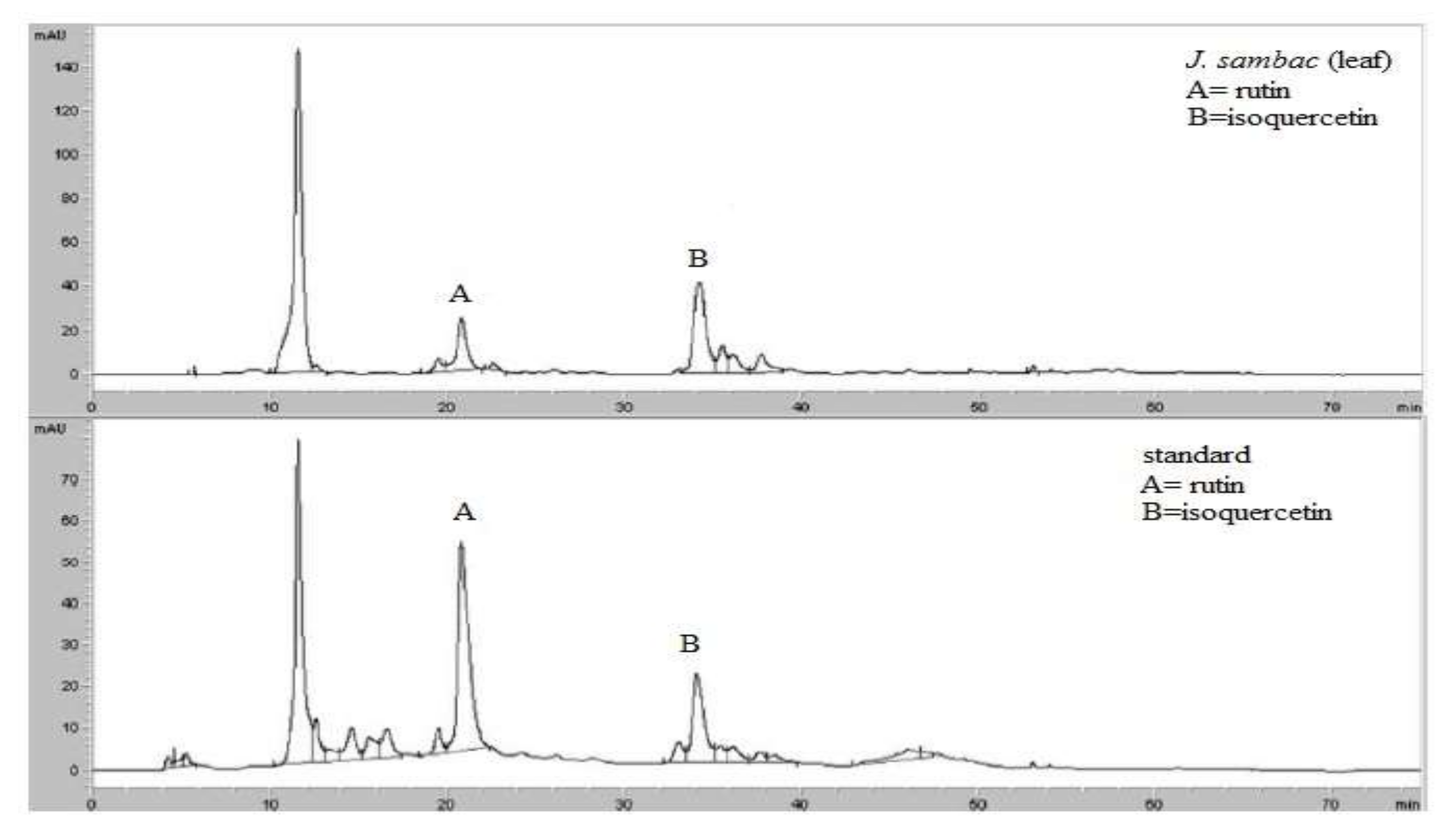
2.2. HPLC Analysis
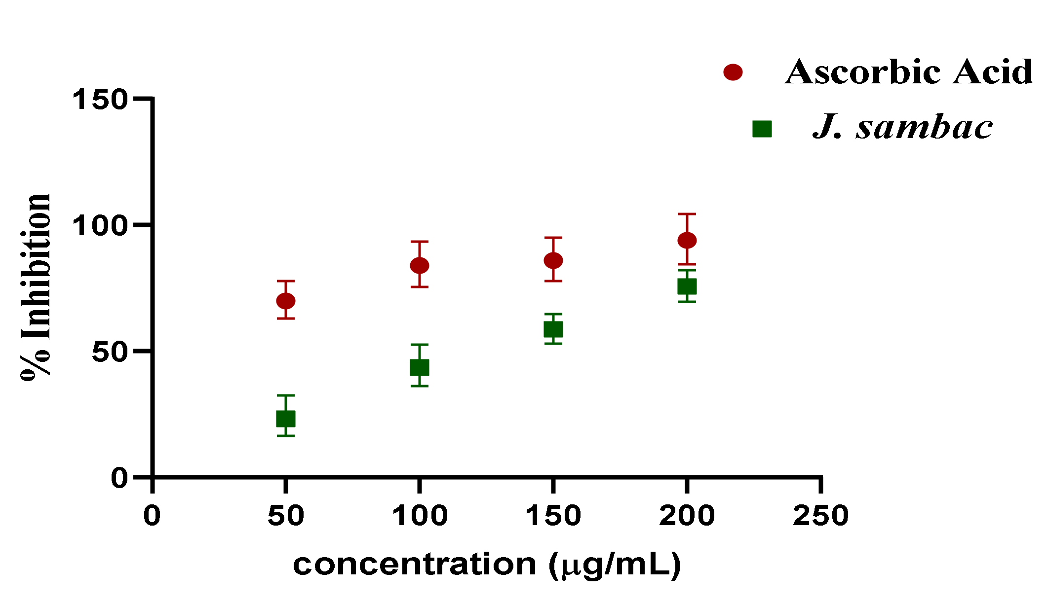
2.3. DPPH Assay
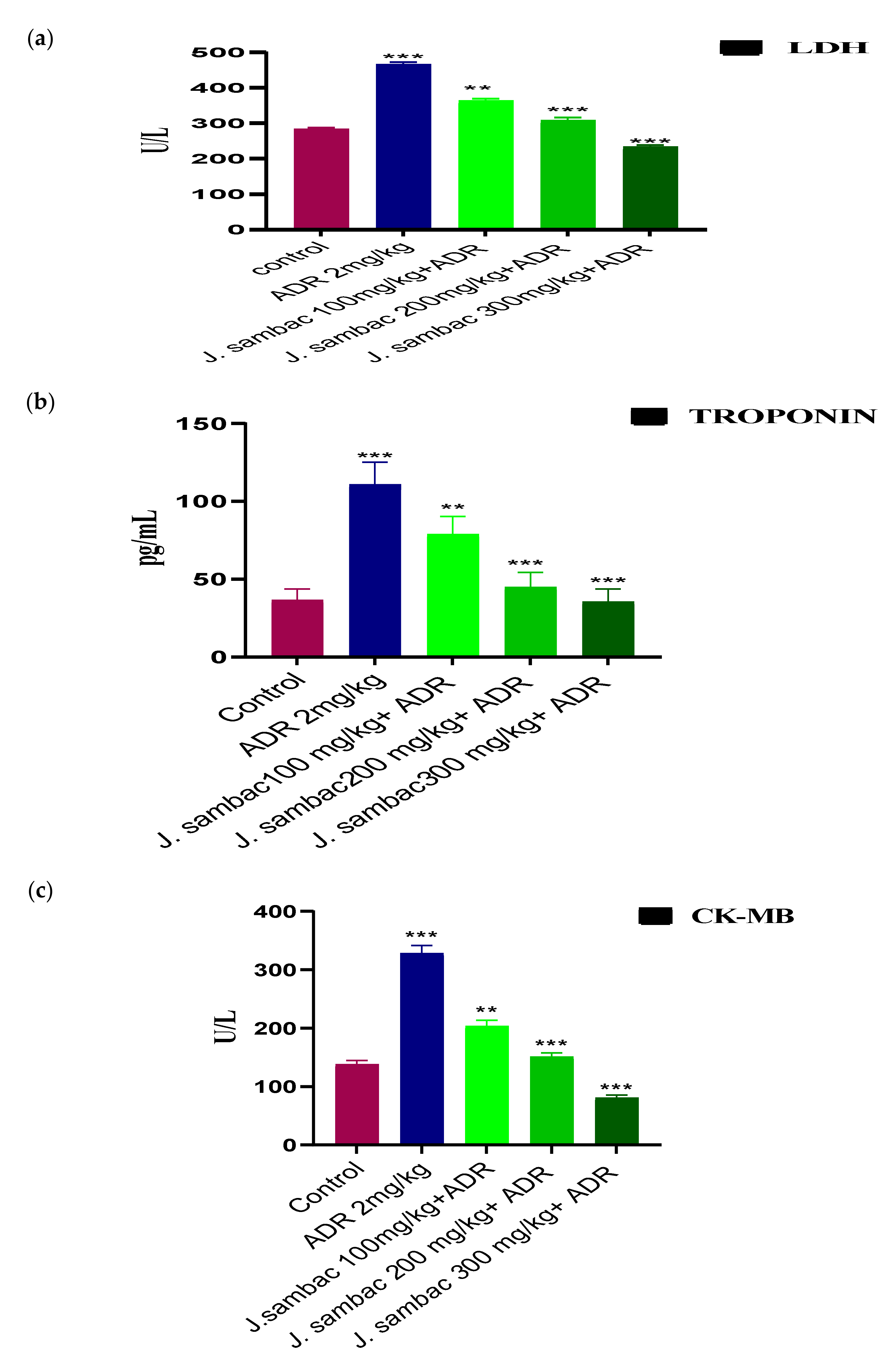
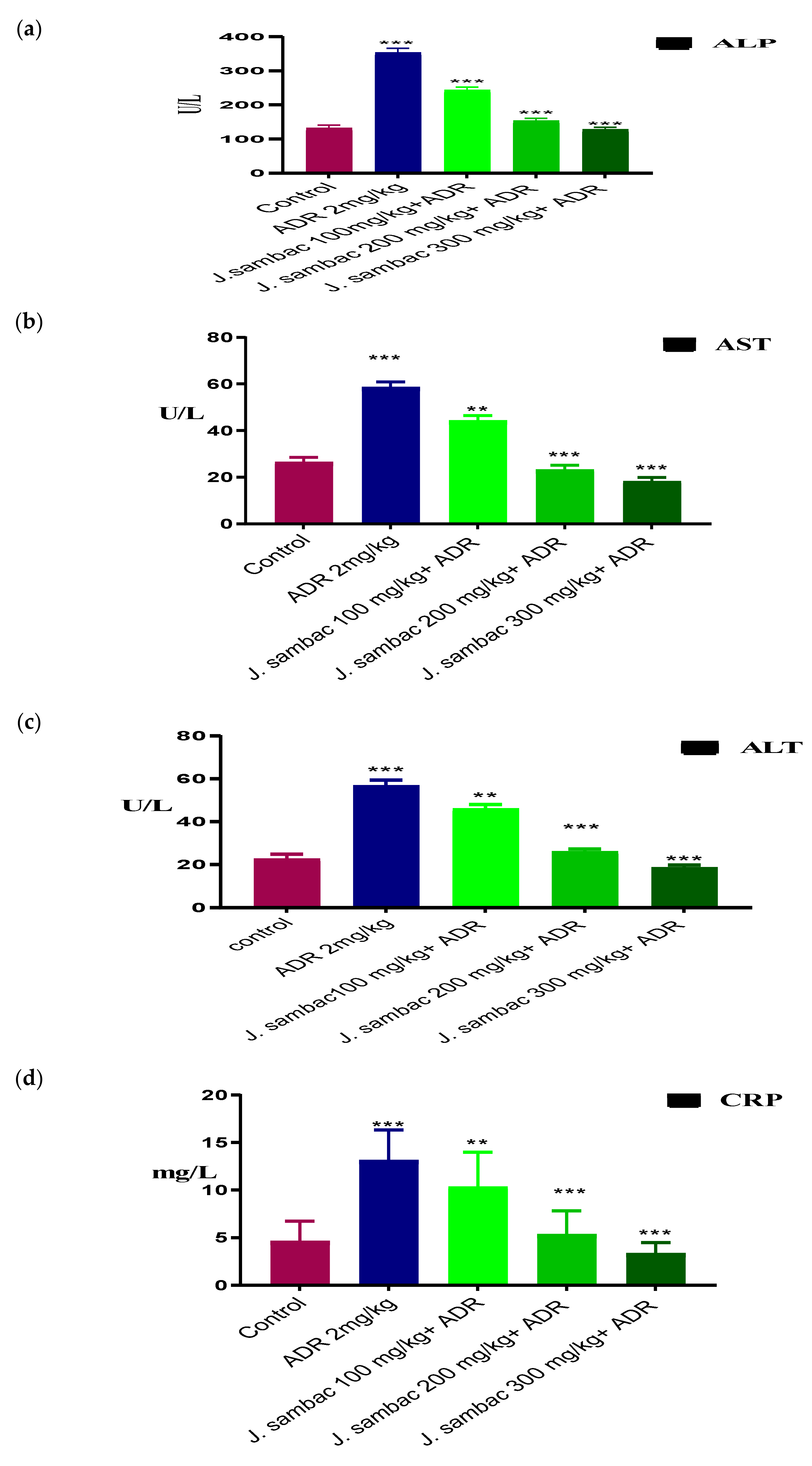
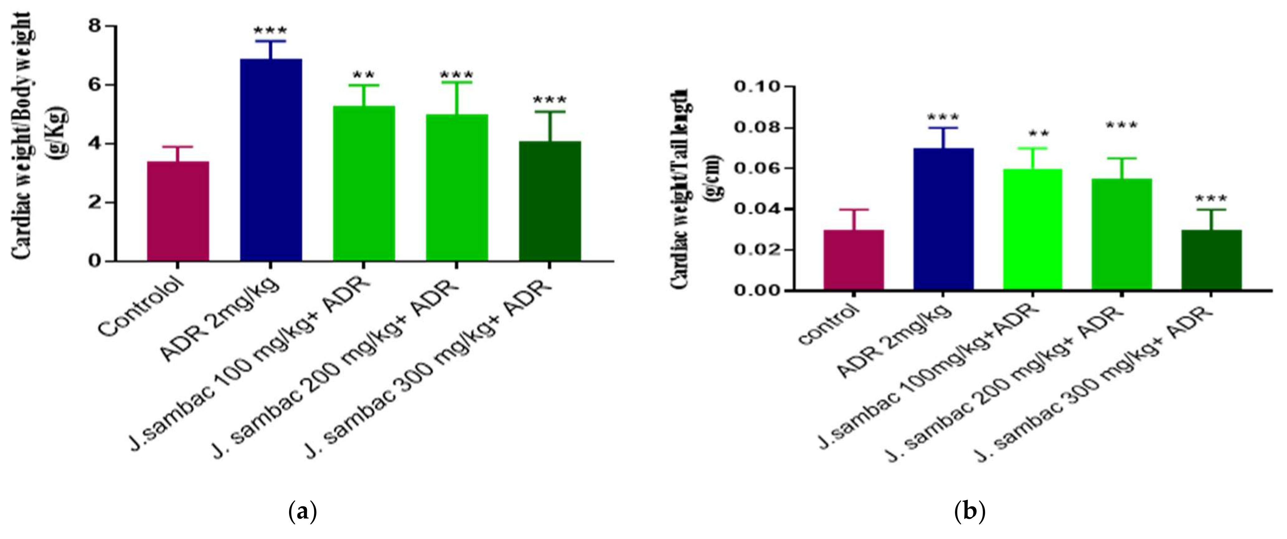
2.4. Acute Myocardial Infarction Study
Effect on Heart to Body Weight, and Weight of Heart to Tail Length Ratios
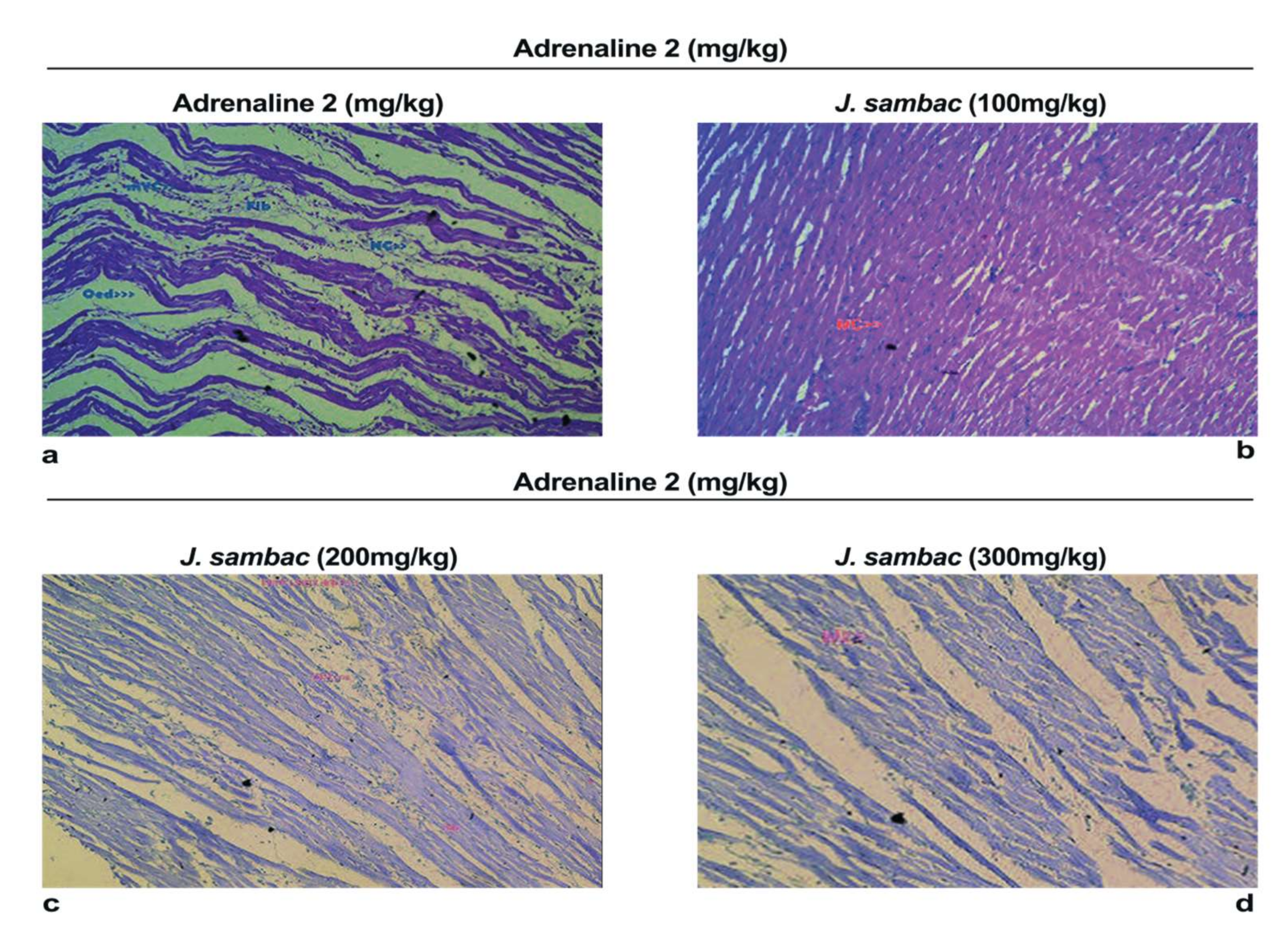
2.5. Histopathology
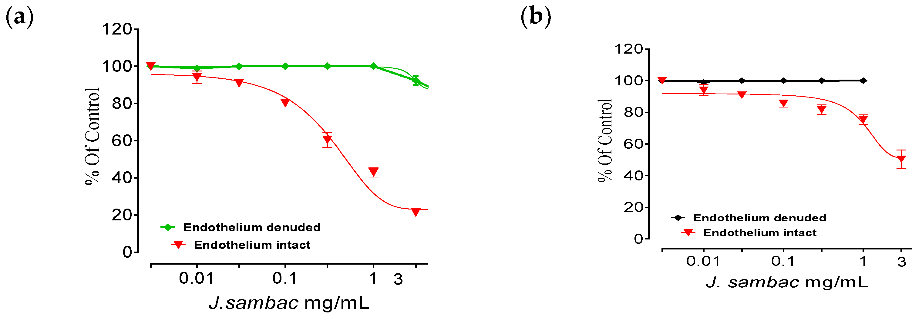
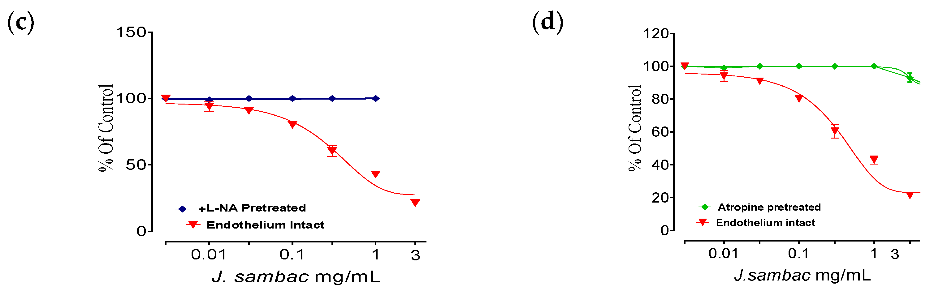
2.6. Isolated Aortic Tissue Preparation and Vasorelaxant Activity
2.7. Effect on Adrenaline Induced Platelet Activation and Aggregation
2.8. Acute Oral Toxicity Dose Test
3. Discussion
4. Materials and Methods
4.1. Plant Materials
4.2. Extract Preparation
4.3. Animals
4.4. Chemicals
4.5. Preliminary Phytochemical Evaluation
4.6. HPLC Analysis
4.7. Acute Oral Toxicity Dose Test
4.8. Determination of DPPH Assay
4.9. Acute Myocardial Infarction Study
Screening of Cardiac to Body Weight and Cardiac Weight to Tail Length Ratio
4.10. Histopathology
4.11. Isolated Aortic Tissue Preparation and Vasorelaxant Activity
4.12. Effect on Adrenaline Induced Platelet Activation and Aggregation
5. Conclusions
Author Contributions
Funding
Institutional Review Board Statement
Informed Consent Statement
Data Availability Statement
Acknowledgments
Conflicts of Interest
Sample Availability
References
- Khan, I.A. Pharmacotherapeutic modifications in cardiopulmonary patients during COVID-19 outbreak. J. Coll. Phy. Surg. Pak. 2020, 30, 15–17. [Google Scholar]
- Quiñones, M.; Miguel, M.; Aleixandre, G. Beneficial effects of polyphenols on cardiovascular disease. Pharmacol. Res. 2013, 68, 125–131. [Google Scholar] [CrossRef]
- Shaito, A.; Thuan, D.T.B.; Nguyen, T.H.D.; Hasan, H.; Halabi, S.; Abdelhady, S.; Nasrallah, G.K.; Pintus, G. Herbal Medicine for Cardiovascular Diseases: Efficacy, Mechanisms, and Safety. Front. Pharmacol. 2020, 11, 422. [Google Scholar] [CrossRef] [Green Version]
- Kumar, S.V.; Saritha, G.; Fareedullah, G. Role of antioxidants and oxidative stress in cardiovascular diseases. Ann. Biol. Res. 2010, 3, 158–173. [Google Scholar]
- Tsoupras, A.; Lordan, R.; Zabetakis, I. Inflammation, not Cholesterol, Is a Cause of Chronic Disease. Nutrients 2018, 10, 604. [Google Scholar] [CrossRef] [Green Version]
- Amin, M.M.; El-Gazayerly, O.N.; Gawad, N.A.; Halim, S.M. Effect of formulation variables on design, in vitro evaluation of valsartan SNEDDS and estimation of its antioxidant effect in adrenaline-induced acute myocardial infarction in rats. Pharmaceut. Dev. Technol. 2016, 21, 909–920. [Google Scholar] [CrossRef]
- Fathiazad, F.; Matlobi, S.; Khorrami, N. Phytochemical screening and evaluation of cardioprotective activity of ethanolic extract of Ocimumbasilicum L. (basil) against isoproterenol induced myocardial infarction in rats. DARU J. Pharma. Sci. 2012, 20, 198–203. [Google Scholar]
- Saqib, F.; Arif Aslam, M.; Mujahid, K.; Marceanu, L.; Moga, M.; Ahmedah, H.T.; Chicea, L. Studies to Elucidate the Mechanism of Cardio Protective and Hypotensive Activities of Anogeissus acuminata (Roxb. ex DC.) in Rodents. Molecules 2020, 25, 3471. [Google Scholar] [CrossRef] [PubMed]
- Koffuor, G.A.; Amoateng, P. Anti-oxidant and anticoagulant properties of Phyllanthus fraternus GL webster (Family: Euphorbiaceae). J. Pharmacol. Toxicol. 2012, 6, 624–626. [Google Scholar] [CrossRef] [Green Version]
- Khare, C.P. Encyclopedia of Indian Medicinal Plants, Rational Western Therapy. Ayurvedic and Other Traditional Usage, Botany; Springer Publications: Berlin/Heidelberg, Germany, 2004; pp. 314–315. [Google Scholar]
- Al-Snafi, A.E. Pharmacological and therapeutic effects of Jasminum sambac—A review. Indo Am. J. Pharma. Sci. 2018, 5, 1766–1778. [Google Scholar]
- Phanukit, K.U.; Chuleratana, B.; Kajsongkram, T.; Amonrat, K.H. Chemical composition, toxicity and vasodilatation Effect of the flowers extract of Jasminum sambac (L.) Ait. “G. Duke of Tuscany”. Evid.-Based Complement. Altern. Med. 2012, 47, 1312–1317. [Google Scholar]
- Bucki, R.; Pastore, J.J.; Giraud, F.; Sulpice, J.C.; Janmey, P.A. Flavonoid inhibition of platelet procoagulant activity and phosphoinositide synthesis. J. Thromb. Haemost. 2003, 1, 1820–1828. [Google Scholar] [CrossRef] [PubMed] [Green Version]
- Kumari, G.; Wong, K.U.; Serr, A.; Joon, S.; Yoon, S.H.; Tam, P. Molecular diversity and function of jasmintides from Jasminum sambac. BMC Plant. Biol. 2018, 18, 144–149. [Google Scholar] [CrossRef] [PubMed] [Green Version]
- Dong, Y.; Duan, L.; Chen, H.W.; Liu, Y.M.; Zhang, Y. Network pharmacology-based prediction and verification of the targets and mechanism for Panax notoginseng saponins against coronary heart disease. Evid.-Based Complement. Altern. Med. 2019, 65, 37–52. [Google Scholar] [CrossRef] [PubMed]
- He, Y.; Hu, Z.; Li, A.; Zhu, Z.; Yang, N.; Ying, Z. Recent advances in biotransformation of saponins. Molecules 2019, 24, 2365. [Google Scholar] [CrossRef] [Green Version]
- Sun, J.; Yu, X.; Huangpu, H.; Yao, F. Ginsenoside Rb3 protects cardiomyocytes against hypoxia/reoxygenation injury via activating the antioxidation signaling pathway of PERK/Nrf2/HMOX1. Biomed. Pharmacother. 2019, 109, 254–261. [Google Scholar] [CrossRef]
- Mundugaru, R.; Udaykumar, P.; Senthilkumar, S.; Bhat, S. Cardioprotective activity of fruit of Garcinia pedunculata on isoprenaline-induced myocardial infarction in rat. Bangladesh J. Pharmacol. 2018, 11, 231–235. [Google Scholar] [CrossRef] [Green Version]
- Kalaiselvi, M.; Narmadha, R.; Ragavendran, P.; Arul, R.; Sophia, D.; Ravi, K.G.; Gomathi, D.; Uma, C. In vivo simulated in vitro model of Jasminum sambac Linn. using mammalian liver slice technique. Asian Pac. J. Trop. Biomed. 2011, 16, 216–219. [Google Scholar] [CrossRef]
- Ittagi, S.; Merugumolu, K.M.; Siddamsetty, S.K. Cardioprotective effect of hydroalcoholic extract of Tecoma stans flowers against isoproterenol induced myocardial infarction in rats. Asian Pac. J. Trop. Biomed. 2014, 4, S378–S384. [Google Scholar] [CrossRef]
- Panchal, S.K.; Brown, L. Cholesterol versus Inflammation as Cause of Chronic Diseases. Nutrients 2019, 11, 2332. [Google Scholar] [CrossRef] [Green Version]
- Gilani, A.H.; Janbaz, K.H.; Zaman, H.; Lateef, A.; Suria, A. Possible Presence of Calcium Channel Blocker(s) in Rubia cordifolia: An Indigenous Medicinal Plant. JPMA 1994, 44, 84–89. [Google Scholar]
- Prince, P.S.; Rajakumar, S.; Dhanasekar, K. Protective effects of vanillic acid on electrocardiogram, lipid peroxidation, antioxidants, proinflammatory markers and histopathology in isoproterenol induced cardiotoxic rats. Eur. J. Pharmacol. 2011, 668, 233–240. [Google Scholar] [CrossRef]
- Wang, X.L. Potential herb-drug interaction in the prevention of cardiovascular diseases during integrated traditional and western medicine treatment. Chin. J. Integr. Med. 2015, 21, 3–9. [Google Scholar] [CrossRef]
- Satoh, S.; Nishida, S. Electropharmacological actions of Ginkgo biloba extract on vascular smooth and heart muscles. Clin. Chim. Acta 2004, 342, 13–22. [Google Scholar] [CrossRef] [PubMed]
- Khan, I.A.; Lodhi, A.H.; Munawar, S.H.; Manzoor, A.; Manzoor, Z.; Raza, M.A.; Iqbal, O. Assessment of ameliorative effect of Aab-e-Shifa polyherbal formulation in experimentally-induced wound in rabbits. Trop. J. Pharm. Res. 2020, 19, 2357–2362. [Google Scholar]
- National Institute of Health. Guide for the Care and Use of Laboratory Animals; No. 85-23; NIH Publication: Bethesda, MD, USA, 1985. [Google Scholar]
- Trease, G.E.; Evans, W.C. Trease and Evans’ Pharmacognosy, 16th ed.; Saunders/Elsevier: Edinburgh, Australia, 2009. [Google Scholar]
- Teresa, F.M.; Lourdes, C.; Mantell, C.; Miguel, R.; Enrique, M.O. Extraction of antioxidant compounds from different varieties of Mangifera indica leaves using green technologies. J. Sup. Fluids 2012, 72, 168–175. [Google Scholar]
- Al-Afifi, N.A.; Alabsi, A.M.; Bakri, M.M. Acute and sub-acute oral toxicity of Dracaena cinnabari resin methanol extract in rats. BMC Complement. Altern. Med. 2018, 18, 50–55. [Google Scholar] [CrossRef] [PubMed] [Green Version]
- Sreejayan, A.; Rao, M.N. Free radicals scavenging activity of curcuminoids. Drug Res. 1996, 46, 169–173. [Google Scholar]
- Salma, A.; El-Marasy, S.A.; Awdan, E.; Hassan, H.; Heba, M.I. Cardioprotective effect of thymol against adrenaline-induced myocardial injury in rats. Heliyon 2020, 7, 35–39. [Google Scholar]
- Veeresh, B.D.; Ramesh, K.; Bhatt, K. Evaluation of Hepatoprotective activity of Jasminum sambac in rats. Int. J. Res. Pharmacol. Pharmacother. 2017, 6, 104–116. [Google Scholar]
- Khan, I.A.; Lodhi, A.H.; Munawar, S.H.; Manzoor, A.; Manzoor, Z.; Raza, M.A. Formulation and evaluation of allicin and curcumin gel improves normal and diabetic ulcers in rabbits. Lat. Am. J. Pharm. 2018, 37, 1602–1607. [Google Scholar]
- Imtiaz, S.M.; Aleem, A.; Saqib, F.; Ormenisan, A.N.; Elena Neculau, A.; Anastasiu, C.V. The Potential involvement of an ATP-Dependent Potassium Channel-Opening Mechanism in the Smooth Muscle Relaxant Properties of Tamarix dioica Roxb. Biomolecules 2019, 9, 722. [Google Scholar] [CrossRef] [PubMed] [Green Version]
- Isik, B.S.; Altay, F.; Capanoglu, E. The uniaxial and coaxial encapsulations of sour cherry (Prunus cerasus L.) concentrate by electrospinning and their in vitro bioaccessibility. Food Chem. 2018, 265, 260–273. [Google Scholar] [CrossRef] [PubMed]
- Singh, S.; Malm, C.J.; Ramström, S.; Hesse, C.; Jeppsson, A. Adrenaline enhances in vitro platelet activation and aggregation in blood samples from ticagrelor-treated patients. Res. Pract. Thromb. Haemost. 2018, 2, 718–725. [Google Scholar] [CrossRef] [PubMed] [Green Version]


| Tests | Observation | Results |
|---|---|---|
| Alkaloid | no ppt | − |
| Saponins | 1 cm froth | + |
| Tannins | No Light purple | − |
| Anthraquinones | No Pink Colour | − |
| Coumarins | Yellow fluorescence. | + |
| Phenols | Light purple | + |
| Flavonoid | Light yellow colour | ++ |
Publisher’s Note: MDPI stays neutral with regard to jurisdictional claims in published maps and institutional affiliations. |
© 2021 by the authors. Licensee MDPI, Basel, Switzerland. This article is an open access article distributed under the terms and conditions of the Creative Commons Attribution (CC BY) license (https://creativecommons.org/licenses/by/4.0/).
Share and Cite
Khan, I.A.; Hussain, M.; Munawar, S.H.; Iqbal, M.O.; Arshad, S.; Manzoor, A.; Shah, M.A.; Abbas, K.; Shakeel, W.; Syed, S.K. Jasminum sambac: A Potential Candidate for Drug Development to Cure Cardiovascular Ailments. Molecules 2021, 26, 5664. https://doi.org/10.3390/molecules26185664
Khan IA, Hussain M, Munawar SH, Iqbal MO, Arshad S, Manzoor A, Shah MA, Abbas K, Shakeel W, Syed SK. Jasminum sambac: A Potential Candidate for Drug Development to Cure Cardiovascular Ailments. Molecules. 2021; 26(18):5664. https://doi.org/10.3390/molecules26185664
Chicago/Turabian StyleKhan, Imran Ahmad, Musaddique Hussain, Shaukat Hussain Munawar, Muhammad Omer Iqbal, Shafia Arshad, Ashira Manzoor, Mazhar Abbas Shah, Khizar Abbas, Waleed Shakeel, and Shahzada Khurram Syed. 2021. "Jasminum sambac: A Potential Candidate for Drug Development to Cure Cardiovascular Ailments" Molecules 26, no. 18: 5664. https://doi.org/10.3390/molecules26185664