
Polyisobutylene—New Opportunities for Medical Applications

 , , , , and
Abstract
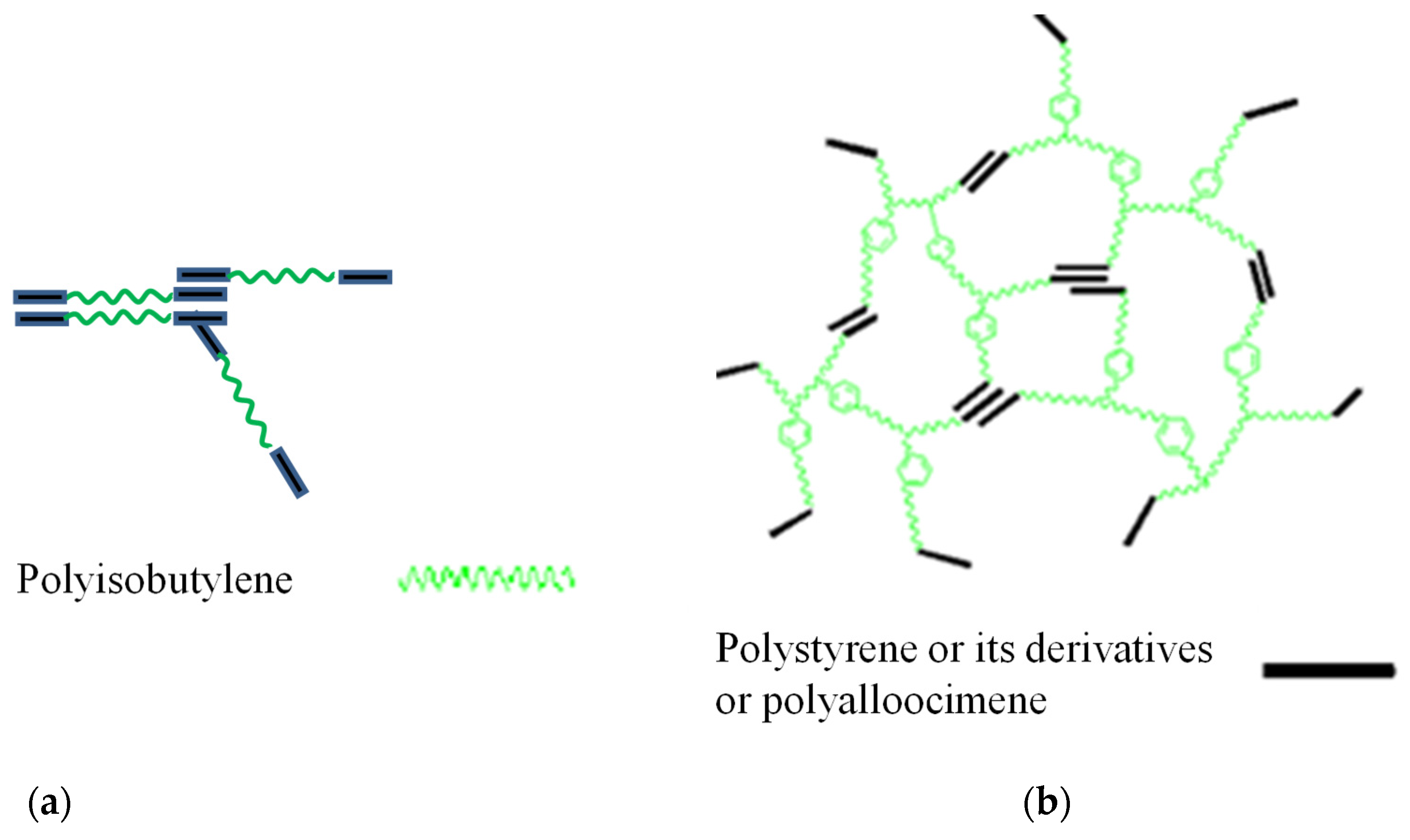
:1. Introduction
2. Results and Discussion
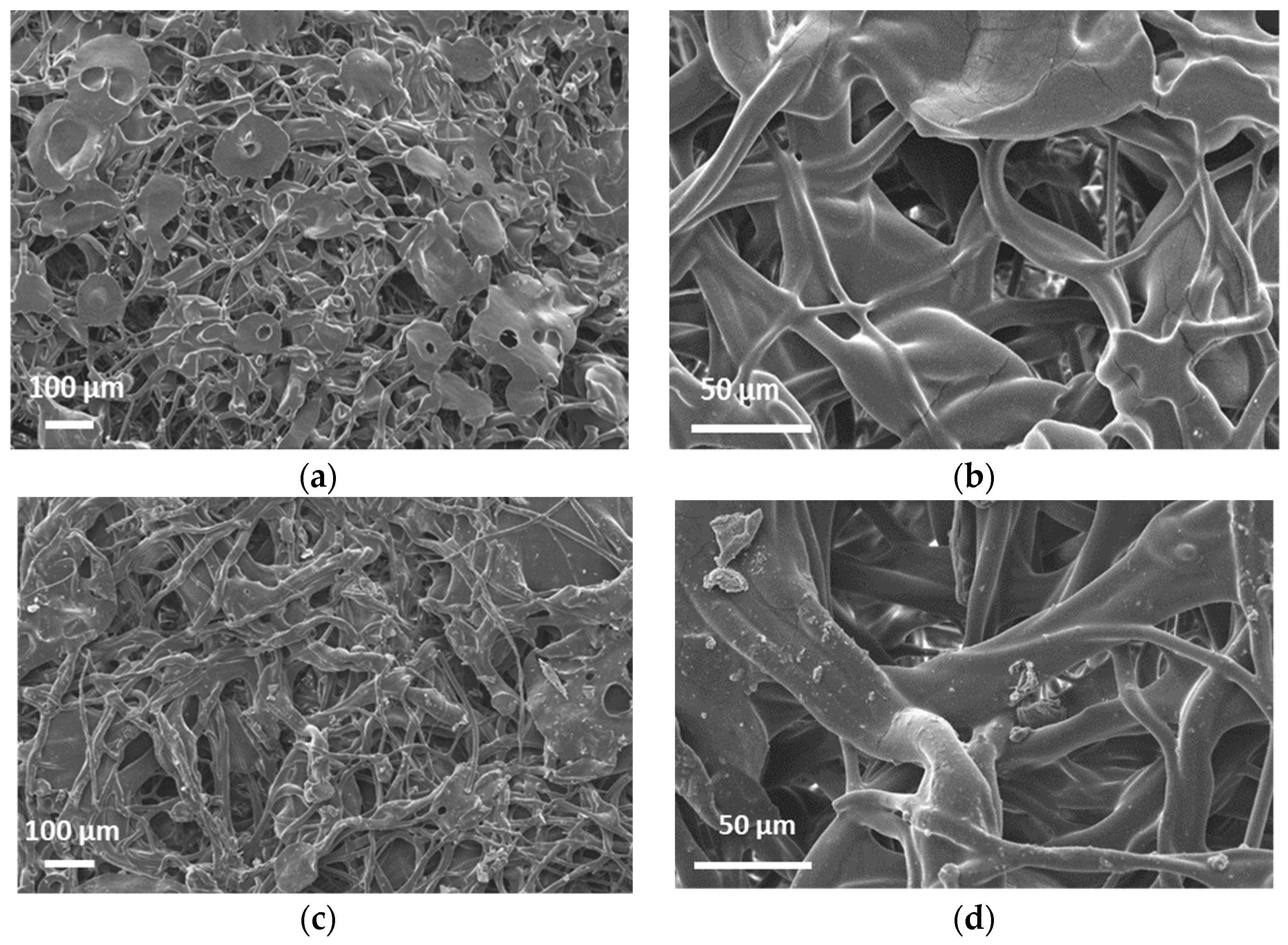
2.1. Characterization of the Mats
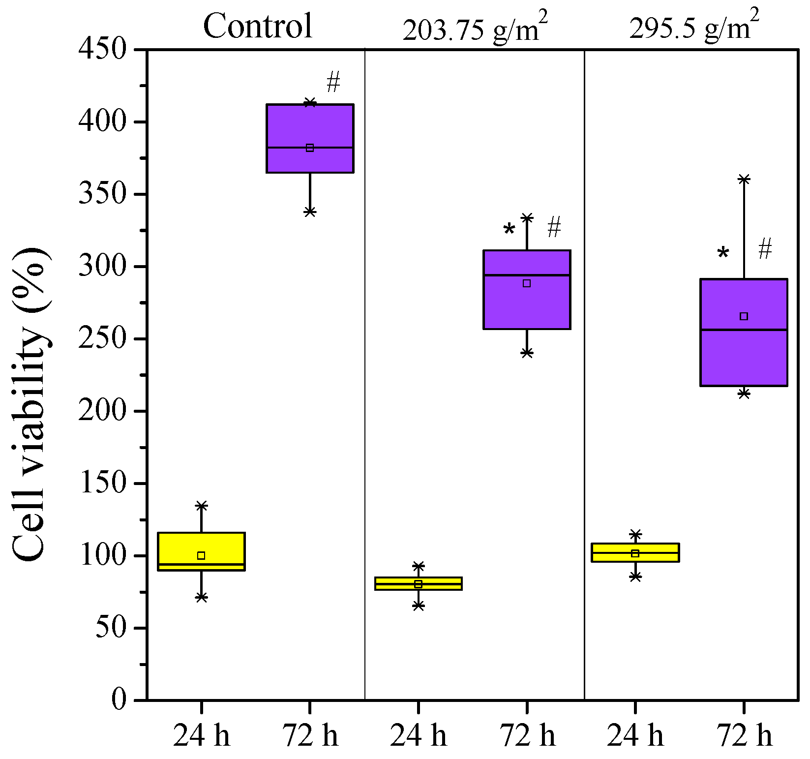
2.2. Cytotoxicity
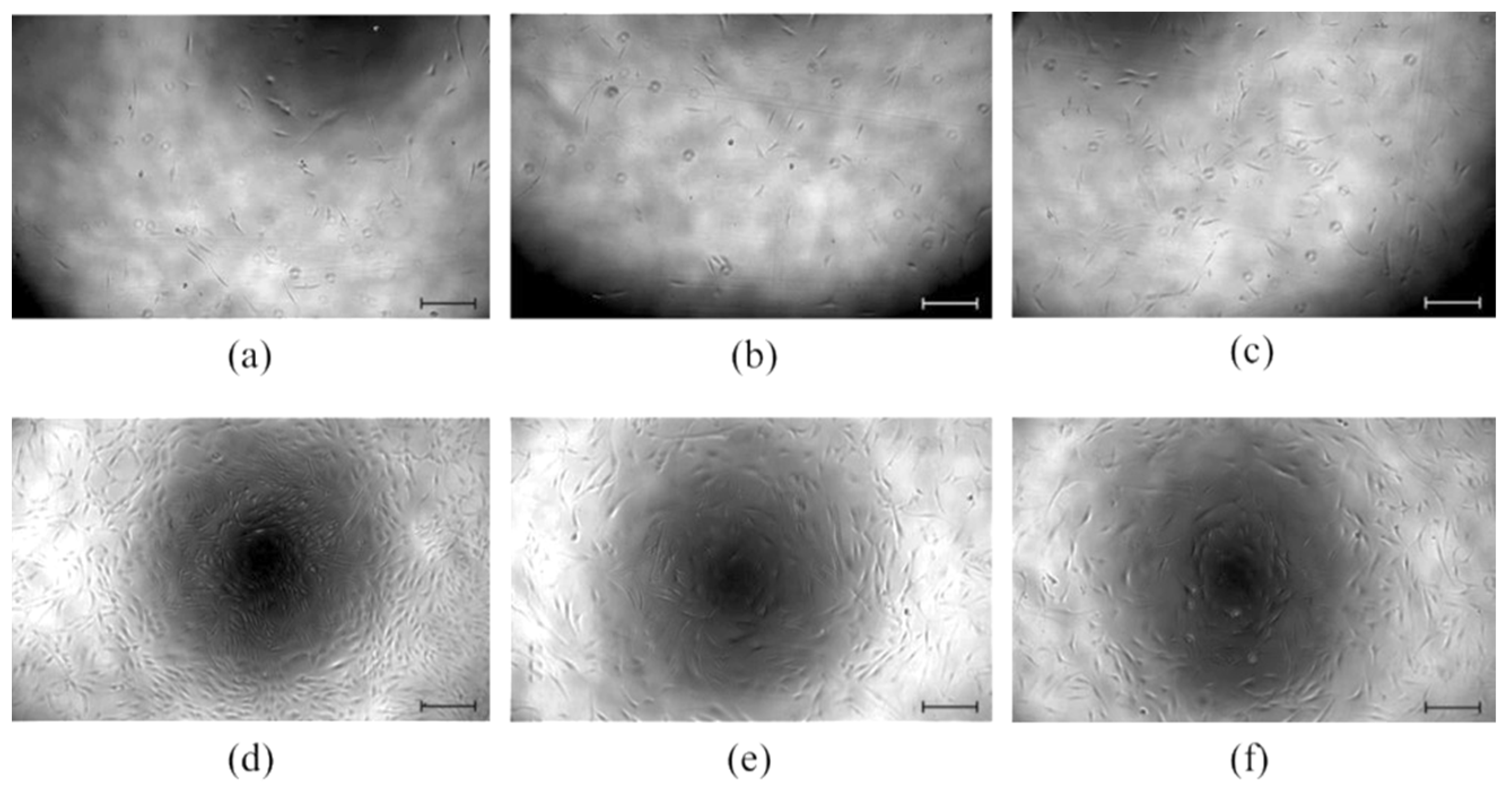
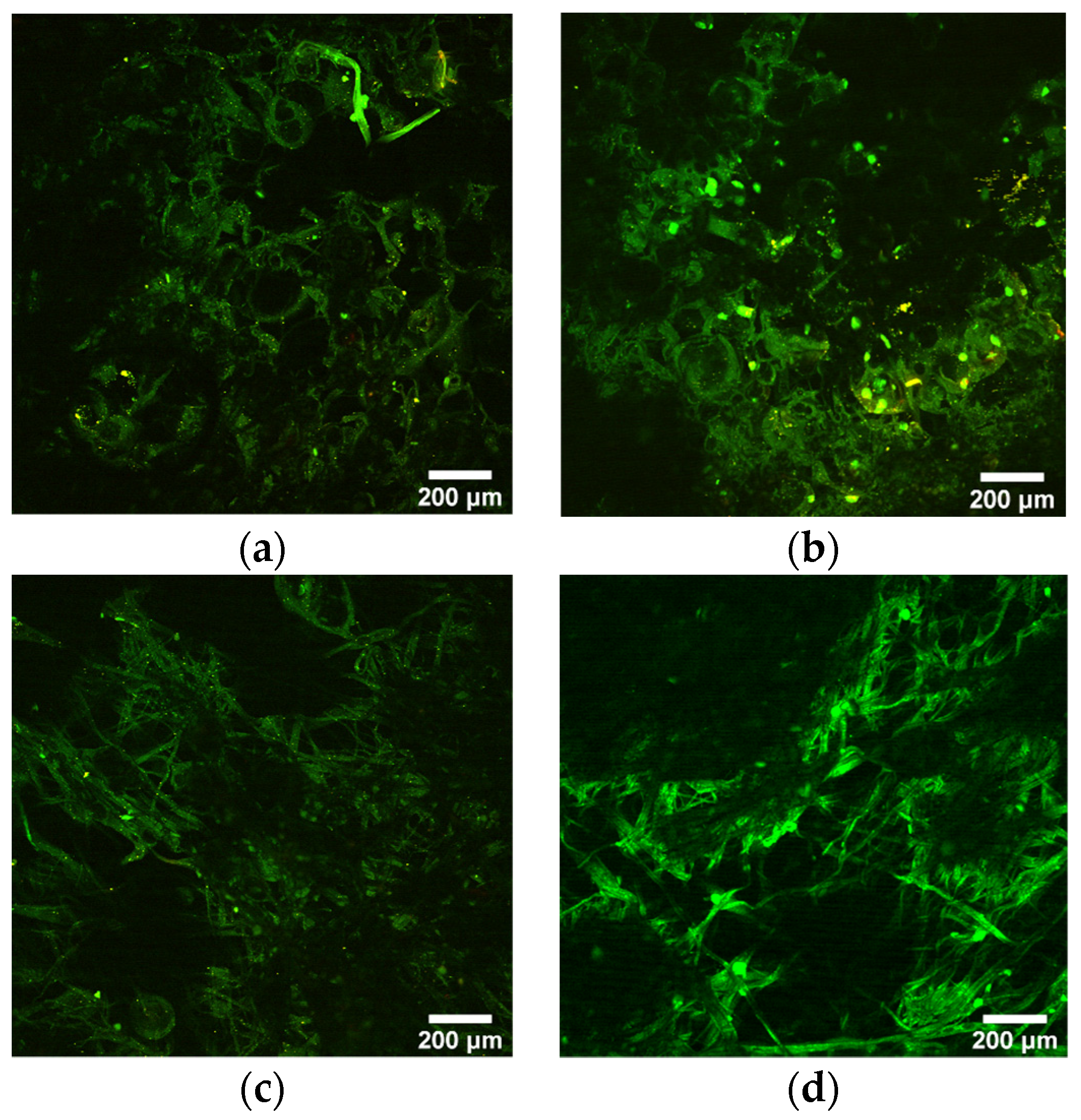
2.3. Cell Adhesion
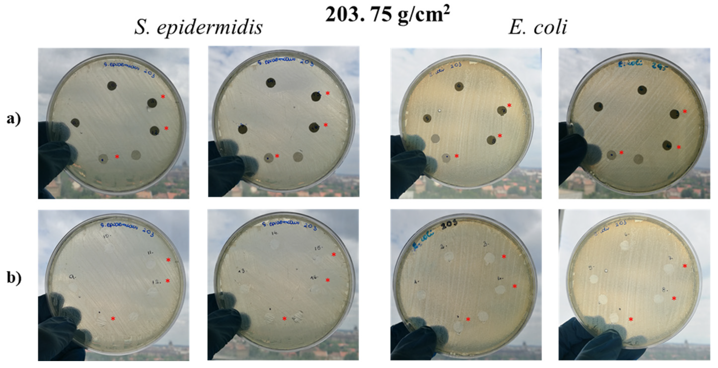
2.4. Antibacterial Activity
2.5. Biofilm Formation
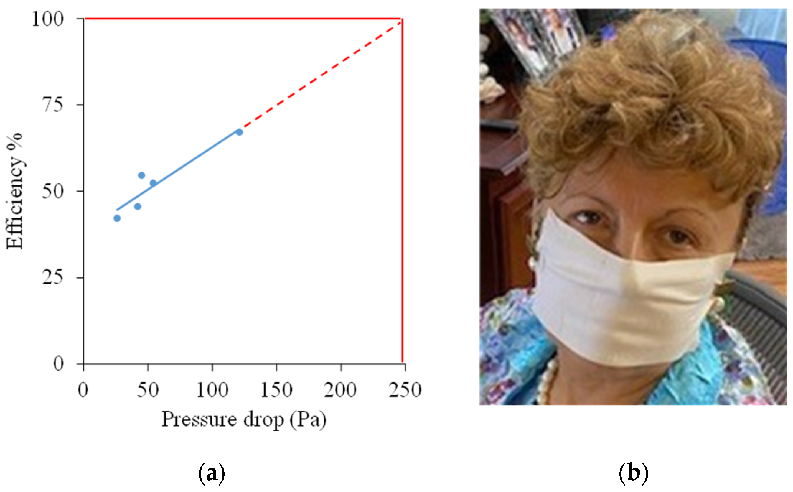
2.6. Potential Applications
3. Materials and Methods
3.1. Materials
3.2. Scanning Electron Microscopy (SEM)
3.3. X-ray Photoelectron Spectroscopy (XPS)
3.4. Water Contact Angle (WCA) Measurement
3.5. Cytotoxicity Assay
3.6. Cell Adhesion
3.7. Antibacterial Testing
3.8. Biofilm Formation Testing
3.9. Filtration Efficiency Tests
4. Conclusions
Supplementary Materials
Author Contributions
Funding
Institutional Review Board Statement
Informed Consent Statement
Data Availability Statement
Acknowledgments
Conflicts of Interest
Sample Availability
References
- Puskas, J.E.; Kaszas, G. Carbocationic Polymerization. In Encyclopedia of Polymer Science and Technology; Wiley: New York, NY, USA, 2016; pp. 1–43. [Google Scholar]
- Puskas, J.E.; Kaszas, G.; Molnar, K.; Helfer, C.A. Polyisobutylene for the rescue: Advanced elastomers for healthcare. In Macromolecular Engineering; Elsevier: Amsterdam, The Netherlands, 2021; pp. 237–253. [Google Scholar]
- US FDA, TAXUSTM Express2TM Paclitaxel-Eluting Coronary Stent System (Monorail or over the Wire), PO30025. 2004. Available online: https://www.accessdata.fda.gov/scripts/cdrh/cfdocs/cfpma/pma.cfm?id=P030025S021 (accessed on 10 June 2021).
- Kamath, K.R.; Barry, J.J.; Miller, K.M. The TaxusTM drug-eluting stent: A new paradigm in controlled drug delivery. Adv. Drug Deliv. Rev. 2006, 58, 412–436. [Google Scholar] [CrossRef] [PubMed]
- Puskas, J.E.; Kwon, Y.; Antony, P.; Bhowmick, A.K. Synthesis and characterization of novel dendritic (arborescent, hyperbranched) polyisobutylene-polystyrene block copolymers. J. Polym. Sci. Part A Polym. Chem. 2005, 43, 1811–1826. [Google Scholar] [CrossRef]
- Teck Lim, G.; Valente, S.A.; Hart-Spicer, C.R.; Evancho-Chapman, M.M.; Puskas, J.E.; Horne, W.I.; Schmidt, S.P. New biomaterial as a promising alternative to silicone breast implants. J. Mech. Behav. Biomed. Mater. 2013, 21, 47–56. [Google Scholar] [CrossRef] [PubMed]
- Puskas, J.E.; Dos Santos, L.M.; Orlowski, E. Polyisobutylene-based thermoplastic biorubbers. Rubber Chem. Technol. 2010, 83, 235–246. [Google Scholar] [CrossRef]
- Puskas, J.E.; Paulo, C.; Antony, P. Arborescent Thermoplastic Elastomers and Products Therefrom. U.S. Patent 6,747,098, 8 June 2004. [Google Scholar]
- Puskas, J.E. Terpene/Isoolefin Copolymers Having Substantially Heterogeneous Compositional Distribution and Displaying Thermoplastic Elastomeric Properties. U.S. Patent 9,790,301, 17 October 2017. [Google Scholar]
- Gergely, A.L.; Puskas, J.E. Synthesis and characterization of thermoplastic elastomers with polyisobutylene and polyalloocimene blocks. J. Polym. Sci. Part A Polym. Chem. 2015, 53, 1567–1574. [Google Scholar] [CrossRef]
- Puskas, J.E. Rubber city girl: The path to the goodyear medal. Rubber Chem. Technol. 2018, 91, 1–26. [Google Scholar] [CrossRef]
- Kantor, J.; Puskas, J.E.; Kaszas, G. The Effect of Reaction Conditions on the Synthesis of Thermoplastic Elastomers Containing Polyalloocimene, Polyisobutylene and Tapered Blocks. Chin. J. Polym. Sci. 2019, 37, 884–890. [Google Scholar] [CrossRef]
- Puskas, J.E.; Foreman-Orlowski, E.A.; Lim, G.T.; Porosky, S.E.; Evancho-Chapman, M.M.; Schmidt, S.P.; El Fray, M.; Piątek, M.; Prowans, P.; Lovejoy, K. A nanostructured carbon-reinforced polyisobutylene-based thermoplastic elastomer. Biomaterials 2010, 31, 2477–2488. [Google Scholar] [CrossRef]
- Puskas, J.E.; Hoerr, R.A. Drug release from novel rubbery coatings. In Macromolecular Symposia; WILEY-VCH Verlag: Weinheim, Germany, 2010; Volume 291, pp. 326–329. [Google Scholar] [CrossRef]
- Zhang, P.; Zhang, S.; Wan, D.; Zhang, P.; Zhang, Z.; Shao, G. Multilevel polarization-fields enhanced capture and photocatalytic conversion of particulate matter over flexible schottky-junction nanofiber membranes. J. Hazard. Mater. 2020, 395, 122639. [Google Scholar] [CrossRef]
- Zhang, M.; Qi, Y.; Zhang, Z. AgBr/BiOBr Nano-Heterostructure-Decorated Polyacrylonitrile Nanofibers: A Recyclable High-Performance Photocatalyst for Dye Degradation under Visible-Light Irradiation. Polymers 2019, 11, 1718. [Google Scholar] [CrossRef] [Green Version]
- Liu, Y.; Liu, X.; Chen, J.; Gilmore, K.J.; Wallace, G.G. 3D Bio-nanofibrous PPy/SIBS mats as platforms for cell culturing. Chem. Commun. 2008, 32, 3729–3731. [Google Scholar] [CrossRef] [PubMed]
- Liu, Y.; Gilmore, K.J.; Chen, J.; Misoska, V.; Wallace, G.G. Bio-nanowebs Based on Poly(styrene-β-isobutylene-β-styrene) (SIBS) Containing Single-Wall Carbon Nanotubes. Chem. Mater. 2007, 19, 2721–2723. [Google Scholar] [CrossRef]
- Lim, G.T.; Puskas, J.E.; Reneker, D.H.; Jàkli, A.; Horton, W.E. Highly hydrophobic electrospun fiber mats from polyisobutylene-based thermoplastic elastomers. Biomacromolecules 2011, 12, 1795–1799. [Google Scholar] [CrossRef] [PubMed]
- Jindal, A.; Puskas, J.E.; McClain, A.; Nedic, K.; Luebbers, M.T.; Baker, J.R.; dos Santos, B.P.; Camassola, M.; Jennings, W.; Einsporn, R.L.; et al. Encapsulation and release of Zafirlukast from electrospun polyisobutylene-based thermoplastic elastomeric fiber mat. Eur. Polym. J. 2018, 98, 254–261. [Google Scholar] [CrossRef]
- Jindal, A.; Molnár, K.; McClain, A.; Paiva dos Santos, B.; Camassola, M.; Puskas, J.E. Electrospun fiber mats from poly(alloocimene-b-isobutylene-b-alloocimene) thermoplastic elastomer. Int. J. Polym. Mater. Polym. Biomater. 2020, 69, 263–267. [Google Scholar] [CrossRef] [Green Version]
- Kim, B.H.; Park, M.; Park, H.J.; Lee, S.H.; Choi, S.Y.; Park, C.G.; Han, S.M.; Heo, C.Y.; Choy, Y. Bin Prolonged, acute suppression of cysteinyl leukotriene to reduce capsular contracture around silicone implants. Acta Biomater. 2017, 51, 209–219. [Google Scholar] [CrossRef]
- Götz, C.; Lim, G.T.; Puskas, J.E.; Altstädt, V. The effect of carbon black reinforcement on the dynamic fatigue and creep of polyisobutylene-based biomaterials. J. Mech. Behav. Biomed. Mater. 2014, 39, 355–365. [Google Scholar] [CrossRef]
- Puskas, J.E.; Kaszas, G.; McClain, A.T.; Helfer, C. Filler Interaction in Thermoplastic Elastomer Composites. In Proceedings of the 194th Technical Meeting, Louisville, KY, USA, 9 October 2018. [Google Scholar]
- Um, I.C.; Fang, D.; Hsiao, B.S.; Okamoto, A.; Chu, B. Electro-Spinning and Electro-Blowing of Hyaluronic Acid. Biomacromolecules 2004, 5, 1428–1436. [Google Scholar] [CrossRef]
- Dowling, D.P.; Miller, I.S.; Ardhaoui, M.; Gallagher, W.M. Effect of surface wettability and topography on the adhesion of osteosarcoma cells on plasma-modified polystyrene. J. Biomater. Appl. 2011, 26, 327–347. [Google Scholar] [CrossRef]
- Alvarez Albarran, A.; Rosenthal-Kim, E.Q.; Kantor, J.; Liu, L.; Nikolov, Z.; Puskas, J.E. Stimuli-responsive antifouling polyisobutylene-based biomaterials via modular surface functionalization. J. Polym. Sci. Part A Polym. Chem. 2017, 55, 1742–1749. [Google Scholar] [CrossRef]
- Puskas, J.E.; Albarran, A.A.; Rosenthal-Kim, E.Q. Modular Surface Functionalization of Polyisobutylene-Based Materials. U.S. Patent 10,314,945, 11 June 2019. [Google Scholar]
- Available online: https://www.goodyearrubber.com/custom-rubber-manufacturing-knowledge-center/rubber-technology-compounding-ingredients/ (accessed on 10 June 2021).
- Alves, M.M.; Bouchami, O.; Tavares, A.; Córdoba, L.; Santos, C.F.; Miragaia, M.; de Fátima Montemor, M. New Insights into Antibiofilm Effect of a Nanosized ZnO Coating against the Pathogenic Methicillin Resistant Staphylococcus aureus. ACS Appl. Mater. Interfaces 2017, 9, 28157–28167. [Google Scholar] [CrossRef]
- Merritt, J.H.; Kadouri, D.E.; O’Toole, G.A. Growing and Analyzing Static Biofilms Judith. Curr. Protoc. Microbiol. 2011, 22, 1B.1.1–1B.1.17. [Google Scholar] [CrossRef] [Green Version]
- Liu, Y.; Zhou, S.; Gao, Y.; Zhai, Y. Electrospun nanofibers as a wound dressing for treating diabetic foot ulcer. Asian J. Pharm. Sci. 2019, 14, 130–143. [Google Scholar] [CrossRef]
- Zhang, Z.; Ji, D.; He, H.; Ramakrishna, S. Electrospun ultrafine fibers for advanced face masks. Mater. Sci. Eng. R Rep. 2021, 143, 100594. [Google Scholar] [CrossRef]
- Seidel, D.L.; Ista, T.K.; Lindquist, T.J.; Tuman, S.J.; 3M Innovative Properties Co. Elastic Nonwoven Fibrous Webs and Methods of Making and Using. U.S. Patent 9,840,794, 12 December 2017. [Google Scholar]
- Kiss, B.; Kis, Z.; Pályi, B.; Kellermayer, M.S.Z. Topography, Spike Dynamics, and Nanomechanics of Individual Native SARS-CoV-2 Virions. Nano Lett. 2021, 21, 2675–2680. [Google Scholar] [CrossRef] [PubMed]
- Bar-On, Y.M.; Flamholz, A.; Phillips, R.; Milo, R. SARS-CoV-2 (COVID-19) by the numbers. Elife 2020, 9, e57309. [Google Scholar] [CrossRef] [PubMed]
- National Institute for Occupational Safety and Health, National Personal Protective Technology Laboratory, n.d.-b; N95DECON Research Document. Available online: https://static1.squarespace.com/static/5e8126f89327941b9453eeef/t/5f2f458117f1700098c7c8c3/1596933505885/N95DECON_Respirator_Mask_Standards_Technical_Report_200803.pdf (accessed on 10 June 2021).
- Noszticzius, Z.; Wittmann, M.; Kály-Kullai, K.; Beregvári, Z.; Kiss, I.; Rosivall, L.; Szegedi, J. Chlorine dioxide is a size-selective antimicrobial agent. PLoS ONE 2013, 8, e79157. [Google Scholar] [CrossRef] [PubMed] [Green Version]
- ImQuest BioSciences Biofilm Protocol Optimization for Pseudomonas aeruginosa: Culture Media, Incubation Time, and Biofilm Measurement. 2016. Available online: http://imquestbio.com/resources/technicaldocuments/ (accessed on 9 May 2019).


| Atoms | Binding Energy (eV) | 203.75 g/m2 At% | 295.5 g/m2 At% |
|---|---|---|---|
| O | 532 | 4.47 | 5.23 |
| C | 285 | 93.38 | 93.00 |
| Si | 102 | 2.15 | 1.77 |
| N95, FFP2, KN95 | Surgical/Medical Mask Material | Cloth Mask Material | ||
|---|---|---|---|---|
| Inhalation | Exhalation | |||
| NIOSH | 343 Pa (85 L/min) | 245 Pa (85 L/min) | --- | --- |
| ASTM | --- | --- | 50/60/60 Pa/cm2 (245/294/294 Pa) (8 L/min) | --- |
| EU | 240 Pa (95 L/min) 70 Pa (30 L/min) | 300 Pa (160 L/min) | 40/40/60 Pa/cm2 (196/196/294 Pa) (8 L/min) | 70 Pa/cm2 |
| China | 350 Pa (85 L/min) | 250 (85 L/min) | 49 Pa/cm2 (240 Pa) (8 L/min) | --- |
Publisher’s Note: MDPI stays neutral with regard to jurisdictional claims in published maps and institutional affiliations. |
© 2021 by the authors. Licensee MDPI, Basel, Switzerland. This article is an open access article distributed under the terms and conditions of the Creative Commons Attribution (CC BY) license (https://creativecommons.org/licenses/by/4.0/).
Share and Cite
Barczikai, D.; Domokos, J.; Szabó, D.; Molnar, K.; Juriga, D.; Krisch, E.; Nagy, K.S.; Kohidai, L.; Helfer, C.A.; Jedlovszky-Hajdu, A.; et al. Polyisobutylene—New Opportunities for Medical Applications. Molecules 2021, 26, 5207. https://doi.org/10.3390/molecules26175207
Barczikai D, Domokos J, Szabó D, Molnar K, Juriga D, Krisch E, Nagy KS, Kohidai L, Helfer CA, Jedlovszky-Hajdu A, et al. Polyisobutylene—New Opportunities for Medical Applications. Molecules. 2021; 26(17):5207. https://doi.org/10.3390/molecules26175207
Chicago/Turabian StyleBarczikai, Dóra, Judit Domokos, Dóra Szabó, Kristof Molnar, David Juriga, Eniko Krisch, Krisztina S. Nagy, Laszlo Kohidai, Carin A. Helfer, Angela Jedlovszky-Hajdu, and et al. 2021. "Polyisobutylene—New Opportunities for Medical Applications" Molecules 26, no. 17: 5207. https://doi.org/10.3390/molecules26175207