
Phages and Enzybiotics in Food Biopreservation
by
 , , and
José Ramos-Vivas
1,2 ,
,
María Elexpuru-Zabaleta
1, 
María Luisa Samano
1,2,
Alina Pascual Barrera
2,
Tamara Y. Forbes-Hernández
3,
Francesca Giampieri
4,5,* and
Maurizio Battino
4,6,* 1
Research Group on Foods, Nutritional Biochemistry and Health, Universidad Europea del Atlántico, 39011 Santander, Spain
2
Department of Project Management, Universidad Internacional Iberoamericana, Campeche 24560, Mexico
3
Department of Analytical and Food Chemistry, CITACA, CACTI, University of Vigo, 36310 Vigo, Spain
4
Department of Clinical Sciences, Polytechnic University of Marche, 60131 Ancona, Italy
5
Department of Biochemistry, Faculty of Sciences, King Abdulaziz University, Jeddah 21589, Saudi Arabia
6
International Research Center for Food Nutrition and Safety, Jiangsu University, Zhenjiang 212013, China
*
Authors to whom correspondence should be addressed.
Molecules 2021, 26(17), 5138; https://doi.org/10.3390/molecules26175138
Submission received: 13 July 2021
/
Revised: 10 August 2021
/
Accepted: 20 August 2021
/
Published: 25 August 2021
(This article belongs to the Special Issue Bioproducts for Health II)
Abstract
:Presently, biopreservation through protective bacterial cultures and their antimicrobial products or using antibacterial compounds derived from plants are proposed as feasible strategies to maintain the long shelf-life of products. Another emerging category of food biopreservatives are bacteriophages or their antibacterial enzymes called “phage lysins” or “enzybiotics”, which can be used directly as antibacterial agents due to their ability to act on the membranes of bacteria and destroy them. Bacteriophages are an alternative to antimicrobials in the fight against bacteria, mainly because they have a practically unique host range that gives them great specificity. In addition to their potential ability to specifically control strains of pathogenic bacteria, their use does not generate a negative environmental impact as in the case of antibiotics. Both phages and their enzymes can favor a reduction in antibiotic use, which is desirable given the alarming increase in resistance to antibiotics used not only in human medicine but also in veterinary medicine, agriculture, and in general all processes of manufacturing, preservation, and distribution of food. We present here an overview of the scientific background of phages and enzybiotics in the food industry, as well as food applications of these biopreservatives.
1. Introduction
Food preservation by suitable means is key in food safety and quality. There are several traditional and well-known physical preservation techniques such as refrigeration and pasteurization, but the modern industry is always looking for new procedures for food preservation to increase the product’s shelf-life by minimizing the loss of nutritional quality and organoleptic properties. Presently, some modern biopreservation techniques rely on naturally occurring microorganisms (i.e., lactic acid bacteria) and their metabolites. These food preservatives are mainly used to produce safer food for the consumer, preventing the action of pernicious microbes which can cause food deterioration or even toxicity and therefore be dangerous to human health.
Moreover, bacteria -including multidrug-resistant bacteria- can reach food at different points in the food supply chain, from farm to postharvest, and processing such as slaughtering, fermentation, packaging and storage [1,2,3,4,5].
As most natural foods are highly perishable, by extending their half-life we can also control their native microbiota for proper preservation, maintaining their safety and quality. As microorganisms produce a long list of molecules ranging from classic antibiotics to antibacterial enzymes, the control of indigenous populations in food can be achieved by adding these products directly. The paradigm of bacterial molecules used in the food industry as biopreservatives is Nisin, a bacteriocin produced by the Gram-positive bacterium Lactococcus lactis, one of the lactic acid bacteria most extensively used for the manufacture of dairy products [6]. Other well-known bacteriocins, such as Pediocin, Natamycin, Enterocin, and Leucocin [7], also have inhibitory properties against other microorganisms which makes them very interesting for use in the food industry. Some bacteria that produce these compounds have been used as probiotics. Current research on probiotics is quite promising and modern fashion trends push probiotics and bacteriocins from modulation of the gut microbiota toward a wide range of other health-promoting activities away from food, such as cancer treatment, skin health care, periodontal health, or allergies [8,9,10,11].
In addition, the use of bacteriocin producing strains or those that can compete against pathogens in the context of the food industry needs new approaches, mainly due to the increase in foodborne infections, the appearance of new production processes, the massive demand for food, and the changing consumer trends. Moreover, the extensive use of antibiotics against animal and human pathogens has also led to an increase in foodborne pathogens resistant to antibiotics, which makes the picture not reassuring at all [12,13,14].
Goodridge and Abedon published an article in 2003 where they proposed to use the terms “phage biocontrol” and “phage bioprocessing” to differentiate the application of bacteriophages in the farm or crops from their use in the food industry [15]. Several years later, Greer published a review of the control of foodborne bacteria using phages, including the effects of these microorganisms on food storage and preservation [16].
At that time, the excellent properties of endolysins to kill bacteria were already known, but their use to protect food from foodborne pathogens had not yet been effectively tested. One of the first murein hydrolases to be studied concerning food-related bacteria was that of the Lactobacillus helveticus bacteriophage 0303 [17]. This endolysin exhibited a broad spectrum of activity, killing different bacterial species such Pediococcus acidilactici, Lactobacillus delbrueckii subsp. bulgaricus, Lactobacillus delbrueckii subsp. lactis, Lactobacillus acidophilus, Bacillus subtilis, Enterococcus faecium, and several strains of Lactobacillus helveticus.
Problems of deterioration of the organoleptic properties have been described after physical treatments; also, consumers are increasingly demanding low-processed foods. One of the advantages of phages over the usual physical treatments is that phages do not modify any organoleptic properties of foods. Moreover, even with common treatments such as heat, team and UV light, a relatively high percentage of food products are lost due to subsequent microbial spoilage or microbial contamination; when food becomes contaminated, it will lead to food spoilage, and such food will no longer be fit for consumption.
Thanks to their ability to control or to inactivate spoilage and/or foodborne bacteria selectively, bacteriophages have great potential as food biopreservatives. Additionally, in terms of food biopreservation, enzybiotics are beginning to be increasingly studied in the field of food microbiology, taking advantage of the pull that in vitro successes have displayed against very important multidrug-resistant human and animal pathogens [18,19,20].
In this review, we discuss the use of phages and their lytic enzymes as a tool to eliminate or reduce spoilage bacteria and common foodborne bacterial pathogens.
2. Why Bacteriophages?
Bacteriophages are an alternative to antimicrobials in the fight against bacteria, mainly because they have a practically unique host range, which gives them great specificity. Apart from their selective activity, bacteriophages have been successfully tested to eliminate or weaken biofilms formed by different classes of both Gram-negative and Gram-positive pathogens in the food industry [21,22,23,24]. Biofilms are consortia of bacteria that persist on different surfaces or pipelines within the food industries that contaminate food at some point in the processing or packaging chain.
In addition to their potential ability to specifically control strains and biofilms of pathogenic bacteria, their use does not generate a negative environmental impact like in the case of antibiotics or disinfectants [25]. Other advantages of these viruses are: (i) safety—as they are not toxic to eukaryotic cells, (ii) the preservation of the organoleptic properties of food, and (iii) the control of multi-resistant bacteria since the tolerance of some strains to phages can often be overcome with the use of phage cocktails [14]. In addition, phages can be used in combination with antibiotics, bacteriocins, or even with probiotics.
The main limitations of bacteriophages as biopreservative tools in foods derive from the scarce knowledge of their genetics since the use of strains that may contain virulence factors, lysogeny, or antibiotic resistance genes is inadvisable. As an example, studies prior to this decade did not have the modern and inexpensive sequencing techniques that almost all laboratories can afford today. Furthermore, in some cases, it is necessary to use phage cocktails that are more difficult to characterize than individual strains. Additionally, we need to learn much more about their behavior within solid and liquid food matrices to optimize the amount of phage to be used in each case. The method of releasing phages on food is also important, since the phages must reach the largest number of bacteria possible so that they can effectively control them and reduce their number to safe values. In other words, phages and bacteria must be in contact with liquid but also with solid foods; moreover, as much bacterial contamination occurs initially at low numbers (a minimum bacterial density is a prerequisite) sometimes we must apply a large number of phages to those foods. Knowing the optimal number of viral particles (multiplicity of infection, MOI) to use for each food, as well as their infection kinetics in each food matrix, it is essential to understand how these phages are acting on their target pathogens [26,27,28,29,30,31,32]. Minimum host threshold requirement has been demonstrated for phages of different food pathogens [33,34]. As successful biopreservative agents, it is also important to consider phages’ stability in food matrices under different environmental conditions such as water activity, salinity, temperature, pH, osmotic shock, and light (visible and UV). According to several authors, phages have a remarkable stability in foods [35,36,37]. Phage propagation on a susceptible host, purification, and phage or cocktail formulation are very relevant parameters too.
In some studies, in which a high number of phages are used, the bacterial lysis ‘from without’ can occur because many viral particles bind to the bacterial surface, leading to the production of numerous holes in the cell wall [38,39]. All these concepts must be better studied and understood in order to apply phages to food pathogens.
Although the application of phages will continue, there is a phenomenon that must always be kept in mind, the emergence of phage-resistant strains. When infecting bacterial cells, phages already face a range of antiviral mechanisms (i.e., restriction modification systems/enzymes), and they have evolved multiple tactics to avoid these mechanisms. In this co-evolution between bacteria and phages, most authors agree that phages can effectively raise a counter-resistance. Therefore, finding a new phage that can infect a bacterium will always be easier than finding an entirely novel family of antibiotics.
We do not know much about how often these resistant variants of phages used in the food industry appear, as few publications include assays to study this phenomenon. It is likely that researchers prioritize the study of efficacy over safety. Moreover, multidrug resistance, where a bacterium has obtained resistance mechanisms against several different families of antibiotics, is increasingly common, but this phenomenon does not occur when phages are used. Additionally, many studies suggest that phage combinations can be optimized to limit the emergence and persistence of resistance, therefore promoting the long-term usefulness of phage therapy. With regards to this issue, enzybiotics offer the advantage that they do not generate resistance because they act on essential targets for the bacteria’s viability, so, it is difficult for bacteria to modify them.
The other most important issue in addition to the development of phage-resistant strains is phage spread. As bacteriophages applied to food can be easily transferred between facilities in the food industry, we must pay particular attention to the number of phages used, and above all, to how they are applied to food. An undesirable effect would be the inactivation of starter cultures that initiate the fermentation processes. Despite the narrow spectrum of a specific phage, the problem of the phages spread within the food industries is real because it is not convenient; for example, to collaterally eliminate some species of lactic acid bacteria that confer characteristic properties to the products in which they are present [40].
As with isolated phages, phage cocktails can be used directly on food or surfaces and food handling tools in chain processing plants. Another advantage of phage cocktails is that they can be modified quickly and conveniently to deal with specific strains that may appear in a particular food manufacturing facility [41]. No articles were reviewed here where more than three bacteriophages or cocktails containing undefined strains were used because in the last few years there have been excellent reviews on that scope [26,41,42,43]. Moreover, Theuretzbacher’s recent article in the currently available weaponry against superbugs indicates that more than 20 different bacteriophage-based products have been approved for the control of pathogenic bacteria related to the food industries or direct food contamination [44].
Our review of approximately 100 bacteriophages indicates that three families (Myoviridae, Siphoviridae, and Podoviridae) account for the majority of virulent phages for the most common food-borne pathogen species. Much work has focused on the biocontrol or biopreservation of foods with six of the most important food-borne pathogens: E. coli (mainly serotype 015:H7), Listeria monocytogenes, Staphylococcus aureus, Clostridium spp., Campilobacter jejuni, and Salmonella spp., (Table 1). In addition to those six important food pathogens, phages against many other bacteria capable of causing foodborne infections should begin to be studied. This would allow us to identify not only new phages but also interesting enzybiotics.
According to the articles analyzed, the phages of the family Myoviridae were preferentially used to control E. coli. Other important food pathogens such as C. jejuni, Salmonella, L. monocytogenes, and S. aureus were controlled by Siphoviridae and Myoviridae. The analyzed studies showed that the Podoviridae family can infect all these species, but fewer phage strains of this family have been found to control bacteria in the different foods tested. Comparative genomics and morphological observation by transmission electron microscopy revealed that the phage LPSEYT, able to infect Salmonella, represents a new genus within the Myoviridae family [42]. This last example shows that if we go a little deeper into the genomic characterization of the isolated strains, we will be able to advance in the knowledge of the taxonomy of phages. Most of the phages used to control these pathogen species in food were isolated from wastewater, sewage, or other environmental samples; but many have also been isolated from different foods. One phage strain (EcpYZU01) of the Corticoviridae family was isolated from sewage samples and tested against Enterobacter cloacae in cucumber juice [43]. Finally, a phage (LPST94) from the Ackermannviridae family isolated from water was effective against Salmonella in foods [108,109]. This newly assigned family was recently added to the list of the International Committee on Taxonomy of Viruses ICTV catalog. The isolation of phages from sewage and water samples is common due to their abundance in these ecosystems. However, Scattolini et al., pointed out that the search and characterization of phages isolated in the same foods in which the pathogens can hide could be a good way “to integrate this control measure in an innovative, cost-effective, safe and environmentally friendly way” [86]. Therefore, it seems like a good idea to use phages in food safety which in turn come from food, especially for the consumer, who can identify fewer drawbacks than when consuming phages or their genetically manipulated enzybiotics.
Bacteriophages can also be used to prevent or to reduce colonization of domesticated livestock with bacterial pathogens before they enter the production chain [148]. After that, phages can be used to decontaminate inanimate surfaces made, for example, of stainless steel or to fight bacterial biofilms. Finally, phages can be used directly on food, both in unprocessed or ready-to-eat foods as well as processed foods, even stored at temperatures ranging from 4 °C to 20 °C.
Several cofactors tested with phages used in the control of L. monocytogenes in the food industry have been recently reviewed by Kawacka and coworkers [26,149]. Among those factors, we can find other bacterial cultures such as Lactobacillus spp., Gluconocbacter assii, the bacteriocins Nisin, Enterocin and Pediocin, and several compounds such as lauric arginate, potassium lactate, sodium diacetate, sucrose monolaurate.
3. Spatial Distribution of Phages
Bacteriophages’ ubiquity is another advantage. It is estimated that there are 10 bacteriophages for every bacterium present on our planet, representing a virtually unlimited source, not only of virions but also of lytic enzymes. Phages are especially abundant in seawater and soil and have also been found in large quantities in wastewater. The potential use of bacteriophages as indicators of environmental contamination has also been investigated in the last few decades [150,151,152,153,154,155]. Perhaps the most impressive figures are that phages kill bacteria at rates of up to 40% of the total population of marine bacteria per day and that carbon flux through phage biomass is estimated at 145 gigatonnes per year, playing a crucial role in our planet’s global carbon cycle [156,157]. They are also easily found on any animal or plant surfaces as they are part of the microbiota of most living things. Phages have also been isolated from a variety of foods, including ready-to-eat foods, fish and shellfish, milk products, meat, and vegetables [33,158,159,160,161,162]. Because of this, consumers are already in contact with food bacteriophages every day. Therefore, if researchers could offer an adequate explanation, it would help consumers to increase their acceptance of the use of food bacteriophages. In other words, they should accept their use as biopreservatives if we can explain well what this class of virus really is and how exactly they are used to fight “bad” bacteria in food.
4. Morphology and Classification
Initially, phages were characterized by transmission electron microscopy (TEM), followed by pulse-field gel electrophoresis and restriction endonuclease analysis. However, although TEM continues to be essential in publications on bacteriophage viruses, the quality of the images in many of the articles is questionable [163]. Most studies use the work of Ackermann or the criteria of the International Committee on Taxonomy of Viruses (ICTV) [164] to identify their phage isolates [165,166,167]. For further taxonomic classification and phage characterization, more detailed information, such as genomic data, has begun to be included in scientific publications [168,169,170,171].
Most phages belong to the order Caudovirales. Based on the tail morphology, Caudovirales are divided into three families: Myoviridae, Siphoviridae, and Podoviridae. Myoviridae phages are characterized by long straight contractile tails, Siphoviridae phages possess long flexible non-contractile tails, and Podoviridae phages have short, non-contractile tails [172].
Alternatively, we can also use the PCR technique and subsequent sequencing to partially characterize the isolated phages. For example, some authors used specific primers to detect the Major Capsid Protein (MCP) of reported Salmonella phages [158,159].
Augustine et al., also used PCR or multiplex PCR to perform a screening of virulence factors in DNA obtained from phages [35]. Tomat et al. used PCR to detect virulence factor genes (from diarrheagenic E. coli toxins) in two phages (DT1 and DT6) isolated from stool samples of patients with diarrhea [72].
Presently, full genome sequencing and analysis provide the key tool for taxonomic classification and for alerting the presence of “dangerous” genes that phage genomes may contain. We believe that it is necessary to sequence phage genomes to obtain information on the presence of antibiotic-resistant genes or virulence factors before determining their suitability for food applications. An outline with the steps followed for the isolation and characterization of phages for food biopreservation is shown in Figure 1.
DNA genomes of Caudovirales range in size from about 15 up to 500 kbp [173]. The study of the genome of phages is crucial today, but most investigations analyzed before to the last 10 years do not include the sequencing or annotation of these genomes. The complete genomes of phages are already included as a technique of characterization and phylogeny, but the in-depth analysis of these genomes has only been carried out very recently; this even allows us to discover new subfamilies and new genera of phages infecting food pathogens [43,125].
5. Phage’s Life Cycle
To perpetuate themselves, phages must infect their host bacteria by binding to specific receptors on them. After injecting their nucleic acid into the bacterium’s cytoplasm, phages can hijack the bacterium’s cellular machinery to induce their own replication, through a process called the “lytic cycle”, giving rise to hundreds or thousands of complete viral particles that will leave the cell after killing it (Figure 2). Alternatively, if the phage nucleic acid is inserted into the chromosome or within a plasmid of the bacterium, it can remain in a kind of dormant state known as the “lysogenic cycle,” which will not produce new virus particles until conditions are favorable, or their genes are activated by some external stimulus. Lytic bacteriophages are the first choice to selectively kill bacteria in foods because lysogenic phages remain in the bacterial chromosome and will not multiply until the environment in which the bacterium is found allows for it, making lysogenic phages difficult to control.
6. Enzybiotics
There are three classes of bacterial cell wall hydrolases: animal lysozymes, bacterial autolysins, and phage lysins. All animal lysozymes share the ability to hydrolyze the β-(1,4)-glycosidic bond between the alternating N-acetylmuramic acid and N-acetylglucosamine residues of the bacterial cell wall polymer called peptidoglycan. Their biological role is mainly antibacterial defense, but some lysozymes also work as food digestive enzymes in animal guts [174]. Bacterial cell wall hydrolases are involved in carefully remodeling the cell wall to maintain cell integrity but also participate actively in processes such as cell division, bacterial surface appendages’ assembly, and the facilitation of bacterial secretion systems’ stabilization [175,176]. Most of these autolysins are peptidoglycan hydrolases (PGHs) that can provoke bacterial autolysis, so their expression and activity need to be tightly regulated.
The third class of cell wall hydrolases are phage endolysins, enzymes that directly target bonds in the peptidoglycan of the bacterial cell wall. These so-called enzybiotics (for ENZYme antiBIOTICS) are synthesized at the end of the bacteriophage lytic cycle to lyse the bacterium they parasitize, producing a lysis “from within” in Gram-negative bacteria [177]. Most endolysins contain one or two enzymatically active domains (EAD) in the N-terminus (which cleave one of the bonds in the bacterial peptidoglycan) and one cell wall-binding domain (CBD) in the C-terminal region (which is involved in host bacterial recognition). Based on their EAD, enzybiotics can be broadly divided into three types: endopeptidases, amidases, and glycosidases.
On the other hand, in Gram-positive bacteria, endolysins are also able to lyse bacteria “from outside” during the phage adsorption at the bacterial surface [178,179].
Endolysins have an extensive structural variation and a diverse cleavage predilection for the molecules with glycosidic, amide, or peptide bonds present in the bacterial peptidoglycan [180,181]. The structure of endolysins can be either globular or modular. Globular endolysins are unique for phages infecting Gram-negative bacteria, whereas modular endolysins are found in phages with a Gram-positive host. Another class of phage enzymes is virion-associated peptidoglycan hydrolases which share a similar mode of action on the bacterial peptidoglycan [182,183,184,185]. A good example of these newly studied antibacterial molecules is the virion-associated peptidoglycan hydrolase HydH5 of Staphylococcus aureus bacteriophage vB_SauS-phiIPLA88 [186]. Additionally, some phages can produce depolymerases to overcome bacterial protective layers such as proteinaceous S-layers [187] or polysaccharide capsules [188].
Among the advantages of enzybiotics, we include the possibility of totally or partially breaking the structure of bacterial biofilms. A biofilm can be defined as a structured community of bacterial cells enclosed in a self-produced polymeric matrix and adherent to an inert or living surface. Growth in biofilms enhances the survival of bacterial populations in the food industry environments, increasing the probability of causing food-borne infections. Due to the presence of extracellular material that protects biofilms, many phages have limited access to bacteria inside these structures. This can be solved using phages expressing exopolysaccharide depolymerases and endolysins. Endolysins can act effectively irrespective of the metabolic status of the cells (exponential and stationary phase cells) and are capable of killing planktonic cells as well as sessile cells. In this way, phage endolysins have been shown to be effective in eliminating biofilms formed by tenacious pathogens on different surfaces commonly used in the food industry [189,190,191,192]. Moreover, endolysins can be evaluated in combination with depolymerases or even with antibiotics to kill the underlying pathogen that formed the biofilm. On the other hand, as many pathogens build their biofilms based on different substances that form the biofilm matrix, it would be advisable to evaluate the activity of endolysins against biofilms that present a different proportion of proteins, nucleic acids, sugars or lipids.
Additionally, endolysins can kill “persister” bacteria that escape conventional antibiotics and even can kill the dreaded multi-resistant strains. Although there are not many studies in this regard, endolysins also offer the possibility of being used in combination with other molecules or with other solutions for the food industry, such as bacteriocins or probiotics. Furthermore, as gene-encoded proteins, enzybiotics are amenable to bioengineering strategies, both to optimize specificity and to increase yields [193,194]. An example is the construction of hybrid proteins consisting of LysSA11 -an endolysin of the S. aureus phage SA11 and the enzymatically active domain of LysB4- and endolysin from the Bacillus cereus phage B4 [195].
The search, characterization, and practical use of these phage-derived lysins have received less attention than phages, basically because they are more difficult for many laboratories to study. However, there is a growing body of work on these enzymes, particularly in the field of human and animal pathogens, which has encouraged researchers in other fields, including food safety, to begin promising work with enzybiotics. Not surprisingly, many enzybiotics have been successfully tested as biopreservatives or have been proposed by their discoverers as good candidates to be used in food against Gram-negative and Gram-positive bacteria (Table 2). The study of these enzymes in phages that do not belong to the “selective group” of food pathogens could provide a wide range of new proteins with different properties and varied spectra.
Furthermore, enzybiotics can improve the narrow host spectrum of phages against both Gram-positive and Gram-negative bacteria. Therefore, the narrow host range of phages should be used to control specific spoilage or pathogenic bacteria, while the broadest spectrum of enzybiotics can be used to control different strains or species. Some of the newly isolated and characterized endolysins have a broad spectrum so they could be candidates for use in the food industry. An example is endolysin M4Lys, which has a peculiar mosaic structure [222].
The main limitation in the use of phage enzybiotics in food is their complicated production and purification, since relatively large amounts of proteins are needed even to be studied in in vitro assays. Another problem is their low resistance to high temperatures used in different processes in the food industry, such as disinfection. However, the search for new enzymes with new properties will make it possible to find thermostable and easy-to-produce forms in heterologous hosts such as E. coli and Lactococcus lactis [21,221,223,224,225].
7. Concluding Remarks
Many natural and eco-friendly methodologies for food preservation have been proposed in the last few years, but only limited data are available about the usefulness of most of them under industrial scale conditions, which needs proper attention to satisfy the requirements of the industry as well as the demand of the consumers [226,227,228,229,230]. Consequently, studies about the ability of the reported biopreservative agents to control the development of undesirable microorganisms when applied at the industrial scale are greatly required.
Studies on the biocontrol of food-borne pathogens in foods have generally produced very good results. However, not all are lights in the use of phages against pathogenic bacteria in food, there are also shadows. There are assays in which it was not possible to reduce the number of pathogenic bacteria in food using bacteriophages [136,231,232].
The use of phages in human and veterinary medicine has received much more attention than their use in the food industry; but the increasing appearance of antibiotic-resistant strains in the food industry has begun to make these viruses be seriously taken into account when seeking their (application for food safety), also in this context. Similarly, their lytic enzymes have not been sufficiently exploited in the food industry to date. However, this is beginning to change; indeed, after the successful use of lysozyme (animal) or Nisin (bacteria), enzymes are beginning to be seriously valued in the food industry. Phages offer new and interesting possibilities when planning the control of annoying microorganisms in food manufacturing, food biopreservation, or food processing. Additionally, their lytic enzymes, easily modifiable through molecular biology processes, offer a very wide range of possibilities both for direct application against bacteria, as well as for inclusion in food matrices or the preparation of antibacterial surfaces generated by biotechnology [233].
Virulent bacteriophages are naturally present in foods, therefore both phages and their enzybiotics would be exploited in different ways for food safety as the consumer demand for the use of ecofriendly biopreservatives is increasing. Contamination of ready-to-eat products with pathogenic bacteria is a more serious problem than the contamination of food that will then be cooked before being consumed since many of the cooking methods reduce the number of these bacteria. In this context, both phage and enzybiotics have been tested in ready-to-eat meals. However, not only is the use of phages and their enzymes in food is not only an area of incipient research, but the whole biology of phages is experiencing a new boom in all domains of research, mainly in human and veterinary health, where spectacular achievements have already been reached in some patients and farm animals.
Along with this increasing amount of isolation and characterization of phage strains capable of controlling important food-borne pathogens—it is always desirable to increase our armament against superbugs—we must make a parallel effort to understand more in-depth their interaction with target pathogens, as well as their biology and ecology in food if we want to apply them in the different stages of the production chain, increasing their biopreservation capacity. At the molecular level, we must better characterize enzybiotics, study the possibility of applying them in different processes, and optimize their production so that their application is profitable for food producers and does not raise the price too much for consumers.
Furthermore, the safety and ubiquity of phages must be well explained to both food producers and consumers to avoid rejection of “the unknown” [234,235]. Bacteriophages are the most abundant microorganisms on the planet and even in our guts, with approximately 1014 phage particles in our body [236]. As we have seen in this review, phages and their enzybiotics can be found in the environment, in animals, and in food we eat every day. Finally, some phage-based products for the control of pathogens in food are already being used in different countries after being approved by competent authorities, even in ready-to-eat products. Those products mainly include a cocktail of phages, for example against E. coli (EcoShield™), L. monocytogenes (ListShield™ and PhageGuard Listex™), and Salmonella spp. (SalmoFresh™) [237].
Author Contributions
Conceptualization, J.R.-V.; writing—original draft preparation, J.R.-V., M.E.-Z., M.L.S.; writing—review and editing, J.R.-V., M.E.-Z., M.L.S., A.P.B.; Visualization: T.Y.F.-H.; supervision F.G., M.B. All authors have read and agreed to the published version of the manuscript.
Funding
Research in our group was supported by SODERCAN (Project RH20-XX-032, FAGOFOOD).
Acknowledgments
Tamara Y. Forbes-Hernández is supported by a “Juan de la Cierva-Formación” post-doctoral contract.
Conflicts of Interest
The authors declare no conflict of interest. The funders had no role in the design of the study, or in the writing of the manuscript.
References
- Lundén, J.; Björkroth, J.; Korkeala, H. Contamination Routes and Analysis in Food Processing Environments. In Handbook of Hygiene Control in the Food Industry; Lelieved, H.L.M., Holah, M.A., Eds.; Woodhead Publishing Series in Food Science, Technology and Nutrition; Woodhead Publishing: Cambridge, UK, 2005; pp. 539–555. [Google Scholar]
- Alegbeleye, O.O.; Singleton, I.; Sant’Ana, A.S. Sources and Contamination Routes of Microbial Pathogens to Fresh Produce during Field Cultivation: A Review. Food Microbiol. 2018, 73, 177–208. [Google Scholar] [CrossRef]
- Olaimat, A.N.; Holley, R.A. Factors Influencing the Microbial Safety of Fresh Produce: A Review. Food Microbiol. 2012, 32, 1–19. [Google Scholar] [CrossRef]
- Kim, N.H.; Cho, T.J.; Rhee, M.S. Current Interventions for Controlling Pathogenic Escherichia coli. Adv. Appl. Microbiol. 2017, 100, 1–47. [Google Scholar] [CrossRef] [PubMed]
- Rajan, K.; Shi, Z.; Ricke, S.C. Current Aspects of Salmonella Contamination in the US Poultry Production Chain and the Potential Application of Risk Strategies in Understanding Emerging Hazards. Crit. Rev. Microbiol. 2017, 43, 370–392. [Google Scholar] [CrossRef] [PubMed]
- Laroute, V.; Tormo, H.; Couderc, C.; Mercier-Bonin, M.; Le Bourgeois, P.; Cocaign-Bousquet, M.; Daveran-Mingot, M.-L. From Genome to Phenotype: An Integrative Approach to Evaluate the Biodiversity of Lactococcus Lactis. Microorganisms 2017, 5, 27. [Google Scholar] [CrossRef] [PubMed]
- Klaenhammer, T.R. Genetics of Bacteriocins Produced by Lactic Acid Bacteria. FEMS Microbiol. Rev. 1993, 12, 39–85. [Google Scholar] [CrossRef]
- Kang, M.-S.; Lee, D.-S.; Lee, S.-A.; Kim, M.-S.; Nam, S.-H. Effects of Probiotic Bacterium Weissella cibaria CMU on Periodontal Health and Microbiota: A Randomised, Double-Blind, Placebo-Controlled Trial. BMC Oral Health 2020, 20, 243. [Google Scholar] [CrossRef] [PubMed]
- Jeong, J.H.; Lee, C.Y.; Chung, D.K. Probiotic Lactic Acid Bacteria and Skin Health. Crit. Rev. Food Sci. Nutr. 2016, 56, 2331–2337. [Google Scholar] [CrossRef] [PubMed]
- Esber, N.; Mauras, A.; Delannoy, J.; Labellie, C.; Mayeur, C.; Caillaud, M.-A.; Kashima, T.; Souchaud, L.; Nicolis, I.; Kapel, N.; et al. Three Candidate Probiotic Strains Impact Gut Microbiota and Induce Anergy in Mice with Cow’s Milk Allergy. Appl. Environ. Microbiol. 2020, 86, e01203-20. [Google Scholar] [CrossRef] [PubMed]
- Paparo, L.; Nocerino, R.; Di Scala, C.; Della Gatta, G.; Di Costanzo, M.; Buono, A.; Bruno, C.; Berni Canani, R. Targeting Food Allergy with Probiotics. Adv. Exp. Med. Biol. 2019, 1125, 57–68. [Google Scholar] [CrossRef]
- de Dapkevicius, M.L.E.; Sgardioli, B.; Câmara, S.P.A.; Poeta, P.; Malcata, F.X. Current Trends of Enterococci in Dairy Products: A Comprehensive Review of Their Multiple Roles. Foods 2021, 10, 821. [Google Scholar] [CrossRef]
- De Silva, L.a.D.S.; Wickramanayake, M.V.K.S.; Heo, G.-J. Virulence and Antimicrobial Resistance Potential of Aeromonas Spp. Associated with Shellfish. Lett. Appl. Microbiol. 2021, 73, 176–186. [Google Scholar] [CrossRef]
- Luque-Sastre, L.; Arroyo, C.; Fox, E.M.; McMahon, B.J.; Bai, L.; Li, F.; Fanning, S. Antimicrobial Resistance in Listeria Species. Microbiol. Spectr. 2018, 6. [Google Scholar] [CrossRef]
- Goodridge, L.; Abedon, S.T. Bacteriophage Biocontrol and Bioprocessing: Application of Phage Therapy to Industry. Soc. Ind. Microbiol. News 2003, 53, 254–262. [Google Scholar]
- Greer, G.G. Bacteriophage Control of Foodborne Bacteriat. J. Food Prot. 2005, 68, 1102–1111. [Google Scholar] [CrossRef] [PubMed]
- Deutsch, S.-M.; Guezenec, S.; Piot, M.; Foster, S.; Lortal, S. Mur-LH, the Broad-Spectrum Endolysin of Lactobacillus helveticus Temperate Bacteriophage Phi-0303. Appl. Environ. Microbiol. 2004, 70, 96–103. [Google Scholar] [CrossRef] [Green Version]
- Röhrig, C.; Huemer, M.; Lorgé, D.; Luterbacher, S.; Phothaworn, P.; Schefer, C.; Sobieraj, A.M.; Zinsli, L.V.; Mairpady Shambat, S.; Leimer, N.; et al. Targeting Hidden Pathogens: Cell-Penetrating Enzybiotics Eradicate Intracellular Drug-Resistant Staphylococcus aureus. mBio 2020, 11, e00209-20. [Google Scholar] [CrossRef] [Green Version]
- Dams, D.; Briers, Y. Enzybiotics: Enzyme-Based Antibacterials as Therapeutics. Adv. Exp. Med. Biol. 2019, 1148, 233–253. [Google Scholar] [CrossRef]
- Gerstmans, H.; Rodríguez-Rubio, L.; Lavigne, R.; Briers, Y. From Endolysins to Artilysin®s: Novel Enzyme-Based Approaches to Kill Drug-Resistant Bacteria. Biochem. Soc. Trans. 2016, 44, 123–128. [Google Scholar] [CrossRef] [Green Version]
- Gutiérrez, D.; Rodríguez-Rubio, L.; Martínez, B.; Rodríguez, A.; García, P. Bacteriophages as Weapons Against Bacterial Biofilms in the Food Industry. Front. Microbiol. 2016, 7, 825. [Google Scholar] [CrossRef]
- Ganegama Arachchi, G.J.; Cridge, A.G.; Dias-Wanigasekera, B.M.; Cruz, C.D.; McIntyre, L.; Liu, R.; Flint, S.H.; Mutukumira, A.N. Effectiveness of Phages in the Decontamination of Listeria monocytogenes Adhered to Clean Stainless Steel, Stainless Steel Coated with Fish Protein, and as a Biofilm. J. Ind. Microbiol. Biotechnol. 2013, 40, 1105–1116. [Google Scholar] [CrossRef]
- Soni, K.A.; Nannapaneni, R. Removal of Listeria monocytogenes Biofilms with Bacteriophage P100. J. Food Prot. 2010, 73, 1519–1524. [Google Scholar] [CrossRef]
- Sillankorva, S.; Neubauer, P.; Azeredo, J. Phage Control of Dual Species Biofilms of Pseudomonas fluorescens and Staphylococcus lentus. Biofouling 2010, 26, 567–575. [Google Scholar] [CrossRef] [Green Version]
- Ghosh, C.; Sarkar, P.; Issa, R.; Haldar, J. Alternatives to Conventional Antibiotics in the Era of Antimicrobial Resistance. Trends Microbiol. 2019, 27, 323–338. [Google Scholar] [CrossRef]
- Kawacka, I.; Olejnik-Schmidt, A.; Schmidt, M.; Sip, A. Effectiveness of Phage-Based Inhibition of Listeria monocytogenes in Food Products and Food Processing Environments. Microorganisms 2020, 8, 1764. [Google Scholar] [CrossRef]
- Zaburlin, D.; Quiberoni, A.; Mercanti, D. Changes in Environmental Conditions Modify Infection Kinetics of Dairy Phages. Food Environ. Virol. 2017, 9, 270–276. [Google Scholar] [CrossRef] [PubMed]
- Payne, R.J.; Phil, D.; Jansen, V.A. Phage Therapy: The Peculiar Kinetics of Self-Replicating Pharmaceuticals. Clin. Pharmacol. Ther. 2000, 68, 225–230. [Google Scholar] [CrossRef] [PubMed] [Green Version]
- Shao, Y.; Wang, I.-N. Bacteriophage Adsorption Rate and Optimal Lysis Time. Genetics 2008, 180, 471–482. [Google Scholar] [CrossRef] [PubMed] [Green Version]
- Gáspár, S.; Rontó, G.; Müller, G. Determination of the Biological Parameters of Bacterium-Phage Complexes. Z. Allg. Mikrobiol. 1979, 19, 163–169. [Google Scholar] [CrossRef] [PubMed]
- Abedon, S.T. Kinetics of Phage-Mediated Biocontrol of Bacteria. Foodborne Pathog. Dis. 2009, 6, 807–815. [Google Scholar] [CrossRef]
- Abedon, S.T.; Katsaounis, T.I. Basic Phage Mathematics. Methods Mol. Biol. 2018, 1681, 3–30. [Google Scholar] [CrossRef] [PubMed]
- Hudson, J.A.; Billington, C.; Carey-Smith, G.; Greening, G. Bacteriophages as Biocontrol Agents in Food. J. Food Prot. 2005, 68, 426–437. [Google Scholar] [CrossRef]
- Hudson, J.A.; Billington, C.; Wilson, T.; On, S.L.W. Effect of Phage and Host Concentration on the Inactivation of Escherichia coli O157:H7 on Cooked and Raw Beef. Food Sci. Technol. Int. 2015, 21, 104–109. [Google Scholar] [CrossRef] [PubMed]
- Augustine, J.; Louis, L.; Varghese, S.M.; Bhat, S.G.; Kishore, A. Isolation and Partial Characterization of ΦSP-1, a Salmonella Specific Lytic Phage from Intestinal Content of Broiler Chicken. J. Basic Microbiol. 2013, 53, 111–120. [Google Scholar] [CrossRef] [PubMed]
- Bigwood, T.; Hudson, J.A.; Billington, C. Influence of Host and Bacteriophage Concentrations on the Inactivation of Food-Borne Pathogenic Bacteria by Two Phages. FEMS Microbiol. Lett. 2009, 291, 59–64. [Google Scholar] [CrossRef]
- Sváb, D.; Falgenhauer, L.; Rohde, M.; Chakraborty, T.; Tóth, I. Identification and Characterization of New Broad Host-Range RV5-like Coliphages C203 and P206 Directed against Enterobacteria. Infect. Genet. Evol. 2018, 64, 254–261. [Google Scholar] [CrossRef] [PubMed] [Green Version]
- Tarahovsky, Y.S.; Ivanitsky, G.R.; Khusainov, A.A. Lysis of Escherichia coli Cells Induced by Bacteriophage T4. FEMS Microbiol. Lett. 1994, 122, 195–199. [Google Scholar] [CrossRef] [Green Version]
- Delbrück, M. The growth of bacteriophage and lysis of the host. J. Gen. Physiol. 1940, 23, 643–660. [Google Scholar] [CrossRef] [Green Version]
- Sommer, J.; Trautner, C.; Witte, A.K.; Fister, S.; Schoder, D.; Rossmanith, P.; Mester, P.-J. Don’t Shut the Stable Door after the Phage Has Bolted-The Importance of Bacteriophage Inactivation in Food Environments. Viruses 2019, 11, 468. [Google Scholar] [CrossRef] [Green Version]
- Molina, F.; Simancas, A.; Ramírez, M.; Tabla, R.; Roa, I.; Rebollo, J.E. A New Pipeline for Designing Phage Cocktails Based on Phage-Bacteria Infection Networks. Front. Microbiol. 2021, 12, 564532. [Google Scholar] [CrossRef] [PubMed]
- Yan, T.; Liang, L.; Yin, P.; Zhou, Y.; Sharoba, A.M.; Lu, Q.; Dong, X.; Liu, K.; Connerton, I.F.; Li, J. Application of a Novel Phage LPSEYT for Biological Control of Salmonella in Foods. Microorganisms 2020, 8, 400. [Google Scholar] [CrossRef] [Green Version]
- Zheng, X.-F.; Yang, Z.-Q.; Zhang, H.; Jin, W.-X.; Xu, C.-W.; Gao, L.; Rao, S.-Q.; Jiao, X.-A. Isolation of Virulent Phages Infecting Dominant Mesophilic Aerobic Bacteria in Cucumber Pickle Fermentation. Food Microbiol. 2020, 86, 103330. [Google Scholar] [CrossRef] [PubMed]
- Theuretzbacher, U.; Outterson, K.; Engel, A.; Karlén, A. The Global Preclinical Antibacterial Pipeline. Nat. Rev. Microbiol. 2020, 18, 275–285. [Google Scholar] [CrossRef] [Green Version]
- Duarte, J.; Pereira, C.; Costa, P.; Almeida, A. Bacteriophages with Potential to Inactivate Aeromonas hydrophila in Cockles: In Vitro and In Vivo Preliminary Studies. Antibiotics 2021, 10, 710. [Google Scholar] [CrossRef] [PubMed]
- Kong, M.; Kim, M.; Ryu, S. Complete Genome Sequence of Bacillus cereus Bacteriophage PBC1. J. Virol. 2012, 86, 6379–6380. [Google Scholar] [CrossRef] [PubMed] [Green Version]
- Kong, M.; Ryu, S. Bacteriophage PBC1 and Its Endolysin as an Antimicrobial Agent against Bacillus cereus. Appl. Environ. Microbiol. 2015, 81, 2274–2283. [Google Scholar] [CrossRef] [Green Version]
- Greer, G.G. Psychrotrophic Brocothrix thermosphacta Bacteriophages Isolated from Beef. Appl. Environ. Microbiol. 1983, 46, 245–251. [Google Scholar] [CrossRef] [PubMed] [Green Version]
- Carey-Smith, G.V. The Use of Bacteriophages as a Biocontrol Mechanism for Campylobacter and Salmonella Contaminants of Food. Master’s Thesis, School of Biological Sciences, University of Canterbury, Christchurch, New Zealand, 2004. [Google Scholar]
- Atterbury, R.J.; Connerton, P.L.; Dodd, C.E.R.; Rees, C.E.D.; Connerton, I.F. Application of Host-Specific Bacteriophages to the Surface of Chicken Skin Leads to a Reduction in Recovery of Campylobacter jejuni. Appl. Environ. Microbiol. 2003, 69, 6302–6306. [Google Scholar] [CrossRef] [PubMed] [Green Version]
- Sails, A.D.; Wareing, D.R.; Bolton, F.J.; Fox, A.J.; Curry, A. Characterisation of 16 Campylobacter jejuni and C. coli Typing Bacteriophages. J. Med. Microbiol. 1998, 47, 123–128. [Google Scholar] [CrossRef]
- Siringan, P.; Connerton, P.L.; Payne, R.J.H.; Connerton, I.F. Bacteriophage-Mediated Dispersal of Campylobacter jejuni Biofilms. Appl. Environ. Microbiol. 2011, 77, 3320–3326. [Google Scholar] [CrossRef] [Green Version]
- Loc Carrillo, C.; Atterbury, R.J.; el-Shibiny, A.; Connerton, P.L.; Dillon, E.; Scott, A.; Connerton, I.F. Bacteriophage Therapy to Reduce Campylobacter jejuni Colonization of Broiler Chickens. Appl. Environ. Microbiol. 2005, 71, 6554–6563. [Google Scholar] [CrossRef] [PubMed] [Green Version]
- Goode, D.; Allen, V.M.; Barrow, P.A. Reduction of Experimental Salmonella and Campylobacter Contamination of Chicken Skin by Application of Lytic Bacteriophages. Appl. Environ. Microbiol. 2003, 69, 5032–5036. [Google Scholar] [CrossRef] [Green Version]
- Kropinski, A.M.; Arutyunov, D.; Foss, M.; Cunningham, A.; Ding, W.; Singh, A.; Pavlov, A.R.; Henry, M.; Evoy, S.; Kelly, J.; et al. Genome and Proteome of Campylobacter jejuni Bacteriophage NCTC 12673. Appl. Environ. Microbiol. 2011, 77, 8265–8271. [Google Scholar] [CrossRef] [Green Version]
- Mayer, M.J.; Payne, J.; Gasson, M.J.; Narbad, A. Genomic Sequence and Characterization of the Virulent Bacteriophage PhiCTP1 from Clostridium tyrobutyricum and Heterologous Expression of Its Endolysin. Appl. Environ. Microbiol. 2010, 76, 5415–5422. [Google Scholar] [CrossRef] [Green Version]
- Kim, K.-P.; Klumpp, J.; Loessner, M.J. Enterobacter sakazakii Bacteriophages Can Prevent Bacterial Growth in Reconstituted Infant Formula. Int. J. Food Microbiol. 2007, 115, 195–203. [Google Scholar] [CrossRef]
- Son, H.M.; Duc, H.M.; Masuda, Y.; Honjoh, K.-I.; Miyamoto, T. Application of Bacteriophages in Simultaneously Controlling Escherichia coli O157:H7 and Extended-Spectrum Beta-Lactamase Producing Escherichia coli. Appl. Microbiol. Biotechnol. 2018, 102, 10259–10271. [Google Scholar] [CrossRef] [PubMed]
- McLean, S.K.; Dunn, L.A.; Palombo, E.A. Phage Inhibition of Escherichia coli in Ultrahigh-Temperature-Treated and Raw Milk. Foodborne Pathog. Dis. 2013, 10, 956–962. [Google Scholar] [CrossRef]
- Liao, Y.-T.; Salvador, A.; Harden, L.A.; Liu, F.; Lavenburg, V.M.; Li, R.W.; Wu, V.C.H. Characterization of a Lytic Bacteriophage as an Antimicrobial Agent for Biocontrol of Shiga Toxin-Producing Escherichia coli O145 Strains. Antibiotics 2019, 8, 74. [Google Scholar] [CrossRef] [PubMed] [Green Version]
- Hong, Y.; Pan, Y.; Ebner, P.D. Meat Science and Muscle Biology Symposium: Development of Bacteriophage Treatments to Reduce Escherichia coli O157:H7 Contamination of Beef Products and Produce. J. Anim. Sci. 2014, 92, 1366–1377. [Google Scholar] [CrossRef]
- Hong, Y.; Pan, Y.; Harman, N.J.; Ebner, P.D. Complete Genome Sequences of Two Escherichia coli O157:H7 Phages Effective in Limiting Contamination of Food Products. Genome Announc. 2014, 2, e00519-14. [Google Scholar] [CrossRef] [Green Version]
- O’Flynn, G.; Ross, R.P.; Fitzgerald, G.F.; Coffey, A. Evaluation of a Cocktail of Three Bacteriophages for Biocontrol of Escherichia coli O157:H7. Appl. Environ. Microbiol. 2004, 70, 3417–3424. [Google Scholar] [CrossRef] [Green Version]
- Akusobi, C.; Chan, B.K.; Williams, E.S.C.P.; Wertz, J.E.; Turner, P.E. Parallel Evolution of Host-Attachment Proteins in Phage PP01 Populations Adapting to Escherichia coli O157:H7. Pharmaceuticals 2018, 11, 60. [Google Scholar] [CrossRef] [PubMed] [Green Version]
- Morita, M.; Tanji, Y.; Mizoguchi, K.; Akitsu, T.; Kijima, N.; Unno, H. Characterization of a Virulent Bacteriophage Specific for Escherichia coli O157:H7 and Analysis of Its Cellular Receptor and Two Tail Fiber Genes. FEMS Microbiol. Lett. 2002, 211, 77–83. [Google Scholar] [CrossRef] [PubMed]
- Hudson, J.A.; Billington, C.; Cornelius, A.J.; Wilson, T.; On, S.L.W.; Premaratne, A.; King, N.J. Use of a Bacteriophage to Inactivate Escherichia coli O157:H7 on Beef. Food Microbiol. 2013, 36, 14–21. [Google Scholar] [CrossRef] [PubMed]
- Kudva, I.T.; Jelacic, S.; Tarr, P.I.; Youderian, P.; Hovde, C.J. Biocontrol of Escherichia coli O157 with O157-Specific Bacteriophages. Appl. Environ. Microbiol. 1999, 65, 3767–3773. [Google Scholar] [CrossRef] [PubMed] [Green Version]
- Sharma, M.; Ryu, J.-H.; Beuchat, L.R. Inactivation of Escherichia coli O157:H7 in Biofilm on Stainless Steel by Treatment with an Alkaline Cleaner and a Bacteriophage. J. Appl. Microbiol. 2005, 99, 449–459. [Google Scholar] [CrossRef]
- Abuladze, T.; Li, M.; Menetrez, M.Y.; Dean, T.; Senecal, A.; Sulakvelidze, A. Bacteriophages Reduce Experimental Contamination of Hard Surfaces, Tomato, Spinach, Broccoli, and Ground Beef by Escherichia coli O157:H7. Appl. Environ. Microbiol. 2008, 74, 6230–6238. [Google Scholar] [CrossRef] [Green Version]
- Ferguson, S.; Roberts, C.; Handy, E.; Sharma, M. Lytic Bacteriophages Reduce Escherichia coli O157: H7 on Fresh Cut Lettuce Introduced through Cross-Contamination. Bacteriophage 2013, 3, e24323. [Google Scholar] [CrossRef] [Green Version]
- Tomat, D.; Mercanti, D.; Balagué, C.; Quiberoni, A. Phage Biocontrol of Enteropathogenic and Shiga Toxin-Producing Escherichia coli during Milk Fermentation. Lett. Appl. Microbiol. 2013, 57, 3–10. [Google Scholar] [CrossRef]
- Tomat, D.; Migliore, L.; Aquili, V.; Quiberoni, A.; Balagué, C. Phage Biocontrol of Enteropathogenic and Shiga Toxin-Producing Escherichia coli in Meat Products. Front. Cell Infect. Microbiol. 2013, 3, 20. [Google Scholar] [CrossRef] [Green Version]
- Snyder, A.B.; Perry, J.J.; Yousef, A.E. Developing and Optimizing Bacteriophage Treatment to Control Enterohemorrhagic Escherichia coli on Fresh Produce. Int. J. Food Microbiol. 2016, 236, 90–97. [Google Scholar] [CrossRef] [Green Version]
- Deasy, T.; Mahony, J.; Neve, H.; Heller, K.J.; van Sinderen, D. Isolation of a Virulent Lactobacillus brevis Phage and Its Application in the Control of Beer Spoilage. J. Food Prot. 2011, 74, 2157–2161. [Google Scholar] [CrossRef] [PubMed]
- Greer, G.G.; Dilts, B.D.; Ackermann, H.-W. Characterization of a Leuconostoc gelidum Bacteriophage from Pork. Int. J. Food Microbiol. 2007, 114, 370–375. [Google Scholar] [CrossRef]
- Hibma, A.M.; Jassim, S.A.; Griffiths, M.W. Infection and Removal of L-Forms of Listeria monocytogenes with Bred Bacteriophage. Int. J. Food Microbiol. 1997, 34, 197–207. [Google Scholar] [CrossRef]
- Klumpp, J.; Loessner, M.J. Listeria Phages: Genomes, Evolution, and Application. Bacteriophage 2013, 3, e26861. [Google Scholar] [CrossRef] [PubMed] [Green Version]
- Roy, B.; Ackermann, H.W.; Pandian, S.; Picard, G.; Goulet, J. Biological Inactivation of Adhering Listeria monocytogenes by Listeriaphages and a Quaternary Ammonium Compound. Appl. Environ. Microbiol. 1993, 59, 2914–2917. [Google Scholar] [CrossRef] [PubMed] [Green Version]
- Ackermann, H.W.; DuBow, M.S. Viruses of Prokaryotes: General Properties of Bacteriophages; CRC Press Inc.: Boca Raton, FL, USA, 1987; pp. 49–85. [Google Scholar]
- Zink, R.; Loessner, M.J. Classification of Virulent and Temperate Bacteriophages of Listeria Spp. on the Basis of Morphology and Protein Analysis. Appl. Environ. Microbiol. 1992, 58, 296–302. [Google Scholar] [CrossRef] [PubMed] [Green Version]
- Loessner, M.J.; Goeppl, S.; Busse, M. Comparative Inducibility of Bacteriophage in Naturally Lysogenic and Lysogenized Strains of Listeria Spp. by u.v. Light and Mitomycin C. Lett. Appl. Microbiol. 1991, 12, 196–199. [Google Scholar] [CrossRef]
- Guenther, S.; Huwyler, D.; Richard, S.; Loessner, M.J. Virulent Bacteriophage for Efficient Biocontrol of Listeria monocytogenes in Ready-to-Eat Foods. Appl. Environ. Microbiol. 2009, 75, 93–100. [Google Scholar] [CrossRef] [Green Version]
- Klumpp, J.; Dorscht, J.; Lurz, R.; Bielmann, R.; Wieland, M.; Zimmer, M.; Calendar, R.; Loessner, M.J. The Terminally Redundant, Nonpermuted Genome of Listeria Bacteriophage A511: A Model for the SPO1-like Myoviruses of Gram-Positive Bacteria. J. Bacteriol. 2008, 190, 5753–5765. [Google Scholar] [CrossRef] [Green Version]
- Guenther, S.; Loessner, M.J. Bacteriophage Biocontrol of Listeria monocytogenes on Soft Ripened White Mold and Red-Smear Cheeses. Bacteriophage 2011, 1, 94–100. [Google Scholar] [CrossRef] [Green Version]
- Bigot, B.; Lee, W.-J.; McIntyre, L.; Wilson, T.; Hudson, J.A.; Billington, C.; Heinemann, J.A. Control of Listeria monocytogenes Growth in a Ready-to-Eat Poultry Product Using a Bacteriophage. Food Microbiol. 2011, 28, 1448–1452. [Google Scholar] [CrossRef] [PubMed]
- Scattolini, S.; D’Angelantonio, D.; Boni, A.; Mangone, I.; Marcacci, M.; Battistelli, N.; D’Agostino, K.; Pomilio, F.; Camma, C.; Migliorati, G.; et al. Characterization and In Vitro Efficacy against Listeria monocytogenes of a Newly Isolated Bacteriophage, ΦIZSAM-1. Microorganisms 2021, 9, 731. [Google Scholar] [CrossRef]
- Aprea, G.; D’Angelo, A.R.; Prencipe, V.A.; Migliorati, G. Bacteriophage Morphological Characterization by Using Transmission Electron Microscopy. J. Life Sci. 2015, 9, 214–220. [Google Scholar]
- Carlton, R.M.; Noordman, W.H.; Biswas, B.; de Meester, E.D.; Loessner, M.J. Bacteriophage P100 for Control of Listeria monocytogenes in Foods: Genome Sequence, Bioinformatic Analyses, Oral Toxicity Study, and Application. Regul. Toxicol. Pharmacol. 2005, 43, 301–312. [Google Scholar] [CrossRef]
- Oliveira, M.; Viñas, I.; Colàs, P.; Anguera, M.; Usall, J.; Abadias, M. Effectiveness of a Bacteriophage in Reducing Listeria monocytogenes on Fresh-Cut Fruits and Fruit Juices. Food Microbiol. 2014, 38, 137–142. [Google Scholar] [CrossRef] [PubMed]
- Iacumin, L.; Manzano, M.; Comi, G. Phage Inactivation of Listeria monocytogenes on San Daniele Dry-Cured Ham and Elimination of Biofilms from Equipment and Working Environments. Microorganisms 2016, 4, 4. [Google Scholar] [CrossRef] [Green Version]
- Soni, K.A.; Nannapaneni, R. Bacteriophage Significantly Reduces Listeria monocytogenes on Raw Salmon Fillet Tissue. J. Food Prot. 2010, 73, 32–38. [Google Scholar] [CrossRef]
- Soni, K.A.; Nannapaneni, R.; Hagens, S. Reduction of Listeria monocytogenes on the Surface of Fresh Channel Catfish Fillets by Bacteriophage Listex P100. Foodborne Pathog. Dis. 2010, 7, 427–434. [Google Scholar] [CrossRef] [Green Version]
- Ellis, D.E.; Whitman, P.A.; Marshall, R.T. Effects of Homologous Bacteriophage on Growth of Pseudomonas fragi WY in Milk. Appl. Microbiol. 1973, 25, 24–25. [Google Scholar] [CrossRef]
- Whitman, P.A.; Marshall, R.T. Isolation of Psychrophilic Bacteriophage-Host Systems from Refrigerated Food Products. Appl. Microbiol. 1971, 22, 220–223. [Google Scholar] [CrossRef]
- Whitman, P.A.; Marshall, R.T. Characterization of Two Psychrophilic Pseudomonas Bacteriophages Isolated from Ground Beef. Appl. Microbiol. 1971, 22, 463–468. [Google Scholar] [CrossRef] [PubMed]
- Greer, G.G. Homologous Bacteriophage Control of Pseudomonas Growth and Beef Spoilage 1, 2. J. Food Prot. 1986, 49, 104–109. [Google Scholar] [CrossRef]
- Greer, G.G. Psychrotrophic Bacteriophages for Beef Spoilage Pseudomonads 1. J. Food Prot. 1982, 45, 1318–1325. [Google Scholar] [CrossRef] [PubMed]
- Tanaka, C.; Yamada, K.; Takeuchi, H.; Inokuchi, Y.; Kashiwagi, A.; Toba, T. A Lytic Bacteriophage for Controlling Pseudomonas lactis in Raw Cow’s Milk. Appl. Environ. Microbiol. 2018, 84, e00111-18. [Google Scholar] [CrossRef] [PubMed] [Green Version]
- Sillankorva, S.; Neubauer, P.; Azeredo, J. Pseudomonas fluorescens Biofilms Subjected to Phage PhiIBB-PF7A. BMC Biotechnol. 2008, 8, 79. [Google Scholar] [CrossRef] [Green Version]
- Sillankorva, S.; Neubauer, P.; Azeredo, J. Isolation and Characterization of a T7-like Lytic Phage for Pseudomonas fluorescens. BMC Biotechnol. 2008, 8, 80. [Google Scholar] [CrossRef] [Green Version]
- Kang, H.-W.; Kim, J.-W.; Jung, T.-S.; Woo, G.-J. Wksl3, a New Biocontrol Agent for Salmonella Enterica Serovars Enteritidis and Typhimurium in Foods: Characterization, Application, Sequence Analysis, and Oral Acute Toxicity Study. Appl. Environ. Microbiol. 2013, 79, 1956–1968. [Google Scholar] [CrossRef] [Green Version]
- Kim, J.H.; Kim, H.J.; Jung, S.J.; Mizan, M.F.R.; Park, S.H.; Ha, S.-D. Characterization of Salmonella Spp.-Specific Bacteriophages and Their Biocontrol Application in Chicken Breast Meat. J. Food Sci. 2020, 85, 526–534. [Google Scholar] [CrossRef]
- Fiorentin, L.; Vieira, N.D.; Barioni, W. Oral Treatment with Bacteriophages Reduces the Concentration of Salmonella Enteritidis PT4 in Caecal Contents of Broilers. Avian Pathol. 2005, 34, 258–263. [Google Scholar] [CrossRef]
- Fiorentin, L.; Vieira, N.D.; Barioni, J.W.; Barros, S. In Vitro Characterization and in Vivo Properties of Salmonellae Lytic Bacteriophages Isolated from Free-Range Layers. Braz. J. Poult. Sci. 2004, 6, 121–128. [Google Scholar] [CrossRef] [Green Version]
- Fiorentin, L.; Vieira, N.D.; Barioni, J.W. Use of Lytic Bacteriophages to Reduce Salmonella Enteritidis in Experimentally Contaminated Chicken Cuts. Braz. J. Poult. Sci. 2005, 7, 255–260. [Google Scholar] [CrossRef] [Green Version]
- Berchieri, A.; Lovell, M.A.; Barrow, P.A. The Activity in the Chicken Alimentary Tract of Bacteriophages Lytic for Salmonella Typhimurium. Res. Microbiol. 1991, 142, 541–549. [Google Scholar] [CrossRef]
- Sonalika, J.; Srujana, A.S.; Akhila, D.S.; Juliet, M.R.; Santhosh, K.S. Application of Bacteriophages to Control Salmonella Enteritidis in Raw Eggs. Iran J. Vet. Res. 2020, 21, 221–225. [Google Scholar] [PubMed]
- Islam, M.S.; Zhou, Y.; Liang, L.; Nime, I.; Liu, K.; Yan, T.; Wang, X.; Li, J. Application of a Phage Cocktail for Control of Salmonella in Foods and Reducing Biofilms. Viruses 2019, 11, 841. [Google Scholar] [CrossRef] [Green Version]
- Islam, M.S.; Zhou, Y.; Liang, L.; Nime, I.; Yan, T.; Willias, S.P.; Mia, M.Z.; Bei, W.; Connerton, I.F.; Fischetti, V.A.; et al. Application of a Broad Range Lytic Phage LPST94 for Biological Control of Salmonella in Foods. Microorganisms 2020, 8, 247. [Google Scholar] [CrossRef] [Green Version]
- Huang, C.; Virk, S.M.; Shi, J.; Zhou, Y.; Willias, S.P.; Morsy, M.K.; Abdelnabby, H.E.; Liu, J.; Wang, X.; Li, J. Isolation, Characterization, and Application of Bacteriophage LPSE1 Against Salmonella Enterica in Ready to Eat (RTE) Foods. Front. Microbiol. 2018, 9, 1046. [Google Scholar] [CrossRef] [PubMed]
- Whichard, J.M.; Sriranganathan, N.; Pierson, F.W. Suppression of Salmonella Growth by Wild-Type and Large-Plaque Variants of Bacteriophage Felix O1 in Liquid Culture and on Chicken Frankfurters. J. Food Prot. 2003, 66, 220–225. [Google Scholar] [CrossRef]
- Whichard, J.M.; Weigt, L.A.; Borris, D.J.; Li, L.L.; Zhang, Q.; Kapur, V.; Pierson, F.W.; Lingohr, E.J.; She, Y.-M.; Kropinski, A.M.; et al. Complete Genomic Sequence of Bacteriophage Felix O1. Viruses 2010, 2, 710–730. [Google Scholar] [CrossRef] [PubMed]
- Felix, A.; Callow, B.R. Typing of Paratyphoid B Bacilli by Vi Bacteriophage. Br. Med. J. 1943, 2, 127–130. [Google Scholar] [CrossRef] [Green Version]
- Guenther, S.; Herzig, O.; Fieseler, L.; Klumpp, J.; Loessner, M.J. Biocontrol of Salmonella Typhimurium in RTE Foods with the Virulent Bacteriophage FO1-E2. Int. J. Food Microbiol. 2012, 154, 66–72. [Google Scholar] [CrossRef]
- Higgins, J.P.; Higgins, S.E.; Guenther, K.L.; Huff, W.; Donoghue, A.M.; Donoghue, D.J.; Hargis, B.M. Use of a Specific Bacteriophage Treatment to Reduce Salmonella in Poultry Products. Poult. Sci. 2005, 84, 1141–1145. [Google Scholar] [CrossRef] [PubMed]
- Wall, S.K.; Zhang, J.; Rostagno, M.H.; Ebner, P.D. Phage Therapy to Reduce Preprocessing Salmonella Infections in Market-Weight Swine. Appl. Environ. Microbiol. 2010, 76, 48–53. [Google Scholar] [CrossRef] [PubMed] [Green Version]
- Zhang, J.; Hong, Y.; Harman, N.J.; Das, A.; Ebner, P.D. Genome Sequence of a Salmonella Phage Used to Control Salmonella Transmission in Swine. Genome Announc. 2014, 2, e00521-14. [Google Scholar] [CrossRef] [PubMed] [Green Version]
- Hong, Y.; Schmidt, K.; Marks, D.; Hatter, S.; Marshall, A.; Albino, L.; Ebner, P. Treatment of Salmonella-Contaminated Eggs and Pork with a Broad-Spectrum, Single Bacteriophage: Assessment of Efficacy and Resistance Development. Foodborne Pathog. Dis. 2016, 13, 679–688. [Google Scholar] [CrossRef] [PubMed]
- Zhang, Y.; Ding, Y.; Li, W.; Zhu, W.; Wang, J.; Wang, X. Application of a Novel Lytic Podoviridae Phage Pu20 for Biological Control of Drug-Resistant Salmonella in Liquid Eggs. Pathogens 2021, 10, 34. [Google Scholar] [CrossRef] [PubMed]
- Li, Z.; Ma, W.; Li, W.; Ding, Y.; Zhang, Y.; Yang, Q.; Wang, J.; Wang, X. A Broad-Spectrum Phage Controls Multidrug-Resistant Salmonella in Liquid Eggs. Food Res. Int. 2020, 132, 109011. [Google Scholar] [CrossRef]
- Ahn, J.; Kim, S.; Jung, L.-S.; Biswas, D. In Vitro Assessment of the Susceptibility of Planktonic and Attached Cells of Foodborne Pathogens to Bacteriophage P22-Mediated Salmonella Lysates. J. Food Prot. 2013, 76, 2057–2062. [Google Scholar] [CrossRef]
- Susskind, M.M.; Botstein, D. Molecular Genetics of Bacteriophage P22. Microbiol. Rev. 1978, 42, 385–413. [Google Scholar] [CrossRef]
- Zorn, G.A.; Gough, M. Morphology of Bacteriophage P22 as Seen in Thin Sections of Pelleted Phage. Virology 1976, 71, 434–443. [Google Scholar] [CrossRef]
- Zinno, P.; Devirgiliis, C.; Ercolini, D.; Ongeng, D.; Mauriello, G. Bacteriophage P22 to Challenge Salmonella in Foods. Int. J. Food Microbiol. 2014, 191, 69–74. [Google Scholar] [CrossRef]
- Islam, M.S.; Hu, Y.; Mizan, M.F.R.; Yan, T.; Nime, I.; Zhou, Y.; Li, J. Characterization of Salmonella Phage LPST153 That Effectively Targets Most Prevalent Salmonella Serovars. Microorganisms 2020, 8, 1089. [Google Scholar] [CrossRef]
- Spricigo, D.A.; Bardina, C.; Cortés, P.; Llagostera, M. Use of a Bacteriophage Cocktail to Control Salmonella in Food and the Food Industry. Int. J. Food Microbiol. 2013, 165, 169–174. [Google Scholar] [CrossRef] [PubMed]
- Bardina, C.; Colom, J.; Spricigo, D.A.; Otero, J.; Sánchez-Osuna, M.; Cortés, P.; Llagostera, M. Genomics of Three New Bacteriophages Useful in the Biocontrol of Salmonella. Front. Microbiol. 2016, 7, 545. [Google Scholar] [CrossRef] [PubMed] [Green Version]
- Bardina, C.; Spricigo, D.A.; Cortés, P.; Llagostera, M. Significance of the Bacteriophage Treatment Schedule in Reducing Salmonella Colonization of Poultry. Appl. Environ. Microbiol. 2012, 78, 6600–6607. [Google Scholar] [CrossRef] [PubMed] [Green Version]
- Augustine, J.; Bhat, S.G. Biocontrol of Salmonella Enteritidis in Spiked Chicken Cuts by Lytic Bacteriophages ΦSP-1 and ΦSP-3. J. Basic Microbiol. 2015, 55, 500–503. [Google Scholar] [CrossRef] [PubMed]
- Augustine, J.; Varghese, S.M.; Bhat, S.G. ΦSP-3, a Salmonella-Specific Lytic Phage Capable of Infecting Its Host under Nutrient-Deprived States. Ann. Microbiol. 2013, 63, 381–386. [Google Scholar] [CrossRef]
- Modi, R.; Hirvi, Y.; Hill, A.; Griffiths, M.W. Effect of Phage on Survival of Salmonella Enteritidis during Manufacture and Storage of Cheddar Cheese Made from Raw and Pasteurized Milk. J. Food Prot. 2001, 64, 927–933. [Google Scholar] [CrossRef]
- Bao, H.; Zhang, P.; Zhang, H.; Zhou, Y.; Zhang, L.; Wang, R. Bio-Control of Salmonella Enteritidis in Foods Using Bacteriophages. Viruses 2015, 7, 4836–4853. [Google Scholar] [CrossRef]
- Bao, H.; Zhou, Y.; Shahin, K.; Zhang, H.; Cao, F.; Pang, M.; Zhang, X.; Zhu, S.; Olaniran, A.; Schmidt, S.; et al. The Complete Genome of Lytic Salmonella Phage VB_SenM-PA13076 and Therapeutic Potency in the Treatment of Lethal Salmonella Enteritidis Infections in Mice. Microbiol. Res. 2020, 237, 126471. [Google Scholar] [CrossRef] [PubMed]
- Duc, H.M.; Son, H.M.; Yi, H.P.S.; Sato, J.; Ngan, P.H.; Masuda, Y.; Honjoh, K.-I.; Miyamoto, T. Isolation, Characterization and Application of a Polyvalent Phage Capable of Controlling Salmonella and Escherichia coli O157:H7 in Different Food Matrices. Food Res. Int. 2020, 131, 108977. [Google Scholar] [CrossRef]
- Kocharunchitt, C.; Ross, T.; McNeil, D.L. Use of Bacteriophages as Biocontrol Agents to Control Salmonella Associated with Seed Sprouts. Int. J. Food Microbiol. 2009, 128, 453–459. [Google Scholar] [CrossRef] [PubMed]
- Pao, S.; Rolph, S.P.; Westbrook, E.W.; Shen, H. Use of Bacteriophages to Control Salmonella in Experimentally Contaminated Sprout Seeds. J. Food Sci. 2006, 69, 127–130. [Google Scholar] [CrossRef]
- Li, J.; Li, Y.; Ding, Y.; Huang, C.; Zhang, Y.; Wang, J.; Wang, X. Characterization of a Novel Siphoviridae Salmonella Bacteriophage T156 and Its Microencapsulation Application in Food Matrix. Food Res. Int. 2021, 140, 110004. [Google Scholar] [CrossRef] [PubMed]
- Kelly, D.; McAuliffe, O.; Ross, R.P.; Coffey, A. Prevention of Staphylococcus aureus Biofilm Formation and Reduction in Established Biofilm Density Using a Combination of Phage K and Modified Derivatives. Lett. Appl. Microbiol. 2012, 54, 286–291. [Google Scholar] [CrossRef]
- Gill, J.J. Revised Genome Sequence of Staphylococcus aureus Bacteriophage K. Genome Announc. 2014, 2, e01173-13. [Google Scholar] [CrossRef] [Green Version]
- Bueno, E.; García, P.; Martínez, B.; Rodríguez, A. Phage Inactivation of Staphylococcus aureus in Fresh and Hard-Type Cheeses. Int. J. Food Microbiol. 2012, 158, 23–27. [Google Scholar] [CrossRef]
- Garcia, P.; Madera, C.; Martinez, B.; Rodriguez, A. Biocontrol of Staphylococcus aureus in Curd Manufacturing Processes Using Bacteriophages. Int. Dairy J. 2007, 17, 1232–1239. [Google Scholar] [CrossRef]
- García, P.; Martínez, B.; Obeso, J.M.; Lavigne, R.; Lurz, R.; Rodríguez, A. Functional Genomic Analysis of Two Staphylococcus aureus Phages Isolated from the Dairy Environment. Appl. Environ. Microbiol. 2009, 75, 7663–7673. [Google Scholar] [CrossRef] [PubMed] [Green Version]
- Duc, H.M.; Son, H.M.; Ngan, P.H.; Sato, J.; Masuda, Y.; Honjoh, K.-I.; Miyamoto, T. Isolation and Application of Bacteriophages Alone or in Combination with Nisin against Planktonic and Biofilm Cells of Staphylococcus aureus. Appl. Microbiol. Biotechnol. 2020, 104, 5145–5158. [Google Scholar] [CrossRef] [PubMed]
- Chang, Y.; Bai, J.; Lee, J.-H.; Ryu, S. Mutation of a Staphylococcus aureus Temperate Bacteriophage to a Virulent One and Evaluation of Its Application. Food Microbiol. 2019, 82, 523–532. [Google Scholar] [CrossRef] [PubMed]
- Yang, Z.-Q.; Tao, X.-Y.; Zhang, H.; Rao, S.-Q.; Gao, L.; Pan, Z.-M.; Jiao, X.-A. Isolation and Characterization of Virulent Phages Infecting Shewanella baltica and Shewanella putrefaciens, and Their Application for Biopreservation of Chilled Channel Catfish (Ictalurus Punctatus). Int. J. Food Microbiol. 2019, 292, 107–117. [Google Scholar] [CrossRef] [PubMed]
- Zhang, H.; Wang, R.; Bao, H. Phage Inactivation of Foodborne Shigella on Ready-to-Eat Spiced Chicken. Poult. Sci. 2013, 92, 211–217. [Google Scholar] [CrossRef]
- Zhang, H.; Yang, Z.; Zhou, Y.; Bao, H.; Wang, R.; Li, T.; Pang, M.; Sun, L.; Zhou, X. Application of a Phage in Decontaminating Vibrio parahaemolyticus in Oysters. Int. J. Food Microbiol. 2018, 275, 24–31. [Google Scholar] [CrossRef]
- Cooper, I.R. A Review of Current Methods Using Bacteriophages in Live Animals, Food and Animal Products Intended for Human Consumption. J. Microbiol. Methods 2016, 130, 38–47. [Google Scholar] [CrossRef] [PubMed] [Green Version]
- Gray, J.A.; Chandry, P.S.; Kaur, M.; Kocharunchitt, C.; Bowman, J.P.; Fox, E.M. Novel Biocontrol Methods for Listeria monocytogenes Biofilms in Food Production Facilities. Front. Microbiol. 2018, 9, 605. [Google Scholar] [CrossRef] [Green Version]
- Hsu, F.C.; Shieh, Y.S.C.; Sobsey, M.D. Enteric Bacteriophages as Potential Fecal Indicators in Ground Beef and Poultry Meat. J. Food Prot. 2002, 65, 93–99. [Google Scholar] [CrossRef] [PubMed]
- Wongsuntornpoj, S.; Moreno Switt, A.I.; Bergholz, P.; Wiedmann, M.; Chaturongakul, S. Salmonella Phages Isolated from Dairy Farms in Thailand Show Wider Host Range than a Comparable Set of Phages Isolated from U.S. Dairy Farms. Vet. Microbiol. 2014, 172, 345–352. [Google Scholar] [CrossRef] [PubMed] [Green Version]
- Muniesa, M.; Jofre, J. Abundance in Sewage of Bacteriophages Infecting Escherichia coli O157:H7. Methods Mol. Biol. 2004, 268, 79–88. [Google Scholar] [CrossRef]
- DePaola, A.; Motes, M.L.; Chan, A.M.; Suttle, C.A. Phages Infecting Vibrio vulnificus Are Abundant and Diverse in Oysters (Crassostrea virginica) Collected from the Gulf of Mexico. Appl. Environ. Microbiol. 1998, 64, 346–351. [Google Scholar] [CrossRef] [Green Version]
- Muniain-Mujika, I.; Calvo, M.; Lucena, F.; Girones, R. Comparative Analysis of Viral Pathogens and Potential Indicators in Shellfish. Int. J. Food Microbiol. 2003, 83, 75–85. [Google Scholar] [CrossRef]
- Leclerc, H.; Edberg, S.; Pierzo, V.; Delattre, J.M. Bacteriophages as Indicators of Enteric Viruses and Public Health Risk in Groundwaters. J. Appl. Microbiol. 2000, 88, 5–21. [Google Scholar] [CrossRef] [Green Version]
- Luo, E.; Aylward, F.O.; Mende, D.R.; DeLong, E.F. Bacteriophage Distributions and Temporal Variability in the Ocean’s Interior. mBio 2017, 8, e01903-17. [Google Scholar] [CrossRef] [Green Version]
- Suttle, C.A. Marine Viruses--Major Players in the Global Ecosystem. Nat. Rev. Microbiol. 2007, 5, 801–812. [Google Scholar] [CrossRef]
- Kennedy, J.E.; Wei, C.I.; Oblinger, J.L. Distribution of Coliphages in Various Foods. J. Food Prot. 1986, 49, 944–951. [Google Scholar] [CrossRef] [PubMed]
- Kennedy, J.E.; Oblinger, J.L.; Bitton, G. Recovery of Coliphages from Chicken, Pork Sausage and Delicatessen Meats. J. Food Prot. 1984, 47, 623–626. [Google Scholar] [CrossRef]
- Kennedy, J.E.; Wei, C.I.; Oblinger, J.L. Methodology for Enumeration of Coliphages in Foods. Appl. Environ. Microbiol. 1986, 51, 956–962. [Google Scholar] [CrossRef] [PubMed] [Green Version]
- Gautier, M.; Rouault, A.; Sommer, P.; Briandet, R. Occurrence of Propionibacterium freudenreichii Bacteriophages in Swiss Cheese. Appl. Environ. Microbiol. 1995, 61, 2572–2576. [Google Scholar] [CrossRef] [PubMed] [Green Version]
- Hagens, S.; Loessner, M.J. Bacteriophage for Biocontrol of Foodborne Pathogens: Calculations and Considerations. Curr. Pharm. Biotechnol. 2010, 11, 58–68. [Google Scholar] [CrossRef]
- Ackermann, H.-W. Sad State of Phage Electron Microscopy. Please Shoot the Messenger. Microorganisms 2013, 2, 1–10. [Google Scholar] [CrossRef] [Green Version]
- Available online: https://talk.ictvonline.org (accessed on 1 August 2021).
- Ackermann, H.W. Classification of Bacteriophages. In The Bacteriophages, 2nd ed.; Calendar, R., Abedon, S.T., Eds.; Oxford University Press: Oxford, UK, 2006. [Google Scholar]
- Ackermann, H.-W. Phage Classification and Characterization. Methods Mol. Biol. 2009, 501, 127–140. [Google Scholar] [CrossRef]
- Ackermann, H.-W. 5500 Phages Examined in the Electron Microscope. Arch. Virol. 2007, 152, 227–243. [Google Scholar] [CrossRef]
- Adriaenssens, E.M.; Edwards, R.; Nash, J.H.E.; Mahadevan, P.; Seto, D.; Ackermann, H.-W.; Lavigne, R.; Kropinski, A.M. Integration of Genomic and Proteomic Analyses in the Classification of the Siphoviridae Family. Virology 2015, 477, 144–154. [Google Scholar] [CrossRef] [Green Version]
- Lavigne, R.; Darius, P.; Summer, E.J.; Seto, D.; Mahadevan, P.; Nilsson, A.S.; Ackermann, H.W.; Kropinski, A.M. Classification of Myoviridae Bacteriophages Using Protein Sequence Similarity. BMC Microbiol. 2009, 9, 224. [Google Scholar] [CrossRef] [Green Version]
- Lavigne, R.; Seto, D.; Mahadevan, P.; Ackermann, H.-W.; Kropinski, A.M. Unifying Classical and Molecular Taxonomic Classification: Analysis of the Podoviridae Using BLASTP-Based Tools. Res. Microbiol. 2008, 159, 406–414. [Google Scholar] [CrossRef]
- Dion, M.B.; Oechslin, F.; Moineau, S. Phage Diversity, Genomics and Phylogeny. Nat. Rev. Microbiol. 2020, 18, 125–138. [Google Scholar] [CrossRef]
- Fokine, A.; Rossmann, M.G. Molecular Architecture of Tailed Double-Stranded DNA Phages. Bacteriophage 2014, 4, e28281. [Google Scholar] [CrossRef] [PubMed] [Green Version]
- Casjens, S.R. Comparative Genomics and Evolution of the Tailed-Bacteriophages. Curr. Opin. Microbiol. 2005, 8, 451–458. [Google Scholar] [CrossRef] [PubMed]
- Callewaert, L.; Michiels, C.W. Lysozymes in the Animal Kingdom. J. Biosci. 2010, 35, 127–160. [Google Scholar] [CrossRef] [PubMed]
- Vollmer, W.; Joris, B.; Charlier, P.; Foster, S. Bacterial Peptidoglycan (Murein) Hydrolases. FEMS Microbiol. Rev. 2008, 32, 259–286. [Google Scholar] [CrossRef] [PubMed] [Green Version]
- Vermassen, A.; Leroy, S.; Talon, R.; Provot, C.; Popowska, M.; Desvaux, M. Cell Wall Hydrolases in Bacteria: Insight on the Diversity of Cell Wall Amidases, Glycosidases and Peptidases Toward Peptidoglycan. Front. Microbiol. 2019, 10, 331. [Google Scholar] [CrossRef]
- Young, I.; Wang, I.; Roof, W.D. Phages Will out: Strategies of Host Cell Lysis. Trends Microbiol. 2000, 8, 120–128. [Google Scholar] [CrossRef]
- Loessner, M.J. Bacteriophage Endolysins--Current State of Research and Applications. Curr. Opin. Microbiol. 2005, 8, 480–487. [Google Scholar] [CrossRef]
- Abedon, S.T. Lysis from Without. Bacteriophage 2011, 1, 46–49. [Google Scholar] [CrossRef]
- Catalão, M.J.; Gil, F.; Moniz-Pereira, J.; São-José, C.; Pimentel, M. Diversity in Bacterial Lysis Systems: Bacteriophages Show the Way. FEMS Microbiol. Rev. 2013, 37, 554–571. [Google Scholar] [CrossRef] [PubMed] [Green Version]
- Gutiérrez, D.; Fernández, L.; Rodríguez, A.; García, P. Are Phage Lytic Proteins the Secret Weapon To Kill Staphylococcus aureus? mBio 2018, 9, e01923-17. [Google Scholar] [CrossRef] [PubMed] [Green Version]
- Rodríguez-Rubio, L.; Martínez, B.; Donovan, D.M.; Rodríguez, A.; García, P. Bacteriophage Virion-Associated Peptidoglycan Hydrolases: Potential New Enzybiotics. Crit. Rev. Microbiol. 2013, 39, 427–434. [Google Scholar] [CrossRef] [PubMed] [Green Version]
- Oliveira, H.; São-José, C.; Azeredo, J. Phage-Derived Peptidoglycan Degrading Enzymes: Challenges and Future Prospects for In Vivo Therapy. Viruses 2018, 10, 292. [Google Scholar] [CrossRef] [Green Version]
- Latka, A.; Maciejewska, B.; Majkowska-Skrobek, G.; Briers, Y.; Drulis-Kawa, Z. Bacteriophage-Encoded Virion-Associated Enzymes to Overcome the Carbohydrate Barriers during the Infection Process. Appl. Microbiol. Biotechnol. 2017, 101, 3103–3119. [Google Scholar] [CrossRef] [PubMed] [Green Version]
- Tan, J.-X.; Dao, F.-Y.; Lv, H.; Feng, P.-M.; Ding, H. Identifying Phage Virion Proteins by Using Two-Step Feature Selection Methods. Molecules 2018, 23, 2000. [Google Scholar] [CrossRef] [PubMed] [Green Version]
- Rodríguez, L.; Martínez, B.; Zhou, Y.; Rodríguez, A.; Donovan, D.M.; García, P. Lytic Activity of the Virion-Associated Peptidoglycan Hydrolase HydH5 of Staphylococcus aureus Bacteriophage VB_SauS-PhiIPLA88. BMC Microbiol. 2011, 11, 138. [Google Scholar] [CrossRef] [Green Version]
- Pires, D.P.; Oliveira, H.; Melo, L.D.R.; Sillankorva, S.; Azeredo, J. Bacteriophage-Encoded Depolymerases: Their Diversity and Biotechnological Applications. Appl. Microbiol. Biotechnol. 2016, 100, 2141–2151. [Google Scholar] [CrossRef] [PubMed] [Green Version]
- Yan, J.; Mao, J.; Mao, J.; Xie, J. Bacteriophage Polysaccharide Depolymerases and Biomedical Applications. BioDrugs 2014, 28, 265–274. [Google Scholar] [CrossRef] [PubMed]
- Fenton, M.; Keary, R.; McAuliffe, O.; Ross, R.P.; O’Mahony, J.; Coffey, A. Bacteriophage-Derived Peptidase CHAP(K) Eliminates and Prevents Staphylococcal Biofilms. Int. J. Microbiol. 2013, 2013, 625341. [Google Scholar] [CrossRef] [Green Version]
- Gutiérrez, D.; Ruas-Madiedo, P.; Martínez, B.; Rodríguez, A.; García, P. Effective Removal of Staphylococcal Biofilms by the Endolysin LysH5. PLoS ONE 2014, 9, e107307. [Google Scholar] [CrossRef] [Green Version]
- Sass, P.; Bierbaum, G. Lytic Activity of Recombinant Bacteriophage Phi11 and Phi12 Endolysins on Whole Cells and Biofilms of Staphylococcus aureus. Appl. Environ. Microbiol. 2007, 73, 347–352. [Google Scholar] [CrossRef] [PubMed] [Green Version]
- Son, J.-S.; Lee, S.-J.; Jun, S.Y.; Yoon, S.J.; Kang, S.H.; Paik, H.R.; Kang, J.O.; Choi, Y.-J. Antibacterial and Biofilm Removal Activity of a Podoviridae Staphylococcus aureus Bacteriophage SAP-2 and a Derived Recombinant Cell-Wall-Degrading Enzyme. Appl. Microbiol. Biotechnol. 2010, 86, 1439–1449. [Google Scholar] [CrossRef] [PubMed]
- Gerstmans, H.; Criel, B.; Briers, Y. Synthetic Biology of Modular Endolysins. Biotechnol. Adv. 2018, 36, 624–640. [Google Scholar] [CrossRef]
- Yang, H.; Yu, J.; Wei, H. Engineered Bacteriophage Lysins as Novel Anti-Infectives. Front. Microbiol. 2014, 5, 542. [Google Scholar] [CrossRef]
- Son, B.; Kong, M.; Cha, Y.; Bai, J.; Ryu, S. Simultaneous Control of Staphylococcus aureus and Bacillus cereus Using a Hybrid Endolysin LysB4EAD-LysSA11. Antibiotics 2020, 9, 906. [Google Scholar] [CrossRef]
- Son, B.; Yun, J.; Lim, J.-A.; Shin, H.; Heu, S.; Ryu, S. Characterization of LysB4, an Endolysin from the Bacillus cereus-Infecting Bacteriophage B4. BMC Microbiol. 2012, 12, 33. [Google Scholar] [CrossRef] [PubMed] [Green Version]
- Garde, S.; Calzada, J.; Sánchez, C.; Gaya, P.; Narbad, A.; Meijers, R.; Mayer, M.J.; Ávila, M. Effect of Lactococcus lactis Expressing Phage Endolysin on the Late Blowing Defect of Cheese Caused by Clostridium tyrobutyricum. Int. J. Food Microbiol. 2020, 329, 108686. [Google Scholar] [CrossRef]
- Mayer, M.J.; Gasson, M.J.; Narbad, A. Genomic Sequence of Bacteriophage ATCC 8074-B1 and Activity of Its Endolysin and Engineered Variants against Clostridium sporogenes. Appl. Environ. Microbiol. 2012, 78, 3685–3692. [Google Scholar] [CrossRef] [Green Version]
- Zimmer, M.; Vukov, N.; Scherer, S.; Loessner, M.J. The Murein Hydrolase of the Bacteriophage Phi3626 Dual Lysis System Is Active against All Tested Clostridium perfringens Strains. Appl. Environ. Microbiol. 2002, 68, 5311–5317. [Google Scholar] [CrossRef] [Green Version]
- Cho, J.-H.; Kwon, J.-G.; O’Sullivan, D.J.; Ryu, S.; Lee, J.-H. Development of an Endolysin Enzyme and Its Cell Wall-Binding Domain Protein and Their Applications for Biocontrol and Rapid Detection of Clostridium perfringens in Food. Food Chem. 2021, 345, 128562. [Google Scholar] [CrossRef] [PubMed]
- Loessner, M.J.; Wendlinger, G.; Scherer, S. Heterogeneous Endolysins in Listeria monocytogenes Bacteriophages: A New Class of Enzymes and Evidence for Conserved Holin Genes within the Siphoviral Lysis Cassettes. Mol. Microbiol. 1995, 16, 1231–1241. [Google Scholar] [CrossRef] [PubMed]
- Gaeng, S.; Scherer, S.; Neve, H.; Loessner, M.J. Gene Cloning and Expression and Secretion of Listeria monocytogenes Bacteriophage-Lytic Enzymes in Lactococcus lactis. Appl. Environ. Microbiol. 2000, 66, 2951–2958. [Google Scholar] [CrossRef] [Green Version]
- Turner, M.S.; Waldherr, F.; Loessner, M.J.; Giffard, P.M. Antimicrobial Activity of Lysostaphin and a Listeria monocytogenes Bacteriophage Endolysin Produced and Secreted by Lactic Acid Bacteria. Syst. Appl. Microbiol. 2007, 30, 58–67. [Google Scholar] [CrossRef] [PubMed]
- Xu, S.; Campisi, E.; Li, J.; Fischetti, V.A. Decontamination of Escherichia coli O157:H7 on Fresh Romaine Lettuce Using a Novel Bacteriophage Lysin. Int. J. Food Microbiol. 2021, 341, 109068. [Google Scholar] [CrossRef] [PubMed]
- Ribelles, P.; Rodríguez, I.; Suárez, J.E. LysA2, the Lactobacillus casei Bacteriophage A2 Lysin Is an Endopeptidase Active on a Wide Spectrum of Lactic Acid Bacteria. Appl. Microbiol. Biotechnol. 2012, 94, 101–110. [Google Scholar] [CrossRef]
- Van Tassell, M.L.; Ibarra-Sánchez, L.A.; Hoepker, G.P.; Miller, M.J. Hot Topic: Antilisterial Activity by Endolysin PlyP100 in Fresh Cheese. J. Dairy Sci. 2017, 100, 2482–2487. [Google Scholar] [CrossRef]
- Zhang, H.; Bao, H.; Billington, C.; Hudson, J.A.; Wang, R. Isolation and Lytic Activity of the Listeria Bacteriophage Endolysin LysZ5 against Listeria monocytogenes in Soya Milk. Food Microbiol. 2012, 31, 133–136. [Google Scholar] [CrossRef] [PubMed]
- Simmons, M.; Morales, C.A.; Oakley, B.B.; Seal, B.S. Recombinant Expression of a Putative Amidase Cloned from the Genome of Listeria monocytogenes That Lyses the Bacterium and Its Monolayer in Conjunction with a Protease. Probiotics Antimicrob. Proteins 2012, 4, 1–10. [Google Scholar] [CrossRef] [PubMed]
- Schmelcher, M.; Waldherr, F.; Loessner, M.J. Listeria Bacteriophage Peptidoglycan Hydrolases Feature High Thermoresistance and Reveal Increased Activity after Divalent Metal Cation Substitution. Appl. Microbiol. Biotechnol. 2012, 93, 633–643. [Google Scholar] [CrossRef] [Green Version]
- Dorscht, J.; Klumpp, J.; Bielmann, R.; Schmelcher, M.; Born, Y.; Zimmer, M.; Calendar, R.; Loessner, M.J. Comparative Genome Analysis of Listeria Bacteriophages Reveals Extensive Mosaicism, Programmed Translational Frameshifting, and a Novel Prophage Insertion Site. J. Bacteriol. 2009, 191, 7206–7215. [Google Scholar] [CrossRef] [PubMed] [Green Version]
- Yan, J.; Yang, R.; Yu, S.; Zhao, W. The Application of the Lytic Domain of Endolysin from Staphylococcus aureus Bacteriophage in Milk. J. Dairy Sci. 2021, 104, 2641–2653. [Google Scholar] [CrossRef]
- Gu, J.; Xu, W.; Lei, L.; Huang, J.; Feng, X.; Sun, C.; Du, C.; Zuo, J.; Li, Y.; Du, T.; et al. LysGH15, a Novel Bacteriophage Lysin, Protects a Murine Bacteremia Model Efficiently against Lethal Methicillin-Resistant Staphylococcus aureus Infection. J. Clin. Microbiol. 2011, 49, 111–117. [Google Scholar] [CrossRef] [Green Version]
- Chang, Y.; Kim, M.; Ryu, S. Characterization of a Novel Endolysin LysSA11 and Its Utility as a Potent Biocontrol Agent against Staphylococcus aureus on Food and Utensils. Food Microbiol. 2017, 68, 112–120. [Google Scholar] [CrossRef] [PubMed]
- García, P.; Martínez, B.; Rodríguez, L.; Rodríguez, A. Synergy between the Phage Endolysin LysH5 and Nisin to Kill Staphylococcus aureus in Pasteurized Milk. Int. J. Food Microbiol. 2010, 141, 151–155. [Google Scholar] [CrossRef] [PubMed] [Green Version]
- Duarte, A.C.; Fernández, L.; De Maesschalck, V.; Gutiérrez, D.; Campelo, A.B.; Briers, Y.; Lavigne, R.; Rodríguez, A.; García, P. Synergistic Action of Phage PhiIPLA-RODI and Lytic Protein CHAPSH3b: A Combination Strategy to Target Staphylococcus aureus Biofilms. NPJ Biofilms Microbiomes 2021, 7, 39. [Google Scholar] [CrossRef]
- Rodríguez-Rubio, L.; Martínez, B.; Donovan, D.M.; García, P.; Rodríguez, A. Potential of the Virion-Associated Peptidoglycan Hydrolase HydH5 and Its Derivative Fusion Proteins in Milk Biopreservation. PLoS ONE 2013, 8, e54828. [Google Scholar] [CrossRef] [Green Version]
- Schmelcher, M.; Powell, A.M.; Camp, M.J.; Pohl, C.S.; Donovan, D.M. Synergistic Streptococcal Phage ΛSA2 and B30 Endolysins Kill Streptococci in Cow Milk and in a Mouse Model of Mastitis. Appl. Microbiol. Biotechnol. 2015, 99, 8475–8486. [Google Scholar] [CrossRef]
- Pritchard, D.G.; Dong, S.; Baker, J.R.; Engler, J.A. The Bifunctional Peptidoglycan Lysin of Streptococcus agalactiae Bacteriophage B30. Microbiology 2004, 150, 2079–2087. [Google Scholar] [CrossRef]
- Zhang, Y.; Huang, H.-H.; Duc, H.M.; Masuda, Y.; Honjoh, K.-I.; Miyamoto, T. Endolysin LysSTG2: Characterization and Application to Control Salmonella Typhimurium Biofilm Alone and in Combination with Slightly Acidic Hypochlorous Water. Food Microbiol. 2021, 98, 103791. [Google Scholar] [CrossRef]
- Ding, Y.; Zhang, Y.; Huang, C.; Wang, J.; Wang, X. An Endolysin LysSE24 by Bacteriophage LPSE1 Confers Specific Bactericidal Activity against Multidrug-Resistant Salmonella Strains. Microorganisms 2020, 8, 737. [Google Scholar] [CrossRef]
- Oliveira, H.; Thiagarajan, V.; Walmagh, M.; Sillankorva, S.; Lavigne, R.; Neves-Petersen, M.T.; Kluskens, L.D.; Azeredo, J. A Thermostable Salmonella Phage Endolysin, Lys68, with Broad Bactericidal Properties against Gram-Negative Pathogens in Presence of Weak Acids. PLoS ONE 2014, 9, e108376. [Google Scholar] [CrossRef] [PubMed] [Green Version]
- Bai, J.; Lee, S.; Ryu, S. Identification and in Vitro Characterization of a Novel Phage Endolysin That Targets Gram-Negative Bacteria. Microorganisms 2020, 8, 447. [Google Scholar] [CrossRef] [Green Version]
- Skorynina, A.V.; Piligrimova, E.G.; Kazantseva, O.A.; Kulyabin, V.A.; Baicher, S.D.; Ryabova, N.A.; Shadrin, A.M. Bacillus-Infecting Bacteriophage Izhevsk Harbors Thermostable Endolysin with Broad Range Specificity. PLoS ONE 2020, 15, e0242657. [Google Scholar] [CrossRef] [PubMed]
- Ha, E.; Son, B.; Ryu, S. Clostridium perfringens Virulent Bacteriophage CPS2 and Its Thermostable Endolysin LysCPS2. Viruses 2018, 10, 251. [Google Scholar] [CrossRef] [Green Version]
- Stentz, R.; Bongaerts, R.J.; Gunning, A.P.; Gasson, M.; Shearman, C. Controlled Release of Protein from Viable Lactococcus lactis Cells. Appl. Environ. Microbiol. 2010, 76, 3026–3031. [Google Scholar] [CrossRef] [PubMed] [Green Version]
- Singh, B.K.; Tiwari, S.; Dubey, N.K. Essential Oils and Their Nanoformulations as Green Preservatives to Boost Food Safety against Mycotoxin Contamination of Food Commodities: A Review. J. Sci. Food Agric. 2021, 101, 4879–4890. [Google Scholar] [CrossRef]
- Tavakoli, S.; Regenstein, J.M.; Daneshvar, E.; Bhatnagar, A.; Luo, Y.; Hong, H. Recent Advances in the Application of Microalgae and Its Derivatives for Preservation, Quality Improvement, and Shelf-Life Extension of Seafood. Crit. Rev. Food Sci. Nutr. 2021, 1–14. [Google Scholar] [CrossRef] [PubMed]
- Beya, M.M.; Netzel, M.E.; Sultanbawa, Y.; Smyth, H.; Hoffman, L.C. Plant-Based Phenolic Molecules as Natural Preservatives in Comminuted Meats: A Review. Antioxidants 2021, 10, 263. [Google Scholar] [CrossRef] [PubMed]
- Davies, C.R.; Wohlgemuth, F.; Young, T.; Violet, J.; Dickinson, M.; Sanders, J.-W.; Vallieres, C.; Avery, S.V. Evolving Challenges and Strategies for Fungal Control in the Food Supply Chain. Fungal Biol. Rev. 2021, 36, 15–26. [Google Scholar] [CrossRef] [PubMed]
- Ritota, M.; Manzi, P. Natural Preservatives from Plant in Cheese Making. Animals 2020, 10, 749. [Google Scholar] [CrossRef] [PubMed]
- Dykes, G.A.; Moorhead, S.M. Combined Antimicrobial Effect of Nisin and a Listeriophage against Listeria monocytogenes in Broth but Not in Buffer or on Raw Beef. Int. J. Food Microbiol. 2002, 73, 71–81. [Google Scholar] [CrossRef]
- Leverentz, B.; Conway, W.S.; Camp, M.J.; Janisiewicz, W.J.; Abuladze, T.; Yang, M.; Saftner, R.; Sulakvelidze, A. Biocontrol of Listeria monocytogenes on Fresh-Cut Produce by Treatment with Lytic Bacteriophages and a Bacteriocin. Appl. Environ. Microbiol. 2003, 69, 4519–4526. [Google Scholar] [CrossRef] [PubMed] [Green Version]
- Rohde, C.; Wittmann, J. Phage Diversity for Research and Application. Antibiotics 2020, 9, 734. [Google Scholar] [CrossRef]
- Sulakvelidze, A. Safety by Nature: Potential Bacteriophage Applications. Microbe 2011, 6, 122–126. [Google Scholar] [CrossRef] [Green Version]
- Naanwaab, C.; Yeboah, O.-A.; Ofori Kyei, F.; Sulakvelidze, A.; Goktepe, I. Evaluation of Consumers’ Perception and Willingness to Pay for Bacteriophage Treated Fresh Produce. Bacteriophage 2014, 4, e979662. [Google Scholar] [CrossRef]
- Dalmasso, M.; Hill, C.; Ross, R.P. Exploiting Gut Bacteriophages for Human Health. Trends Microbiol. 2014, 22, 399–405. [Google Scholar] [CrossRef] [PubMed]
- Moye, Z.D.; Woolston, J.; Sulakvelidze, A. Bacteriophage Applications for Food Production and Processing. Viruses 2018, 10, 205. [Google Scholar] [CrossRef] [PubMed] [Green Version]
Figure 1.
Steps followed for the isolation and characterization of phages.

Figure 2.
Gram-negative bacterium after lysis by phages. Numerous complete or incomplete phage heads and tails can be seen in the image. Inset: Detail of the boxed area showing two phages of the Siphoviridae family. Original magnification ×25,000.
Figure 2.
Gram-negative bacterium after lysis by phages. Numerous complete or incomplete phage heads and tails can be seen in the image. Inset: Detail of the boxed area showing two phages of the Siphoviridae family. Original magnification ×25,000.


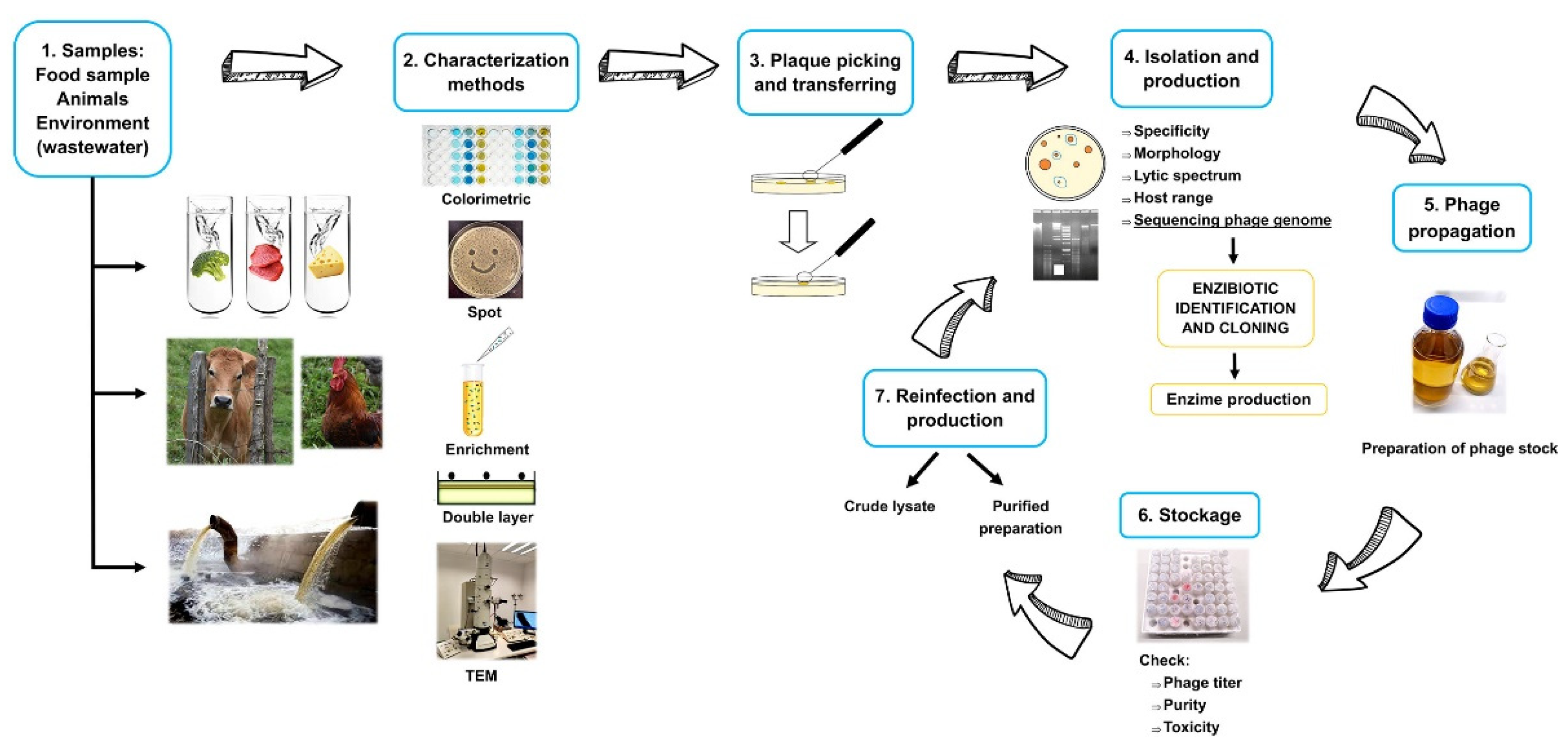
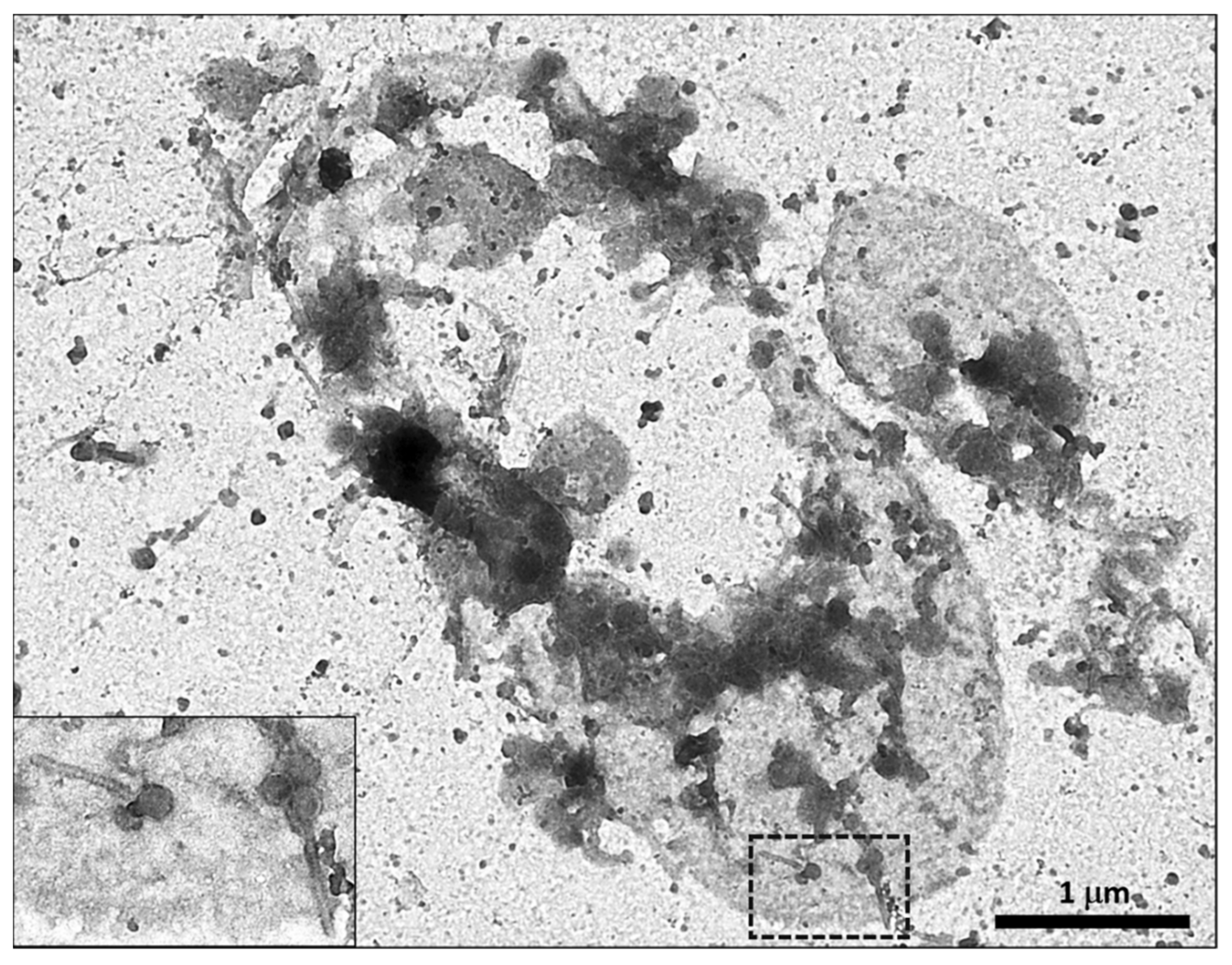
Table 1.
Phages tested against food-borne pathogens and their proposed use as food biopreservatives.
Table 1.
Phages tested against food-borne pathogens and their proposed use as food biopreservatives.
| Target Bacteria | Phage/s | Source | Characterization Method | Genome Length | 2 Family | Food Application | Reference |
|---|---|---|---|---|---|---|---|
| Aeromonas hydrophila | AH-1 | Sewage samples | TEM | ND | Myoviridae | Depuration of artificially contaminated cockles | [45] |
| AH-4 | |||||||
| AH-5 | |||||||
| Bacillus cereus | PBC1 | Sewage sample | TEM, sequencing | 41,164 bp | Siphoviridae | Inhibition of B. cereus growth in boiled rice | [46,47] |
| Brochothrix thermosphacta | A3 | Spoiled retail rib steaks | TEM | ND | ND | Control of bacterial strains during refrigerated storage | [16,48] |
| Campylobacter jejuni | Cj6 | Unknown | - | ND | ND | Control of pathogens in liquid foods | [36,49] |
| C. jejuni | 2 | Unknown | 1 dsDNA | ~140 kb | Myoviridae | Reduction of C. jejuni contamination of retail poultry products | [50,51] |
| C. jejuni | CP8 CP30 | Poultry excreta | TEM, dsDNA | ~140 kb | Myoviridae | Reduction of food-borne bacteria and biofilms | [52,53] |
| C. jejuni | 12673 | NCTC (UK) | TEM, DNA sequencing | ~135 kb | Myoviridae | Reduction of bacterial contamination on chicken carcass surfaces | [54,55] |
| Clostridium tyrobutyricum and C. sporogenes | CTP1 | Landfill | TEM, DNA sequencing | 59,199 bp | Siphoviridae | Cheese manufacturing | [56] |
| Cronobacter (Enterobacter) sakazakii | ESP 1–3 | Sewage treatment plant | TEM, dsDNA | ND | Siphoviridae | Control of E. sakazakii in reconstituted infant formula | [57] |
| ESP 732–1 | Myoviridae | ||||||
| Escherichia coli | PE37 | Bovine intestine samples | TEM, DNA sequencing | 166,423 bp | Myoviridae | Biocontrol of E. coli STEC O157:H7 and ESBLEC. | [58] |
| E. coli | EC6 | Sewage | TEM, dsDNA | ND | Siphoviridae | Biocontrol against E. coli in UHT and raw bovine milk | [59] |
| EC9 | Myoviridae | ||||||
| EC11 | Podoviridae | ||||||
| E. coli (STEC) O145 | Ro145clw | Non-fecal compost samples | TEM, DNA sequencing | 42,031 bp | Siphoviridae | Control of foodborne STEC O145 | [60] |
| E. coli O157:H7 | vB_EcoS_FFH_1 | Wastewater treatment plants | TEM, sequencing | 108,483 bp 139,020 bp | Siphoviridae | Reduction of contamination in ground beef | [61,62] |
| _vB_EcoS_FFH_1 | Myoviridae | ||||||
| E. coli O157:H7 | e11/2 | Bovine farmyard Slurry samples Swine stool samples | TEM, dsDNA | ND ND ~140 kb | Myoviridae | Elimination or reduction of E. coli O157:H7 bacteria from meat carcasses | [63,64,65] |
| e4/1c | Siphoviridae | ||||||
| PP01 | Myoviridae | ||||||
| E. coli O157:H7 | FAHEc1 | Raw screened sewage | TEM, dsDNA | ~90 kb | Myoviridae | Inactivation of E. coli O157:H7 on beef | [34,66] |
| E. coli O157:H7 | KH1 | Cattle and sheep fecal samples | - | ND | ND | Elimination of O157:H7 from foods under refrigerated conditions. Reduction of E. coli on surfaces. | [67,68] |
| KH4 | |||||||
| KH5 | |||||||
| E. coli O157:H7 | ECML-4 | Fresh and salt water environments | TEM, DNA sequencing | 157,308 bp 66,854 bp 166,783 bp | Myoviridae | Reduction of contamination of hard surfaces and foods contaminated by E. coli O157:H7 | [69,70] |
| ECML-117 | |||||||
| ECML-134 | |||||||
| E. coli strains, Salmonella and Shigella spp. | C203 | Cottage cheese and from poultry liver | TEM, DNA sequencing | 138,073 bp | Myoviridae | Biocontrol agent against E. coli EHEC O157 | [37] |
| P206 | |||||||
| Shigatoxigenic E. coli Enteropathogenic E. coli | DT1 | Stool samples of patients with diarrhea | TEM | ND | Myoviridae | Control of pathogenic E.coli in meat products and during milk fermentation | [71,72] |
| DT5 | |||||||
| DT6 | |||||||
| E. coli strains including serotype O157:H7 | OSY-SP | Manure from cattle, sheep, and horse farms | Pulsed-field gel electrophoresis (PFGE) | ~150 Kb | Myoviridae | Reduction of E. coli in fresh produce type (cut green pepper or spinach leaves) | [73] |
| Lactobacillus brevis | SA-C12 | fresh silage | TEM | ND | Myoviridae | Control of L. brevis beer-spoilage | [74] |
| Leuconostoc gelidum | ggg | vacuum-packaged pork | TEM | ND | Siphoviridae | Inhibition of Leuconostoc in raw pork | [75] |
| Listeria monocytogenes | A500 ATCC® 23074-B1™ | L. monocytogenes isolated from Guinea pig | TEM | 38,867 bp | Siphoviridae | Control of L-forms of L. monocytogenes on surfaces | [76,77] |
| L. monocytogenes | H387 H387-A 2671 | - | TEM | ND | Siphoviridae | Disinfection of working surfaces of food processing plants | [78,79] |
| L. monocytogenes | LiMN4L | Seafood waste water treatment unit | ND | ND | ND | Control of L. monocytogenes on stainless steel in seafood processing environments | [22] |
| LiMN4p | |||||||
| LiMN17 | |||||||
| L. monocytogenes | A511 | Sewage from a sewage treatment plant | Phage typing, TEM, sequencing | 134,494 bp | Myoviridae | Ready-to-eat foods from plant and animal origin including cheeses | [80,81,82,83,84] |
| L. monocytogenes | FWLLm1 | Sheep feces | TEM, | ND | ND | Reduction of L. monocytogenes growth in ready-to-eat poultry products | [85] |
| L. monocytogenes | IZSAM-1 | Floor drain-water from an Italian blue cheese dairy factory | TEM, sequencing | ~50 kb | Siphoviridae | Biocontrol of L. monocytogenes within cheese industrial facilities | [86,87] |
| Listeria spp. | P100 | Sewage effluent from a dairy plant | TEM, sequencing | 131,384 bp | Myoviridae | Biocontrol of contaminated surfaces, the surface of soft cheeses, ready-to-eat foods, fresh-cut fruit, and fruit juices, raw fish fillets, | [88,89,90,91,92] |
| Pseudomonas fragi | Wy | Ground Beef | TEM, dsDNA | ND | - | Reduction of P. fragi in refrigerated raw milk | [93,94,95] |
| Pseudomonas sp. | C35 | spoiled retail beef | - | ND | - | Biological control of beef spoilage | [96,97] |
| Pseudomonas lactis | HU1 | sludge obtained after passing raw cow’s milk through a centrifugal clarifier | TEM, dsDNA | ~48 Kb | Podoviridae | Control of P. lactis in Raw Cow’s Milk | [98] |
| Pseudomonas fluorescens E. cloacae strains | PspYZU5415 | Sewage samples | TEM, sequencing | 39,636 bp | Siphoviridae Corticoviridae | Growth inhibition of E. cloacae and P. fluorescens in cucumber juice with different salt concentrations | [43] |
| EcpYZU01 | 39,767 bp | ||||||
| P. fluorescens | IBB-PF7A | Sewage treatment plant | TEM, dsDNA | ~42 kbp | Podoviridae | Biocontrol of P. fluorescens in dairy and other food industries | [99,100] |
| Salmonella Enteritidis, S. Typhimurium | wksl3 | Chicken by-product samples | TEM, sequencing | 42,766 bp | Siphoviridae | Control Salmonella on chicken skin. from broiler carcasses | [101] |
| Salmonella serovars | LPSEYT | Water samples | TEM, sequencing | 53,387 bp | Myoviridae | Biocontrol of Salmonella in food matrices including milk and lettuce | [42] |
| Salmonella Enteritidis | CAU-SEP-1 | River water samples | TEM | ND | Myoviridae and Siphoviridae | Control of S. Enteritidis in chicken breast meat | [102] |
| CAU-SEP-2 | |||||||
| CAU-SEP-3 | |||||||
| CAU-SEP-4 | |||||||
| Salmonella Enteritidis | CNPSA 1 CNPSA3 CNPSA4 | free-range chickens | TEM, dsDNA | ND | tailed dsDNA phages | Reduction of Salmonella Enteritidis in Contaminated Chicken Cuts | [103,104,105] |
| Salmonella Enteritidis | P29C | Raw human sewage | - | ND | Siphoviridae | Reduction of bacterial contamination on chicken carcass surfaces | [54,106] |
| Salmonella spp. | PSE5 | Poultry slaughterhouse wastewater | plaque morphology and RAPD analysis | ND | ND | Reduction of contamination in raw chicken eggs | [107] |
| Salmonella spp. | LPSTLL | Environmentally water samples | TEM | ND | Siphoviridae | Reduction of Salmonella counts in milk and chicken breast and on stainless still surfaces | [108,109] |
| LPST94 | Ackermannviridae | ||||||
| LPST153 | Podoviridae | ||||||
| Salmonella strains | LPSE1 | Environmental samples | TEM, dsDNA, sequencing | 41,854 bp | Siphoviridae | Control of Salmonella in ready-to-eat foods | [110] |
| Salmonella strains | Felix O1/Felix O1-E2 | Feces of paratyphoid B patients | TEM, Sequencing | 86,155 bp/~84 kb | Myoviridae | Suppression of Salmonella growth on chicken frankfurters, poultry products, and ready-to-eat foods | [111,112,113,114] |
| Salmonella strains | PHL4 | Wastewater treatment plant | - | ND | ND | Reduction of Salmonella growth poultry products | [115] |
| Salmonella strains | vB_SalS_SJ_3 | Wastewater | TEM, DNA sequencing | 162,910 bp | Siphoviridae | Biocontrol of Salmonella in contaminated Eggs and Pork | [116,117,118] |
| Salmonella strains | Pu20 | Sewage samples | TEM, sequencing | 59,435 bp | Podoviridae | Growth inhibition of Salmonella strains in liquid egg white and yolk | [119] |
| Salmonella strains | D1-2 | Environmental samples | TEM, sequencing | 86,878 bp | Myoviridae | Growth inhibition of Salmonella strains in liquid egg white and yolk | [120] |
| Salmonella Typhimurium | P22 [Argo4] | Salmonella enterica subsp. enterica serovar Typhimurium | TEM, sequencing. Reference strain ATCC® 97540™ | 41,724 bp | Podoviridae | Prevention of attachment to food surfaces and food matrices | [121,122,123,124] |
| Salmonella Typhimurium | P7 | Unknown | - | ND | ND | Control of pathogens in liquid foods | [36] |
| Salmonella serovars | LPST153 | Water samples | TEM, sequencing | 39,176 bp | Autographivirinae | Control of Salmonella in raw milk and raw beef sausages | [125] |
| S. enterica serovar Typhimurium S. enterica serovar Enteritidis | UAB_Phi 20 | Chicken | TEM, dsDNA, sequencing | 41,809 bp 44,110 bp 87,669 bp | Podoviridae Podoviridae Myoviridae | Reduction of Salmonella on foods and reduction of Salmonella Colonization of poultry | [126,127,128] |
| UAB_Phi78 | Chicken | ||||||
| UAB_Phi87 | pig | ||||||
| Salmonella Enteritidis | SP-1 | Intestinal content of broiler chickens | TEM, dsDNA, PCR amplification | ~86 kb | Podoviridae | Biocontrol of Salmonella in cooked chicken meat | [35,129,130] |
| SP-3 | ~88 kb | Siphoviridae | |||||
| Salmonella Enteritidis | SJ2 | Chicken egg | ND | ND | ND | Reduction of Salmonella counts in Cheddar cheese made from both raw and pasteurized milk, and in contaminated eggs and pork | [131] |
| Salmonella Enteritidis | vBSenM-PA13076 (PA13076) vBSenM-PC2184 (PC2184) | Chicken sewage | TEM | 52,474 bp ND | Myoviridae | Biocontrol of Salmonella in foods (chicken breast, pasteurized whole milk, Chinese cabbage) | [132,133] |
| Salmonella and E. coli O157:H7 | PS5 | Raw chicken products | TEM, sequencing | 158,400 bp | Myoviridae | Reduction of viable counts on solid and liquid foods | [134] |
| Salmonella Oranienburg | SSP5 SSP6 | Sewage samples | TEM | ND | Myoviridae Siphoviridae | Control of Salmonella Oranienburg on alfalfa seeds | [135] |
| S. Typhimurium S. Enteritidis S. Montevideo | A B | sewage treatment plant | TEM | ND | Myoviridae Siphoviridae | Control of Salmonella in mustard and broccoli seeds | [136] |
| Salmonella strains, including MDR Salmonella | T156 | Waste water | TEM, dsDNA, sequencing | 123,849 bp | Siphoviridae | Microencapsulated bacteriophage applied in skim milk and lettuce for biocontrol of Salmonella | [137] |
| Staphylococcus aureus | K | Deposited by EA Asheshov | ATCC® 19685-B1™ | 139,831 bp | Myoviridae | Removing S. aureus biofilms | [138,139] |
| S. aureus | H5 (phiPLA88) A72 (phiPLA35) | Raw milk | TEM, dsDNA, sequencing | 42,526 bp 45,344 bp | Siphoviridae | Curd manufacturing, fresh and hard-type cheeses | [140,141,142] |
| S. aureus | SA46-CTH2 | Food samples | TEM | 17,505 bp | Podoviridae | Inactivation of S. aureus planktonic cells in pasteurized milk and biofilms on stainless steel surfaces | [143] |
| S. aureus | SA13m | Temperate phage SA13 isolated from a goat fecal sample | TEM, sequencing | 42,652 bp | Siphoviridae | Biocontrol of S. aureus in pasteurized whole milk at refrigeration and ambient temperatures | [144] |
| Shewanella baltica and S. putrefaciens | SppYZU01 to SppYZU10 | Wastewater from freshwater and marine product marketplaces | TEM, sequencing | SppYZU01 (43,567 bp) SppYZU05 (54,319 bp) | Myoviridae Siphoviridae | Biopreservation of chilled channel catfish | [145] |
| Shigella spp. | SF-A2 | Spiced chicken | TEM | ND | Myoviridae | inactivation of foodborne Shigella on ready-to-eat chicken | [146] |
| SD-11 | Pig farm effluent | ||||||
| SS-92 | Pig farm effluent | ||||||
| Vibrio parahaemolyticus | vB_VpaS_OMN (designated as phage OMN) | Sea water | TEM, sequencing | 42,202 bp | Podoviridae | Inactivation of V. parahaemolyticus in oyster meat | [147] |
1 Nuclease digestion tests and/or Random Amplified Polymorphic DNA Analyses (RAPD), 2 Family designated by the authors, ND: not determined, TEM: Transmission Electron Microscopy.
Table 2.
Enzybiotics tested against food-borne pathogens and their proposed use in foods.
| Target Bacteria | Enzybiotic | Source | Food Application | Reference |
|---|---|---|---|---|
| Bacillus cereus, B. subtilis and L. monocytogenes | LysB4 | B. cereus phage B4 | antibacterial agent to control foodborne pathogens. | [196] |
| Clostridium tyrobutyricum and C. sporogenes | Ctp1L | Bacteriophage CTP1 isolated from landfill | Cheese manufacture, reduction of clostridial activity in cheese | [56,197] |
| C. tyrobutyricum C. acetobutylicum | CS74L | Lytic bacteriophage (ATCC® 8074-B1TM) of C. sporogenes | Biocontrol of clostridia strains in foods | [198] |
| C. perfringens | Ply3626 | C. perfringens ATCC 3626 | Control of anaerobic spore-formers | [199] |
| C. perfringens | LysCPAS15 | C. perfringens phage CPAS-15 | Inhibition of C. perfringens in sterilized milk | [200] |
| Bacillus subtilis B. megaterium L. monocytogenes | PLY118, PLY500 PLY 511 | Phages from Listeria monocytogenes | Production of airy starter cultures with biopreservation properties | [201,202,203] |
| E. coli O157:H7 | PlyEc2 | Phage from E. coli | Reduction of E. coli O157:H7 on contaminated lettuce | [204] |
| Lactococcus lactis, Pediococcus acidilactici and P. pentosaceus | LysA2 | L. casei bacteriophage A2 | Ripening of fermented products | [205] |
| Lactobacilli, lactococci, pediococci, B. Subtilis Brevibacterium linens Enterococcus faecium | Mur-LH | Phage 0303 from Lactobacillus helveticus CNRZ 303 | Preventing the growth of spoilage microbes | [17] |
| L. monocytogenes B. subtilis | PlyP100 | Phage from L. monocytogenes | Antimicrobial biopreservative in fresh cheese. | [206] |
| L. monocytogenes | LysZ5 | Phage from L. monocytogenes | Control pathogens in soya milk | [207] |
| L. monocytogenes | PlyLM | Phage from L. monocytogenes strain 4b | Proposed control of L. monocytogenes in food matrices and processing facilities | [208] |
| L. monocytogenes | HPL118 HPL500 HPL511 HPLP35 | Recombinant endolysins from L. monocytogenes phages | Reduction of L. monocytogenes viable counts in iceberg lettuce. Promising perspectives in production and packaging environments | [201,209,210] |
| Methicillin-resistant Staphylococcus aureus | LysGH15 | Phage isolated from Sewage samples | Biopreservative in whole and skim milk | [211,212] |
| methicillin-resistant S. aureus | LysSA11 | Staphylococcus aureus phage SA11 | Biocontrol of S. aureus on strain in pasteurized milk or ham and utensils | [213] |
| S. aureus Bacillus cereus | Hybrid LysB4EAD-LysSA11 | Phage SA11 from S. aureus phage B4 S from B. cereus | Biocontrol of S. aureus and B. cereus in boiled rice | [195] |
| S. aureus | LysH5 | Staphylococcal bacteriophage phi-SauS-IPLA88 | Disinfection process of industrial food facilities. Elimination of S. aureus in pasteurized milk | [190,214] |
| S. aureus | CHAPSH3b | Chimeric protein (CHAP domain from peptidoglycan hydrolase HydH5 and the SH3b cell wall-binding domain from lysostaphin) | S. aureus biofilm elimination | [215] |
| S. aureus | CHAPK | Truncated derivative of the phage lysin LysK from the staphylococcal bacteriophage K | Reduction of biofilm formation in processing systems | [189] |
| S. aureus | HydH5 HydH5Lyso HydH5SH3b CHAPSH3b and lysostaphin | S. aureus bacteriophage vB_SauS-phiIPLA88 | Biocontrol of S. aureus in dairy products | [216] |
| Streptococcus spp. | λSA2 | Streptococcus agalactiae (serotype III GBS strain 3330) bacteriophage B30 | Inactivation of Strepcococcus spp. in cow milk | [217,218] |
| S. Typhimurium | LysSTG2 | Salmonella-lytic bacteriophage STG2 | Combating S. Typhimurium biofilms in food industries | [219] |
| Salmonella strains | LysSE24 | Salmonella phage LPSE1 | Food Control of Salmonella strains | [220] |
| Several Gram-negative pathogens, particularly against Salmonella Typhimurium | Lys68 | Salmonella phage phi68 isolated from feces from a poultry farm | Combat Gram-negative pathogens in the food industry | [221] |
Publisher’s Note: MDPI stays neutral with regard to jurisdictional claims in published maps and institutional affiliations. |
© 2021 by the authors. Licensee MDPI, Basel, Switzerland. This article is an open access article distributed under the terms and conditions of the Creative Commons Attribution (CC BY) license (https://creativecommons.org/licenses/by/4.0/).
Share and Cite
MDPI and ACS Style
Ramos-Vivas, J.; Elexpuru-Zabaleta, M.; Samano, M.L.; Barrera, A.P.; Forbes-Hernández, T.Y.; Giampieri, F.; Battino, M. Phages and Enzybiotics in Food Biopreservation. Molecules 2021, 26, 5138. https://doi.org/10.3390/molecules26175138
AMA Style
Ramos-Vivas J, Elexpuru-Zabaleta M, Samano ML, Barrera AP, Forbes-Hernández TY, Giampieri F, Battino M. Phages and Enzybiotics in Food Biopreservation. Molecules. 2021; 26(17):5138. https://doi.org/10.3390/molecules26175138
Chicago/Turabian StyleRamos-Vivas, José, María Elexpuru-Zabaleta, María Luisa Samano, Alina Pascual Barrera, Tamara Y. Forbes-Hernández, Francesca Giampieri, and Maurizio Battino. 2021. "Phages and Enzybiotics in Food Biopreservation" Molecules 26, no. 17: 5138. https://doi.org/10.3390/molecules26175138