
The Effects of Medicinal Plants and Bioactive Natural Compounds on Homocysteine
by
Mohammad Amin Atazadegan
1, 
Mohammad Bagherniya
2,3,4,*,
Gholamreza Askari
2,3,4,
Aida Tasbandi
5 and
Amirhossein Sahebkar
5,6,7,* 1
Department of Clinical Nutrition, School of Nutrition and Food Science, Isfahan University of Medical Sciences, Isfahan 8174673461, Iran
2
Food Security Research Center, Isfahan University of Medical Sciences, Isfahan 8174673461, Iran
3
Anesthesia and Critical Care Research Center, Isfahan University of Medical Sciences, Isfahan 8174673461, Iran
4
Department of Community Nutrition, School of Nutrition and Food Science, Isfahan University of Medical Sciences, Isfahan 8174673461, Iran
5
Applied Biomedical Research Center, Mashhad University of Medical Sciences, Mashhad 9177948564, Iran
6
Biotechnology Research Center, Pharmaceutical Technology Institute, Mashhad University of Medical Sciences, Mashhad 9177948564, Iran
7
School of Pharmacy, Mashhad University of Medical Sciences, Mashhad 9177948954, Iran
*
Authors to whom correspondence should be addressed.
Molecules 2021, 26(11), 3081; https://doi.org/10.3390/molecules26113081
Submission received: 19 March 2021
/
Revised: 11 May 2021
/
Accepted: 18 May 2021
/
Published: 21 May 2021
(This article belongs to the Special Issue Natural Products for Cardiovascular Disease)
Abstract
:Background: Among non-communicable diseases, cardiovascular diseases (CVDs) are the leading cause of mortality and morbidity in global communities. By 2030, CVD-related deaths are projected to reach a global rise of 25 million. Obesity, smoking, alcohol, hyperlipidemia, hypertension, and hyperhomocysteinemia are several known risk factors for CVDs. Elevated homocysteine is tightly related to CVDs through multiple mechanisms, including inflammation of the vascular endothelium. The strategies for appropriate management of CVDs are constantly evolving; medicinal plants have received remarkable attention in recent researches, since these natural products have promising effects on the prevention and treatment of various chronic diseases. The effects of nutraceuticals and herbal products on CVD/dyslipidemia have been previously studied. However, to our knowledge, the association between herbal bioactive compounds and homocysteine has not been reviewed in details. Thus, the main objective of this study is to review the efficacy of bioactive natural compounds on homocysteine levels according to clinical trials and animal studies. Results: Based on animal studies, black and green tea, cinnamon, resveratrol, curcumin, garlic extract, ginger, and soy significantly reduced the homocysteine levels. According to the clinical trials, curcumin and resveratrol showed favorable effects on serum homocysteine. In conclusion, this review highlighted the beneficial effects of medicinal plants as natural, inexpensive, and accessible agents on homocysteine levels based on animal studies. Nevertheless, the results of the clinical trials were not uniform, suggesting that more well-designed trials are warranted.
1. Introduction
In recent decades, we have experienced a shift in the disease burden towards non-communicable diseases, which is likely due to the improved efficacy of treatments and lifestyle changes [1,2]. According to the World Health Organization (WHO), non-communicable diseases account for 75% of global deaths in 2020; of these, cardiovascular diseases (CVDs) are the leading cause of mortality and morbidity in communities [3,4,5]. By 2030, CVD-related deaths are projected to reach a global increase of 25 million [6,7]. The documented studies show that non-communicable diseases are responsible for $7.28 trillion loss from 2011 to 2025, half of which is related to CVDs. It is estimated that a 10% reduction in CVDs will reduce the economic loss by $378 billion over the years [8,9]. The known risk factors of CVDs include obesity, smoking, alcohol consumption, hyperlipidemia, hypertension, hyperhomocysteinemia, among others [10,11]. Homocysteine is a mediator in methylation cycle that acts as a cysteine and methionine precursor, a substrate for tissue folate recycling, and methyl receptor in choline catabolism [12,13,14]. Methionine is first converted to S-adenosylmethionine (SAM), which is, in turn, converted to S-adenosyl homocysteine (SAH) in a reversible reaction. Subsequently, S-adenosine homocysteine breaks down into adenosine and homocysteine. In all tissues, 5-Methyltetrahydrofolate (as methyl group donor; in the liver, betaine acts as a methyl donor) and methylcobalamin (as coenzyme) are required to convert homocysteine to methionine with the help of vitamin B12. Homocysteine itself is first converted to cystathionine and then to cysteine (with the help of vitamin B6), which is finally excreted in the urine as sulfur-containing compounds or converted to taurine [15,16,17]. Homocysteine and its derivatives are bound to intracellular proteins and their presence in the blood may be due to their removal from the cells to be used by other tissues [12,18,19]. In 1962, the association between homocysteine and CVDs was established [20]. The normal homocysteine levels are considered to be less than 10 micrograms per liter. Hyperhomocysteinemia is defined as mild (12–30 micrograms per liter), intermediate (31–100 micrograms per liter), and severe (more than 100 micrograms per liter) [21,22]. In the general population, the prevalence of hypercysteinemia is approximately five to 10 percent and 30 to 40 percent in the elderly [23]. In population studies, an inverse relationship was found between homocysteine concentrations and vitamin B12 or abdominal aortic diameter in the elderly [24,25]. Several meta-analyzes have shown that hyperhomocysteinemia is a strong predictor of CVDs [26,27,28,29,30]. A five micromole per liter increase in blood homocysteine levels is associated with a 32% increase in myocardial ischemia and a 59% increase in myocardial infarction [31]. Additionally, wild-type and mutant methyltetrahydrofolate increase the risk of cardiovascular disease by 25% and the homocysteine level by 16% [31]. Although the mechanism of action of homocysteine on blood vessels is not fully understood, it is thought to damage different layers of arteries [32]. Hyperhomocysteinemia has negative impacts on endothelial cells by affecting the production of nitric oxide, which regulates vascular tone [33,34,35,36,37,38]. It may also impair the maintenance of vascular homeostasis by interfering with the hydrogen sulfide signaling pathway that is closely related to nitric oxide [39]. Evidence suggests that hyperhomocysteinemia promotes inflammation of vascular endothelium by inducing inflammatory factors, such as interleukin-6, interleukin-8, and tumor necrosis factor alpha [40,41,42]. It is shown that high levels of homocysteine are associated with increased endoplasmic reticulum stress, which, in turn, leads to vascular inflammation [43] and oxidative stress in mice [44]. Additionally, homocysteine causes endothelial cell apoptosis, which is a primary sign of atherosclerosis [44,45]. In a study conducted in 2015, it was indicted that homocysteine metabolites (homocysteine thiolactone and N-homocysteine) could disrupt vascular homeostasis [46]. Another effect of homocysteine on the dysregulation of vascular homeostasis is through impairing the function of smooth muscle cells and inducing their proliferation [47,48,49,50]. Homocysteine causes arterial stiffness by the deposition of collagen in the vascular wall through the induction of connective tissue growth factor (CTGF). It also triggers the formation of aneurysm in coronary arteries by inducing the production of elastin degrading enzymes [51,52,53,54]. The preventive efforts are the best way of managing CVDs, and they are achieved by a proper lifestyle, including a balanced diet, and adequate physical activity [55], which may be not be favorable to some individuals. Although the strategies for the appropriate treatment of CVDs are constantly evolving; they are often ineffective and could have serious side effects [55,56]. Hence, the recent focus is on natural or low-risk supplements, such as herbal medicines or functional foods [57]. The protective effects of nutraceuticals on CVDs have been previously identified [58,59]. The term nutraceutical was first coined in 1989 by Stephen Deflis. Nutraceuticals are defined as “foods or parts of a food that provide medical or health benefits including prevention or treatment of a disease” [60]. Nutraceuticals are substances that can be used instead of a medication as dietary supplements to control, prevent, or even treat diseases, such as CVDs [61]. These are known as food drugs and they are particularly helpful in patients who do not have access to a medication or for treatment of chronic diseases, such as osteoporosis, heart diseases, among others [62]. Recently, medicinal plants are becoming the focus of several researches, since these natural products have shown promising effects on the prevention and treatment of various diseases, particularly CVDs [63,64,65,66], diabetes mellitus [67,68,69], hypertension [70,71,72], and non-alcoholic fatty liver disease (NAFLD) [56]. The beneficial effects of herbal bioactive compounds on 3-hydroxy-3-methyl-glutaryl-coenzyme A reductase (HMG-CoA reductase) [73], Apolipoprotein B (Apo B) [74], and small dense LDL [74] have recently been reviewed. These are three important factors with unfavorable effects on CVDs. The effects of nutraceuticals and herbal products on CVDs/dyslipidemia have been previously studied. However, to our knowledge, the association between herbal bioactive compounds and homocysteine has not been reviewed in detail [63,64,65,66,75,76,77,78,79]. Thus, the main objective of this study is to address the efficacy of bioactive natural compounds on homocysteine levels, which was assessed through clinical trials and animal studies (Figure 1).
2. Results
2.1. Plants
2.1.1. Black Tea
Tea is widely consumed around the world, and all the popular types of tea, like black tea, are manufactured from the leaves of Camellia sinensis (L.) Kuntze from Theaceae family. Several studies have shown that tea and its bioactive polyphenolic constituents have numerous beneficial effects on the prevention of diseases, like cancer, diabetes, arthritis, CVDs, stroke, and obesity [80,81,82,83]. These effects are due to antioxidative, anti-inflammatory, antihypertensive, cholesterol-lowering, antimicrobial, anticarcinogenic, neuroprotective, and thermogenic properties of the tea [83]. The favorable effects of tea on CVDs have been demonstrated in epidemiological studies and clinical trials [83]. Its effect on homocysteine level is one proposed mechanism. In one experiment, the rats were assigned into three groups: (1) Vehicle (saline), (2) angiotensin (Ang) II (50 ng/kg/min.) to induce hypertension, and (3) Ang II + Black tea extract (BT) in which animals were given a 15 mg/kg/day of black tea extract (starting from Day 1 after Ang II pump insertion) for two weeks. Initially, angiotensin II infusion enhanced the plasma homocysteine level and it resulted in endoplasmic reticulum (ER) and oxidative stress, which, in turn, triggered endothelial dysfunction. However, black tea extract dramatically decreased the blood pressure and plasma homocysteine and, consequently, protected arteries of hypertensive rats from ER stress and endothelial dysfunction [84]. According to a clinical trial conducted by Hodgson et al., 20 adults with CAD were randomly assigned to four groups: (I) water and no meal, (II) black tea and no meal, (III) meal with water, or (IV) meal with black tea. The participants were asked to drink a cup of black tea (contained 2.2 g of tea leaves at times 0, 1.5 h, and 3 h) or three cups of hot water with and without a meal (comprised a sausage, egg, bacon, McMuffins, and two hash browns). The total homocysteine was measured at baseline and 3.5 h after drinking black tea or hot water with and without a meal. In the end, an acute increase in homocysteine was observed after drinking black tea. Although the meal caused an acute decline in homocysteine level, it did not alter the homocysteine-raising effect of tea [85]. Similarly, in a randomized crossover study, 22 subjects were divided into two groups to drink 1250 mL black tea/d (five cups each containing 2 g tea leaves in 250 mL boiled water) or 1250 mL hot water/d for four weeks. For the next four weeks, the participants consumed the alternate drink. The findings demonstrated that black tea did not significantly alter the mean homocysteine concentrations [86]. Twenty healthy subjects in another crossover study received a diet low in polyphenols and they were randomly assigned into four groups of supplemented regimens: (I) 2 g chlorogenic acid (a compound found in coffee and black tea), (II) 4 g black tea solids (III) 440 mg quercetin-3-rutinoside, or (IV) 0.5 g citric acid as a placebo. The duration of each trial was seven days (a four-week trial). The results showed that, after 4–5 h of supplementation, chlorogenic acid and black tea both raised total homocysteine concentrations in plasma when compared to the placebo. Quercetin-3-rutinoside exerted no effect on plasma homocysteine [87]. The different results between the animal and clinical studies might be due to the fact that, in a previous animal study, black tea was administered in experimentally hypertensive rats. It was suggested that homocysteine levels are reduced after the intake of black tea polyphenols in hypertension, which might be mediated and attributed to the promotion of homocysteine metabolism [84]. On the other hand, in the clinical trials, a small sample size of healthy subjects without hypertension were recruited, which might be a potential explanation for reporting the different results between animal and human studies [86,87]. In addition, it is suggested that tea and coffee, and their major constituents, such as polyphenols and caffeine, might increase homocysteine by acting as acceptors of methyl groups during the metabolism of methionine to homocysteine [86,87,88,89]. When considering these controversial findings and the fact that several studies suggest that tea consumption has a protective role against CVDs [90,91,92], there have been calls to conduct more studies in the future related to both the mechanistical and clinical aspects to explore the role of black tea on homocysteine.
2.1.2. Green Tea
Green tea is a non-fermented tea [93] that is traditionally used as a natural medicine. It is a rich source of polyphenols, mainly epigallocatechin-3-gallate (EGCG) (Figure 2) [94,95], which is proved to have favorable effects on neurological diseases, cancer, inflammation [94,96,97,98,99], and homocysteine-induced cerebrovascular injury [100]. In a study on adult male Wister rats, six groups were randomly assigned: (1st) normal laboratory diet, (2nd) 2.5 mg/kg body weight EGCG, (3rd) 5 mg/kg body weight EGCG, (4th) exposure to 4 Gy of γ radiation, (5th) 2.5 mg/kg body weight EGCG + exposure to 4 Gy of γ radiation, and (6th) 5 mg/kg body weight EGCG + exposure to 4 Gy of γ radiation. The intervention was performed for three consecutive days in the 2nd and 3rd groups, and for two days in 5th and 6th groups. The third dose was administered for 30 min. before irradiation. In rats that were pretreated with EGCG at a dose of 2.5 and 5 mg/kg, plasma homocysteine was significantly decreased when compared to the first group. The homocysteine levels were significantly decreased in the second and third groups as compared to the first group [101].
2.1.3. Cinnamon
Cinnamon (Cinnamomum verum J.Presl), a plant from the Lauraceae family, is mostly used as a spice [102]. It is a herbal medicine used for conditions, such as flatulence, amenorrhea, diarrhea, toothache, fever, leukorrhea, common cold, and headache [103,104]. Cinnamon and its main component, Cinnamaldehyde (Figure 3), have insulin sensitizer, antioxidant, and anti-inflammatory properties [105,106]. This herbal medicine was also traditionally recommended for the treatment of impotency, frigidity, dyspnea, eye inflammation, vaginitis, cough, rheumatism, neuralgia, and CVDs [107]. A two-phase clinical trial conducted by Amin et al. was conducted on 48 male albino rats for 5–8 weeks. Initially, hypercholesterolemia (the addition of 1% cholesterol powder, 0.25% bile salts, and beef tallow in a percentage of 4% to basal normal diet for 15 days) was induced. Subsequently, during the treatment period (starting from the third week and continued for six weeks), the hypercholesterolemic rats were divided into three subgroups (12 rats per group) according to the type of treatment. These regimens included HCD, HCD + Atorvastatin (0.2 mg/kg body weight), and HCD + cinnamon (C. zeylanicum Blume) (20 mg/day/rat). It was showed that cinnamon extract could reduce hypercholesterolemia and modulate oxidative stress and homocysteine [108].
2.1.4. Anthocyanin
The word anthocyanin is derived from two Greek words, plant (Anthos) and blue (kianos), which are the most important pigments in vascular plants [109]. Like chlorophyll, they are natural pigments that are non-toxic, water-soluble, and widely present in plant cells [110]. Anthocyanins are the most colorful compounds among the flavonoids that are responsible for different colors found in many fruits, vegetables, and flowers [111,112]. They play essential roles in the management of CADs due to their high antioxidant effect [113,114]. In one clinical trial, 20 healthy female volunteers were randomly assigned into two groups to receive a placebo beverage or cranberry juice (750 mL/day (3 × 250 mL), which contained 2.80 mg/L anthocyanins) for two weeks. In the end, the plasma total homocysteine remained unchanged [115].
2.1.5. Garlic Extract
Garlic (Allium sativum L. Liliaceae) is a well-recognized medicinal plant. Several pharmacological implications of A. sativum and its organosulfur compounds, especially Allicin (Figure 4), include antibacterial, antiviral, antifungal, antiparasitic, anticancer, anti-inflammatory, and cardiovascular protective properties [116,117,118,119,120,121,122,123]. It has beneficial roles on dyslipidemia, which lowers the total cholesterol concentrations by approximately 10% and favorably alters HDL/LDL ratios. Additionally, it acts as a mild anti-hypertensive that reduces blood pressure by 5–7% [124]. A group of 60 subjects were randomly assigned to two groups: (I) intervention (received a daily capsule of aged garlic-extract (AGE) (250 mg) plus vitamin-B12 (100 μg), folic-acid (300 μg), vitamin-B6 (12.5 mg), and L-arginine (100 mg) for 12 months) and (II) placebo. Finally, a reduced level of homocysteine was observed in the intervention group [125]. In a study conducted by Budoff et al., 65 patients with an intermediate risk for CVDs were randomly allocated to two groups: (I) the daily administration of a capsule containing AGE (250 mg) plus Vitamin B12 (100 μg), folic acid (300 μg), Vitamin B6 (12.5 mg), and L-arginine (100 mg) or (II) placebo. After one year of treatment, the homocysteine level was decreased in the AGE group [126]. In another experiment, 40 rats were employed to receive one of the following diets for six weeks (four groups of 10 each): (I) AIN-93G folic-acid sufficient (2 mg/kg of diet); (II) AIN-93G folic-acid deficient; (III) AIN-93G folic-acid sufficient that was supplemented with AGE (4% of diet, wt:wt); and, (IV) AIN-93G folic-acid deficient supplemented with AGE. The results showed that the addition of AGE to the severely folate-deficient diet decreased the total plasma homocysteine concentration by 30% [127]. Based on a similar study, 23 patients with known CAD were randomly assigned to two groups: (I) received AGE (4 mL) or (II) received placebo for one year. Following intervention, the homocysteine levels showed no significant improvement [128]. In another randomized controlled trial (RCT), 30 postmenopausal women were randomly assigned to four groups: (1) placebo, (2) consumption of AGE (5 × 65 mg per week), (3) exercise (60% of maximum heart rate) and placebo, and (4) exercise and aged garlic extract. After 12 weeks, homocysteine was significantly decreased with intervention in the second and fourth groups [129]. Ried et al., conducted an RCT on 30 pediatric (aged 8 to 18 years old) patients who were administered 300 mg AGE (three capsules/ daily) or placebo. After eight weeks of treatment, no significant difference was observed in homocysteine levels between groups [130]. In a similar RCT, 88 uncontrolled hypertensive patients were given 1.2 gr of AGE powder+ 1.2 mg S-allylcysteine each day or placebo for 12 weeks. Following intervention, no significant change was detected in the homocysteine level in both groups [131].
2.1.6. Ginseng
Ginseng (Panax ginseng C.A.Mey., Araliaceae) is a medicinal plant with favorable pharmacological effects in cancer, diabetes, and CVDs, which also improves the immune system and central nervous system (CNS) function, relieves stress, and possesses antioxidant properties, and these benefits are mainly attributed to the presence of ginsenosides (Figure 5) [132,133,134,135]. A group of 40 wistar male rats were randomly assigned to one of the following four groups: (1st) control group (tap water), (2nd) Methionine (Met) (1 g/kg per day) and succinyl sulfathiazole (SSL) (0.5 g/kg per day), (3rd) ginsenosides total saponins (GTS) (Korean ginseng) (50 mg/kg every 12 h), and (4th) Met + GTS + SSL. The homocysteine levels were measured within 30 and 60 days of the intervention, and they were found to be reduced in the fourth group as compared to the second group, whereas the third group had no significant change when compared to the first group [136].
2.1.7. Chlorella
Chlorella is a genus of approximately thirteen species of single-celled green algae belonging to the division Chlorophyta [137]. Chlorella pyrenoidosa, a single cell alga that is found in freshwater, has a rich content of chlorophyll and high concentration of nucleic acids, minerals, amino acids, dietary fiber, and vitamins. It has a strong cell wall that can only be digested by humans during a Dyno-Mill process. Studies showed that injection or oral intake of Chlorella following its cell wall destruction improves the immune function against infection and anti-cancer activity [138,139,140,141,142,143]. In study conducted by Merchant et al., 17 vegetarian/vegan (aged 26–57 years old) with a vitamin B12 deficiency were asked to add 9 gr/day of C. pyrenoidosa to their routine diet for 60 days. The findings reveled that the homocysteine level was reduced by an average of 10% [144].
2.1.8. Ginger
Ginger (Zingiber Officinale Roscoe, Zingiberaceae) is a part of a family of plants, including cardamom and turmeric. It has a strong aroma due to the presence of gingerols (pungent ketones) (Figure 6). The rhizome part of the plant is traditionally used in Asia and tropical areas for fever, common cold, digestive problems, among others. Ginger is an appetite stimulant, and it is categorized by the U.S. FDA as a food additive [145]. Its beneficial effects on nausea/vomiting and arthritis have been previously reported [146,147,148,149,150,151,152,153]. In one experiment, 24 male Wistar rats were randomized into three groups: (1) non-diabetic control (tap water), (2) non-treated diabetic (tap water), and (3) ginger (Z. Officinale) extract treated diabetic (50 mg/kg of hydroalcoholic ginger + tap water). After six weeks, the homocysteine level was notably enhanced in the second group when compared to the first group, whereas it was significantly declined in the third group as compared to the second group [154]. In another experiment by Akbari et al., 28 adult male Sprague Dawley rats were randomized into four groups: (1) control (2 mL/day corn oil), (2) ginger (Z. Officinale) (1 g/kg body weight daily), (3) ethanol (4 g/kg body weight daily), and (4) ethanol-ginger (Z. Officinale). After 28 days, the homocysteine level was significantly increased in the third group when compared to the first group, whereas it was significantly decreased in the fourth group as compared to the third group [155].
2.1.9. Soy
Soybean (Glycine max (L.) Merr., Fabaceae) is a traditional plant that is native to East Asia, which is a good source of phytochemicals (e.g., isoflavones: daidzein, and genistein, Figure 7), fiber, and plant sterols [156,157,158]. Various studies have revealed its beneficial effects on blood lipids, CVDs, fertility, and menopause [159,160,161]. The result of a systematic review and meta-analysis (2016), which reviewed 19 randomized controlled studies, showed that Soy or isoflavones had no effect on homocysteine levels [162]. In a randomized cohort study, 87 healthy postmenopausal women were assigned to one of these groups: (1) 1200 Kcal diet + exercise or (2) 1200 Kcal diet + exercise + 200 mg of Glycine max (80 mg of soy isoflavone: 60.8 mg of genistein, 16 mg of daidzein, and 3.2 mg of glicitein) for six months. Following intervention, the homocysteine level remained unchanged in both of the groups [163]. A group of 117 patients with hypercholesterolemia were also divided into three groups to receive: 15 gr/day soy protein (containing SuproSoy from Solae, Saint Louis, MO, USA) and 25 gr/day soy protein or placebo. No change in homocysteine level as compared to the baseline was found in all groups [164]. Similarly, in a cross over study on 34 postmenopausal women, the participants received 26 ± 5 g/day of isolated soy protein (containing 44 ± 8 mg isoflavones per day) or 26 ± 5 g/day of milk protein isolate for six weeks each and two weeks washout. The homocysteine level was not different between the two groups [165]. Forty patients on peritoneal dialysis were studied in a clinical trial; two groups were, as follows: received 28 gr/day of textured soy flour (contained 14 g of soy protein) or followed their regular diet for eight weeks. The concentration of homocysteine had no significant difference between the two groups [166]. In another randomized crossover study on forty-one hyperlipidemic men and postmenopausal women, three groups were assigned to receive one of these regimens: (1) a low-fat dairy control diet, (2) low-isoflavone soy food diet (10 mg isoflavones/day), and (3) high- isoflavone soy food diet (73 mg isoflavones/day). Intervention was conducted for 3* one months, allowing a two-week washout period between interventions. It was showed that the homocysteine level was lower in both isoflavones groups when compared to the control group [167]. A total of 55 postmenopausal women (aged 42–72 years) in a double blind clinical trial were randomly recruited to receive one of the four soy protein isolate treatments (40 g/d): (1st) normal phytate and isoflavone (Phytate: 0.78 g and Aglycone isoflavones: 84.6 mg), (2nd) normal phytate and low isoflavone (Phytate: 0.64 g and Aglycone isoflavones: 1.2 mg), (3rd) low phytate and normal isoflavone (Phytate: 0.22 g and Aglycone isoflavones: 85.5 mg), and (4th) low phytate and isoflavone (Phytate: 0.22 g and Aglycone isoflavones: 1.2 mg). After six weeks, the homocysteine levels were significantly reduced in soy protein normal phytate groups, whilst no significant change was detected in the soy protein normal isoflavone groups [168]. In another recent randomized crossover study, 89 postmenopausal women were randomly assigned to consume (1) two fruit cereal bar,s each including 25 mg soy isoflavons (genistein:daidzein ratio of 2:1) or (2) only two fruit cereal bars with no isoflavones. The intervention was conducted for eight weeks with an eight-week washout. The results showed that homocysteine did not change in both groups [169]. In a recent double-blind RCT, 38 postmenopausal women complaining of insomnia were asked to either consume 80 mg/day of isoflavones (60.8 mg of genistein, 16 mg of daidzein, and 3.2 mg of glicitein) daily or placebo for four months. Following intervention, the homocysteine showed no significant change in both groups [170]. Similarly, 30 female wild-type mice were randomly allocated to three groups: (I) the control group (modified standard maintenance chow), (II) soy group (55% total energy), or (III) casein group (55% total energy). After 12 weeks, the homocysteine level was higher in casein group when compared to the control group, while the homocysteine level remained the same in the soy and control groups [171]. A total of 24 adult female Sprague Dawley rats were also randomized into four groups: (1) 2% cholesterol diet (2) 2% cholesterol diet + fresh soy oil, (3) 2% cholesterol diet + one-heated soy oil, and (4) 2% cholesterol diet + five-time-heated soy oil. After four months, 2% cholesterol diet + fresh soy oil significantly reduced the homocysteine level when compared to the others [172].
2.1.10. Emblica Officinals (Amla)
Emblica officinalis L. (Phyllanthaceae), which is known as Indian Gooseberry or Amla, is a fruit that has high levels of ascorbic acid (from 1100 to 1700 mg/100 g of fruit extract) and a high density of ellagitannins including emblicanin A (37%) (Figure 8), emblicanin B (33%), punigluconin (12%), and pedunculagin. Amla is often consumed as a functional food due to its physiological features, such as radioprotection [173,174,175], antioxidant activity [176,177,178,179,180], hepatoprotection [181,182,183,184], cytoprotection [185,186], and hypolipidemic effects [176,187,188,189]. In a recent double-blind RCT, 98 patients affected with dyslipidemia were asked to either consume 500 mg capsule of Amla extract (from Indian gooseberry) each day or placebo for 12 weeks. Following intervention, the homocysteine level did not change significantly in both groups [190]. Similarly, 17 uremic patients were selected to receive Amla extract tablets (300 mg, 50% dextrin + 50% amla extract) four times a day. Based on the findings, the homocysteine level did not change after four months of intervention [191].
2.1.11. Nuts
Nuts have scant amounts of bioactive compounds (e.g., phytosterols, unsaturated FAs, fiber, protein, vitamins, calcium, magnesium, sodium, and potassium) and they are a good source of antioxidants [192,193,194]. In a study on 15 hypercholesterolemic (serum cholesterol level > 200 mg/dl) adult males (aged 33–59 years), the patients received hazelnut (Corylus avellana L., Betulaceae) enriched diet (control diet (low-fat, low-cholesterol, and high-carbohydrate) + 40 g/d hazelnut) for four weeks directly after four weeks of a control diet. Following intervention, no change in homocysteine level was detected between the groups [195]. In a two-phase single blind crossover study on 67 patients (serum total cholesterol > 5.2 mmol/L), the participants first consumed low-fat, low-cholesterol diet for six weeks. On the second phase, two groups were randomly selected to either consume 64 g/d walnut (Juglans regia L., Juglandaceae) with their diet or continue the same diet for six weeks. Finally, all of the patients were crossed over into the opposite treatment arm for another six weeks. The findings reveled no statistically significant change on the homocysteine levels [196].
2.1.12. Olive Oil
Olive oil, which is known as the elixir of health and youth, consists almost one-third to two-thirds of the fat used in the Mediterranean diet [197,198,199,200,201]. Many studies have shown its beneficial effects in improving heart diseases or cancer [202,203,204,205]. In one clinical trial, 121 obese (BMI ≥ 35 kg/m2) adults (aged 18–65 years) randomly received one of these three diets: (I) 52 mL/d extra virgin olive oil (EVOO), (II) traditional Brazilian diet (DieTBra), or (III) 52 mL/d EVOO + (DieTBra). After 12 weeks, the homocysteine levels reduced in the EVOO group by nearly 10% (mean), whilst the homocysteine levels did not change significantly between groups two-by-two [206].
2.2. Phytochemicals
2.2.1. Berberine
Berberine, a benzylisoquinoline alkaloid (Figure 9), is an active constituent in numerous medicinal plants with many pharmacological properties. It has been largely used in Ayurvedic and Chinese medicine for its antimicrobial, antiprotozoal, antidiarrheal, and antitrachoma activities. Several clinical and preclinical studies have indicated the promising effects of berberine on metabolic, neurological, and cardiological disorders [207,208,209,210,211,212]. In a study, healthy male rats (weighing 190–210 g) randomly received a standard diet or a high-fat diet (HFD) for 24 weeks. After eight weeks of feeding, rats that were fed with HFD were randomly assigned into two groups: (I) berberine (extracted from Coptis chinensis Franch.) (200 mg/kg/day) or (II) vehicle by gavage for 16 weeks (n = eight per each group). The results showed that the berberine consumption led to a significant reduction in serum homocysteine by about 60% when compared to the vehicle in rats fed with a HFD [213]. In a double-blind RCT, 31 diabetic patients were randomly assigned into two groups to receive Berberis vulgaris L. fruit extract (3 g/d) (which contains the berberine alkaloid) or placebo for three months. No significant change was observed in the serum homocysteine level between the two groups [214].
2.2.2. Curcumin
Curcumin, a turmeric-derived polyphenol (Curcuma longa L., Zingiberaceae) (Figure 10), is known for its safety and medicinal properties against a variety of diseases [215,216,217,218,219,220,221,222,223,224]. It also has beneficial effects in metabolic syndrome and obesity [225,226]. In a recent double-blind RCT, 22 obese men were administered a 500 mg curcumin supplement (193 mg of curcuminoids in the form of 81.8% curcumin, 15.3% demethoxycurcumin (C20H18O5) (Figure 11), and 2.8% bisdemethoxycurcumin(C19H16O4) (Figure 12) were infused into 60% soluble fiber from fenugreek to improve bioavailability) or placebo supplement (comprised of equal parts of soluble fiber from fenugreek) every day for 12 weeks. After intervention, the plasma homocysteine concentration was significantly reduced in the intervention group when compared to the placebo group [227]. In a clinical trial, female Wistar–Furth rats were randomly divided into low ω-3 PUFA (LFO, n = 12) and high ω-3 PUFA (HFO, n = 12) groups and they were further divided into three subgroups: LFO or HFO (controls), LFOE or HFOE (LFO or HFO plus ethanol, 35% of dietary calories derived from ethanol), and LFOEC or HFOEC (LFOE or HFOE supplemented with curcumin 150 mg/kg body weight/day). All of these groups were pair-fed for eight weeks. Curcumin caused a significant increase in homocysteine thiolactonase activities as compared to the high ω-3 PUFA and ethanol groups [228]. In a group of fifty healthy men who were randomly selected, a two month consumption of biscuits with a bioactive complex, such as organic selenium (115 mg), quercetin (6 g dried selenized onion), curcumin (1.3 g curcuma), and catechins (2 g green tea), was associated with decreased homocysteine levels [229]. In a clinical trial on 32 adult male Wister rats, four groups were selected: (1) control group (no injection), (2) vehicle of homocysteine (2 μmol/μL), (3) vehicle of curcumin (50 mg/kg), and (4) homocysteine-curcumin group. Curcumin was injected intraperitoneally once daily for 10 days, beginning five days prior to homocysteine intracerebroventricular injection. In the end, curcumin significantly reversed the behavioral and biochemical changes caused by exposure of homocysteine in the control mice. Similar to homocysteine, curcumin could be considered to be a therapeutic agent in preventing the progression of neurotoxicity [230]. A total of 50 female Sprague–Dawley rats (weighing 220–250 gr) were randomly divided to the following groups: (1) control (0.3 mL of the vehicle), (2) sham (0.3 mL of the vehicle + surgery stress), (3) danazol treatment (7.2 mg/kg BW), (4) curcumin treatment (48 mg/kg BW), and (5) test (0.3 mL of the vehicle). After four weeks of treatment, no significant difference was observed between all groups [231].
2.2.3. Resveratrol
Resveratrol (3,5,4′-trihydroxy-trans-stilbene) (Figure 13), a natural polyphenol that is found in numerous fruits and vegetables, has several properties, including anti-aging, anticancer, anti-inflammatory, and antidiabetic. Although the positive effects of resveratrol against selected cardiovascular risk factors have been a subject of debate over recent years [232,233], there are numerous effects against oxidative stress, apoptosis, mitochondrial dysfunction, endothelial dysfunction, and angiogenesis that might still support the potential role of this phytochemical in the prevention of CVDs [234,235,236]. A total of 24 rats were randomly assigned to three groups: (I) control group (received standard rat food), (II) homocysteine group (received 1 g/kg bodyweight/day methionine in drinking water), and (III) homocysteine + resveratrol group (received same amount of methionine + 20 mg/kg/day resveratrol intraperitoneally). After 30 days, the results indicated that the plasma homocysteine level was significantly reduced in a group that was treated with resveratrol [237]. In a study conducted by Noll et al., the mice were randomly divided into four groups and maintained on the following diets for three months: (I) control group (received standard rodent diet), (II) high methionine (received standard diet plus 0.5% L-methionine (36 mg/day) in drinking water), (III) methionine/resveratrol (received high-methionine diet with 0.001% trans-resveratrol (50 µg/day) in the last month, and (IV) resveratrol (received standard diet with 0.001% trans-resveratrol in the last month). The findings showed that plasma total homocysteine concentration in high methionine group was four times higher than the control group. In the methionine/resveratrol group, plasma total homocysteine levels were non-significantly increased when compared to the methionine group. However, in methionine/resveratrol group plasma homocysteine levels were 1.3 times higher as compared to the methionine group. Additionally, in the resveratrol group, the homocysteine levels were significantly increased by 1.7 (uM) when compared to the control group. Based on these results, resveratrol had detrimental effects on homocysteine levels [238]. Similarly, an experiment was performed on 30 female rats that were randomly allocated to three groups (I) control group, (II) potassium bromate group (KBrO3 80 mg/kg), and (III) resveratrol (33 mg/kg four times a week) + KBrO3 (80 mg/kg). These treatments were continued for five weeks. The findings demonstrated that the serum homocysteine levels in the resveratrol+ KBrO3 group were significantly lower than the control group [239]. According to a study conducted by Schroecksnadel et al., peripheral blood mononuclear cells (PBMC) were isolated from healthy volunteer blood donors by density centrifugation. In order to examine the effects of resveratrol on PBMCs, the cells were either pre-incubated with 10–100 mM resveratrol or stimulated with mitogens after 30 min., or resveratrol was added 2 h after stimulation. The cells were incubated at 37°C in 5% CO2 for 72 h and supernatants were harvested by centrifugation and then frozen at –208 °C until measurement. The results showed that unstimulated PBMCs produced small amounts of homocysteine and pretreatment of unstimulated cells with 10–100 mM resveratrol only slightly decreased the homocysteine production of resting cells [240].
2.3. Other
Soluble Fiber
Water-insoluble fibers enhance the stool bulk and improve bowel movements. The viscous or gel-forming fibers (e.g., gums, mucilages, pectins, algal polysaccharides, some hemicelluloses, and some storage polysaccharides) are water soluble. Good sources of water-soluble fiber include oats, dried beans, barley, some vegetables, and fruits [241,242]. The positive effects of soluble fibers on cardiac diseases, hyperlipidemia, diabetes mellitus, insulin resistance, and obesity are well known [243]. In a double-blind randomized parallel controlled study, 29 overweight men (aged 20–69 years, BMI: 25–35 kg/m2) were asked to consume 3 g soluble fiber every day or placebo for 12 weeks along with a carbohydrate restricted diet and standard daily multivitamins. In the end, the homocysteine level did not significantly increase in the fiber group as compared to the placebo group [244]. A total of 119 subjects were randomly recruited to two groups: (I) multivitamin and 4g soluble fiber or (II) placebo. After eight weeks of intervention, the homocysteine levels were significantly reduced in the fiber blend group when compared to the placebo group [245].
3. Conclusions and Future Perspective
This review demonstrated that medicinal plants and herbal bioactive compounds have promising effects on reducing the homocysteine levels. According to the clinical trials, resveratrol and curcumin had favorable effects on serum homocysteine levels, whilst the results regarding other compounds were inconclusive (Table 1). In some of the previous studies, homocysteine was evaluated as a secondary outcome, and its effects on treatment or prevention of diseases were not elucidated. Moreover, factors, which included small sample size, diverse population in terms of age, being healthy or having underlying chronic diseases, different herbs with dissimilar dosages, and different duration of interventions, made it difficult to draw a strong evidence-based conclusion. Based on almost all animal studies, medicinal plants showed promising effects on homocysteine. Black and green tea, cinnamon, resveratrol, curcumin, garlic extract, ginger, and soy significantly reduced the homocysteine concentrations (Table 2). Altogether, this review highlighted the beneficial effects of medicinal plants as natural, inexpensive, and accessible agents without any considerable adverse effects on homocysteine levels. Nevertheless, the results of the clinical trials were not uniform, which suggested that more well-designed comprehensive clinical trials are warranted.
Author Contributions
Conceptualization, M.A.A., M.B. and A.S.; writing—original draft preparation, M.A.A. and M.B.; writing—review and editing, A.S., A.T. and G.A. All authors have read and agreed to the published version of the manuscript.
Funding
This research received no external funding.
Data Availability Statement
There is no raw data associated with this review article.
Conflicts of Interest
No author of us has to disclosure any conflict of interest.
References
- Kavey, R.-E.W.; Daniels, S.R.; Lauer, R.M.; Atkins, D.L.; Hayman, L.L.; Taubert, K. American Heart Association Guidelines for Primary Prevention of Atherosclerotic Cardiovascular Disease Beginning in Childhood. Circulation 2003, 107, 1562–1566. [Google Scholar] [CrossRef] [PubMed] [Green Version]
- Williams, C.L.; Hayman, L.L.; Daniels, S.R.; Robinson, T.N.; Steinberger, J.; Paridon, S.; Bazzarre, T.J.C. Cardiovascular health in childhood: A statement for health professionals from the Committee on Atherosclerosis, Hypertension, and Obesity in the Young (AHOY) of the Council on Cardiovascular Disease in the Young, American Heart Association. Circulation 2002, 106, 143–160. [Google Scholar] [CrossRef] [PubMed] [Green Version]
- Murray, C.J.; Lopez, A.D.; W.H.O. The Global Burden of Disease: A Comprehensive Assessment of Mortality and Disability from Diseases, Injuries, and Risk Factors in 1990 and Projected to 2020: Summary; World Health Organization: Geneva, Switzerland, 1996. [Google Scholar]
- Abubakar, I.; Tillmann, T.; Banerjee, A.J.L. Global, regional, and national age-sex specific all-cause and cause-specific mortality for 240 causes of death, 1990-2013: A systematic analysis for the Global Burden of Disease Study 2013. Lancet 2015, 385, 117–171. [Google Scholar]
- Yusuf, S.; Wood, D.; Ralston, J.; Reddy, K.S.J.T.L. The World Heart Federation’s vision for worldwide cardiovascular disease prevention. Lancet 2015, 386, 399–402. [Google Scholar] [CrossRef]
- Levenson, J.W.; Ms, P.J.S.; Gaziano, J.M. Reducing the Global Burden of Cardiovascular Disease: The Role of Risk Factors. Prev. Cardiol. 2002, 5, 188–199. [Google Scholar] [CrossRef]
- Okwuosa, I.S.; Lewsey, S.C.; Adesiyun, T.; Blumenthal, R.S.; Yancy, C.W. Worldwide disparities in cardiovascular disease: Challenges and solutions. Int. J. Cardiol. 2016, 202, 433–440. [Google Scholar] [CrossRef]
- W.H.O.; World Economic Forum. From Burden to “Best Buys”: Reducing the Economic Impact of Non-Communicable Diseases in Low-and Middle-Income Countries; World Health Organization: Geneva, Switzerland; World Economic Forum: Cologny, Switzerland, 2011. [Google Scholar]
- G.H.S.I. Shifting Paradigm: How BRICS are Reshaping Global Health and Development; Global Health Strategies Initiatives: New York, NY, USA, 2012. [Google Scholar]
- Benjamin, E.J.; Virani, S.S.; Callaway, C.W.; Chamberlain, A.M.; Chang, A.R.; Cheng, S.; Chiuve, S.E.; Cushman, M.; Delling, F.N.; Deo, R.J.C. Heart disease and stroke statistics—2018 update: A report from the American Heart Association. Circulation 2018, 137, e67–e492. [Google Scholar] [CrossRef]
- Laslett, L.J.; Alagona, P.; Clark, B.A.; Drozda, J.P.; Saldivar, F.; Wilson, S.R.; Poe, C.; Hart, M. The worldwide environment of cardiovascular disease: Prevalence, diagnosis, therapy, and policy issues: A report from the American College of Cardiology. J. Am. Coll. Cardiol. 2012, 60, S1–S49. [Google Scholar] [CrossRef] [Green Version]
- Finkelstein, J.D.; Martin, J.J. Homocysteine. Int. J. Biochem. Cell Biol. 2000, 32, 385–389. [Google Scholar] [CrossRef]
- Mudd, S.; Ebert, M.H.; Scriver, C.R. Labile methyl group balances in the human: The role of sarcosine. Metabolism 1980, 29, 707–720. [Google Scholar] [CrossRef]
- Mudd, S.; Poole, J.R. Labile methyl balances for normal humans on various dietary regimens. Metabolism 1975, 24, 721–735. [Google Scholar] [CrossRef]
- Finkelstein, J.D.J. Methionine metabolism in mammals. J. Nutr. Biochem. 1990, 1, 228–237. [Google Scholar] [CrossRef]
- McKeever, M.P.; Weir, D.G.; Molloy, A.; Scott, J.M. Betaine-homocysteine methyltransferase: Organ distribution in man, pig and rat and subcellular distribution in the rat. Clin. Sci. 1991, 81, 551–556. [Google Scholar] [CrossRef] [PubMed] [Green Version]
- Selhub, J. Homocysteine metabolism. Annu. Rev. Nutr. 1999, 19, 217–246. [Google Scholar] [CrossRef] [PubMed] [Green Version]
- Christensen, B.; Refsum, H.; Vintermyr, O.; Ueland, P.M. Homocysteine export from cells cultured in the presence of physiological or superfluous levels of methionine: Methionine loading of non-transformed, transformed, proliferating, and quiescent cells in culture. J. Cell. Physiol. 1991, 146, 52–62. [Google Scholar] [CrossRef] [PubMed]
- Ueland, P.M.; Refsum, H.; Male, R.; Lillehaug, J.R. Disposition of endogenous homocysteine by mouse fibroblast C3H/10T1/2 CI 8 and the chemically transformed C3H/10T1/2 MCA CI 16 cells following methotrexate exposure. J. Natl. Cancer Inst. 1986, 77, 283–289. [Google Scholar] [PubMed]
- Refsum, H.; Ueland, P.M. Recent data are not in conflict with homocysteine as a cardiovascular risk factor. Curr. Opin. Lipidol. 1998, 9, 533–539. [Google Scholar] [CrossRef] [PubMed]
- Kang, S.-S.; Wong, P.W.; Malinow, M.R. Hyperhomocyst (e) inemia as a risk factor for occlusive vascular disease. Annu. Rev. Nutr. 1992, 12, 279–298. [Google Scholar] [CrossRef]
- Maron, B.A.; Loscalzo, J. The treatment of hyperhomocysteinemia. Annu. Rev. Med. 2009, 60, 39–54. [Google Scholar] [CrossRef] [Green Version]
- Martí-Carvajal, A.J.; Solà, I.; Lathyris, D.; Dayer, M. Homocysteine-lowering interventions for preventing cardiovascular events. Cochrane Database Syst. Rev. 2017, 8, CD006612. [Google Scholar] [CrossRef]
- Lindqvist, M.; Hellström, A.; Henriksson, A.E. Abdominal aortic aneurysm and the association with serum levels of Homocysteine, vitamins B6, B12 and Folate. Am. J. Cardiovasc. Dis. 2012, 2, 318. [Google Scholar] [PubMed]
- Warsi, A.; Davies, B.; Morris-Stiff, G.; Hullin, D.; Lewis, M. Abdominal Aortic Aneurysm and its Correlation to Plasma Homocysteine, and Vitamins. Eur. J. Vasc. Endovasc. Surg. 2004, 27, 75–79. [Google Scholar] [CrossRef]
- Wald, D.S.; Law, M.; Morris, J. Homocysteine and cardiovascular disease: Evidence on causality from a meta-analysis. BMJ 2002, 325, 1202–1206. [Google Scholar] [CrossRef] [Green Version]
- Humphrey, L.L.; Fu, R.; Rogers, K.; Freeman, M.; Helfand, M. Homocysteine Level and Coronary Heart Disease Incidence: A Systematic Review and Meta-analysis. Mayo Clin. Proc. 2008, 83, 1203–1212. [Google Scholar] [CrossRef] [Green Version]
- Homocysteine Studies Collaboration. Homocysteine and risk of ischemic heart disease and stroke. JAMA 2002, 288, 2015–2022. [Google Scholar] [CrossRef]
- Maciel, F.R.; Punaro, G.R.; Rodrigues, A.M.; Bogsan, C.S.; Rogero, M.M.; Oliveira, M.N.; Mouro, M.G.; Higa, E.M. Immunomodulation and nitric oxide restoration by a probiotic and its activity in gut and peritoneal macrophages in diabetic rats. Clin. Nutr. 2016, 35, 1066–1072. [Google Scholar] [CrossRef]
- Klerk, M.; Verhoef, P.; Clarke, R.; Blom, H.J.; Kok, F.J.; Schouten, E.G. MTHFR 677C→ T polymorphism and risk of coronary heart disease: A meta-analysis. JAMA 2002, 288, 2023–2031. [Google Scholar] [CrossRef]
- Azad, A.K.; Huang, P.; Liu, G.; Ren, W.; Teklebrh, T.; Yan, W.; Zhou, X.; Yin, Y. Hyperhomocysteinemia and cardiovascular disease in animal model. Amino Acids 2018, 50, 3–9. [Google Scholar] [CrossRef]
- Balint, B.; Jepchumba, V.K.; Guéant, J.-L.; Guéant-Rodriguez, R.-M. Mechanisms of homocysteine-induced damage to the endothelial, medial and adventitial layers of the arterial wall. Biochimie 2020, 173, 100–106. [Google Scholar] [CrossRef]
- Dayal, S.; Bottiglieri, T.; Arning, E.; Maeda, N.; Malinow, M.R.; Sigmund, C.D.; Heistad, D.D.; Faraci, F.M.; Lentz, S.R. Endothelial Dysfunction and Elevation of S-Adenosylhomocysteine in Cystathionine β-Synthase–Deficient Mice. Circ. Res. 2001, 88, 1203–1209. [Google Scholar] [CrossRef] [Green Version]
- Eberhardt, R.T.; Forgione, M.A.; Cap, A.; Leopold, J.A.; Rudd, M.A.; Trolliet, M.; Heydrick, S.; Stark, R.; Klings, E.S.; Moldovan, N.I.; et al. Endothelial dysfunction in a murine model of mild hyperhomocyst(e)inemia. J. Clin. Investig. 2000, 106, 483–491. [Google Scholar] [CrossRef] [PubMed] [Green Version]
- Jiang, X.; Yang, F.; Tan, H.; Liao, D.; Bryan, R.M., Jr.; Randhawa, J.K.; Rumbaut, R.E.; Durante, W.; Schafer, A.I.; Yang, X.J. Hyperhomocystinemia impairs endothelial function and eNOS activity via PKC activation. Arterioscler. Thromb. Vasc. Biol. 2005, 25, 2515–2521. [Google Scholar] [CrossRef] [PubMed] [Green Version]
- Lentz, S.R.; Erger, R.A.; Dayal, S.; Maeda, N.; Malinow, M.R.; Heistad, D.D.; Faraci, F.M. Folate dependence of hyperhomocysteinemia and vascular dysfunction in cystathionine β-synthase-deficient mice. Am. J. Physiol. Circ. Physiol. 2000, 279, H970–H975. [Google Scholar] [CrossRef] [PubMed]
- Li, L.; Hasegawa, H.; Inaba, N.; Yoshioka, W.; Chang, D.; Liu, J.; Ichida, K. Diet-induced hyperhomocysteinemia impairs vasodilation in 5/6-nephrectomized rats. Amino Acids 2018, 50, 1485–1494. [Google Scholar] [CrossRef]
- Liu, L.-H.; Guo, Z.; Feng, M.; Wu, Z.-Z.; He, Z.-M.; Xiong, Y. Protection of DDAH2 Overexpression Against Homocysteine-Induced Impairments of DDAH/ADMA/NOS/NO Pathway in Endothelial Cells. Cell. Physiol. Biochem. 2012, 30, 1413–1422. [Google Scholar] [CrossRef]
- Szabo, C.J. Hydrogen sulfide, an enhancer of vascular nitric oxide signaling: Mechanisms and implications. Am. J. Physiol. Cell Physiol. 2017, 312, C3–C15. [Google Scholar] [CrossRef]
- Han, S.; Wu, H.; Li, W.; Gao, P. Protective effects of genistein in homocysteine-induced endothelial cell inflammatory injury. Mol. Cell. Biochem. 2015, 403, 43–49. [Google Scholar] [CrossRef]
- Kamat, P.; Kalani, A.; Givvimani, S.; Sathnur, P.; Tyagi, S.; Tyagi, N. Hydrogen sulfide attenuates neurodegeneration and neurovascular dysfunction induced by intracerebral-administered homocysteine in mice. Neuroscience 2013, 252, 302–319. [Google Scholar] [CrossRef] [Green Version]
- Li, J.; Luo, M.; Xie, N.; Wang, J.; Chen, L. Curcumin protects endothelial cells against homocysteine induced injury through inhibiting inflammation. Am. J. Transl. Res. 2016, 8, 4598–4604. [Google Scholar]
- Wu, X.; Zhang, L.; Miao, Y.; Yang, J.; Wang, X.; Wang, C.-C.; Feng, J.; Wang, L. Homocysteine causes vascular endothelial dysfunction by disrupting endoplasmic reticulum redox homeostasis. Redox Biol. 2019, 20, 46–59. [Google Scholar] [CrossRef]
- Zulli, A.; Widdop, R.E.; Hare, D.L.; Buxton, B.F.; Black, M.J. High Methionine and Cholesterol Diet Abolishes Endothelial Relaxation. Arter. Thromb. Vasc. Biol. 2003, 23, 1358–1363. [Google Scholar] [CrossRef]
- Sipkens, J.A.; Hahn, N.; Brand, C.S.V.D.; Meischl, C.; Cillessen, S.A.G.M.; Smith, D.E.C.; Juffermans, L.J.M.; Musters, R.J.P.; Roos, D.; Jakobs, C.; et al. Homocysteine-Induced Apoptosis in Endothelial Cells Coincides with Nuclear NOX2 and Peri-nuclear NOX4 Activity. Cell Biophys. 2011, 67, 341–352. [Google Scholar] [CrossRef] [Green Version]
- Gurda, D.; Handschuh, L.; Kotkowiak, W.; Jakubowski, H. Homocysteine thiolactone and N-homocysteinylated protein induce pro-atherogenic changes in gene expression in human vascular endothelial cells. Amino Acids 2015, 47, 1319–1339. [Google Scholar] [CrossRef] [Green Version]
- Buemi, M.; Marino, D.; Di Pasquale, G.; Floccari, F.; Ruello, A.; Aloisi, C.; Corica, F.; Senatore, M.; Romeo, A.; Frisina, N. Effects of Homocysteine on Proliferation, Necrosis, and Apoptosis of Vascular Smooth Muscle Cells in Culture and Influence of Folic Acid. Thromb. Res. 2001, 104, 207–213. [Google Scholar] [CrossRef]
- Tsai, J.C.; Wang, H.; Perrella, M.A.; Yoshizumi, M.; Sibinga, N.E.; Tan, L.C.; Haber, E.; Chang, T.H.; Schlegel, R.; Lee, M.E. Induction of cyclin A gene expression by homocysteine in vascular smooth muscle cells. J. Clin. Investig. 1996, 97, 146–153. [Google Scholar] [CrossRef] [Green Version]
- Küskü-Kiraz, Z.; Genc, S.; Bekpınar, S.; Ünlücerci, Y.; Çevik, A.; Olgaç, V.; Gürdöl, F.; Uysal, M. Effects of betaine supplementation on nitric oxide metabolism, atherosclerotic parameters, and fatty liver in guinea pigs fed a high cholesterol plus methionine diet. Nutrients 2018, 45, 41–48. [Google Scholar] [CrossRef]
- Majesky, M.W. Developmental basis of vascular smooth muscle diversity. Arterioscler. Thromb. Vasc. Biol. 2007, 27, 1248–1258. [Google Scholar] [CrossRef] [Green Version]
- Hofmann, M.A.; Lalla, E.; Lu, Y.; Gleason, M.R.; Wolf, B.M.; Tanji, N.; Ferran, L.J.; Kohl, B.; Rao, V.; Kisiel, W.; et al. Hyperhomocysteinemia enhances vascular inflammation and accelerates atherosclerosis in a murine model. J. Clin. Investig. 2001, 107, 675–683. [Google Scholar] [CrossRef] [Green Version]
- Liu, X.; Luo, F.; Li, J.; Wu, W.; Li, L.; Chen, H. Homocysteine induces connective tissue growth factor expression in vascular smooth muscle cells. J. Thromb. Haemost. 2007, 6, 184–192. [Google Scholar] [CrossRef]
- Ovechkin, A.V.; Tyagi, N.; Sen, U.; Lominadze, D.; Steed, M.M.; Moshal, K.S.; Tyagi, S.C. 3-Deazaadenosine mitigates arterial remodeling and hypertension in hyperhomocysteinemic mice. Am. J. Physiol. Cell. Mol. Physiol. 2006, 291, L905–L911. [Google Scholar] [CrossRef]
- Wilson, K.; Lindholt, J.; Hoskins, P.; Heickendorff, L.; Vammen, S.; Bradbury, A. The Relationship Between Abdominal Aortic Aneurysm Distensibility and Serum Markers of Elastin and Collagen Metabolism. Eur. J. Vasc. Endovasc. Surg. 2001, 21, 175–178. [Google Scholar] [CrossRef] [PubMed] [Green Version]
- Leon, A.S.; Bronas, U.G. Dyslipidemia and risk of coronary heart disease: Role of lifestyle approaches for its management. Am. J. Lifestyle Med. 2009, 3, 257–273. [Google Scholar] [CrossRef]
- Bagherniya, M.; Nobili, V.; Blesso, C.N.; Sahebkar, A. Medicinal plants and bioactive natural compounds in the treatment of non-alcoholic fatty liver disease: A clinical review. Pharmacol. Res. 2018, 130, 213–240. [Google Scholar] [CrossRef] [PubMed]
- Gurib-Fakim, A. Medicinal plants: Traditions of yesterday and drugs of tomorrow. Mol. Asp. Med. 2006, 27, 1–93. [Google Scholar] [CrossRef]
- Moss, J.W.; Ramji, D.P. Nutraceutical therapies for atherosclerosis. Nat. Rev. Cardiol. 2016, 13, 513–532. [Google Scholar] [CrossRef]
- Sosnowska, B.; Penson, P.; Banach, M. The role of nutraceuticals in the prevention of cardiovascular disease. Cardiovasc. Diagn. Ther. 2017, 67, S21–S31. [Google Scholar] [CrossRef] [Green Version]
- DeFelice, S.L. The nutraceutical revolution: Its impact on food industry R&D. Trends Food Sci. Technol. 1995, 6, 59–61. [Google Scholar]
- Santini, A.; Novellino, E. Nutraceuticals: Beyond the diet before the drugs. Curr. Bioact. Compd. 2014, 10, 1–12. [Google Scholar] [CrossRef]
- Santini, A.; Novellino, E. Nutraceuticals in hypercholesterolaemia: An overview. Br. J. Pharmacol. 2017, 174, 1450–1463. [Google Scholar] [CrossRef] [Green Version]
- Alissa, E.M.; Ferns, G.A. Functional Foods and Nutraceuticals in the Primary Prevention of Cardiovascular Diseases. J. Nutr. Metab. 2012, 2012, 1–16. [Google Scholar] [CrossRef] [Green Version]
- Ramaa, C.S.; Shirode, A.R.; Mundada, A.S.; Kadam, V.J. Nutraceuticals—An Emerging Era in the Treatment and Prevention of Cardiovascular Diseases. Curr. Pharm. Biotechnol. 2006, 7, 15–23. [Google Scholar] [CrossRef]
- Zuchi, C.; Ambrosio, G.; Lüscher, T.F.; Landmesser, U. Nutraceuticals in Cardiovascular Prevention: Lessons from Studies on Endothelial Function. Cardiovasc. Ther. 2010, 28, 187–201. [Google Scholar] [CrossRef]
- Badimon, L.; Vilahur, G.; Padro, T. Nutraceuticals and Atherosclerosis: Human Trials. Cardiovasc. Ther. 2010, 28, 202–215. [Google Scholar] [CrossRef]
- McCarty, M.F. Nutraceutical resources for diabetes prevention—An update. Med. Hypotheses 2005, 64, 151–158. [Google Scholar] [CrossRef]
- Davì, G.; Santilli, F.; Patrono, C. Nutraceuticals in Diabetes and Metabolic Syndrome. Cardiovasc. Ther. 2010, 28, 216–226. [Google Scholar] [CrossRef]
- Bahadoran, Z.; Mirmiran, P.; Azizi, F. Dietary polyphenols as potential nutraceuticals in management of diabetes: A review. J. Diabetes Metab. Disord. 2013, 12, 43. [Google Scholar] [CrossRef] [PubMed] [Green Version]
- Houston, M. The role of nutrition and nutraceutical supplements in the treatment of hypertension. World J. Cardiol. 2014, 6, 38–66. [Google Scholar] [CrossRef] [Green Version]
- Houston, M.C. Nutraceuticals, Vitamins, Antioxidants, and Minerals in the Prevention and Treatment of Hypertension. Prog. Cardiovasc. Dis. 2005, 47, 396–449. [Google Scholar] [CrossRef]
- Houston, M.C. Nutrition and nutraceutical supplements in the treatment of hypertension. Expert Rev. Cardiovasc. Ther. 2010, 8, 821–833. [Google Scholar] [CrossRef]
- Mahdavi, A.; Bagherniya, M.; Fakheran, O.; Reiner, Ž.; Xu, S.; Sahebkar, A. Medicinal plants and bioactive natural compounds as inhibitors of HMG-CoA reductase: A literature review. BioFactors 2020, 46, 906–926. [Google Scholar] [CrossRef]
- Talebi, S.; Bagherniya, M.; Atkin, S.L.; Askari, G.; Orafai, H.M.; Sahebkar, A. The beneficial effects of nutraceuticals and natural products on small dense LDL levels, LDL particle number and LDL particle size: A clinical review. Lipids Health Dis. 2020, 19, 1–21. [Google Scholar] [CrossRef] [PubMed] [Green Version]
- Izzo, R.; de Simone, G.; Giudice, R.; Chinali, M.; Trimarco, V.; De Luca, N.; Trimarco, B. Effects of nutraceuticals on prevalence of metabolic syndrome and on calculated Framingham Risk Score in individuals with dyslipidemia. J. Hypertens. 2010, 28, 1482–1487. [Google Scholar] [CrossRef]
- Houston, M. The Role of Nutraceutical Supplements in the Treatment of Dyslipidemia. J. Clin. Hypertens. 2012, 14, 121–132. [Google Scholar] [CrossRef]
- Sirtori, C.R.; Galli, C.; Anderson, J.W.; Arnoldi, A. Nutritional and nutraceutical approaches to dyslipidemia and atherosclerosis prevention: Focus on dietary proteins. Atherosclerosis 2009, 203, 8–17. [Google Scholar] [CrossRef] [PubMed]
- Mannarino, M.R.; Ministrini, S.; Pirro, M. Nutraceuticals for the treatment of hypercholesterolemia. Eur. J. Intern. Med. 2014, 25, 592–599. [Google Scholar] [CrossRef] [PubMed]
- Scicchitano, P.; Cameli, M.; Maiello, M.; Modesti, P.A.; Muiesan, M.L.; Novo, S.; Palmiero, P.; Saba, P.S.; Pedrinelli, R.; Ciccone, M.M. Nutraceuticals and dyslipidaemia: Beyond the common therapeutics. J. Funct. Foods 2014, 6, 11–32. [Google Scholar] [CrossRef]
- Arab, L.; Liu, W.; Elashoff, D. Green and black tea consumption and risk of stroke: A meta-analysis. Stroke 2009, 40, 1786–1792. [Google Scholar] [CrossRef] [PubMed] [Green Version]
- Bahorun, T.; Luximon-Ramma, A.; Gunness, T.K.; Sookar, D.; Bhoyroo, S.; Jugessur, R.; Reebye, D.; Googoolye, K.; Crozier, A.; Aruoma, O.I. Black tea reduces uric acid and C-reactive protein levels in humans susceptible to cardiovascular diseases. Toxicology 2010, 278, 68–74. [Google Scholar] [CrossRef]
- Vasisht, K. Study to Promote the Industrial Exploitation of Green Tea Polyphenols in India; UNIDO: Vienna, Austria, 2003. [Google Scholar]
- Hayat, K.; Iqbal, H.; Malik, U.; Bilal, U.; Mushtaq, S. Tea and Its Consumption: Benefits and Risks. Crit. Rev. Food Sci. Nutr. 2015, 55, 939–954. [Google Scholar] [CrossRef]
- Cheang, W.S.; Ngai, C.Y.; Tam, Y.Y.; Tian, X.Y.; Wong, W.T.; Zhang, Y.; Lau, C.W.; Chen, Z.Y.; Bian, Z.-X.; Huang, Y.; et al. Black tea protects against hypertension-associated endothelial dysfunction through alleviation of endoplasmic reticulum stress. Sci. Rep. 2015, 5, 10340. [Google Scholar] [CrossRef]
- Hodgson, J.M.; Puddey, I.B.; Van Bockxmeer, F.M.; Burke, V. Acute effects of tea on fasting and non-fasting plasma total homocysteine concentrations in human subjects. Br. J. Nutr. 2007, 97, 842–846. [Google Scholar] [CrossRef] [Green Version]
- Hodgson, J.M.; Burke, V.; Beilin, L.J.; Croft, K.D.; Puddey, I.B. Can black tea influence plasma total homocysteine concentrations? Am. J. Clin. Nutr. 2003, 77, 907–911. [Google Scholar] [CrossRef]
- Olthof, M.R.; Hollman, P.C.; Zock, P.L.; Katan, M.B. Consumption of high doses of chlorogenic acid, present in coffee, or of black tea increases plasma total homocysteine concentrations in humans. Am. J. Clin. Nutr. 2001, 73, 532–538. [Google Scholar] [CrossRef]
- Urgert, R.; Van Vliet, T.; Zock, P.L.; Katan, M.B. Heavy coffee consumption and plasma homocysteine: A randomized controlled trial in healthy volunteers. Am. J. Clin. Nutr. 2000, 72, 1107–1110. [Google Scholar] [CrossRef]
- Verhoef, P.; Pasman, W.J.; Van Vliet, T.; Urgert, R.; Katan, M.B. Contribution of caffeine to the homocysteine-raising effect of coffee: A randomized controlled trial in humans. Am. J. Clin. Nutr. 2002, 76, 1244–1248. [Google Scholar] [CrossRef] [Green Version]
- Hartley, L.; Flowers, N.; Holmes, J.; Clarke, A.; Stranges, S.; Hooper, L.; Rees, K. Green and black tea for the primary prevention of cardiovascular disease. Cochrane Database Syst. Rev. 2013. [Google Scholar] [CrossRef]
- Mineharu, Y.; Koizumi, A.; Wada, Y.; Iso, H.; Watanabe, Y.; Date, C.; Yamamoto, A.; Kikuchi, S.; Inaba, Y.; Toyoshima, H.; et al. Coffee, green tea, black tea and oolong tea consumption and risk of mortality from cardiovascular disease in Japanese men and women. J. Epidemiol. Community Health 2009, 65, 230–240. [Google Scholar] [CrossRef]
- Tang, J.; Zheng, J.-S.; Fang, L.; Jin, Y.; Cai, W.; Li, D. Tea consumption and mortality of all cancers, CVD and all causes: A meta-analysis of eighteen prospective cohort studies. Br. J. Nutr. 2015, 114, 673–683. [Google Scholar] [CrossRef]
- Cabrera, C.; Artacho, R.; Giménez, R. Beneficial effects of green tea—A review. J. Am. Coll. Nutr. 2006, 25, 79–99. [Google Scholar] [CrossRef]
- Goszcz, K.; Duthie, G.G.; Stewart, D.; Leslie, S.J.; Megson, I.L. Bioactive polyphenols and cardiovascular disease: Chemical antagonists, pharmacological agents or xenobiotics that drive an adaptive response? Br. J. Pharmacol. 2017, 174, 1209–1225. [Google Scholar] [CrossRef] [Green Version]
- Šilarová, P.; Česlová, L.; Meloun, M. Fast gradient HPLC/MS separation of phenolics in green tea to monitor their degradation. Food Chem. 2017, 237, 471–480. [Google Scholar] [CrossRef] [PubMed]
- Assi, M. The Impact of Physical Activity and Antioxidants on Tumor-Skeletal Muscle Crosstalk during Cancer: Deciphering Signaling Pathways Involved in Tumor Growth and Muscle Wasting. Ph.D. Thesis, University Rennes 2, Rennes, France, 2016. [Google Scholar]
- Lee, S.-R.; Suh, S.-I.; Kim, S.-P. Protective effects of the green tea polyphenol (−)-epigallocatechin gallate against hippocampal neuronal damage after transient global ischemia in gerbils. Neurosci. Lett. 2000, 287, 191–194. [Google Scholar] [CrossRef]
- Ortiz-López, L.; Márquez-Valadez, B.; Gómez-Sánchez, A.; Silva-Lucero, M.; Torres-Pérez, M.; Téllez-Ballesteros, R.; Ichwan, M.; Meraz-Ríos, M.; Kempermann, G.; Ramírez-Rodríguez, G. Green tea compound epigallo-catechin-3-gallate (EGCG) increases neuronal survival in adult hippocampal neurogenesis in vivo and in vitro. Neuroscience 2016, 322, 208–220. [Google Scholar] [CrossRef] [PubMed]
- Bagherniya, M.; Khedmatgozar, H.; Fakheran, O.; Xu, S.; Johnston, T.P.; Sahebkar, A. Medicinal plants and bioactive natural products as inhibitors of NLRP3 inflammasome. Phytother. Res. 2021. [Google Scholar] [CrossRef]
- Wang, L.; Tian, X. Epigallocatechin-3-Gallate Protects against Homocysteine-Induced Brain Damage in Rats. Planta Med. 2017, 84, 34–41. [Google Scholar] [CrossRef]
- El-Missiry, M.A.; Othman, A.I.; El-Sawy, M.R.; Lebede, M.F. Neuroprotective effect of epigallocatechin-3-gallate (EGCG) on radiation-induced damage and apoptosis in the rat hippocampus. Int. J. Radiat. Biol. 2018, 94, 798–808. [Google Scholar] [CrossRef]
- Ranasinghe, P.; Pigera, S.; Premakumara, G.A.S.; Galappaththy, P.; Constantine, G.R.; Katulanda, P. Medicinal properties of ‘true’ cinnamon (Cinnamomum zeylanicum): A systematic review. BMC Complement. Altern. Med. 2013, 13, 275. [Google Scholar] [CrossRef] [Green Version]
- Bandara, T.; Uluwaduge, I.; Jansz, E.R. Bioactivity of cinnamon with special emphasis on diabetes mellitus: A review. Int. J. Food Sci. Nutr. 2011, 63, 380–386. [Google Scholar] [CrossRef]
- Deng, R. A review of the hypoglycemic effects of five commonly used herbal food supplements. Recent Pat. Foodnutrition Agric. 2012, 4, 50–60. [Google Scholar]
- Mousavi, S.M.; Rahmani, J.; Kord-Varkaneh, H.; Sheikhi, A.; Larijani, B.; Esmaillzadeh, A. Cinnamon supplementation positively affects obesity: A systematic review and dose-response meta-analysis of randomized controlled trials. Clin. Nutr. 2020, 39, 123–133. [Google Scholar] [CrossRef]
- Mahdavi, A.; Bagherniya, M.; Mirenayat, M.S.; Atkin, S.L.; Sahebkar, A. Medicinal Plants and Phytochemicals Regulating Insulin Resistance and Glucose Homeostasis in Type 2 Diabetic Patients: A Clinical Review. Adv. Exp. Med. Biol. 2021, 1308, 161–183. [Google Scholar] [CrossRef]
- Barceloux, D.G. Cinnamon (Cinnamomum Species). Dis. Mon. 2009, 55, 327–335. [Google Scholar] [CrossRef]
- Amin, K.A.; El-Twab, T.M.A. Oxidative markers, nitric oxide and homocysteine alteration in hypercholesterolimic rats: Role of atorvastatine and cinnamon. Int. J. Clin. Exp. Med. 2009, 2, 254–265. [Google Scholar]
- Waterhouse, A.L. Determination of total phenolics. Curr. Protoc. Food Anal. Chem. 2002, 6. [Google Scholar] [CrossRef]
- Buchert, J.; Koponen, J.M.; Suutarinen, M.; Mustranta, A.; Lille, M.; Törrönen, R.; Poutanen, K. Effect of enzyme-aided pressing on anthocyanin yield and profiles in bilberry and blackcurrant juices. J. Sci. Food Agric. 2005, 85, 2548–2556. [Google Scholar] [CrossRef]
- Valenti, L.; Riso, P.; Mazzocchi, A.; Porrini, M.; Fargion, S.; Agostoni, C. Dietary Anthocyanins as Nutritional Therapy for Nonalcoholic Fatty Liver Disease. Oxidative Med. Cell. Longev. 2013, 2013, 1–8. [Google Scholar] [CrossRef] [Green Version]
- Lee, J.; Durst, R.W.; Wrolstad, R.E. Determination of Total Monomeric Anthocyanin Pigment Content of Fruit Juices, Beverages, Natural Colorants, and Wines by the pH Differential Method: Collaborative Study. J. AOAC Int. 2005, 88, 1269–1278. [Google Scholar] [CrossRef] [Green Version]
- Chandrasekhar, J.; Madhusudhan, M.; Raghavarao, K. Extraction of anthocyanins from red cabbage and purification using adsorption. Food Bioprod. Process. 2012, 90, 615–623. [Google Scholar] [CrossRef]
- Ho, S.-C.; Hwang, L.S.; Shen, Y.-J.; Lin, C.-C. Suppressive Effect of a Proanthocyanidin-rich Extract from Longan (Dimocarpus longan Lour.) Flowers on Nitric Oxide Production in LPS-Stimulated Macrophage Cells. J. Agric. Food Chem. 2007, 55, 10664–10670. [Google Scholar] [CrossRef]
- Duthie, S.J.; Jenkinson, A.M.E.; Crozier, A.; Mullen, W.; Pirie, L.; Kyle, J.; Yap, L.S.; Christen, P.; Duthie, G.G. The effects of cranberry juice consumption on antioxidant status and biomarkers relating to heart disease and cancer in healthy human volunteers. Eur. J. Nutr. 2006, 45, 113–122. [Google Scholar] [CrossRef]
- Mikaili, P.; Maadirad, S.; Moloudizargari, M.; Aghajanshakeri, S.; Sarahroodi, S. Therapeutic Uses and Pharmacological Properties of Garlic, Shallot, and Their Biologically Active Compounds. Iran J. Basic Med. Sci. 2013, 16, 1031–1048. [Google Scholar]
- Ankri, S.; Mirelman, D. Antimicrobial properties of allicin from garlic. Microbes Infect. 1999, 1, 125–129. [Google Scholar] [CrossRef]
- Josling, P. Preventing the common cold with a garlic supplement: A double-blind, placebo-controlled survey. Adv. Ther. 2001, 18, 189–193. [Google Scholar] [CrossRef] [PubMed]
- Khodavandi, A.; Alizadeh, F.; Aala, F.; Sekawi, Z.; Chong, P.P. In Vitro Investigation of Antifungal Activity of Allicin Alone and in Combination with Azoles Against Candida Species. Mycopathology 2009, 169, 287–295. [Google Scholar] [CrossRef] [PubMed]
- Lima, C.M.B.L.; Freitas, F.I.D.S.; Morais, L.C.S.L.D.; Cavalcanti, M.G.D.S.; Silva, L.F.D.; Padilha, R.J.R.; Barbosa, C.G.S.; Santos, F.A.B.D.; Alves, L.C.; Diniz, M.D.F.F.M. Ultrastructural study on the morphological changes to male worms of Schistosoma mansoni after in vitro exposure to allicin. Revista Sociedade Brasileira Medicina Tropical 2011, 44, 327–330. [Google Scholar] [CrossRef] [PubMed]
- Younis, F.; Mirelman, D.; Rabinkov, A.; Rosenthal, T. S-Allyl-Mercapto-Captopril: A Novel Compound in the Treatment of Cohen-Rosenthal Diabetic Hypertensive Rats. J. Clin. Hypertens. 2010, 12, 451–455. [Google Scholar] [CrossRef]
- Krishna, A.; Yadav, A. Lead compound design for TPR/COX dual inhibition. J. Mol. Model. 2012, 18, 4397–4408. [Google Scholar] [CrossRef]
- Park, H.S.; Kim, G.-Y.; Choi, I.-W.; Kim, N.D.; Hwang, H.J.; Choi, Y.-W.; Choi, Y.H. Inhibition of Matrix Metalloproteinase Activities and Tightening of Tight Junctions by Diallyl Disulfide in AGS Human Gastric Carcinoma Cells. J. Food Sci. 2011, 76, T105–T111. [Google Scholar] [CrossRef]
- Tattelman, E. Health effects of garlic. Am. Fam. Physician 2005, 72, 103–106. [Google Scholar]
- Ahmadi, N.; Nabavi, V.; Hajsadeghi, F.; Zeb, I.; Flores, F.; Ebrahimi, R.; Budoff, M. Aged garlic extract with supplement is associated with increase in brown adipose, decrease in white adipose tissue and predict lack of progression in coronary atherosclerosis. Int. J. Cardiol. 2013, 168, 2310–2314. [Google Scholar] [CrossRef]
- Budoff, M.J.; Ahmadi, N.; Gul, K.M.; Liu, S.T.; Flores, F.R.; Tiano, J.; Takasu, J.; Miller, E.; Tsimikas, S. Aged garlic extract supplemented with B vitamins, folic acid and l-arginine retards the progression of subclinical atherosclerosis: A randomized clinical trial. Prev. Med. 2009, 49, 101–107. [Google Scholar] [CrossRef]
- Yeh, Y.-Y.; Yeh, S.-M. Homocysteine-Lowering Action Is Another Potential Cardiovascular Protective Factor of Aged Garlic Extract. J. Nutr. 2006, 136, 745S–749S. [Google Scholar] [CrossRef] [Green Version]
- Budoff, M.J.; Takasu, J.; Flores, F.R.; Niihara, Y.; Lu, B.; Lau, B.H.; Rosen, R.T.; Amagase, H. Inhibiting progression of coronary calcification using Aged Garlic Extract in patients receiving statin therapy: A preliminary study. Prev. Med. 2004, 39, 985–991. [Google Scholar] [CrossRef] [PubMed]
- Seo, D.Y.; Lee, S.R.; Kim, H.K.; Baek, Y.H.; Kwak, Y.S.; Ko, T.H.; Kim, N.; Rhee, B.D.; Ko, K.S.; Park, B.J.; et al. Independent beneficial effects of aged garlic extract intake with regular exercise on cardiovascular risk in postmenopausal women. Nutr. Res. Pr. 2012, 6, 226–231. [Google Scholar] [CrossRef] [Green Version]
- McCrindle, B.W.; Helden, E.; Conner, W.T. Garlic Extract Therapy in Children with Hypercholesterolemia. Arch. Pediatr. Adolesc. Med. 1998, 152, 1089–1094. [Google Scholar] [CrossRef]
- Ried, K.; Travica, N.; Sali, A. The effect of aged garlic extract on blood pressure and other cardiovascular risk factors in uncontrolled hypertensives: The AGE at Heart trial. Integr. Blood Press. Control. 2016, 9, 9–21. [Google Scholar] [CrossRef] [Green Version]
- Wee, J.J.; Park, K.M.; Chung, A.-S. Biological activities of ginseng and its application to human health. Herb. Med. Biomol. Clin. Asp. 2011, 2, 157–174. [Google Scholar]
- Park, S.E.; Park, C.; Kim, S.H.; Hossain, M.A.; Kim, M.Y.; Chung, H.Y.; Son, W.S.; Kim, G.-Y.; Choi, Y.H.; Kim, N.D. Korean red ginseng extract induces apoptosis and decreases telomerase activity in human leukemia cells. J. Ethnopharmacol. 2009, 121, 304–312. [Google Scholar] [CrossRef]
- Vuksan, V.; Sung, M.-K.; Sievenpiper, J.L.; Stavro, P.M.; Jenkins, A.L.; Di Buono, M.; Lee, K.-S.; Leiter, L.A.; Nam, K.Y.; Arnason, J.T.; et al. Korean red ginseng (Panax ginseng) improves glucose and insulin regulation in well-controlled, type 2 diabetes: Results of a randomized, double-blind, placebo-controlled study of efficacy and safety. Nutr. Metab. Cardiovasc. Dis. 2008, 18, 46–56. [Google Scholar] [CrossRef]
- Kim, J.-H.; Cho, S.Y.; Lee, J.-H.; Jeong, S.M.; Yoon, I.-S.; Lee, B.-H.; Lee, J.-H.; Pyo, M.K.; Lee, S.-M.; Chung, J.-M.; et al. Neuroprotective effects of ginsenoside Rg3 against homocysteine-induced excitotoxicity in rat hippocampus. Brain Res. 2007, 1136, 190–199. [Google Scholar] [CrossRef]
- Kim, J.-H. Ginseng Total Saponin Attenuate Cardiac Hypertrophy Induced by Homocysteine in Rats. J. Ginseng Res. 2009, 33, 260–267. [Google Scholar] [CrossRef] [Green Version]
- Belasco, W. Algae Burgers for a Hungry World? The Rise and Fall of Chlorella Cuisine. Technol. Cult. 1997, 38, 608. [Google Scholar] [CrossRef]
- Komiyama, K.; Hirokawa, Y.; Mocota, T. Chlorella in cancer therapy. Chemotherapy 1986, 34, 302–307. [Google Scholar]
- Konishi, F.; Tanaka, K.; Himeno, K.; Taniguchi, K.; Nomoto, K. Antitumor effect induced by a hot water extract of Chlorella vulgaris (CE): Resistance to meth-A tumor growth mediated by CE-induced polymorphonuclear leukocytes. Cancer Immunol. Immunother. 1985, 19, 73–78. [Google Scholar] [CrossRef]
- Merchant, R.E.; Rice, C.D.; Young, H.F. Dietary Chlorella pyrenoidosa for patients with malignant glioma: Effects on immunocompetence, quality of life, and survival. Phytother. Res. 1990, 4, 220–231. [Google Scholar] [CrossRef]
- Mitsuda, H.; Nishikawa, Y.; Higuchi, M.; Nakajima, K.; Kawai, F. Effect of the Breaking of Chlorella Cells on the Digestibility of Chlorella Protein. Eiyo Shokuryo 1977, 30, 93–98. [Google Scholar] [CrossRef]
- Miyazawa, Y.; Murayama, T.; Ooya, N.; Wang, L.; Tung, Y.; Yamaguchi, N. Immunomodulation by a unicellular green algae (Chlorella pyrenoidosa) in tumor-bearing mice. J. Ethnopharmacol. 1988, 24, 135–146. [Google Scholar] [CrossRef]
- Tanaka, K.; Koga, T.; Konishi, F.; Nakamura, M.; Mitsuyama, M.; Himeno, K.; Nomoto, K. Augmentation of host defense by a unicellular green alga, Chlorella vulgaris, to Escherichia coli infection. Infect. Immun. 1986, 53, 267–271. [Google Scholar] [CrossRef] [Green Version]
- Merchant, R.E.; Phillips, T.W.; Udani, J. Nutritional Supplementation with Chlorella pyrenoidosa Lowers Serum Methylmalonic Acid in Vegans and Vegetarians with a Suspected Vitamin B12 Deficiency. J. Med. Food 2015, 18, 1357–1362. [Google Scholar] [CrossRef]
- White, B. Ginger: An overview. Am. Fam. Physician 2007, 75, 1689–1691. [Google Scholar]
- Borrelli, F.; Capasso, R.; Aviello, G.; Pittler, M.H.; Izzo, A. Effectiveness and Safety of Ginger in the Treatment of Pregnancy-Induced Nausea and Vomiting. Obstet. Gynecol. 2005, 105, 849–856. [Google Scholar] [CrossRef] [Green Version]
- Chaiyakunapruk, N.; Kitikannakorn, N.; Nathisuwan, S.; Leeprakobboon, K.; Leelasettagool, C. The efficacy of ginger for the prevention of postoperative nausea and vomiting: A meta-analysis. Am. J. Obstet. Gynecol. 2006, 194, 95–99. [Google Scholar] [CrossRef] [PubMed]
- Ernst, E.; Pittler, M.H. Efficacy of ginger for nausea and vomiting: A systematic review of randomized clinical trials. Br. J. Anaesth. 2000, 84, 367–371. [Google Scholar] [CrossRef]
- Jewell, D.; Young, G. Interventions for nausea and vomiting in early pregnancy. Cochrane Database Syst. Rev. 2010, CD000145. [Google Scholar] [CrossRef]
- Manusirivithaya, S.; Sripramote, M.; Tangjitgamol, S.; Sheanakul, C.; Leelahakorn, S.; Thavaramara, T.; Tangcharoenpanich, K. Antiemetic effect of ginger in gynecologic oncology patients receiving cisplatin. Int. J. Gynecol. Cancer 2004, 14, 1063–1069. [Google Scholar] [CrossRef]
- Altman, R.D.; Marcussen, K.C. Effects of a ginger extract on knee pain in patients with osteoarthritis. Arthritis Rheum. 2001, 44, 2531–2538. [Google Scholar] [CrossRef]
- Bliddal, H.; Rosetzsky, A.; Schlichting, P.; Weidner, M.; Andersen, L.; Ibfelt, H.-H.; Christensen, K.; Jensen, O.; Barslev, J. A randomized, placebo-controlled, cross-over study of ginger extracts and Ibuprofen in osteoarthritis. Osteoarthr. Cartil. 2000, 8, 9–12. [Google Scholar] [CrossRef] [Green Version]
- Srivastava, K.; Mustafa, T. Ginger (Zingiber officinale) in rheumatism and musculoskeletal disorders. Med. Hypotheses 1992, 39, 342–348. [Google Scholar] [CrossRef]
- Ilkhanizadeh, B.; Shirpoor, A.; Ansari, M.H.K.; Nemati, S.; Rasmi, Y. Protective Effects of Ginger (Zingiber officinale) Extract against Diabetes-Induced Heart Abnormality in Rats. Diabetes Metab. J. 2016, 40, 46–53. [Google Scholar] [CrossRef]
- Akbari, A.; Nasiri, K.; Heydari, M.; Mosavat, S.H.; Iraji, A. The Protective Effect of Hydroalcoholic Extract of Zingiber officinale Roscoe (Ginger) on Ethanol-Induced Reproductive Toxicity in Male Rats. J. Evid. Based Integr. Med. 2017, 22, 609–617. [Google Scholar] [CrossRef] [Green Version]
- Golbitz, P. Traditional soyfoods: Processing and products. J. Nutr. 1995, 125, 570–572. [Google Scholar]
- Lee, G.-A.; Crawford, G.W.; Liu, L.; Sasaki, Y.; Chen, X. Archaeological Soybean (Glycine max) in East Asia: Does Size Matter? PLoS ONE 2011, 6, e26720. [Google Scholar] [CrossRef] [PubMed]
- Abuajah, C.I.; Ogbonna, A.C.; Osuji, C.M. Functional components and medicinal properties of food: A review. J. Food Sci. Technol. 2015, 52, 2522–2529. [Google Scholar] [CrossRef] [PubMed] [Green Version]
- Hooper, L.; Ryder, J.; Kurzer, M.; Lampe, J.; Messina, M.; Phipps, W.; Cassidy, A. Effects of soy protein and isoflavones on circulating hormone concentrations in pre- and post-menopausal women: A systematic review and meta-analysis. Hum. Reprod. Updat. 2009, 15, 423–440. [Google Scholar] [CrossRef] [Green Version]
- Kohama, T.; Kobayashi, H.; Inoue, M. The Effect of Soybeans on the Anovulatory Cycle. J. Med. Food 2005, 8, 550–551. [Google Scholar] [CrossRef]
- Tokede, O.A.; Onabanjo, T.A.; Yansane, A.; Gaziano, J.M.; Djoussé, L. Soya products and serum lipids: A meta-analysis of randomised controlled trials. Br. J. Nutr. 2015, 114, 831–843. [Google Scholar] [CrossRef]
- Song, X.; Zeng, R.; Ni, L.; Liu, C. The effect of soy or isoflavones on homocysteine levels: A meta-analysis of randomised controlled trials. J. Hum. Nutr. Diet. 2016, 29, 797–804. [Google Scholar] [CrossRef]
- Llaneza, P.; González, C.; Fernandez-Iñarrea, J.; Alonso, A.; Diaz, F.; Arnott, I.; Ferrer-Barriendos, J. Soy isoflavones, diet and physical exercise modify serum cytokines in healthy obese postmenopausal women. Phytomedicine 2011, 18, 245–250. [Google Scholar] [CrossRef]
- Høie, L.H.; Graubaum, H.-J.; Harde, A.; Gruenwald, J.; Wernecke, K.-D. Lipid-lowering effect of 2 dosages of a soy protein supplement in Hypercholesterolemia. Adv. Ther. 2005, 22, 175–186. [Google Scholar] [CrossRef]
- Greany, K.A.; Nettleton, J.A.; Wangen, K.E.; Thomas, W.; Kurzer, M.S. Consumption of isoflavone-rich soy protein does not alter homocysteine or markers of inflammation in postmenopausal women. Eur. J. Clin. Nutr. 2007, 62, 1419–1425. [Google Scholar] [CrossRef] [Green Version]
- Imani, H.; Tabibi, H.; Atabak, S.; Rahmani, L.; Ahmadinejad, M.; Hedayati, M. Effects of Soy Consumption on Oxidative Stress, Blood Homocysteine, Coagulation Factors, and Phosphorus in Peritoneal Dialysis Patients. J. Ren. Nutr. 2009, 19, 389–395. [Google Scholar] [CrossRef]
- Jenkins, D.J.A.; Kendall, C.W.C.; Jackson, C.-J.C.; Connelly, P.W.; Parker, T.; Faulkner, D.; Vidgen, E.; Cunnane, S.C.; Leiter, L.A.; Josse, R.G. Effects of high- and low-isoflavone soyfoods on blood lipids, oxidized LDL, homocysteine, and blood pressure in hyperlipidemic men and women. Am. J. Clin. Nutr. 2002, 76, 365–372. [Google Scholar] [CrossRef] [Green Version]
- Hanson, L.N.; Engelman, H.M.; Alekel, D.L.; Schalinske, K.L.; Kohut, M.L.; Reddy, M.B. Effects of soy isoflavones and phytate on homocysteine, C-reactive protein, and iron status in postmenopausal women. Am. J. Clin. Nutr. 2006, 84, 774–780. [Google Scholar] [CrossRef] [Green Version]
- Reimann, M.; Dierkes, J.; Carlsohn, A.; Talbot, D.; Ferrari, M.; Hallund, J.; Hall, W.L.; Vafeiadou, K.; Huebner, U.; Branca, F.; et al. Consumption of Soy Isoflavones Does Not Affect Plasma Total Homocysteine or Asymmetric Dimethylarginine Concentrations in Healthy Postmenopausal Women. J. Nutr. 2006, 136, 100–105. [Google Scholar] [CrossRef]
- Brandão, L.C.; Hachul, H.; Bittencourt, L.R.; Baracat, E.C.; Tufik, S.; D’Almeida, V. Effects of isoflavone on oxidative stress parameters and homocysteine in postmenopausal women complaining of insomnia. Biol. Res. 2009, 42, 281–287. [Google Scholar] [CrossRef]
- Snelson, M.; Mamo, J.C.L.; Lam, V.; Giles, C.; Takechi, R. Differential Effects of High-Protein Diets Derived from Soy and Casein on Blood–Brain Barrier Integrity in Wild-type Mice. Front. Nutr. 2017, 4, 35. [Google Scholar] [CrossRef] [Green Version]
- Adam, S.K.; Das, S.; Soelaiman, I.N.; Umar, N.A.; Jaarin, K. Consumption of Repeatedly Heated Soy Oil Increases the Serum Parameters Related to Atherosclerosis in Ovariectomized Rats. Tohoku J. Exp. Med. 2008, 215, 219–226. [Google Scholar] [CrossRef] [Green Version]
- Kumar, K.B.H.; Sabu, M.C.; Lima, P.S.; Kuttan, R. Modulation of Haematopoetic System and Antioxidant Enzymes by Emblica Officinalis Gaertn and its Protective Role Against γ-radiation Induced Damages in Mice. J. Radiat. Res. 2004, 45, 549–555. [Google Scholar] [CrossRef] [Green Version]
- Singh, I.; Sharma, A.; Nunia, V.; Goyal, P.K. Radioprotection of Swiss albino mice by Emblica officinalis. Phytother. Res. 2005, 19, 444–446. [Google Scholar] [CrossRef]
- Singh, I.; Soyal, D.; Goyal, P.K. Emblica officinalis (Linn.) Fruit Extract Provides Protection against Radiation-Induced Hematological and Biochemical Alterations in Mice. J. Environ. Pathol. Toxicol. Oncol. 2006, 25, 643–654. [Google Scholar] [CrossRef]
- Kim, H.Y.; Yokozawa, T.; Tohda, C.; Rao, T.P.; Juneja, L.R. Influence of Amla (Emblica officinalis Gaertn.) on Hypercholesterolemia and Lipid Peroxidation in Cholesterol-Fed Rats. J. Nutr. Sci. Vitaminol. 2005, 51, 413–418. [Google Scholar] [CrossRef]
- Panda, S.; Kar, A. Fruit extract of Emblica officinalis ameliorates hyperthyroidism and hepatic lipid peroxidation in mice. Die Pharmazie 2003, 58, 753–755. [Google Scholar] [PubMed]
- Rajak, S.; Banerjee, S.; Sood, S.; Dinda, A.; Gupta, Y.; Gupta, S.; Maulik, S. Emblica officinalis causes myocardial adaptation and protects against oxidative stress in ischemic-reperfusion injury in rats. Phytother. Res. 2004, 18, 54–60. [Google Scholar] [CrossRef] [PubMed]
- Rao, T.; Sakaguchi, N.; Juneja, L.; Wada, E.; Yokozawa, T. Amla (Emblica officinalis Gaertn.) Extracts Reduce Oxidative Stress in Streptozotocin-Induced Diabetic Rats. J. Med. Food 2005, 8, 362–368. [Google Scholar] [CrossRef]
- Yokozawa, T.; Kim, H.Y.; Okubo, T.; Chu, D.-C.; Juneja, L.R. Amla (Emblica officinalis Gaertn.) prevents dyslipidaemia and oxidative stress in the ageing process. Br. J. Nutr. 2007, 97, 1187–1195. [Google Scholar] [CrossRef] [PubMed] [Green Version]
- Anilakumar, K.R.; Nagaraj, N.S.; Santhanam, K. Reduction of hexachlorocyclohexane-induced oxidative stress and cytotoxicity in rat liver by Emblica officinalis gaertn. Indian J. Exp. Biol. 2007, 45, 450–454. [Google Scholar] [PubMed]
- Sultana, S.; Ahmad, S.; Khan, N.; Jahangir, T. Effect of Emblica officinalis (Gaertn) on CCl4 induced hepatic toxicity and DNA synthesis in Wistar rats. Indian J. Exp. Biol. 2005, 43, 430–436. [Google Scholar]
- Sultana, S.; Ahmed, S.; Sharma, S.; Jahangir, T. Emblica officinalis reverses thioacetamide-induced oxidative stress and early promotional events of primary hepatocarcinogenesis. J. Pharm. Pharmacol. 2010, 56, 1573–1579. [Google Scholar] [CrossRef]
- Tasduq, S.A.; Kaisar, P.; Gupta, D.K.; Kapahi, B.K.; Jyotsna, S.; Maheshwari, H.S.; Johri, R.K. Protective effect of a 50% hydroalcoholic fruit extract of Emblica officinalis against anti-tuberculosis drugs induced liver toxicity. Phytother. Res. 2005, 19, 193–197. [Google Scholar] [CrossRef]
- Ram, M.S.; Neetu, D.; Yogesh, B.; Anju, B.; Dipti, P.; Pauline, T.; Sharma, S.; Sarada, S.; Ilavazhagan, G.; Kumar, D.; et al. Cytoprotective and immunomodulating properties of Amla (Emblica officinalis) on lymphocytes: An in-vitro study. J. Ethnopharmacol. 2002, 81, 5–10. [Google Scholar] [CrossRef]
- Sai Ram, M.; Neetu, D.; Deepti, P.; Vandana, M.; Ilavazhagan, G.; Kumar, D.; Selvamurthy, W. Cytoprotective activity of Amla (Emblica officinalis) against chromium (VI) induced oxidative injury in murine macrophages. Phytother. Res. 2003, 17, 430–433. [Google Scholar] [CrossRef]
- Mathur, R.; Sharma, A.; Dixit, V.; Varma, M. Hypolipidaemic effect of fruit juice of Emblica officinalis in cholesterol-fed rabbits. J. Ethnopharmacol. 1996, 50, 61–68. [Google Scholar] [CrossRef]
- Brown, R.P.; Gerbarg, P.L.; Ramazanov, Z. Rhodiola rosea: A phytomedicinal overview. HerbalGram 2002, 56, 40–52. [Google Scholar]
- Winston, D. Harmony Remedies: An Overview of Adaptogens; Herbal Therapeutics Research Library: Washington, NJ, USA, 2004. [Google Scholar]
- Upadya, H.; Prabhu, S.; Prasad, A.; Subramanian, D.; Gupta, S.; Goel, A. A randomized, double blind, placebo controlled, multicenter clinical trial to assess the efficacy and safety of Emblica officinalis extract in patients with dyslipidemia. BMC Complement. Altern. Med. 2019, 19, 27. [Google Scholar] [CrossRef] [Green Version]
- Chen, T.-S.; Liou, S.-Y.; Chang, Y.-L. Supplementation of Emblica Officinalis (Amla) Extract Reduces Oxidative Stress in Uremic Patients. Am. J. Chin. Med. 2009, 37, 19–25. [Google Scholar] [CrossRef]
- Rainey, C.; Nyquist, L. Nuts—Nutrition and Health Benefits of Daily Use. Nutr. Today 1997, 32, 157–163. [Google Scholar] [CrossRef]
- Segura, R.; Javierre, C.; Lizarraga, M.A.; Ros, E. Other relevant components of nuts: Phytosterols, folate and minerals. Br. J. Nutr. 2006, 96, S36–S44. [Google Scholar] [CrossRef] [Green Version]
- Yang, J.; Liu, R.H.; Halim, L. Antioxidant and antiproliferative activities of common edible nut seeds. LWT 2009, 42, 1–8. [Google Scholar] [CrossRef]
- Mercanlıgil, S.M.; Arslan, P.; Alasalvar, C.; Okut, E.; Akgül, E.; Pınar, A.; Geyik, P.Ö.; Tokgözoğlu, L.; Shahidi, F. Effects of hazelnut-enriched diet on plasma cholesterol and lipoprotein profiles in hypercholesterolemic adult men. Eur. J. Clin. Nutr. 2006, 61, 212–220. [Google Scholar] [CrossRef] [Green Version]
- Morgan, J.M.; Horton, K.; Reese, D.; Carey, C.; Walker, K.; Capuzzi, D.M. Effects of Walnut Consumption as Part of a Low-Fat, Low-Cholesterol Diet on Serum Cardiovascular Risk Factors. Int. J. Vitam. Nutr. Res. 2002, 72, 341–347. [Google Scholar] [CrossRef]
- Caramia, G. Virgin olive oil. From legend to scientific knowledge of the nutraceutical aspects. Med Surg. Pediatrics 2006, 28, 9. [Google Scholar]
- Covas, M.-I.; Konstantinidou, V.; Fitó, M. Olive Oil and Cardiovascular Health. J. Cardiovasc. Pharmacol. 2009, 54, 477–482. [Google Scholar] [CrossRef] [PubMed]
- Kipnis, V.; Freedman, L.S.; Brown, C.C.; Hartman, A.; Schatzkin, A.; Wacholder, S. Interpretation of Energy Adjustment Models for Nutritional Epidemiology. Am. J. Epidemiol. 1993, 137, 1376–1380. [Google Scholar] [CrossRef] [PubMed]
- Trichopoulou, A.; Dilis, V. Olive oil and longevity. Mol. Nutr. Food Res. 2007, 51, 1275–1278. [Google Scholar] [CrossRef]
- Trichopoulou, A.; Lagiou, P. Healthy Traditional Mediterranean Diet: An Expression of Culture, History, and Lifestyle. Nutr. Rev. 1997, 55, 383–389. [Google Scholar] [CrossRef] [PubMed]
- Braga, C.; Vecchia, C.L.; Franceschi, S.; Negri, E.; Parpinel, M.; Decarli, A.; Giacosa, A.; Trichopoulos, D. Olive oil, other seasoning fats, and the risk of colorectal carcinoma. Cancer 1998, 82, 448–453. [Google Scholar] [CrossRef]
- Buckland, G.; Gonzalez, C.A. The role of olive oil in disease prevention: A focus on the recent epidemiological evidence from cohort studies and dietary intervention trials. Br. J. Nutr. 2015, 113, S94–S101. [Google Scholar] [CrossRef] [PubMed]
- Martin-Moreno, J.M.; Willett, W.C.; Gorgojo, L.; Banegas, J.R.; Rodriguez-Artalejo, F.; Fernandez-Rodriguez, J.C.; Maisonneuve, P.; Boyle, P. Dietary fat, olive oil intake and breast cancer risk. Int. J. Cancer 1994, 58, 774–780. [Google Scholar] [CrossRef]
- Tzonou, A.; Kalandidi, A.; Trichopoulou, A.; Hsieh, C.C.; Toupadaki, N.; Willett, W.; Trichopoulos, D. Diet and coronary heart disease: A case-control study in Athens, Greece. Epidemiology 1993, 4, 511–516. [Google Scholar] [CrossRef]
- Rodrigues, A.P.D.S.; Rosa, L.P.D.S.; Noll, M.; Silveira, E.A. Traditional Brazilian Diet and Olive Oil Reduce Cardiometabolic Risk Factors in Severely Obese Individuals: A Randomized Trial. Nutrients 2020, 12, 1413. [Google Scholar] [CrossRef]
- Battu, S.K.; Repka, M.A.; Maddineni, S.; Chittiboyina, A.G.; Avery, M.A.; Majumdar, S. Physicochemical Characterization of Berberine Chloride: A Perspective in the Development of a Solution Dosage Form for Oral Delivery. AAPS PharmSciTech 2010, 11, 1466–1475. [Google Scholar] [CrossRef] [Green Version]
- Bhutada, P.; Mundhada, Y.; Bansod, K.; Dixit, P.; Umathe, S.; Mundhada, D. Anticonvulsant activity of berberine, an isoquinoline alkaloid in mice. Epilepsy Behav. 2010, 18, 207–210. [Google Scholar] [CrossRef]
- Domitrović, R.; Cvijanović, O.; Pernjak-Pugel, E.; Škoda, M.; Mikelić, L.; Crnčević-Orlić, Ž. Berberine exerts nephroprotective effect against cisplatin-induced kidney damage through inhibition of oxidative/nitrosative stress, inflammation, autophagy and apoptosis. Food Chem. Toxicol. 2013, 62, 397–406. [Google Scholar] [CrossRef]
- Kulkarni, S.K.; Dhir, A. Berberine: A plant alkaloid with therapeutic potential for central nervous system disorders. Phytother. Res. 2009, 24, 317–324. [Google Scholar] [CrossRef]
- Kumar, A.; Ekavali; Chopra, K.; Mukherjee, M.; Pottabathini, R.; Dhull, D.K. Current knowledge and pharmacological profile of berberine: An update. Eur. J. Pharmacol. 2015, 761, 288–297. [Google Scholar] [CrossRef]
- Li, M.-H.; Zhang, Y.-J.; Yu, Y.-H.; Yang, S.-H.; Iqbal, J.; Mi, Q.-Y.; Li, B.; Wang, Z.-M.; Mao, W.-X.; Xie, H.-G.; et al. Berberine improves pressure overload-induced cardiac hypertrophy and dysfunction through enhanced autophagy. Eur. J. Pharmacol. 2014, 728, 67–76. [Google Scholar] [CrossRef]
- Chang, X.-X.; Yan, H.-M.; Xu, Q.; Xia, M.-F.; Bian, H.; Zhu, T.-F.; Gao, X. The effects of berberine on hyperhomocysteinemia and hyperlipidemia in rats fed with a long-term high-fat diet. Lipids Health Dis. 2012, 11, 86. [Google Scholar] [CrossRef] [Green Version]
- Shidfar, F.; Ebrahimi, S.S.; Hosseini, S.; Heydari, I.; Shidfar, S.; Hajhassani, G. The Effects of Berberis vulgaris Fruit Extract on Serum Lipoproteins, apoB, apoA-I, Homocysteine, Glycemic Control and Total Antioxidant Capacity in Type 2 Diabetic Patients. Iran. J. Pharm. Res. 2012, 11, 643–652. [Google Scholar]
- Alikiaii, B.; Bagherniya, M.; Askari, G.; Johnston, T.P.; Sahebkar, A. The role of phytochemicals in sepsis: A mechanistic and therapeutic perspective. BioFactors 2021, 47, 19–40. [Google Scholar] [CrossRef]
- Alikiaii, B.; Bagherniya, M.; Askari, G.; Sathyapalan, T.; Sahebkar, A. Evaluation of the effect of curcumin on pneumonia: A systematic review of preclinical studies. Phytother. Res. 2020. [Google Scholar] [CrossRef]
- Ghandadi, M.; Sahebkar, A. Curcumin: An Effective Inhibitor of Interleukin-6. Curr. Pharm. Des. 2017, 23, 921–931. [Google Scholar] [CrossRef] [PubMed]
- Momtazi, A.A.; Derosa, G.; Maffioli, P.; Banach, M.; Sahebkar, A. Role of microRNAs in the Therapeutic Effects of Curcumin in Non-Cancer Diseases. Mol. Diagn. Ther. 2016, 20, 335–345. [Google Scholar] [CrossRef] [PubMed]
- Panahi, Y.; Ahmadi, Y.; Teymouri, M.; Johnston, T.P.; Sahebkar, A. Curcumin as a potential candidate for treating hyperlipidemia: A review of cellular and metabolic mechanisms. J. Cell. Physiol. 2018, 233, 141–152. [Google Scholar] [CrossRef]
- Teymouri, M.; Pirro, M.; Johnston, T.P.; Sahebkar, A. Curcumin as a multifaceted compound against human papilloma virus infection and cervical cancers: A review of chemistry, cellular, molecular, and preclinical features. BioFactors 2017, 43, 331–346. [Google Scholar] [CrossRef]
- Iranshahi, M.; Sahebkar, A.; Hosseini, S.; Takasaki, M.; Konoshima, T.; Tokuda, H. Cancer chemopreventive activity of diversin from Ferula diversivittata in vitro and in vivo. Phytomedicine 2010, 17, 269–273. [Google Scholar] [CrossRef] [PubMed]
- Panahi, Y.; Khalili, N.; Sahebi, E.; Namazi, S.; Simental-Mendía, L.E.; Majeed, M.; Sahebkar, A.; Simental-Mendía, L. Effects of Curcuminoids Plus Piperine on Glycemic, Hepatic and Inflammatory Biomarkers in Patients with Type 2 Diabetes Mellitus: A Randomized Double-Blind Placebo-Controlled Trial. Drug Res. 2018, 68, 403–409. [Google Scholar] [CrossRef] [PubMed]
- Bianconi, V.; Sahebkar, A.; Atkin, S.L.; Pirro, M. The regulation and importance of monocyte chemoattractant protein-1. Curr. Opin. Hematol. 2018, 25, 44–51. [Google Scholar] [CrossRef]
- Ghasemi, F.; Shafiee, M.; Banikazemi, Z.; Pourhanifeh, M.H.; Khanbabaei, H.; Shamshirian, A.; Moghadam, S.A.; ArefNezhad, R.; Sahebkar, A.; Avan, A.; et al. Curcumin inhibits NF-kB and Wnt/β-catenin pathways in cervical cancer cells. Pathol. Res. Pr. 2019, 215, 152556. [Google Scholar] [CrossRef]
- Martin, R.C.; Aiyer, H.S.; Malik, D.J.; Li, Y. Effect on pro-inflammatory and antioxidant genes and bioavailable distribution of whole turmeric vs curcumin: Similar root but different effects. Food Chem. Toxicol. 2012, 50, 227–231. [Google Scholar] [CrossRef] [Green Version]
- Sahebkar, A. Why it is necessary to translate curcumin into clinical practice for the prevention and treatment of metabolic syndrome? BioFactors 2013, 39, 197–208. [Google Scholar] [CrossRef]
- Campbell, M.S.; Ouyang, A.; Krishnakumar, I.M.; Charnigo, R.J.; Westgate, P.M.; Fleenor, B.S. Influence of enhanced bioavailable curcumin on obesity-associated cardiovascular disease risk factors and arterial function: A double-blinded, randomized, controlled trial. Nutrients 2019, 62, 135–139. [Google Scholar] [CrossRef] [PubMed]
- Varatharajalu, R.; Garige, M.; Leckey, L.C.; Reyes-Gordillo, K.; Shah, R.; Lakshman, M.R. Protective Role of Dietary Curcumin in the Prevention of the Oxidative Stress Induced by Chronic Alcohol with respect to Hepatic Injury and Antiatherogenic Markers. Oxidative Med. Cell. Longev. 2016, 2016, 1–10. [Google Scholar] [CrossRef] [Green Version]
- Madaric, A.; Kadrabova, J.; Krajcovicova-Kudlackova, M.; Valachovicova, M.; Spustova, V.; Mislanova, C.; Kajaba, I.; Blazicek, P. The effect of bioactive complex of quercetin, selenium, catechins and curcumin on cardiovascular risk markers in healthy population after a two month consumption. Bratislavske Lekarske Listy 2013, 114, 84–87. [Google Scholar] [CrossRef] [PubMed] [Green Version]
- Mansouri, Z.; Sabetkasaei, M.; Moradi, F.; Masoudnia, F.; Ataie, A. Curcumin has Neuroprotection Effect on Homocysteine Rat Model of Parkinson. J. Mol. Neurosci. 2012, 47, 234–242. [Google Scholar] [CrossRef] [PubMed]
- Jelodar, G.; Azimifar, A. Evaluation of serum cancer antigen 125, resistin, leptin, homocysteine, and total antioxidant capacity in rat model of endometriosis treated with Curcumin. Physiol. Rep. 2019, 7, e14016. [Google Scholar] [CrossRef] [Green Version]
- Sahebkar, A. Effects of resveratrol supplementation on plasma lipids: A systematic review and meta-analysis of randomized controlled trials. Nutr. Rev. 2013, 71, 822–835. [Google Scholar] [CrossRef] [PubMed]
- Sahebkar, A.; Serban, C.; Ursoniu, S.; Wong, N.D.; Muntner, P.; Graham, I.M.; Mikhailidis, D.P.; Rizzo, M.; Rysz, J.; Sperling, L.S.; et al. Lack of efficacy of resveratrol on C-reactive protein and selected cardiovascular risk factors—Results from a systematic review and meta-analysis of randomized controlled trials. Int. J. Cardiol. 2015, 189, 47–55. [Google Scholar] [CrossRef] [PubMed]
- Abu-Amero, K.K.; Kondkar, A.A.; Chalam, K.V. Resveratrol and Ophthalmic Diseases. Nutrients 2016, 8, 200. [Google Scholar] [CrossRef] [Green Version]
- Limmongkon, A.; Janhom, P.; Amthong, A.; Kawpanuk, M.; Nopprang, P.; Poohadsuan, J.; Somboon, T.; Saijeen, S.; Surangkul, D.; Srikummool, M.; et al. Antioxidant activity, total phenolic, and resveratrol content in five cultivars of peanut sprouts. Asian Pac. J. Trop. Biomed. 2017, 7, 332–338. [Google Scholar] [CrossRef]
- Rauf, A.; Imran, M.; Suleria, H.A.R.; Ahmad, B.; Peters, D.G.; Mubarak, M.S. A comprehensive review of the health perspectives of resveratrol. Food Funct. 2017, 8, 4284–4305. [Google Scholar] [CrossRef]
- Koz, S.T.; Etem, E.O.; Baydas, G.; Yuce, H.; Ozercan, H.I.; Kuloğlu, T.; Koz, S.; Etem, A.; Demir, N. Effects of resveratrol on blood homocysteine level, on homocysteine induced oxidative stress, apoptosis and cognitive dysfunctions in rats. Brain Res. 2012, 1484, 29–38. [Google Scholar] [CrossRef]
- Noll, C.; Hamelet, J.; Ducros, V.; Belin, N.; Paul, J.-L.; Delabar, J.-M.; Janel, N. Resveratrol supplementation worsen the dysregulation of genes involved in hepatic lipid homeostasis observed in hyperhomocysteinemic mice. Food Chem. Toxicol. 2009, 47, 230–236. [Google Scholar] [CrossRef]
- Yilmaz, O.; Keser, S.; Tuzcu, M.; Çetintaş, B. Resveratrol (trans-3,4′,5-trihydroxystilbene) decreases lipid peroxidation level and protects antioxidant capacity in sera and erythrocytes of old female Wistar rats induced by the kidney carcinogen potassium bromate. Environ. Toxicol. Pharmacol. 2007, 24, 79–85. [Google Scholar] [CrossRef]
- Schroecksnadel, K.; Winkler, C.; Wirleitner, B.; Schennach, H.; Weiss, G.; Fuchs, D.; Weiss, G. Anti-inflammatory compound resveratrol suppresses homocysteine formation in stimulated human peripheral blood mononuclear cells in vitro. Clin. Chem. Lab. Med. 2005, 43, 1084–1088. [Google Scholar] [CrossRef]
- Andreson, J. Dietary fiber: Hyperlipidemia, hypertension, and coronary heart disease. Am. J. Gastroenterol. 1986, 81, 907–909. [Google Scholar]
- Theuwissen, E.; Mensink, R.P. Water-soluble dietary fibers and cardiovascular disease. Physiol. Behav. 2008, 94, 285–292. [Google Scholar] [CrossRef]
- Anderson, J.W.; Baird, P.; Davis, R.H.; Ferreri, S.; Knudtson, M.; Koraym, A.; Waters, V.; Williams, C.L. Health benefits of dietary fiber. Nutr. Rev. 2009, 67, 188–205. [Google Scholar] [CrossRef]
- Wood, R.J.; Volek, J.S.; Davis, S.R.; Dell’Ova, C.; Fernandez, M.L. Effects of a carbohydrate-restricted diet on emerging plasma markers for cardiovascular disease. Nutr. Metab. 2006, 3, 19. [Google Scholar] [CrossRef] [Green Version]
- Sprecher, D.L.; Pearce, G.L. Fiber-multivitamin combination therapy: A beneficial influence on low-density lipoprotein and homocysteine. Metabolism 2002, 51, 1166–1170. [Google Scholar] [CrossRef]
Figure 1.
A schematic summary of pathways depicting the beneficial effects of herbal medicine on serum homocysteine (based on animal studies) and the possible mechanisms occurred after reduction in homocysteine level resulted in favorable outcomes; reduction in cardiovascular diseases (CVD).
Figure 1.
A schematic summary of pathways depicting the beneficial effects of herbal medicine on serum homocysteine (based on animal studies) and the possible mechanisms occurred after reduction in homocysteine level resulted in favorable outcomes; reduction in cardiovascular diseases (CVD).

Figure 2.
Chemical structure of (-)-epigallocatechin gallate.

Figure 3.
Chemical structure of cinnamaldehyde.

Figure 4.
Chemical structure of allicin.

Figure 5.
Chemical structure of ginsenoside Rg3.

Figure 6.
Chemical structure of [6]-gingerol.

Figure 7.
Chemical structures of daidzein and genistein.

Figure 8.
Chemical structure of emblicanin A.

Figure 9.
Chemical structure of berberine.

Figure 10.
Chemical structure of curcumin.

Figure 11.
Chemical structure of Demethoxycurcumin.

Figure 12.
Chemical structure of Bisdemethoxycurcumin.

Figure 13.
Chemical structure of resveratrol.


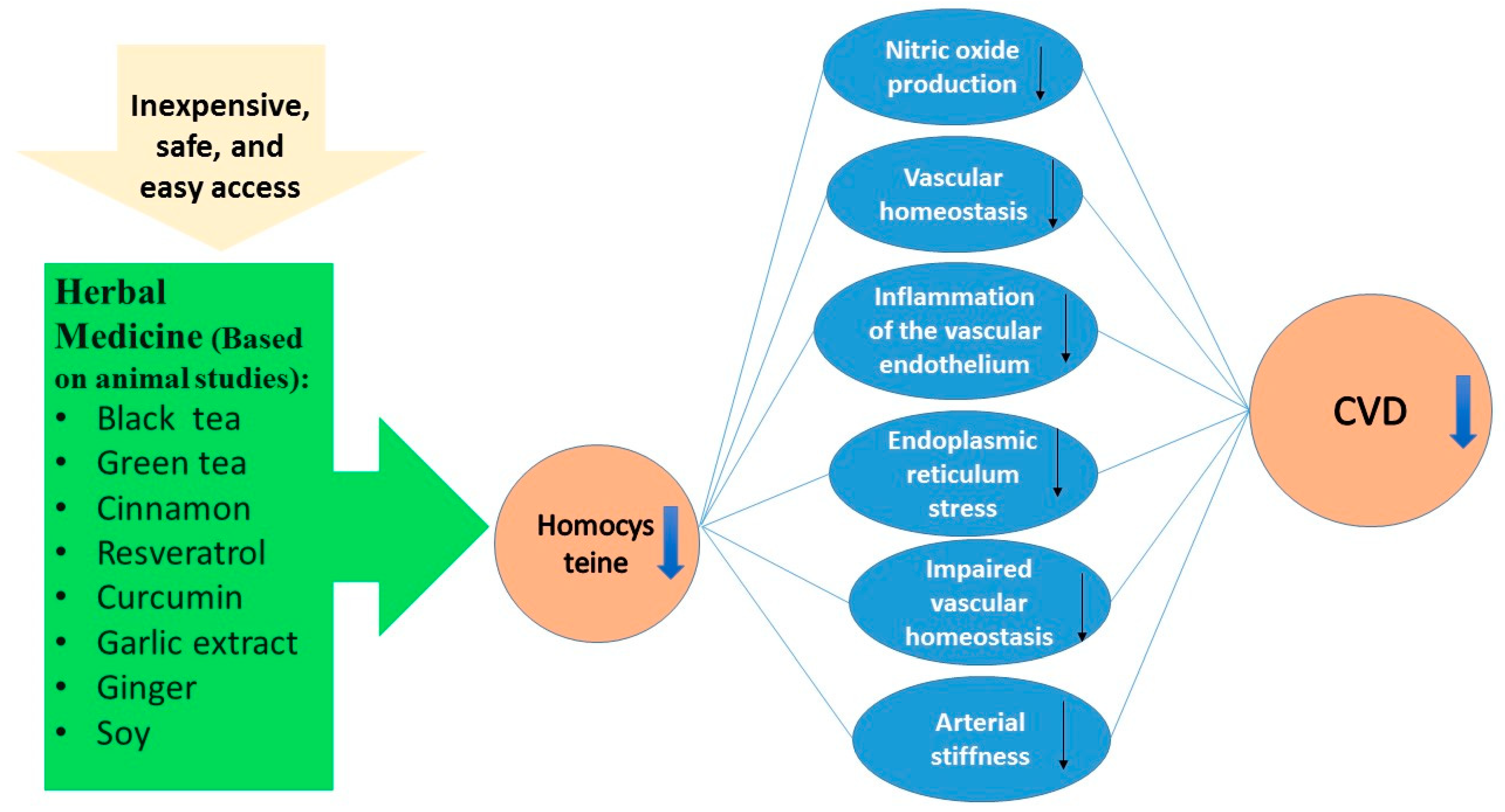
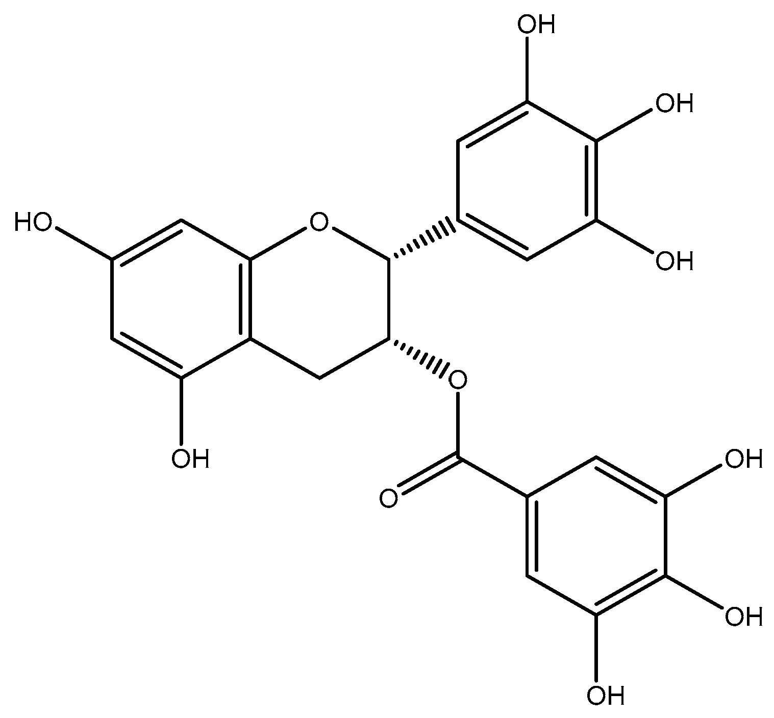
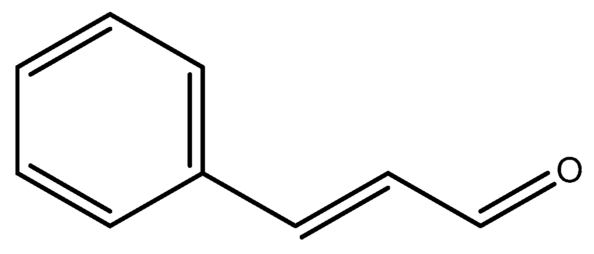
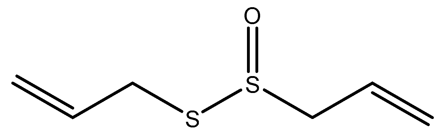
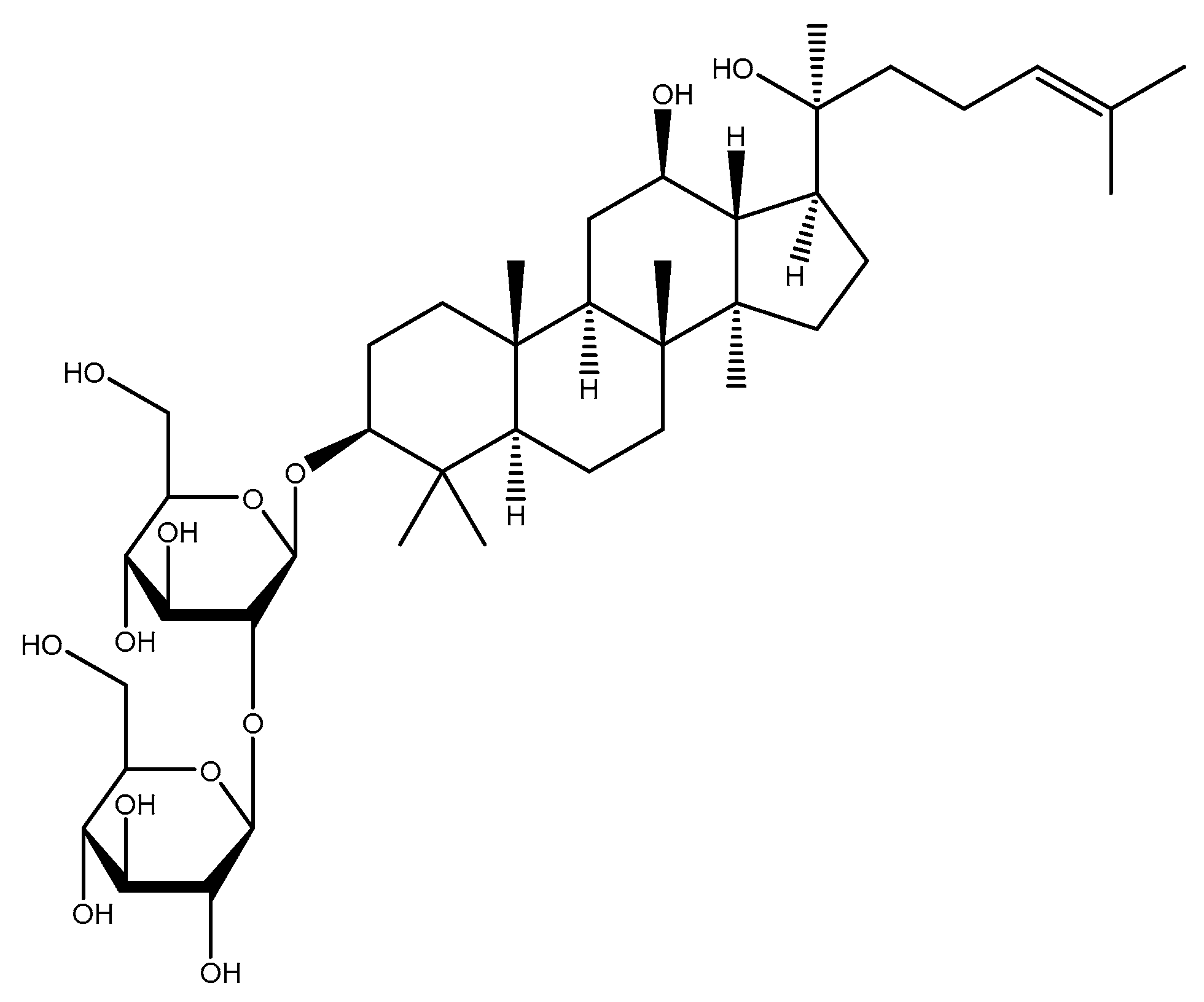
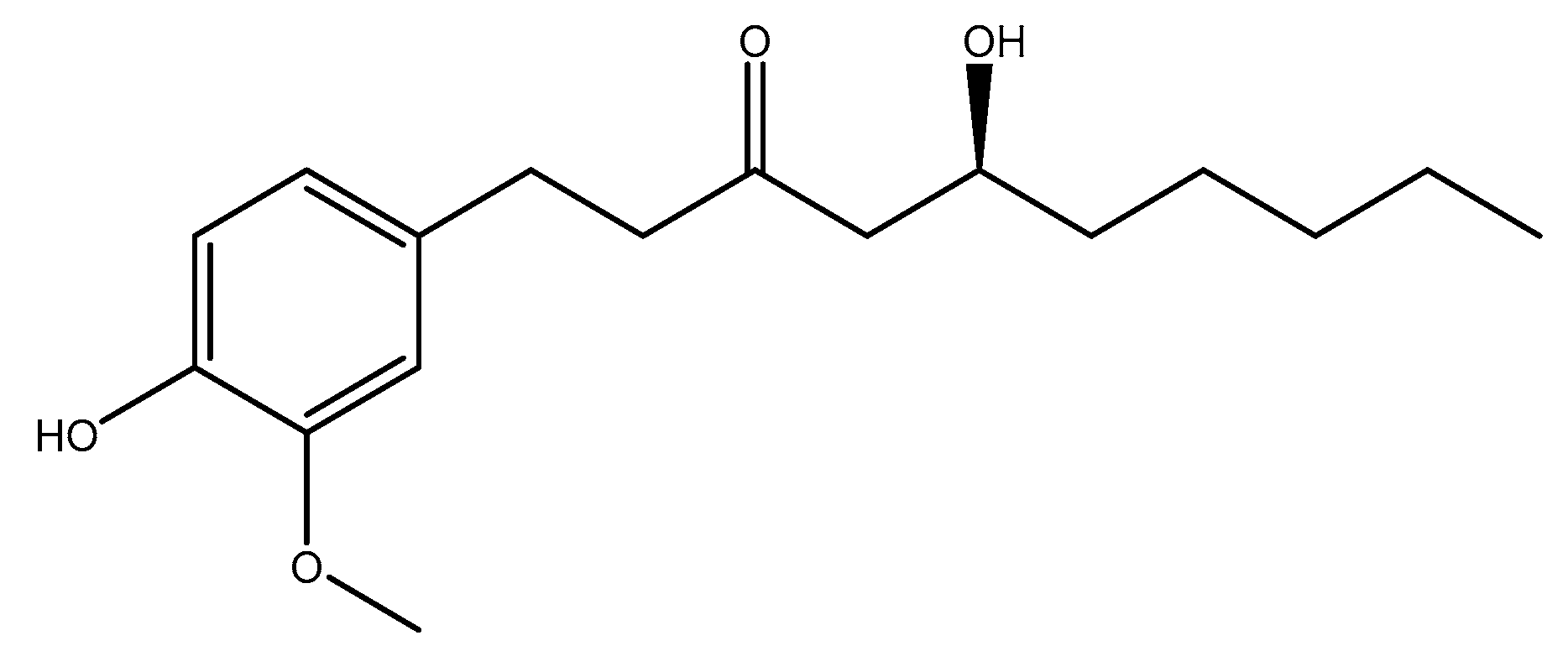
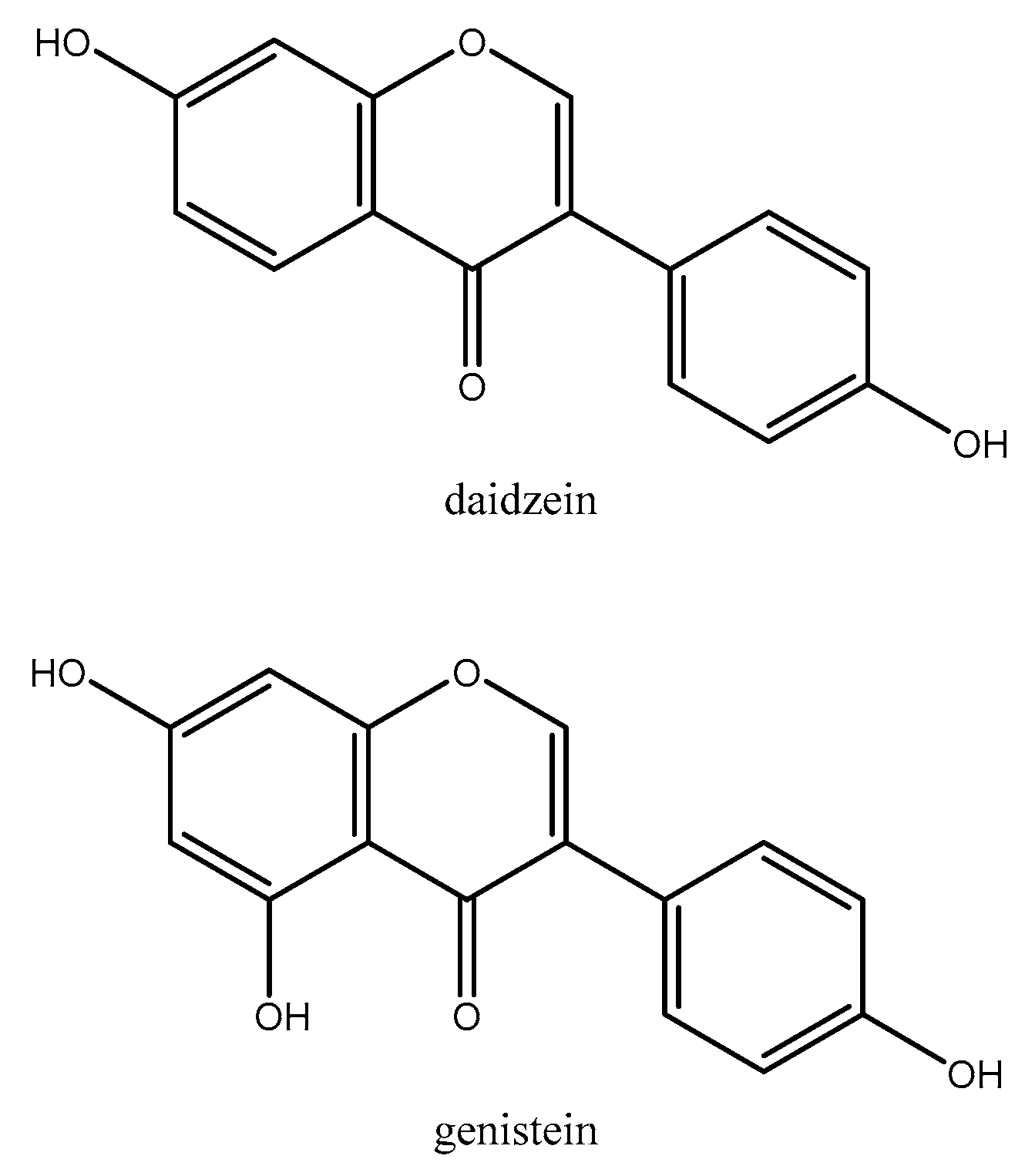
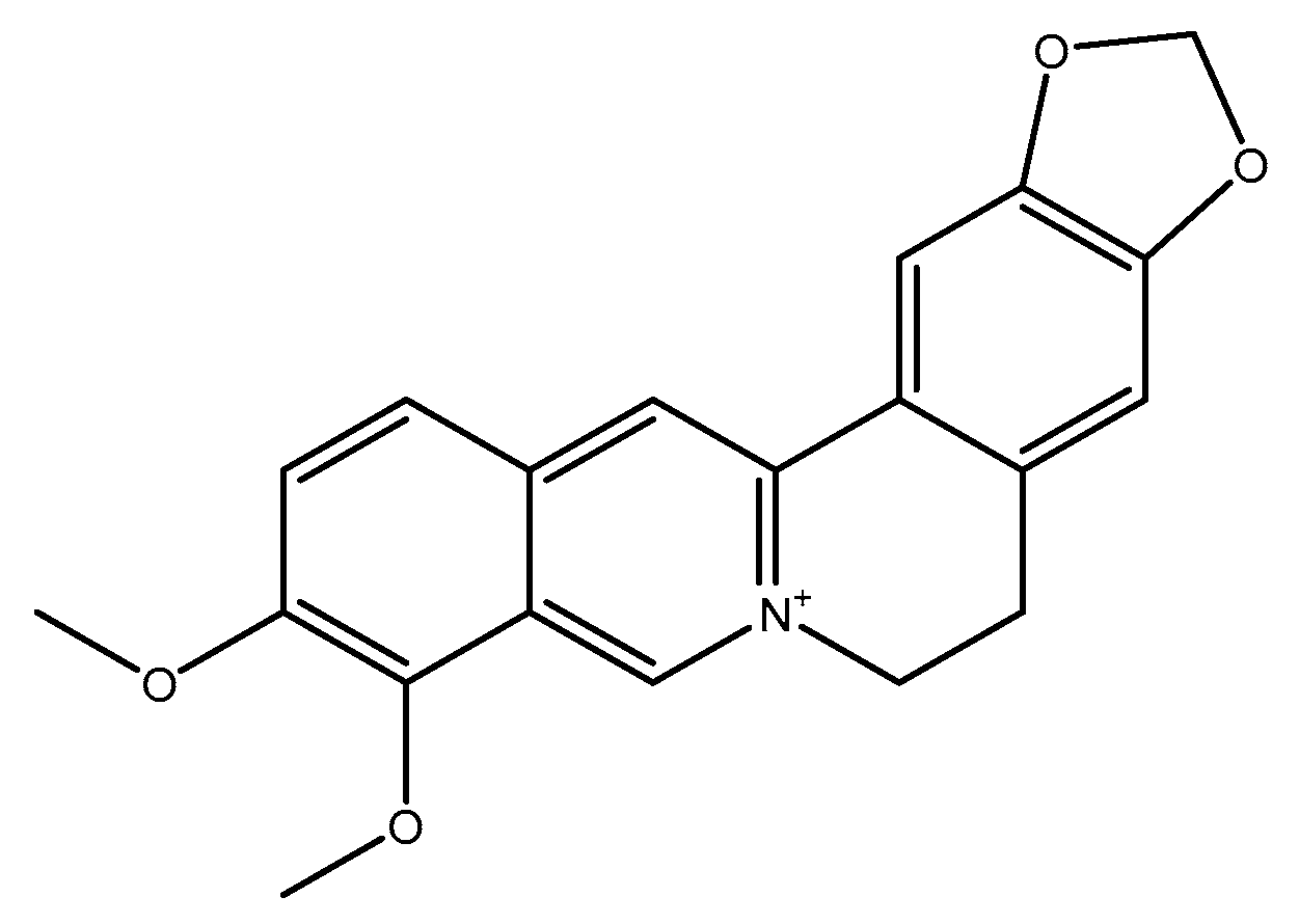
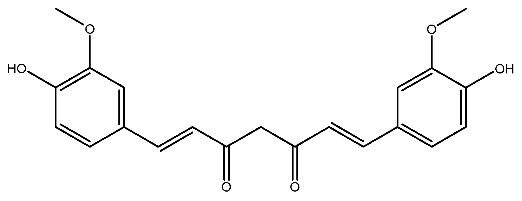
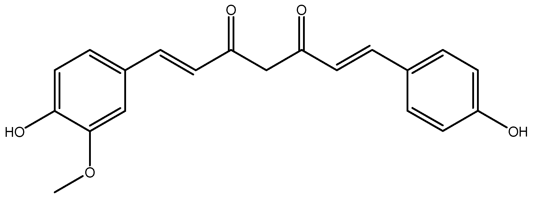
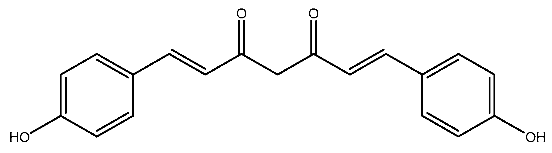
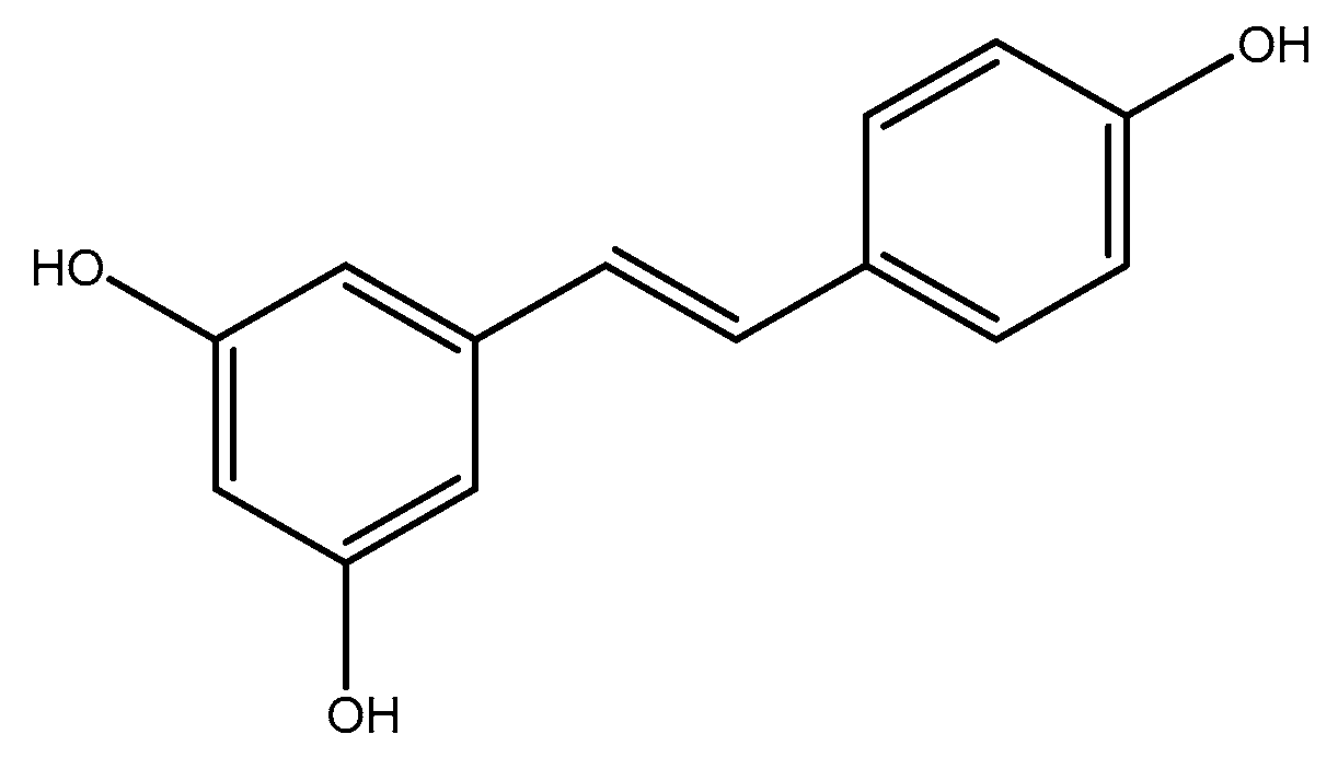
Table 1.
The effects of herbal bioactive compounds on homocysteine levels according to clinical trials.
Table 1.
The effects of herbal bioactive compounds on homocysteine levels according to clinical trials.
| Number | Author, Year | Agent | Dose per Day | Treatment Duration | Subjects | Main Outcome(s) | Final Effects of Nutraceuticals on Homocysteine Level |
|---|---|---|---|---|---|---|---|
| 1 | Hodgson et al., 2007 [85] | Black Tea | 2.2 g tea leaves | 3.5 h | Adults with CAD | Black tea significantly increased plasma homocysteine levels | ↑ |
| 2 | Hodgson JM et al., 2003 [86] | Black Tea | 2 g tea leave/250 mL boiled water | 12 weeks | Healthy subjects | Black tea did not significantly alter mean homocysteine concentrations | No effect |
| 3 | Olthof MR et al., 2001 [87] | Black tea | 4 g black tea | 4 weeks | Healthy subjects | Black tea raised total plasma homocysteine concentrations | ↑ |
| 4 | Duthie et al., 2006 [115] | Anthocyanins | 750 mL/day cranberry juice (2.80 mg/L anthocyanins) | 2 weeks | Healthy volunteer females | Cranberry juice had no effect on plasma homocysteine levels | No effect |
| 5 | Ahmadi N et al., 2013 [125] | Garlic extract | Aged garlic extract (AGE) 250 mg + B12 100 μg + B9 300 μg + B6 12.5 mg + L-arginine 100 mg | 12 months | Healthy subjects | Garlic extract plus other supplements reduced homocysteine level | ↓ |
| 6 | Budoff MJ et al., 2009 [126] | Garlic extract | AGE 250 mg + B12 100 μg + B9 300 μg + B6 12.5 mg + L-arginine 100 mg | 1 year | Healthy subjects | Garlic extract plus other supplements reduced homocysteine level | ↓ |
| 7 | Budoff MJ et al., 2004 [128] | Garlic extract | 4 mL | 1 year | Patients with CAD | Garlic extract did not significantly improve homocysteine level | No effect |
| 8 | Seo DY et al., 2012 [129] | Garlic extract | 5 × 65 mg per week | 12 weeks | 30 postmenopausal women | Homocysteine was significantly decreased | ↓ |
| 9 | McCrindle BW et al., 1998 [130] | Garlic extract | 3 × 300 mg per day | 8weeks | 30 pediatric (8 to 18 years old) | No significant difference between the groups | No effect |
| 10 | Ried K et al., 2016 [131] | Garlic extract | 1.2 g powder per day + 1.2 mg S-allylcysteine | 12 weeks | 88 uncontrolled hypertensive patients | No significant differences between the groups | No effect |
| 11 | Merchant RE et al., 2015 [144] | Chorella | 9 g per day | 60 days | 17 vegetarian or vegan | Homocysteine level decreased by an average of 10% | ↑ |
| 12 | Llaneza P et al., 2011 [163] | Soy | 80 mg of soy isoflavone | 6 months | 87 healthy postmenopausal women | No change in both groups | No effect |
| 13 | Høie LH et al., 2005 [164] | Soy | 15 or 25 g of soy protein | 8 weeks | 117 Hypercholesterolemic patients | No change in all groups | No effect |
| 14 | Greany K et al., 2008 [165] | Soy | 26 ± 5 g/day of soy isolated protein | 2 × 4 weeks + 2 weeks washout | 34 postmenopausal women | No change between both groups | No effect |
| 15 | Imani H et al., 2009 [166] | Soy | 28 g/day of soy textured soy flour | 8 weeks | 40 peritoneal dialysis patients | No change between groups | No effect |
| 16 | Jenkins DJ et al., 2002 [167] | Soy | Low fat dairy food control diet, with low or high isoflavone soy food diets | 3 × 1 months + 2 weeks washout | 41 hyperlipidemic men and postmenopausal women | Homocysteine was lower in both isoflavone groups | ↓ |
| 17 | Hanson LN et al., 2006 [168] | Soy | 40 g/day soy protein isolate | 6 weeks | 55 postmenopausal women | homocysteine was significantly reduced in soy protein normal phytate group, while no significant change was detected in soy protein normal isoflavone group | soy protein normal phytat: ↓ soy protein normal isoflavone: no effect |
| 18 | Reimann M et al., 2006 [169] | Soy | 50 mg soy isoflavone | 8 weeks | 89 postmenopausal women | Homocysteine level did not change in both groups | No effect |
| 19 | Brandao LC et al., 2009 [170] | Soy | 80 mg soy isoflavone | 4 months | 38 postmenopausal women | Homocysteine level did not change in both groups | No effect |
| 20 | Upadya H et al., 2019 [190] | Amla | 500 mg capsule of Amla extract | 12 weeks | 98 patients with dyslipidemia | Homocysteine level did not change significantly between the groups | No effect |
| 21 | Chen T-S et al., 2009 [191] | Amla | amla extract tablets (300 mg, 50% dextrin + 50% amla extract) four times a day | 4 months | 17 uremic patients | Homocysteine level did not change in subjects | No effect |
| 22 | Mercanlıgil S et al., 2007 [195] | Nut | 40 g/day hazeinut | 8 weeks | 15 hypercholesterolemic patients | Homocysteine level did not change between the groups | No effect |
| 23 | Morgan J et al., 2002 [196] | Nut | 64 g/day walnut | 18 weeks | 67 patients (serum total cholesterol > 5.2 mmol/L) | No statically significant effects were observed | No effect |
| 24 | Rodrigues APdS et al., 2020 [206] | Olive oil | 52 mL/d EVOO | 12 weeks | 121 obese adult subjects | Homocysteine levels did not change significantly between the groups | No effect |
| 25 | Shidfar F et al., 2012 [214] | Berberis vulgaris Fruit Extract | 3 g/d | 3 months | Diabetic patients | Berberine did not significantly alter mean serum homocysteine concentration | No effect |
| 26 | Campbell MS et al., 2019 [227] | Curcumin | 500 mg | 12 weeks | Obese men | Homocysteine was significantly reduced in the curcumin group | ↓ |
| 27 | Madaric A et al., 2013 [229] | Curcumin | 100 g of biscuits per day with 1.3 g curcuma | 2 months | Healthy men | Curcumin significantly decreased homocysteine level | ↓ |
| 28 | Schroecksnadel K et al., 2005 [240] | Resveratrol | 1–100 µg | 72 h | Healthy voluntary blood donors | Pretreatment of unstimulated cells with 10–100 mM resveratrol only slightly decreased homocysteine production in the resting cells | ↓ |
| 29 | Wood RJ et al., 2006 [244] | soluble fiber | 3 g soluble fiber | 12 weeks | 29 overweight men | Homocysteine level did not significantly increase in fiber group compared to the placebo group | No effect |
| 30 | Sprecher DL et al., 2002 [245] | soluble fiber | 4 g soluble fiber | 8 weeks | 119 subjects | Homocysteine levels significantly reduced in fiber blend group compared to the placebo group | ↓ |
↑: Increasing effect; ↓: decreasing effect.
Table 2.
The effect of herbal bioactive compounds on homocysteine levels based on animal studies.
| Number | Author, Year | Agent | Dose per Day | Treatment Duration | Animals | Main Outcome(s) | Final Effects of Nutraceuticals on Homocysteine Level |
|---|---|---|---|---|---|---|---|
| 1 | San Cheang et al., 2015 [84] | Black Tea extract | 15 mg/kg/day | 2 weeks | Rats | Black tea extract significantly reduced plasma homocysteine levels | ↓ |
| 2 | El-Missiry MA et al., 2018 [101] | Green tea | 2.5 or 5 mg/kg body weight EGCG | 3 days | Adult male Wister rats | EGCG at a dose of 2.5 and 5 mg/kg significantly decreased plasma homocysteine | ↓ |
| 3 | Amin KA et al., 2009 [108] | Cinnamon extract | 20 mg/day/rat | 5–8 weeks | Male rats | Cinnamon extract reduced homocysteine levels | ↓ |
| 4 | Yeh YY et al., 2006 [127] | Garlic extract | 4% of diet | 6 weeks | Rats | Garlic extract significantly reduced homocysteine level | ↓ |
| 5 | Kim JH, 2019 [136] | ginsenoside | 50 mg/kg every 12 h | 60 days | 40 wistar male rats | Met reduced plasma Homocysteine level, whereas GTS did not affect basal plasma levels | GTS alone: No effect Met: ↓ |
| 6 | Ilkhanizadeh B et al., 2016 [154] | ginger | 50 mg/kg body weight daily | 6 weeks | 24 male Wistar rats | Significant decrease in homocystein level was found in the ginger extract-treated diabetic group | ↓ |
| 7 | Akbari A et al., 2017 [155] | ginger | 1 g/kg body weight daily | 28 days | 28 adult male Sprague-Dawley | In ginger-ethanol group, ginger improved antioxidant enzymes’ activity and reduced tHcy and MDA compared to the ethanol group | ↓ |
| 8 | Snelson M et al., 2017 [171] | Soy | 55% total energy | 12 weeks | 30 female wild-type mice | homocysteine level was the same in soy and control group | No effect |
| 9 | Adam SK et al., 2008 [172] | Soy | Fresh soy oil One-heated soy oil Five-time-heated soy oil | 4 months | 24 adult female Sprague Dawley rats | fresh soy oil significantly reduced homocysteine level compared to the other groups | ↓ |
| 10 | Chang X-x et al., 2012 [213] | Berberine | 200 mg/kg/day | 24 weeks | Healthy male rats | Serum homocysteine level was significantly decreased after berberine consumption in rats fed with a high-fat diet | ↓ |
| 11 | Varatharajalu R et al., 2016 [228] | Curcumin | 150 mg/kg body weight/day | 8 weeks | Female Wistar-Furth rats | Curcumin significantly increased homocysteine thiolactonase activity | ↓ |
| 12 | Mansouri Z et al., 2012 [230] | Curcumin | 50 mg/kg | 10 days | Adult male Wister rats | Investigated the neuroprotective effects of curcumin against homocysteine neurotoxicity | ↓ |
| 13 | Jelodar G et al., 2019 [231] | Curcumin | 48 mg/kg | 4 weeks | female Sprague-Dawley rats | no significant difference was observed between all groups | No effect |
| 14 | Koz ST et al.2012 [237] | Resveratrol | 20 mg/kg/day | 30 days | Rats | Resveratrol significantly reduced plasma Homocysteine levels | ↓ |
| 15 | Noll C et al., 2009 [238] | Resveratrol | 50 µg/day | 3 months | Mice | Resveratrol significantly increased homocysteine levels compared to the control group | ↑ |
| 16 | Yilmaz Ö et al., 2007 [239] | Resveratrol | 33mg/kg four times per week | 5 weeks | Old female rats | Resveratrol significantly decreased homocysteine levels | ↓ |
↑: Increasing effect; ↓: decreasing effect.
Publisher’s Note: MDPI stays neutral with regard to jurisdictional claims in published maps and institutional affiliations. |
© 2021 by the authors. Licensee MDPI, Basel, Switzerland. This article is an open access article distributed under the terms and conditions of the Creative Commons Attribution (CC BY) license (https://creativecommons.org/licenses/by/4.0/).
Share and Cite
MDPI and ACS Style
Atazadegan, M.A.; Bagherniya, M.; Askari, G.; Tasbandi, A.; Sahebkar, A. The Effects of Medicinal Plants and Bioactive Natural Compounds on Homocysteine. Molecules 2021, 26, 3081. https://doi.org/10.3390/molecules26113081
AMA Style
Atazadegan MA, Bagherniya M, Askari G, Tasbandi A, Sahebkar A. The Effects of Medicinal Plants and Bioactive Natural Compounds on Homocysteine. Molecules. 2021; 26(11):3081. https://doi.org/10.3390/molecules26113081
Chicago/Turabian StyleAtazadegan, Mohammad Amin, Mohammad Bagherniya, Gholamreza Askari, Aida Tasbandi, and Amirhossein Sahebkar. 2021. "The Effects of Medicinal Plants and Bioactive Natural Compounds on Homocysteine" Molecules 26, no. 11: 3081. https://doi.org/10.3390/molecules26113081