
Activity of Experimental Mouthwashes and Gels Containing DNA-RNA and Bioactive Molecules against the Oxidative Stress of Oral Soft Tissues: The Importance of Formulations. A Bioreactor-Based Reconstituted Human Oral Epithelium Model

 , , and
Abstract
:1. Introduction
2. Results
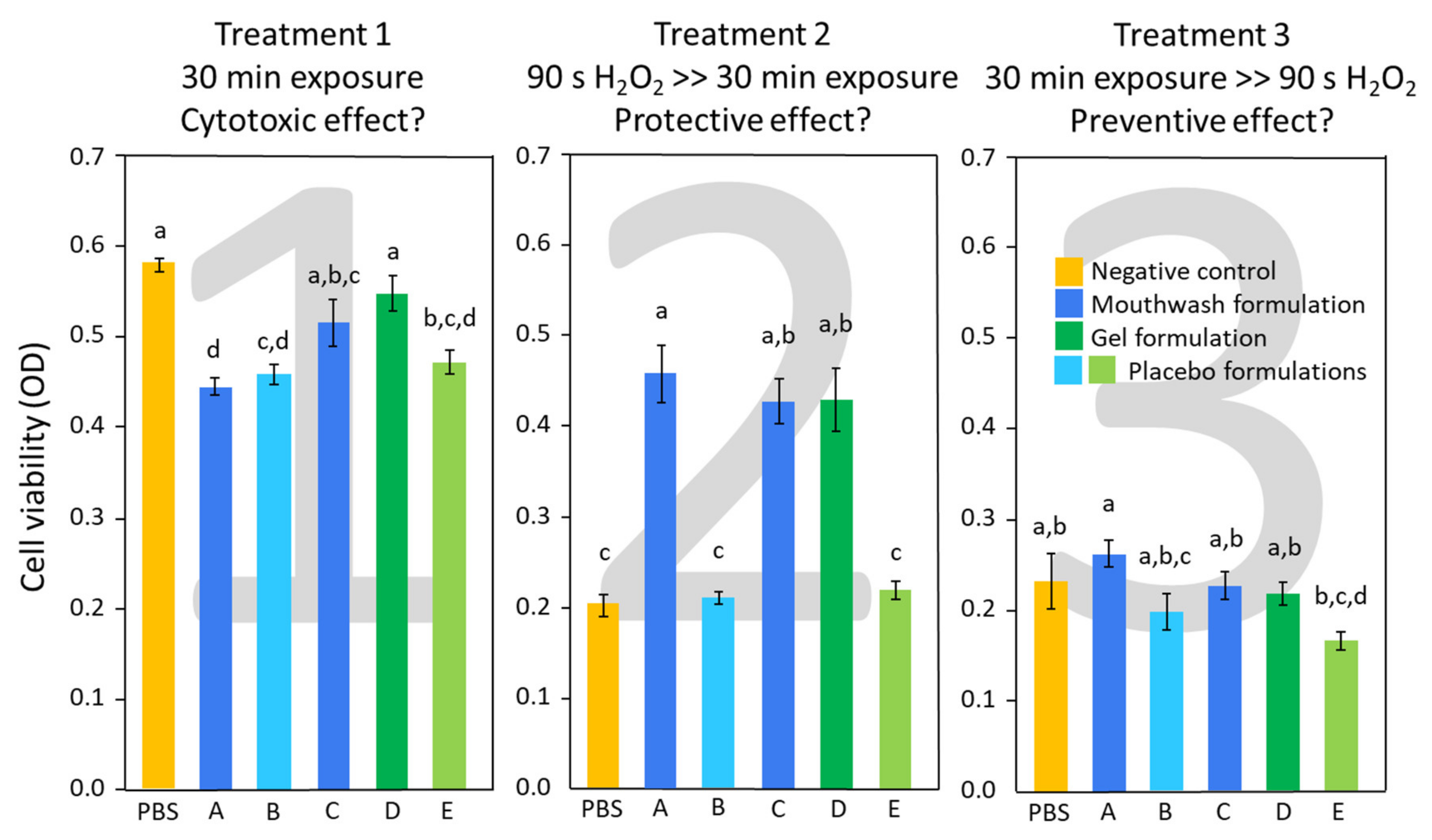
2.1. MTT Assay
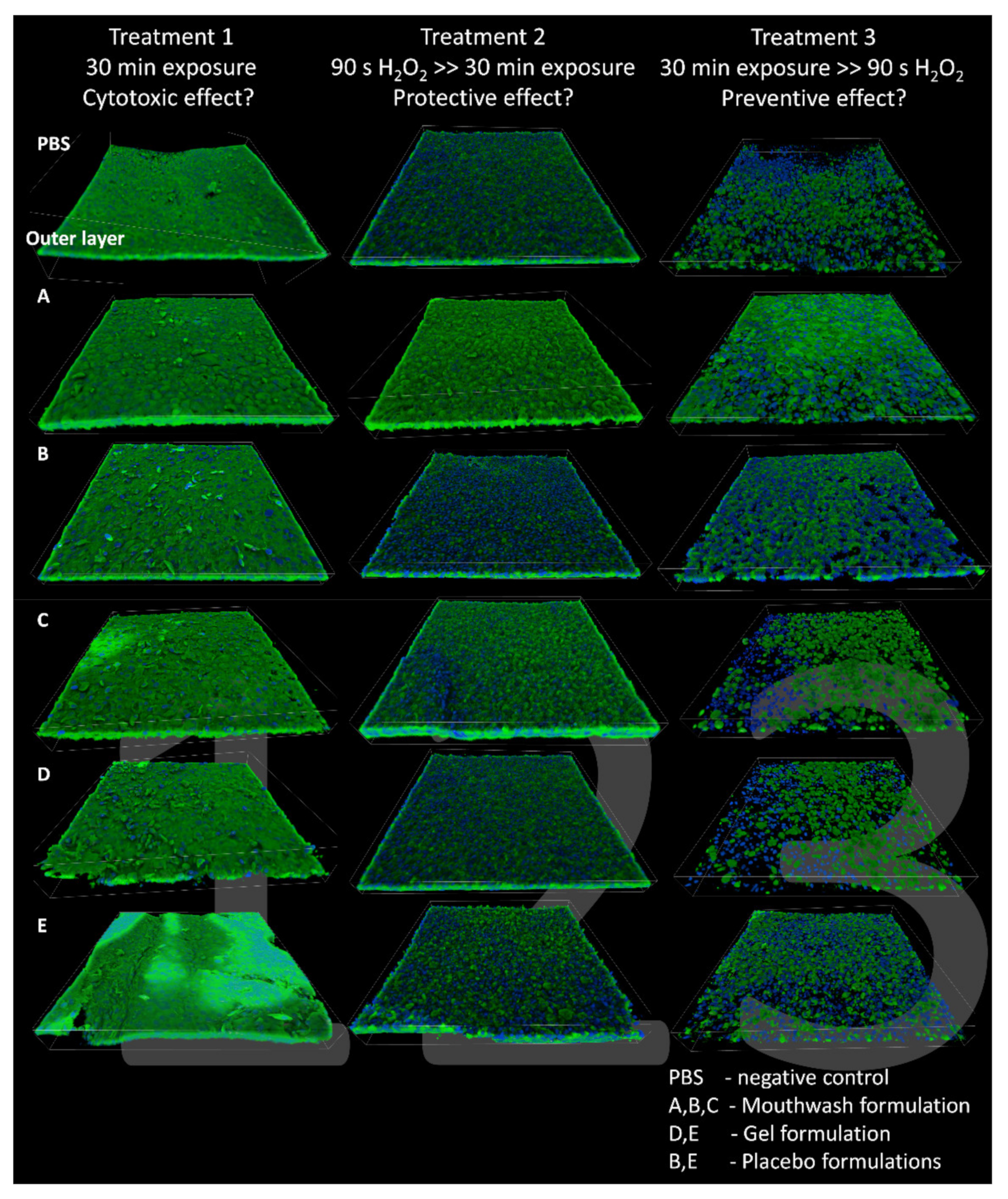
2.2. CLSM Observations
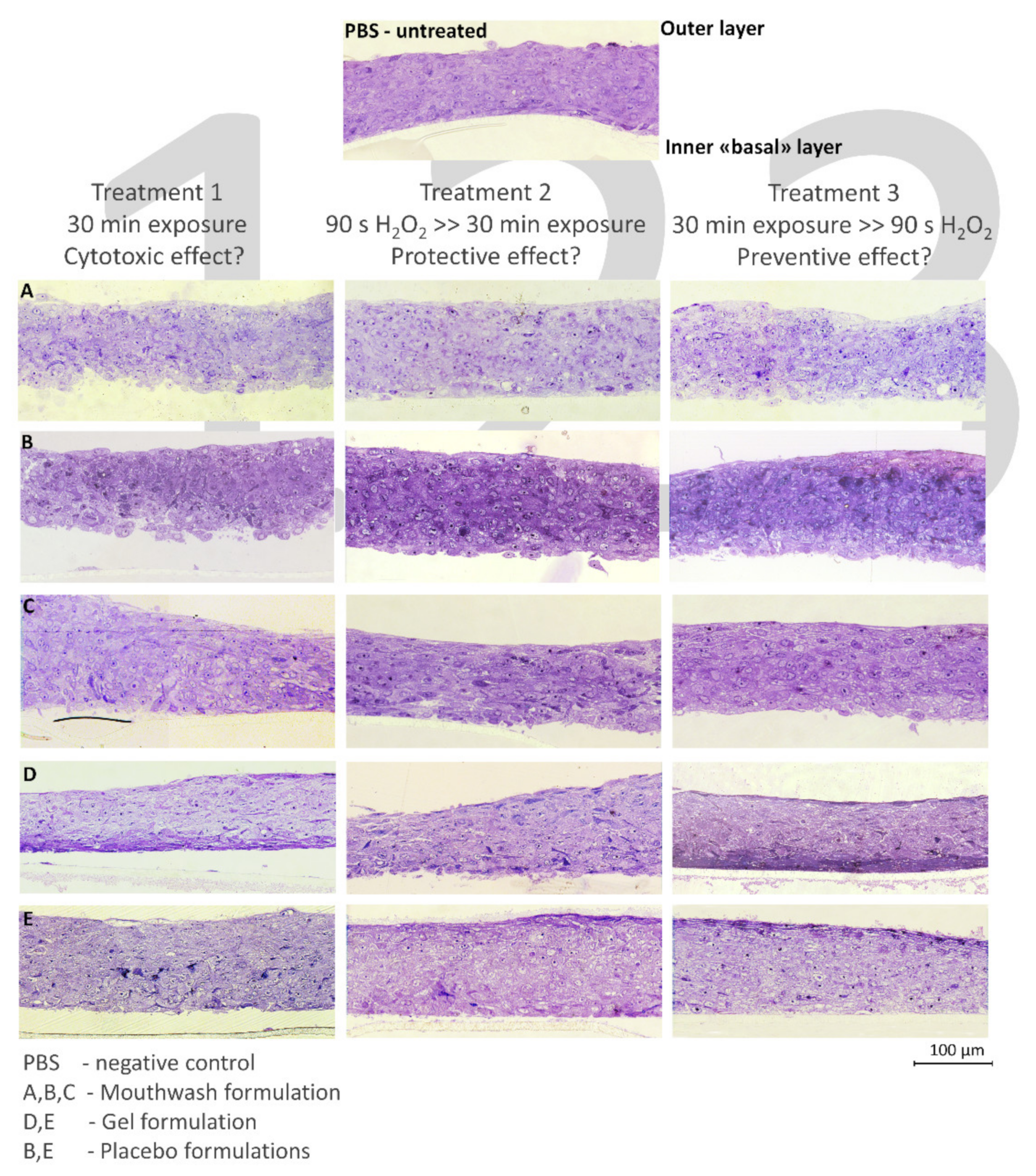
2.3. Histological Evaluation
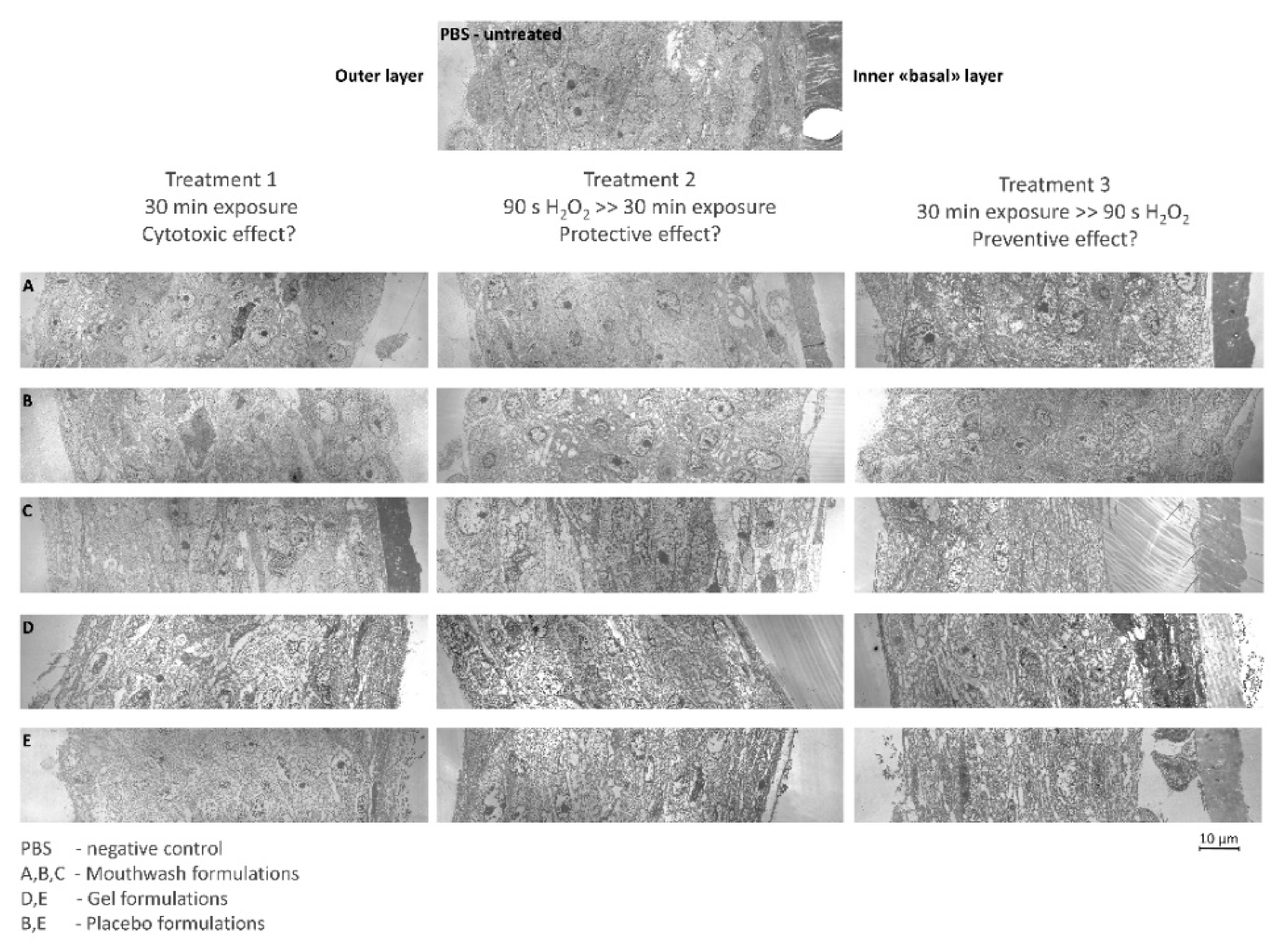
2.4. TEM Analysis
3. Discussion

| Categories | Compounds | Description | Supposed Positive Effects | Supposed Negative Effects | A | B | C | D | E |
|---|---|---|---|---|---|---|---|---|---|
| Active Principles | Hydrolysed DNA RNA | Contains microbial-hydrolyzed low molecular weight fragments of ribonucleic acid (RNA) and deoxyribonucleic acid (DNA) of vegetal origin. | Anti-inflammatory and protective against ROS [17]. | ✓ | ✓ | ✓ | |||
| Hyaluronic acid | Anionic, non-sulfated glycosaminoglycan. | Extracellular matrix regeneration, epithelial regeneration, wound healing [20]. | ✓ | ✓ | |||||
| Beta-Glucan | High-molecular weight β-D-glucose polysaccharide. Water-soluble natural extract. | Antioxidants and anti-aging activity [21]. | ✓ | ✓ | |||||
| Allantoin | 2,5-Dioxo-4-imidazolidinyl urea. Synthetically produced from uric acid. | Promotes cell proliferation and facilitates wound healing [22,23]. | ✓ | ✓ | |||||
| Bisabolol | Monocyclic sesquiterpene alcohol. First isolated from Matricaria chamomilla (Asteraceae). | Anti-irritant, anti-inflammatory, and antimicrobial activity [24,25,26,27,28]. | ✓ | ✓ | |||||
| Ruscogenin | First isolated from Ruscus aculeatus, also a major steroidal sapogenin of the traditional Chinese herb Radix Ophiopogon japonicus. | Anti-inflammatory and protective against ROS [29,30]. | ✓ | ✓ | |||||
| Essential Oils | Glycyrrhetinic Acid | Oleanoic acid derived from shredded Glychirriza (licorice) roots. | Antioxidant, anti-inflammatory activities [31]. | ✓ | ✓ | ||||
| Leptospermum Scoparium Branch/Leaf Oil | Essential oil coming from the Manuka tree native to New Zealand. | Antibacterial and antifungal activity [32,33,34]. | ✓ | ✓ | |||||
| Melaleuca Alternifolia Leaf Oil | Tea tree oil, essential oil distilled from the leaves of a native Australian plant. | Antibacterial, Antimicrobial and Antiviral activity [32]. | ✓ | ✓ | |||||
| Surfactant Agent | Ceteareth-12/20 | Polyethylene glycol (PEG) ethers of Cetearyl Alcohol | Dermal irritation [38]. | ✓ | ✓ | ✓ | |||
| Cetyl Palmitate | Ester derived from hexadecanoic acid and hexadecanol | Dermal toxicity Dermal irritation Dermal sensitization [37]. | ✓ | ✓ | ✓ | ||||
| Cetearyl Alcohol | Straight-chain alcohol. Mixture of mostly Cetyl and Stearyl Alcohols, which are fatty alcohols that occur naturally in small quantities in plants and animals. | Dermal irritation Cytotoxicity [36]. | ✓ | ✓ | ✓ | ||||
| PEG-40 Hydrogenated Castor Oil | Polyethylene glycol derivative of hydrogenated castor oil | Alterations of the plasma membranes of epithelial cells Tight junction opening Cytotoxicity [39,40]. | ✓ | ✓ | |||||
| Caprylyl Glycol | 1,2-glycol compound with 8 carbons in the carbon chain. Also used as humectant and preservative agent. | Safe for use, no negative effect reported [44]. | ✓ | ✓ | |||||
| 1,2-Hexanediol | 1,2-glycol compound with 6 carbons in the carbon chain. Also used as humectant and emollient and preservative agent. | Safe for use, no negative effect reported [44]. | ✓ | ✓ | |||||
| Emulsifier/Emollient | Dicaprylyl Ether | Derived from the dehydration of octane. Used as skin conditioner, emollient and solvent. | Safe for use, no negative effect reported [45]. | ✓ | ✓ | ✓ | |||
| Coco-caprylate/caprate | Made by combining esters from coconut-derived fatty alcohol with caprylic and capric acids, also from coconut. Emollient. | Safe for use, no negative effect reported [46]. | ✓ | ✓ | ✓ | ||||
| Glyceryl Stearate | Ester of stearic acid and ethylene glycol. Monoglyceride commonly used as an emulsifier in foods. | Safe for use, no negative effect reported [47]. | ✓ | ✓ | ✓ | ||||
| Humectant | Propylene Glycol | Propanediol: propane where the hydrogens at positions 1 and 2 are substituted by hydroxyl groups. Used as an organic solvent and diluent, and to absorb extra water and maintain moisture | Safe for use, no negative effect reported [48]. | ✓ | ✓ | ✓ | ✓ | ||
| Glycerin | Simple polyol compound with three alcohol hydroxyl groups. | Safe for use, no negative effect reported [49]. | ✓ | ✓ | |||||
| Thickener | Cellulose Gum | Carboxymethyl cellulose | Non cytotoxic [50]. | ✓ | ✓ | ||||
| Carbomer | Poly-acrylic acid | Non cytotoxic, improved wound healing [51]. | ✓ | ✓ | |||||
| Film-Former | Calcium/Sodium PVM/MA Copolymer | PVM/MA Copolymer is a copolymer of methyl vinyl ether and maleic anhydride or maleic acid. Used as binder and film-former. | Safe for use, no negative effect reported [52]. | ✓ | |||||
| VP/VA Copolymer | Large molecule made from vinyl pyrrolidone (VP) and vinyl acetate (VA) monomers. | Safe for use, no negative effect reported [53]. | ✓ | ||||||
| Sweetener | Xylitol | Polyol, artificial sweetener | Safe for use, no negative effect reported [54]. | ✓ | ✓ | ✓ | |||
| Sodium saccharin | Artificial sweetener | Safe for use, no negative effect reported [55]. | ✓ | ✓ | ✓ | ✓ | ✓ | ||
| Ammonium Glycyrrhizate | Natural extract from Glychirrizia plant | Antiviral, anti-inflammatory [56]. | Gap-junction inhibitor, cytotoxic [56]. | ✓ | |||||
| Preservative | o-Cymen-5-ol | Substitute of parabens | Safe for use, no negative effect reported at the tested concentration (0.1%) [57]. | ✓ | ✓ | ||||
| Sodium Benzoate | Sodium benzoate is the sodium salt of benzoic acid. It is an aromatic compound with antimicrobial activity, therefore is used as a preservative in food products. | Safe for use, no negative effect reported [58]. | ✓ | ✓ | ✓ | ✓ | ✓ | ||
| Phenoxyethanol | Ether alcohol, aromatic compound with antimicrobial activity. Extensively used as preservative in pharmaceuticals, cosmetics and lubricants. | Safe for use, no negative effect reported [59]. | ✓ | ✓ | ✓ | ✓ | ✓ | ||
| Citric acid | Tricarboxylic acid found in citrus fruits, Used as a preservative due to its antioxidant properties. | Safe for use, no negative effect reported [60]. | ✓ | ✓ | ✓ | ||||
| Cosmetic Colorant | CI 16255 | Ponceau 4R, synthetic colourant used for food colouring | Safe for use, no negative effect reported [61]. | ✓ | ✓ | ✓ | |||
| CI 42090 | Brilliant Blue FCF (Blue 1) is a synthetic organic compound used as a colorant for cosmetics and food. | Safe for use, no negative effect reported [61]. | ✓ | ✓ | |||||
4. Materials and Methods
4.1. Reagents
4.2. Reconstituted Human Oral Epithelium (RHOE)
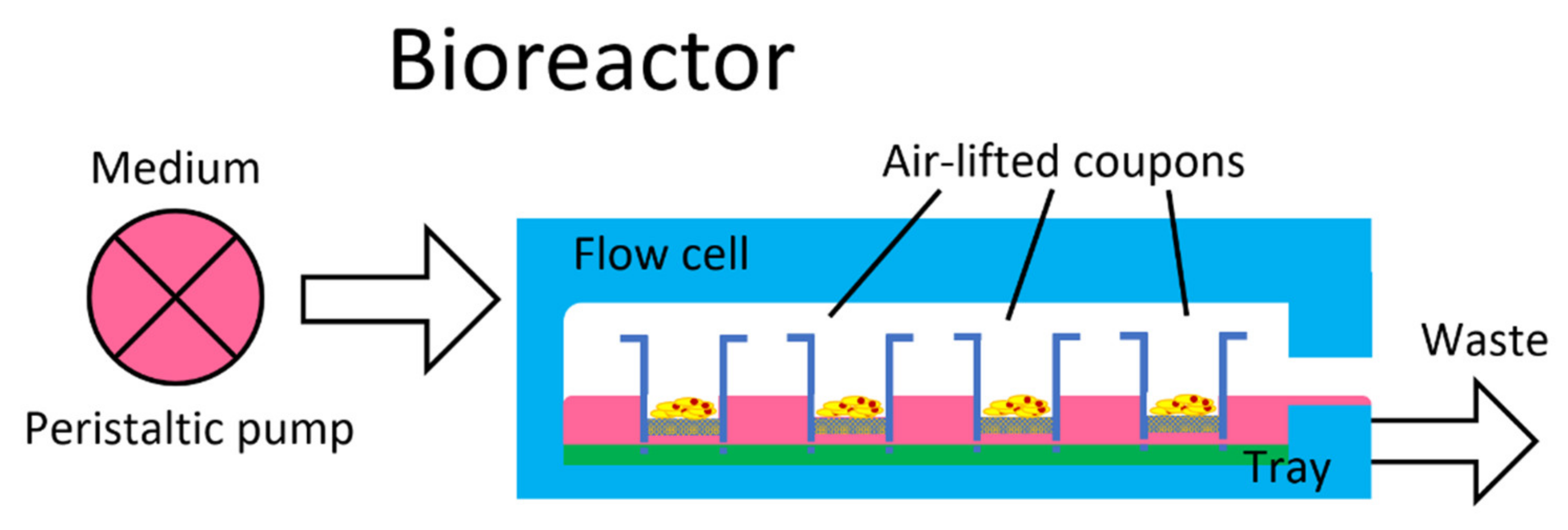
4.3. Bioreactor
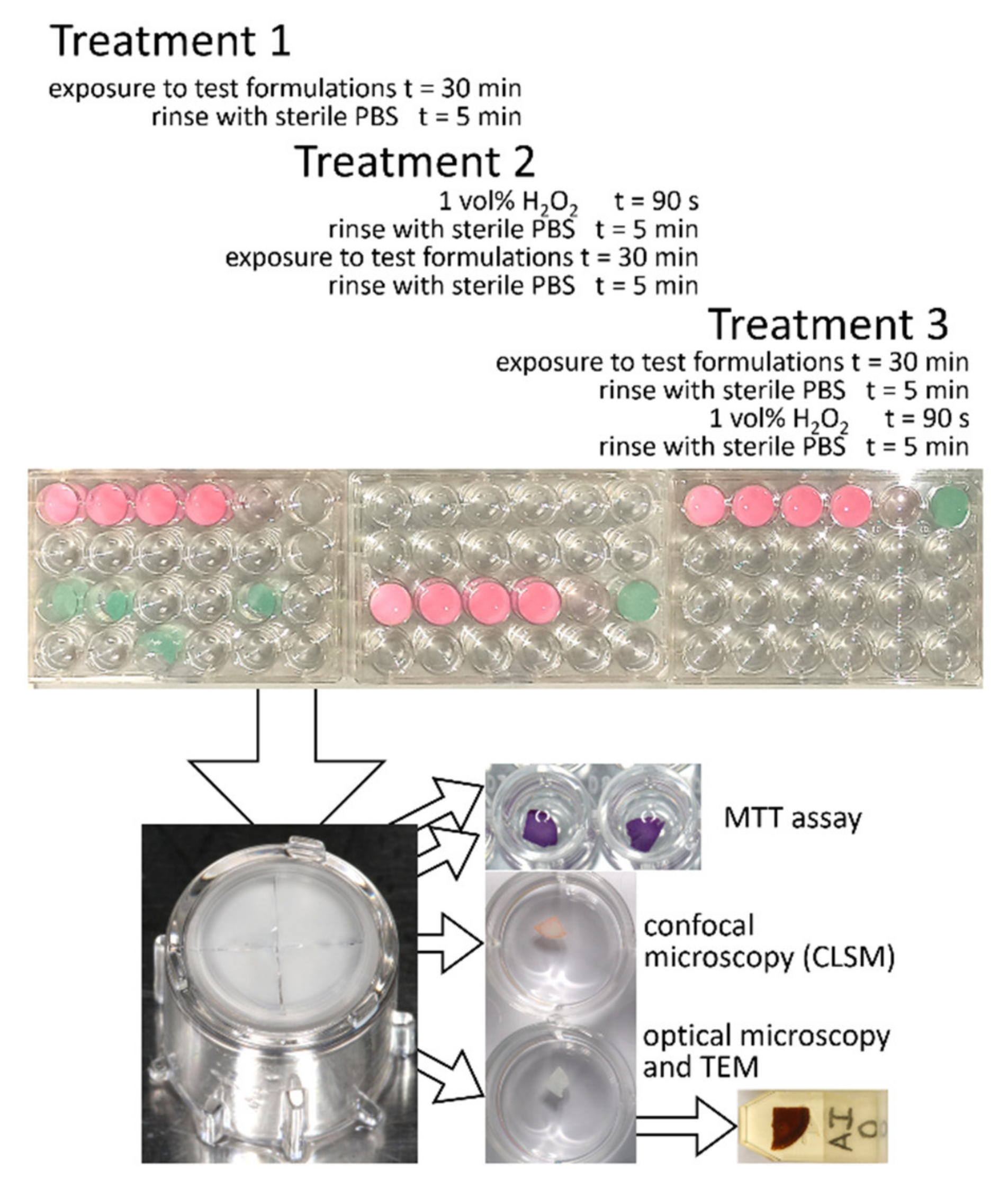
4.4. Test Procedures
4.5. Specimen Evaluation
4.6. MTT Assay
4.7. CLSM Observations
4.8. Histological Evaluation
4.9. Statistical Analysis
5. Conclusions
Author Contributions
Funding
Institutional Review Board Statement
Informed Consent Statement
Data Availability Statement
Conflicts of Interest
Sample Availability
References
- Sies, H. On the history of oxidative stress: Concept and some aspects of current development. Curr. Opin. Toxicol. 2018, 7, 122–126. [Google Scholar] [CrossRef]
- McCord, J.M. The evolution of free radicals and oxidative stress. Am. J. Med. 2000, 108, 652–659. [Google Scholar] [CrossRef]
- Frenkel, K. Carcinogen-mediated oxidant formation and oxidative DNA damage. Pharmac. Ther. 1992, 53, 127–166. [Google Scholar] [CrossRef]
- Halliwell, B.; Aruoma, O.I. DNA damage by oxygen-derived species Its mechanism and measurement in mammalian systems. FEBS Lett. 1991, 281, 9–19. [Google Scholar] [CrossRef] [Green Version]
- Breen, A.P.; Murphy, J.A. Reactions of oxyl radicals with DNA. Free Rad. Biol. Med. 1995, 18, 1033–1077. [Google Scholar] [CrossRef]
- Ames, B.N. Endogenous oxidative DNA damage, aging, and cancer. Free Rad. Res. Commun. 1989, 7, 121–128. [Google Scholar] [CrossRef]
- Cerutti, P.; Trump, B.F. Inflammation and oxidative stress in carcinogenesis. Cancer Cells 1991, 3, 1–7. [Google Scholar]
- De La Haba, C.; Palacio, J.R.; Martínez, P.; Morros, A. Effect of oxidative stress on plasma membrane fluidity of THP-1 induced macrophages. Biochim. Biophys. Acta 2013, 1828, 357–364. [Google Scholar] [CrossRef] [Green Version]
- Li, Z.; Wu, J.; DeLeo, C. RNA damage and surveillance under oxidative stress. IUBMB Life 2006, 58, 581–588. [Google Scholar] [CrossRef]
- Liguori, I.; Russo, G.; Curcio, F.; Bulli, G.; Aran, L.; Della-Morte, D.; Gargiulo, G.; Testa, G.; Cacciatore, F.; Bonaduce, D. Oxidative stress, aging, and diseases. Clin. Interv. Aging 2018, 13, 757. [Google Scholar] [CrossRef] [Green Version]
- Avezov, K.; Reznick, A.Z.; Aizenbud, D. Oxidative stress in the oral cavity: Sources and pathological outcomes. Respir. Physiol. Neurobiol. 2015, 209, 91–94. [Google Scholar] [CrossRef]
- Buffoli, B.; Favero, G.; Borsani, E.; Boninsegna, R.; Sancassani, G.; Labanca, M.; Rezzani, R.; Nocini, P.F.; Albanese, M.; Rodella, L.F. Sodium-DNA for bone tissue regeneration: An experimental study in rat calvaria. Biomed. Res. Int. 2017, 2017. [Google Scholar] [CrossRef] [Green Version]
- Bowler, W.; Buckley, K.; Gartland, A.; Hipskind, R.; Bilbe, G.; Gallagher, J. Extracellular nucleotide signaling: A mechanism for integrating local and systemic responses in the activation of bone remodeling. Bone 2001, 28, 507–512. [Google Scholar] [CrossRef]
- Muratore, O.; Schito, A.P.; Cattarini, G.; Tonoli, E.; Gianoglio, S.; Schiappacasse, S.; Felli, L.; Picchetta, F.; Schito, G. Evaluation of the trophic effect of human placental polydeoxyribonucleotide on human knee skin fibroblasts in primary culture. Cell. Mol. Life Sci. 1997, 53, 279–285. [Google Scholar] [CrossRef]
- Rathbone, M.P.; Middlemiss, P.J.; Gysbers, J.W.; DeForge, S.; Costello, P.; Del Maestro, R.F. Purine nucleosides and nucleotides stimulate proliferation of a wide range of cell types. In Vitro Cell. Dev. Biol. 1992, 28, 529–536. [Google Scholar] [CrossRef]
- Thellung, S.; Florio, T.; Maragliano, A.; Cattarini, G.; Schettini, G. Polydeoxyribonucleotides enhance the proliferation of human skin fibroblasts: Involvement of A2 purinergic receptor subtypes. Life Sci. 1999, 64, 1661–1674. [Google Scholar] [CrossRef]
- Ionescu, A.C.; Vezzoli, E.; Conte, V.; Procacci, P.; Garcia-Godoy, F.; Brambilla, E. Effects of Na-DNA mouthwash solutions on oral soft tissues. A bioreactor-based reconstituted human oral epithelium model. Am. J. Dent. 2020, 33, 277–284. [Google Scholar]
- Belletti, S.; Uggeri, J.; Gatti, R.; Govoni, P.; Guizzardi, S. Polydeoxyribonucleotide promotes cyclobutane pyrimidine dimer repair in UVB-exposed dermal fibroblasts. Photodermatol. Photoimmunol. Photomed. 2007, 23, 242–249. [Google Scholar] [CrossRef]
- Raposio, E.; Guida, C.; Coradeghini, R.; Scanarotti, C.; Parodi, A.; Baldelli, I.; Fiocca, R.; Santi, P. In vitro polydeoxyribonucleotide effects on human pre-adipocytes. Cell Prolif. 2008, 41, 739–754. [Google Scholar] [CrossRef]
- Price, R.D.; Berry, M.; Navsaria, H. Hyaluronic acid: The scientific and clinical evidence. J Plast. Reconstr. Aesthet. Surg. 2007, 60, 1110–1119. [Google Scholar] [CrossRef]
- Du, B.; Bian, Z.; Xu, B. Skin health promotion effects of natural beta-glucan derived from cereals and microorganisms: A review. Phytother. Res. 2014, 28, 159–166. [Google Scholar] [CrossRef] [PubMed]
- Thornfeldt, C. Cosmeceuticals containing herbs: Fact, fiction, and future. Dermatol. Surg. 2005, 31, 873–881. [Google Scholar] [CrossRef]
- Araújo, L.U.; Grabe-Guimarães, A.; Mosqueira, V.C.F.; Carneiro, C.M.; Silva-Barcellos, N.M. Profile of wound healing process induced by allantoin. Acta Cir. Bras. 2010, 25, 460–461. [Google Scholar] [CrossRef]
- Stallings, A.F.; Lupo, M.P. Practical uses of botanicals in skin care. J. Clin. Aesthet. Dermatol. 2009, 2, 36. [Google Scholar] [PubMed]
- Kamatou, G.P.; Viljoen, A.M. A review of the application and pharmacological properties of α-Bisabolol and α-Bisabolol-rich oils. J. Clin. Aesthet. Dermatol. 2010, 87, 1–7. [Google Scholar] [CrossRef]
- Nascimento, A.M.; Brandao, M.G.; Oliveira, G.B.; Fortes, I.C.; Chartone-Souza, E. Synergistic bactericidal activity of Eremanthus erythropappus oil or β-bisabolene with ampicillin against Staphylococcus aureus. Antonie Van Leeuwenhoek 2007, 92, 95–100. [Google Scholar] [CrossRef] [PubMed]
- Rocha, N.F.M.; Rios, E.R.V.; Carvalho, A.M.R.; Cerqueira, G.S.; de Araújo Lopes, A.; Leal, L.K.A.M.; Dias, M.L.; de Sousa, D.P.; de Sousa, F.C.F. Anti-nociceptive and anti-inflammatory activities of (−)-α-bisabolol in rodents. Naunyn Schmiedebergs Arch. Pharmacol. 2011, 384, 525–533. [Google Scholar] [CrossRef] [PubMed]
- Kim, S.; Jung, E.; Kim, J.-H.; Park, Y.-H.; Lee, J.; Park, D. Inhibitory effects of (−)-α-bisabolol on LPS-induced inflammatory response in RAW264. 7 macrophages. Food Chem. Toxicol. 2011, 49, 2580–2585. [Google Scholar] [CrossRef]
- Chen, N.-D.; Yue, L.; Zhang, J.; Kou, J.-P.; Yu, B.-Y. One unique steroidal sapogenin obtained through the microbial transformation of ruscogenin by Phytophthora cactorum ATCC 32134 and its potential inhibitory effect on tissue factor (TF) procoagulant activity. Bioorg. Med. Chem. Lett. 2010, 20, 4015–4017. [Google Scholar] [CrossRef]
- Yavuz, E.; Karagulle, O.O.; Ercan, G.; Celik, A.; Yigitbas, H.; Bayrak, B.Y.; Tartar, R.; Kusaslan, R.; Altinel, Y.; Gulcicek, O.B. Evaluation of prophylactic and therapeutic effects of ruscogenin on acute radiation proctitis: An experimental rat model. Ann. Surg. Treat. Res. 2018, 94, 174. [Google Scholar] [CrossRef] [Green Version]
- Maitraie, D.; Hung, C.-F.; Tu, H.-Y.; Liou, Y.-T.; Wei, B.-L.; Yang, S.-C.; Wang, J.-P.; Lin, C.-N. Synthesis, anti-inflammatory, and antioxidant activities of 18β-glycyrrhetinic acid derivatives as chemical mediators and xanthine oxidase inhibitors. Bioorg. Med. Chem. 2009, 17, 2785–2792. [Google Scholar] [CrossRef] [PubMed]
- Reichling, J.; Schnitzler, P.; Suschke, U.; Saller, R. Essential oils of aromatic plants with antibacterial, antifungal, antiviral, and cytotoxic properties–an overview. Complement. Med. Res. 2009, 16, 79–90. [Google Scholar] [CrossRef] [Green Version]
- Christoph, F.; Kubeczka, K.-H.; Stahl-Biskup, E. The composition of commercial manuka oils from New Zealand. J. Essent. Oil Res. 1999, 11, 705–710. [Google Scholar] [CrossRef]
- Lis-Balchin, M.; Hart, S.; Deans, S. Pharmacological and antimicrobial studies on different tea-tree oils (Melaleuca alternifolia, Leptospermum scoparium or Manuka and Kunzea ericoides or Kanuka), originating in Australia and New Zealand. Phytother. Res. 2000, 14, 623–629. [Google Scholar] [CrossRef]
- Vranic, E.; Lacevic, A.; Mehmedagic, A.; Uzunovic, A. Formulation ingredients for toothpastes and mouthwashes. Bosn. J. Basic Med. Sci. 2004, 4, 51. [Google Scholar] [CrossRef] [PubMed]
- Kojima, H.; Sato, A.; Hanamura, A.; Katada, T.; Konishi, H. Evaluation of skin irritation in a reconstituted human dermal model (3-D model) using water insoluble fatty acids, fatty alcohols and hydrocarbons. Altern. Animal Test. Experiment. 1998, 5, 201–210. [Google Scholar]
- Andersen, F.A. Final Report On the Safety Assessment of Cetyl Esters1. Int. J. Toxicol. 1997, 16, 123–130. [Google Scholar] [CrossRef]
- Nawale, L.; Dubey, P.; Chaudhari, B.; Sarkar, D.; Prabhune, A. Anti-proliferative effect of novel primary cetyl alcohol derived sophorolipids against human cervical cancer cells HeLa. PLoS ONE 2017, 12, e0174241. [Google Scholar] [CrossRef]
- Kristen, U.; Friedrich, R.E. Toxicity screening of mouthwashes in the pollen tube growth test: Safety assessment of recommended dilutions of twenty brands. In Vivo 2004, 18, 803–808. [Google Scholar] [CrossRef] [Green Version]
- Liu, G.; Li, Y.; Yang, L.; Wei, Y.; Wang, X.; Wang, Z.; Tao, L. Cytotoxicity study of polyethylene glycol derivatives. RSC Adv. 2017, 7, 18252–18259. [Google Scholar] [CrossRef] [Green Version]
- Jacobsen, J.; van Deurs, B.; Pedersen, M.; Rassing, M.R. TR146 cells grown on filters as a model for human buccal epithelium: I. Morphology, growth, barrier properties, and permeability. Int. J. Pharm. 1995, 125, 165–184. [Google Scholar] [CrossRef]
- Giannola, L.I.; De Caro, V.; Giandalia, G.; Siragusa, M.G.; Campisi, G.; Florena, A.M.; Ciach, T. Diffusion of naltrexone across reconstituted human oral epithelium and histomorphological features. Eur. J. Pharm. Biopharm. 2007, 65, 238–246. [Google Scholar] [CrossRef]
- Schaller, M.; Zakikhany, K.; Naglik, J.R.; Weindl, G.; Hube, B. Models of oral and vaginal candidiasis based on in vitro reconstituted human epithelia. Nat Protoc. 2006, 1, 2767–2773. [Google Scholar] [CrossRef] [PubMed]
- Hill, R.A.; Klaassen, C.D.; Lieblerz, D.; Snyder, P.W.; Andersen, F.A. Safety Assessment of l, 2-Glycols as Used in Cosmetics. Int. J. Toxicol. 2012, 3, I475–I685. [Google Scholar]
- Johnson, W., Jr.; Bergfeld, W.F.; Belsito, D.V.; Hill, R.A.; Klaassen, C.D.; Liebler, D.; Marks, J.G., Jr.; Shank, R.C.; Slaga, T.J.; Snyder, P.W. Safety assessment of alkyl glyceryl ethers as used in cosmetics. Int. J. Toxicol. 2013, 32, 5S–21S. [Google Scholar] [CrossRef]
- Fiume, M.M.; Heldreth, B.A.; Bergfeld, W.F.; Belsito, D.V.; Hill, R.A.; Klaassen, C.D.; Liebler, D.C.; Marks, J.G., Jr.; Shank, R.C.; Slaga, T. Safety assessment of alkyl esters as used in cosmetics. Int. J. Toxicol. 2015, 34, 5S–69S. [Google Scholar] [CrossRef] [PubMed]
- Johnson, W., Jr. Final report on the safety assessment of trilaurin, triarachidin, tribehenin, tricaprin, tricaprylin, trierucin, triheptanoin, triheptylundecanoin, triisononanoin, triisopalmitin, triisostearin, trilinolein, trimyristin, trioctanoin, triolein, tripalmitin, tripalmitolein, triricinolein, tristearin, triundecanoin, glyceryl triacetyl hydroxystearate, glyceryl triacetyl ricinoleate, and glyceryl stearate diacetate. Int. J. Toxicol. 2001, 20, 61–94. [Google Scholar]
- Fiume, M.M.; Bergfeld, W.F.; Belsito, D.V.; Hill, R.A.; Klaassen, C.D.; Liebler, D.; Marks, J.G., Jr.; Shank, R.C.; Slaga, T.J.; Snyder, P.W. Safety assessment of propylene glycol, tripropylene glycol, and PPGs as used in cosmetics. Int. J. Toxicol. 2012, 31, 245S–260S. [Google Scholar] [CrossRef] [PubMed]
- Becker, L.C.; Bergfeld, W.F.; Belsito, D.V.; Hill, R.A.; Klaassen, C.D.; Liebler, D.C.; Marks, J.G., Jr.; Shank, R.C.; Slaga, T.J.; Snyder, P.W. Safety assessment of glycerin as used in cosmetics. Int. J. Toxicol. 2019, 38, 6S–22S. [Google Scholar] [CrossRef] [PubMed]
- Kanikireddy, V.; Varaprasad, K.; Jayaramudu, T.; Karthikeyan, C.; Sadiku, R. Carboxymethyl cellulose-based materials for infection control and wound healing: A review. Int. J. Biol. Macromol. 2020. [Google Scholar] [CrossRef]
- Hayati, F.; Ghamsari, S.M.; Dehghan, M.M.; Oryan, A. Effects of carbomer 940 hydrogel on burn wounds: An in vitro and in vivo study. J. Dermatolog. Treat. 2018, 29, 593–599. [Google Scholar] [CrossRef] [PubMed]
- Burnett, C.L.; Bergfeld, W.F.; Belsito, D.V.; Hill, R.A.; Klaassen, C.D.; Liebler, D.C.; Marks, J.G., Jr.; Shank, R.C.; Slaga, T.J.; Snyder, P.W. Final report of the Amended Safety Assessment of PVM/MA copolymer and its related salts and esters as used in cosmetics. Int. J. Toxicol. 2011, 30, 128S–144S. [Google Scholar] [CrossRef] [PubMed]
- Belsito, M.; Hill, R.A.; Klaassen, C.D.; Liebler, D.C.; Marks, J.G., Jr.; Shank, R.C.; Slaga, T.J.; Snyder, P.W. Safety Assessment of Vinylpyrrolidone Polymers as Used in Cosmetics; Cosmetic Ingredient Review: Washington, DC, USA, 2018. [Google Scholar]
- Ur-Rehman, S.; Mushtaq, Z.; Zahoor, T.; Jamil, A.; Murtaza, M.A. Xylitol: A review on bioproduction, application, health benefits, and related safety issues. Crit. Rev. Food Sci. Nutr. 2015, 55, 1514–1528. [Google Scholar] [CrossRef] [PubMed]
- Chappel, C.I. A review and biological risk assessement of sodium saccharin. Regul. Toxicol. Pharmacol. 1992, 15, 253–270. [Google Scholar] [CrossRef]
- Cosmetic Ingredient Review Expert Panel. Final report on the safety assessment of glycyrrhetinic acid, potassium glycyrrhetinate, disodium succinoyl glycyrrhetinate, glyceryl glycyrrhetinate, glycyrrhetinyl stearate, stearyl glycyrrhetinate, glycyrrhizic acid, ammonium glycyrrhizate, dipotassium glycyrrhizate, disodium glycyrrhizate, trisodium glycyrrhizate, methyl glycyrrhizate, and potassium glycyrrhizinate. Int. J. Toxicol. 2007, 26, 79–112. [Google Scholar]
- Andersen, A. Final report on the safety assessment of sodium p-chloro-m-cresol, p-chloro-m-cresol, chlorothymol, mixed cresols, m-cresol, o-cresol, p-cresol, isopropyl cresols, thymol, o-cymen-5-ol, and carvacrol. Int. J. Toxicol. 2006, 25, 29–127. [Google Scholar]
- Johnson, W.; Bergfeld, W.F.; Belsito, D.V.; Hill, R.A.; Klaassen, C.D.; Liebler, D.C.; Marks, J.G.; Shank, R.C.; Slaga, T.J.; Snyder, P.W. Safety assessment of benzyl alcohol, benzoic acid and its salts, and benzyl benzoate. Int. J. Toxicol. 2017, 36, 5S–30S. [Google Scholar] [CrossRef] [PubMed]
- Dréno, B.; Zuberbier, T.; Gelmetti, C.; Gontijo, G.; Marinovich, M. Safety review of phenoxyethanol when used as a preservative in cosmetics. J. Eur. Acad. Dermatol. Venereol. 2019, 33, 15–24. [Google Scholar] [CrossRef] [Green Version]
- Fiume, M.M.; Heldreth, B.A.; Bergfeld, W.F.; Belsito, D.V.; Hill, R.A.; Klaassen, C.D.; Liebler, D.C.; Marks, J.G., Jr.; Shank, R.C.; Slaga, T.J. Safety assessment of citric acid, inorganic citrate salts, and alkyl citrate esters as used in cosmetics. Int. J. Toxicol. 2014, 33, 16S–46S. [Google Scholar] [CrossRef]
- Amchova, P.; Kotolova, H.; Ruda-Kucerova, J. Health safety issues of synthetic food colorants. Regul. Toxicol. Pharmacol. 2015, 73, 914–922. [Google Scholar] [CrossRef]


| A (DNA-RNA Mouthwash Solution) | B (Placebo Mouthwash Solution) | C (Market-Ready DNA-RNA Mouthwash Solution) | D (Market-Ready DNA-RNA Gel Formulation) | E (Placebo Gel Formulation) |
|---|---|---|---|---|
| AQUA | AQUA | AQUA | AQUA | AQUA |
| DICAPRYLYL ETHER | DICAPRYLYL ETHER | DICAPRYLYL ETHER | PROPYLENE GLYCOL | PROPYLENE GLYCOL |
| COCO-CAPRYLATE/ CAPRATE | COCO-CAPRYLATE/ CAPRATE | COCO-CAPRYLATE/ CAPRATE | VP/VA COPOLYMER | |
| CETEARETH-20 | CETEARETH-20 | CETEARETH-20 | CARBOMER | CARBOMER |
| CETYL PALMITATE | CETYL PALMITATE | CETYL PALMITATE | CELLULOSE GUM | CELLULOSE GUM |
| CETEARYL ALCOHOL | CETEARYL ALCOHOL | CETEARYL ALCOHOL | CALCIUM/SODIUM PVM/MA COPOLYMER | |
| CETEARETH-12 | CETEARETH-12 | CETEARETH-12 | ||
| GLYCERYL STEARATE | GLYCERYL STEARATE | GLYCERYL STEARATE | ||
| XYLITOL | XYLITOL | XYLITOL | ||
| PROPYLENE GLYCOL | PROPYLENE GLYCOL | |||
| HYDROLYZED RNA | HYDROLYZED RNA | HYDROLYZED RNA | ||
| HYDROLYZED DNA | HYDROLYZED DNA | HYDROLYZED DNA | ||
| HYALURONIC ACID | HYALURONIC ACID | |||
| ALLANTOIN | ALLANTOIN | |||
| GLYCYRRHETINIC ACID | GLYCYRRHETINIC ACID | |||
| BETA-GLUCAN | 1,2-HEXANEDIOL | |||
| GLYCERIN | GLYCERIN | |||
| 1,2-HEXANEDIOL | CAPRYLYL GLYCOL | |||
| CAPRYLYL GLYCOL | BETA-GLUCAN | |||
| RUSCOGENIN | RUSCOGENIN | |||
| BISABOLOL | BISABOLOL | |||
| LEPTOSPERMUM SCOPARIUM BRANCH/LEAF OIL | LEPTOSPERMUM SCOPARIUM BRANCH/LEAF OIL | |||
| MELALEUCA ALTERNIFOLIA LEAF OIL | MELALEUCA ALTERNIFOLIA LEAF OIL | |||
| O-CYMEN-5-OL | O-CYMEN-5-OL | |||
| PHENOXYETHANOL | PHENOXYETHANOL | PHENOXYETHANOL | PHENOXYETHANOL | PHENOXYETHANOL |
| SODIUM BENZOATE | SODIUM BENZOATE | SODIUM BENZOATE | SODIUM BENZOATE | SODIUM BENZOATE |
| SODIUM SACCHARIN | SODIUM SACCHARIN | SODIUM SACCHARIN | SODIUM SACCHARIN | SODIUM SACCHARIN |
| AMMONIUM GLYCYRRHIZATE | ||||
| CITRIC ACID | CITRIC ACID | CITRIC ACID | ||
| PEG-40 HYDROGENATED CASTOR OIL | PEG-40 HYDROGENATED CASTOR OIL | |||
| AROMA | AROMA | AROMA | AROMA | AROMA |
| C.I.16255 | C.I.16255 | C.I.16255 | C.I. 42090 | C.I. 42090 |
Publisher’s Note: MDPI stays neutral with regard to jurisdictional claims in published maps and institutional affiliations. |
© 2021 by the authors. Licensee MDPI, Basel, Switzerland. This article is an open access article distributed under the terms and conditions of the Creative Commons Attribution (CC BY) license (https://creativecommons.org/licenses/by/4.0/).
Share and Cite
Ionescu, A.C.; Vezzoli, E.; Conte, V.; Sartori, P.; Procacci, P.; Brambilla, E. Activity of Experimental Mouthwashes and Gels Containing DNA-RNA and Bioactive Molecules against the Oxidative Stress of Oral Soft Tissues: The Importance of Formulations. A Bioreactor-Based Reconstituted Human Oral Epithelium Model. Molecules 2021, 26, 2976. https://doi.org/10.3390/molecules26102976
Ionescu AC, Vezzoli E, Conte V, Sartori P, Procacci P, Brambilla E. Activity of Experimental Mouthwashes and Gels Containing DNA-RNA and Bioactive Molecules against the Oxidative Stress of Oral Soft Tissues: The Importance of Formulations. A Bioreactor-Based Reconstituted Human Oral Epithelium Model. Molecules. 2021; 26(10):2976. https://doi.org/10.3390/molecules26102976
Chicago/Turabian StyleIonescu, Andrei C., Elena Vezzoli, Vincenzo Conte, Patrizia Sartori, Patrizia Procacci, and Eugenio Brambilla. 2021. "Activity of Experimental Mouthwashes and Gels Containing DNA-RNA and Bioactive Molecules against the Oxidative Stress of Oral Soft Tissues: The Importance of Formulations. A Bioreactor-Based Reconstituted Human Oral Epithelium Model" Molecules 26, no. 10: 2976. https://doi.org/10.3390/molecules26102976