
Ageratum conyzoides L. and Its Secondary Metabolites in the Management of Different Fungal Pathogens

 , ,
, ,  , , , and
Abstract
:1. Introduction
2. Methodology
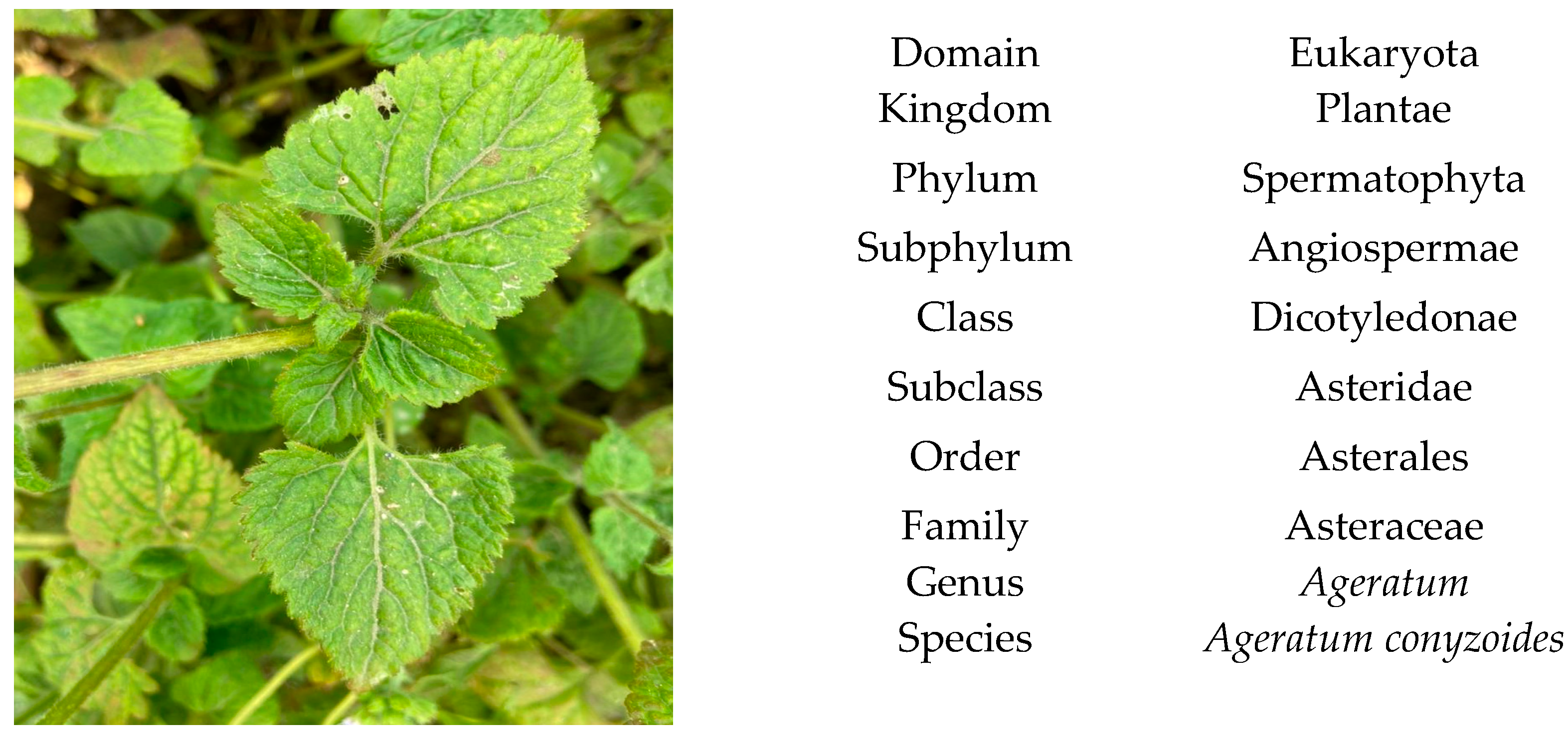
3. Taxonomy, Description and Botanical Characteristics
3.1. Taxonomical Classification
3.2. Description
3.3. Botanical Characteristics
4. Ethnopharmacology
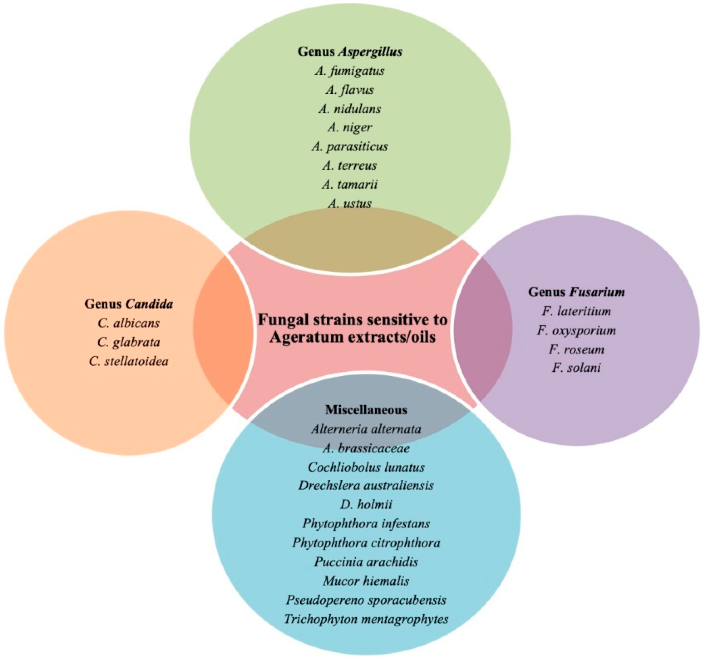
5. Pathogenic Fungal Strains vs. Antifungal Constituents Isolated from Ageratum conyzoides
5.1. Ageratum conyzoides against Fungal Genus Aspergillus
5.2. Ageratum conyzoides against Genus Fusarium
5.3. Ageratum conyzoides against Candida Pathogen
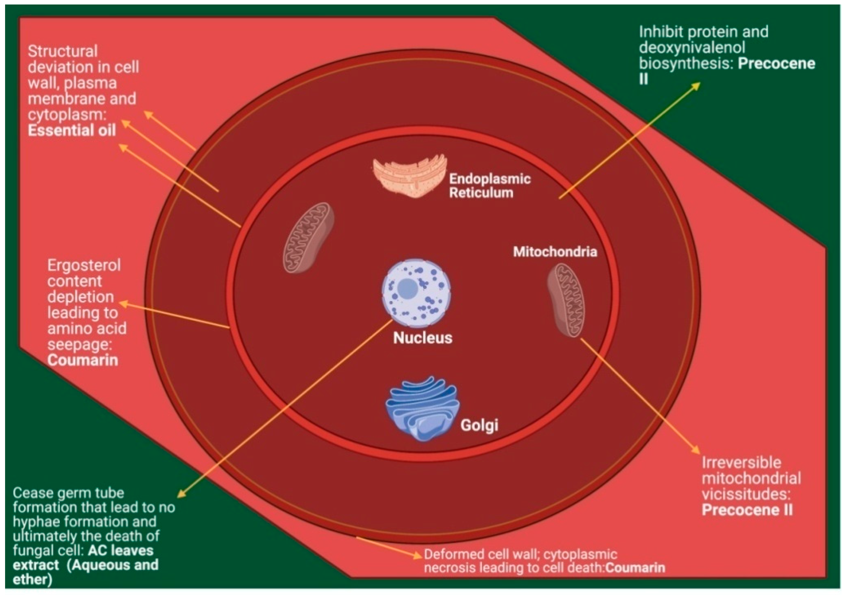
6. Mechanism of Action (MOA)
7. Safety Assessment
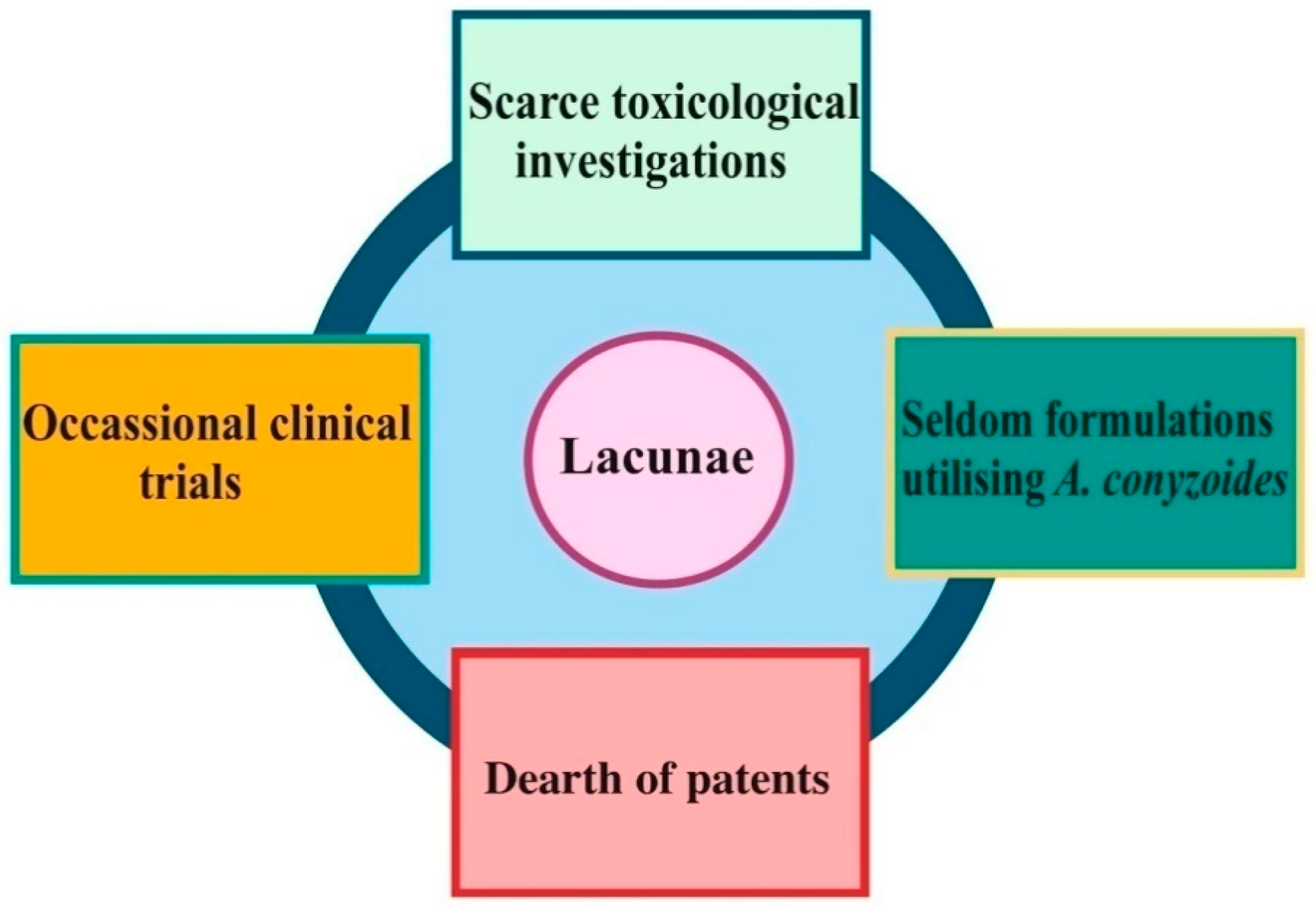
8. Future Research Prospective
9. Conclusions
Author Contributions
Funding
Institutional Review Board Statement
Informed Consent Statement
Data Availability Statement
Acknowledgments
Conflicts of Interest
References
- Kanafani, Z.A.; Perfect, J.R. Resistance to antifungal agents: Mechanisms and clinical impact. Clin. Infect. Dis. 2008, 46, 120–128. [Google Scholar] [CrossRef] [PubMed] [Green Version]
- Prasad, T.; Sethumadhavan, S.; Fatima, Z. Altered ergosterol biosynthetic pathway–an alternate multidrug resistance mechanism independent of drug efflux pump in human pathogenic fungi C. albicans. In Science Against Microbial Pathogens: Communicating Current Research and Technological Advances; Méndez Vilas, A., Ed.; Microbiology Series; Formatex Research Center: Badajoz, Spain, 2011; pp. 757–768. [Google Scholar]
- Samadi, F.M.; Suhail, S.; Sonam, M.; Sharma, N.; Singh, S.; Gupta, S.; Dobhal, A.; Pradhan, H. Antifungal efficacy of herbs. J. Oral Biol. Craniofacial Res. 2019, 9, 8–32. [Google Scholar] [CrossRef] [PubMed]
- Aldholmi, M.; Marchand, P.; Ourliac-Garnier, I.; LePape, P.; Ganesan, A. A Decade of Antifungal Leads from Natural Products: 2010–2019. Pharmaceuticals 2019, 12, 182. [Google Scholar] [CrossRef] [Green Version]
- Arif, T.; Bhosale, J.D.; Kumar, N.; Mandal, T.K.; Bendre, R.S.; Lavekar, G.S.; Dabur, R. Natural products–antifungal agents derived from plants. J. Asian. Nat. Prod. Res. 2009, 11, 621–638. [Google Scholar] [CrossRef] [PubMed]
- White, T.C.; Marr, K.A.; Bowden, R.A. Clinical, cellular, and molecular factors that contribute to antifungal drug resistance. Clin. Microbiol. Rev. 1998, 11, 382–402. [Google Scholar] [CrossRef] [PubMed] [Green Version]
- Vicente, M.F.; Basilio, A.; Cabello, A.; Peláez, F. Microbial natural products as a source of antifungals. Clin. Microbiol. Infect. 2003, 9, 15–32. [Google Scholar] [CrossRef] [Green Version]
- Vengurlekar, S.; Sharma, R.; Trivedi, P. Efficacy of some natural compounds as antifungal agents. Pharmacogn. Rev. 2012, 6, 91–99. [Google Scholar] [CrossRef] [Green Version]
- Mishra, K.K.; Kaur, C.D.; Sahu, A.K.; Panik, R.; Kashyap, P.; Mishra, S.P.; Dutta, S. Medicinal Plants Having Antifungal Properties. In Medicinal Plants-Use in Prevention and Treatment of Diseases; IntechOpen: London, UK, 2020; Available online: https://www.intechopen.com/books/medicinal-plants-use-in-prevention-and-treatment-of-diseases/medicinal-plants-having-antifungal-properties (accessed on 12 October 2020).
- Barrett, D. From natural products to clinically useful antifungals. Biochim. Biophys. Acta (BBA) Mol. Basis Dis. 2002, 1587, 224–233. [Google Scholar] [CrossRef] [Green Version]
- Efferth, T.; Li, P.C.; Konkimalla, V.S.B.; Kaina, B. From traditional Chinese medicine to rational cancer therapy. Trends Mol. Med. 2007, 13, 353–361. [Google Scholar] [CrossRef]
- Khan, S.; Shinwari, M.I.; Haq, A.; Ali, K.W.; Rana, T.; Badshah, M.; Khan, S.A. Fourier-transform infrared spectroscopy analysis and antifungal activity of methanolic extracts of Medicago parviflora, Solanum nigrum, Melilotus alba and Melilotus indicus on soil-borne phytopathogenic fungi. Pak. J. Bot. 2018, 50, 1591–1598. [Google Scholar]
- Khurshid, S.; Shoaib, A.; Javaid, A.; Qaisar, U. Potencial Fungicida de Grama Alelopática Cenchrus pennisetiformis no Crescimentode Fusarium oxysporum sp. Lycopersici sob Estressede Cromo. Planta Daninha 2016, 34, 453–463. [Google Scholar] [CrossRef] [Green Version]
- Shanab, S.M.; Shalaby, E.A.; Lightfoot, D.A.; El-Shemy, H.A. Allelopathic effects of water hyacinth [Eichhornia crassipes]. PLoS ONE 2010, 5, e13200. [Google Scholar] [CrossRef] [PubMed]
- Samarth, R.M.; Samarth, M.; Matsumoto, Y. Medicinally important aromatic plants with radioprotective activity. Future Sci. Oa 2017, 3, FSO247. [Google Scholar] [CrossRef] [PubMed] [Green Version]
- Singh, S.B.; Devi, W.R.; Marina, A.; Devi, W.I.; Swapana, N.; Singh, C.B. Ethnobotany, phytochemistry and pharmacology of Ageratum conyzoides Linn (Asteraceae). J. Med. Plant. Res. 2013, 7, 371–385. [Google Scholar] [CrossRef]
- Thorat, V.H.; Ghorpade, S.S.; Patole, T. Ageratum conyzoides Linn.: A review. Int. J. Pharmacogn. 2018, 5, 213–221. [Google Scholar]
- Lin, Z.; Lin, Y.; Shen, J.; Jiang, M.; Hou, Y. Flavonoids in Ageratum conyzoides L. Exert potent antitumor effects on human cervical adenocarcinoma HeLa cells in vitro and in vivo. Biomed Res. Int. 2020, 2020. [Google Scholar] [CrossRef]
- Singh, V.; Singh, H.; Sharma, G.P.; Raghubanshi, A.S. Eco-physiological performance of two invasive weed congeners (Ageratum conyzoides L. and Ageratum houstonianum Mill.) in the Indo-Gangetic plains of India. Environ. Monit. Assess. 2011, 178, 415–422. [Google Scholar] [CrossRef]
- Negi, B.; Bargali, S.S.; Bargali, K.; Khatri, K. Allelopathic interference of Ageratum conyzoides L. Against Rice Varieties. Curr. Agric. Res. J. 2020, 8, 69–76. [Google Scholar] [CrossRef]
- CABI. Ageratum conyzoides (billy goat weed). In Invasive Species Compendium; CAB International: Wallingford, UK, 2020; Available online: https://www.cabi.org (accessed on 30 October 2020).
- Shekhar, T.C.; Anju, G. A comprehensive review on Ageratum conyzoides Linn. (Goatweed). Int. J. Pharm. Phytopharmacol. Res. 2012, 1, 391–395. Available online: https://eijppr.com/cmvhDWP (accessed on 15 November 2020).
- Chauhan, A.; Rijhwani, S. A comprehensive review on phytochemistry of Ageratum conyzoides Linn. (Goatweed). Int. J. Eng. Technol. Manag. Appl. Sci. 2015, 3, 348–358. [Google Scholar]
- Ming, L.C. Ageratum conyzoides: A tropical source of medicinal and agricultural products. In Perspectives on New Crops and New Uses; Janick, J., Ed.; ASHS Press: Alexandria, VA, USA, 1999; pp. 469–473. [Google Scholar]
- Kong, C. Allelochemicals from Ageratum conyzoides L. and Oryza sativa L. and their effects on related pathogens. In Allelochemicals: Biological Control of Plant Pathogens and Diseases; Springer: Dordrecht, The Netherlands, 2006; Volume 2, pp. 193–206. [Google Scholar] [CrossRef]
- Das, S.K.; Mukherjee, S. Comparative morphological, anatomical and palynological observation in Ageratum conyzoides and Ageratum houstonianum of the family Compositae. Int. J. Pharm. Res. Bio-Pharma Sci. 2013, 2, 48–62. [Google Scholar]
- Kasali, A.A.; Winterhalter, P.; Adio, A.M.; Knapp, H.; Bonnlander, B. Chromenesin Ageratum conyzoides L. Flavour Fragr. J. 2002, 17, 247–250. [Google Scholar] [CrossRef]
- Okunade, A.L. Ageratum conyzoides L. (asteraceae). Fitoterapia 2002, 73, 1–16. [Google Scholar] [CrossRef]
- Usman, L.A.; Zubair, M.F.; Olawore, N.O.; Muhammad, N.O.; M’Civer, F.A.; Ismaeel, R.O. Chemical constituents of floweressential oil of Ageratum conyzoides growing in Nigeria. Elixir Org. Chem. 2013, 54, 12463–12465. [Google Scholar]
- Odeleye, O.P.; Oluyege, J.O.; Aregbesola, O.A.; Odeleye, P.O. Evaluation of preliminary phytochemical and antibacterial activity of Ageratum conyzoides (L.) on some clinical bacterial isolates. Int. J. Eng. Sci. 2014, 3, 1–5. [Google Scholar]
- Okereke, S.C.; Chukwudoruo, C.S.; Nwaokezie, C.O. Phytochemical screening using GC-FID and sub-chronic assessment of Hydroethanolic leaf extract of Ageratum conyzoides Linn. on albino rats. J. Med. Plants Stud. 2017, 5, 282–287. [Google Scholar]
- Amadi, B.A.; Duru, M.K.C.; Agomuo, E.N. Chemical profiles of leaf, stem, root and flower of Ageratum conyzoides. Asian J. Plant Sci. 2012, 2, 428–432. [Google Scholar]
- Agbafor, K.N.; Engwa, G.A.; Obiudu, I.K. Analysis of chemical composition of leaves and roots of Ageratum conyzoides. Int. J. Curr. Res. Acad. Rev. 2015, 3, 60–65. [Google Scholar]
- Santos, R.F.; Nunes, B.M.; Sá, R.D.; Soares, L.A.; Randau, K.P. Morpho-anatomical study of Ageratum conyzoides. Rev. De Farmacogn. 2016, 26, 679–687. [Google Scholar] [CrossRef] [Green Version]
- Janarthanan, L.; Karthikeyan, V.; Jaykar, B.; Balakrishnan, B.R.; Senthilkumar, K.L.; Anandharaj, G. Pharmacognostic studies on the whole plants of Ageratum conyzoides Linn. (Asteraceae). Eur. J. Pharm. Med. Res. 2016, 3, 618–626. [Google Scholar]
- Yadav, N.; Ganie, S.A.; Singh, B.; Chhillar, A.K.; Yadav, S.S. Phytochemical constituents and ethnopharmacological properties of Ageratum conyzoides L. Phytother. Res. 2019, 33, 2163–2178. [Google Scholar] [CrossRef]
- González, A.G.; Aguiar, Z.E.; Grillo, T.A.; Luis, J.G.; Rivera, A.; Calle, J. Methoxyflavones from Ageratum conyzoides. Phytochemistry 1991, 30, 1269–1271. [Google Scholar] [CrossRef]
- Morah, F.N.; Ogie, T. Ichthyotoxic effect of Ageratum Conyzoides leaf. Int. J. Adv. Sci. Res. 2016, 1, 19–20. [Google Scholar]
- Igoli, J.O.; Ogaji, O.G.; Tor-Anyiin, T.A.; Igoli, N.P. Traditional medicinal practice among the Igede people of Nigeria. Part II. Afr. J. Trad. Cam 2005, 2, 134–152. [Google Scholar]
- Okeke, E.C.; Eneobong, H.N.; Uzuegbunam, A.O.; Ozioko, A.O.; Kuhnlein, H. Igbo traditional food system: Documentation, uses and research needs. Pak. J. Nutr. 2008, 7, 365–376. [Google Scholar] [CrossRef] [Green Version]
- Bosi, C.F.; Rosa, D.W.; Grougnet, R.; Lemonakis, N.; Halabalaki, M.; Skaltsounis, A.L.; Biavatti, M.W. Pyrrolizidine alkaloids in medicinal tea of Ageratum conyzoides. Rev. De Farmacogn. 2013, 23, 425–432. [Google Scholar] [CrossRef] [Green Version]
- Yamamoto, L.A.; Soldera, J.C.; Emim, J.A.; Godinho, R.O.; Souccar, C.; Lapa, A.J. Pharmacological screening of Ageratum conyzoides L. (mentrasto). Memórias Do Inst. Oswaldo Cruz. 1991, 86, 145–147. [Google Scholar] [CrossRef]
- Kaur, R.; Dogra, N.K. A review on traditional uses, chemical constituents and pharmacology of Ageratum conyzoides L. (Asteraceae). Int. J. Pharm. Biol. Arch. 2014, 5, 33–45. [Google Scholar]
- Kamboj, A.; Saluja, A.K. Ageratum conyzoides L.: A review on its phytochemical and pharmacological profile. Int. J. Green Pharm. 2008, 59–68. [Google Scholar] [CrossRef]
- Appiah, K.S.; Oppong, C.P.; Mardani, H.K.; Omari, R.A.; Kpabitey, S.; Amoatey, C.A.; Onwona-Agyeman, S.; Oikawa, Y.; Katsura, K.; Fujii, Y. Medicinal plants used in the Ejisu-Juaben Municipality, Southern Ghana: An ethnobotanical study. Medicines 2018, 6, 1. [Google Scholar] [CrossRef] [Green Version]
- Hoffman, B.R.; DelasAlas, H.; Blanco, K.; Wiederhold, N.; Lewis, R.E.; Williams, L. Screening of antibacterial and antifungal activities of ten medicinal plants from Ghana. Pharm. Biol. 2004, 42, 13–17. [Google Scholar] [CrossRef]
- Burkill, H.M. The Useful Plants of West Tropical Africa. Vol. 1. Families AD. Royal Botanic Gardens. 1985. Available online: https://plants.jstor.org/stable/10.5555/al.ap.upwta.1_861 (accessed on 20 August 2020).
- Satyal, P.; Poudel, A.; Setzer, W.N. Variation in the volatile phytochemistry of Ageratum conyzoides. Am. J. Essent. Oils Nat. Prod. 2018, 6, 07–10. [Google Scholar]
- Ndob, I.B.; Mengome, L.E.; Bourobou, H.P.B.; Banfora, Y.L.; Bivigou, F. Ethnobotanical survey of medicinal plants used as anthelmintic remedies in Gabon. J. Ethnopharmacol. 2016, 191, 360–371. [Google Scholar] [CrossRef]
- Betti, J.L.; Iponga, D.M.; Yongo, O.D.; Mbomio, D.O.; Yobo, C.M.; Ngoy, A. Ethnobotanical study of medicinal plants of the Ipassa-Makokou Biosphere Reserve, Gabon: Plants used for treating malaria. J. Med. Plant. Res. 2013, 7, 2300–2318. [Google Scholar]
- Andissa, N.O.; Moussoungou, A.S.; Koloungous, B.C.; Abena, A.A. Topical Anti-inflammatory effect of aqueous extract ointment of Ageratum conyzoïdes L. in wistarrat. Int. J. Phytopharm. 2015, 5, 37–41. [Google Scholar]
- Kaur, R.; Kaur, S. Anxiolytic potential of methanol extract from Ageratum conyzoides Linn. Leaves. Phcog. J. 2015, 7, 236–241. [Google Scholar] [CrossRef]
- Chhabra, S.C.; Mahunnah, R.L.A.; Mshiu, E.N. Plants used in traditional medicine in Eastern Tanzania. II. Angiosperms (Capparidaceae to Ebenaceae). J. Ethnopharmacol. 1989, 25, 339–359. [Google Scholar] [CrossRef] [Green Version]
- Khastini, R.O.; Saraswati, I.; Sulaiman, F.; Alimuddin; Sari, I.J. Antifungal Activity of JukutBatau AgeratumConyzoides Leaves Extract on Candida Albicans In Vitro. Int. J. Sci. Technol. Res. 2019, 8, 2494–2497. [Google Scholar]
- Achmad, H.; Adam, A.M.; Aulia, A.; Sukmana, B.I.; Huldani Khera, S.N.; Ramadhany, Y.F. A Review of Bandotan Leaf Extract (Ageratum conyzoides L.) in Inhibition Test to the Growth of Bacteria (Porphyromonasgingivalis) Case of Periodontitis Disease. Syst. Rev. Pharm. 2020, 11, 390–395. [Google Scholar] [CrossRef]
- Mehra, A.; Bajpai, O.; Joshi, H. Diversity, utilization and sacred values of Ethno-medicinal plants of Kumaun Himalaya. Trop. Plant Res. 2014, 1, 80–86. [Google Scholar]
- Perme, N.; Choudhury, S.N.; Choudhury, R.; Natung, T.; De, B. Medicinal plants in traditional use at Arunachal Pradesh, India. Int. J. Phytopharm. 2015, 5, 86–98. [Google Scholar]
- Tamuli, P.; Ghosal, A. Ethnomedicinal plants used by major ethnic groups of Assam (India) for curing skin diseases. Int. J. Herb. Med. 2017, 5, 140–144. [Google Scholar]
- Arulprakash, K.; Murugan, R.; Ponrasu, T.; Iyappan, K.; Gayathri, V.S.; Suguna, L. Efficacy of Ageratum conyzoides on tissue repair and collagen formation in rats. Clin. Exp. Dermatol. 2012, 37, 418–424. [Google Scholar] [CrossRef]
- Jayasundera, M.; Florentine, S.; Tennakoon, K.U.; Chauhan, B.S. Medicinal Value of Three Agricultural Weed Species of the Asteraceae Family: A Review. Pharmacogn. J. 2021, 13, 264–277. [Google Scholar] [CrossRef]
- Ghosh, G.; Narayan, B.; Bengal, W. Traditional use of plants against leprosy in India: A review of the recent literature. J. Innov. Pharm. Biol. Sci. 2017, 4, 55–64. [Google Scholar]
- Shailajan, S.; Wadke, P.; Joshi, H.; Tiwari, B. Evaluation of quality and efficacy of an ethnomedicinal plant Ageratum conyzoides L. in the management of pediculosis. J. Young Pharm. 2013, 5, 139–143. [Google Scholar] [CrossRef] [Green Version]
- Lans, C. Ethnomedicines used in Trinidad and Tobago for reproductive problems. J. Ethnobiol. Ethnomed. 2007, 3. [Google Scholar] [CrossRef] [Green Version]
- Rafe, M.R. A review of five traditionally used anti-diabetic plants of Bangladesh and their pharmacological activities. Asian Pac. J. Trop. Med. 2017, 10, 933–939. [Google Scholar] [CrossRef]
- Namsa, N.D.; Tag, H.; Mandal, M.; Kalita, P.; Das, A.K. An ethnobotanical study of traditional anti-inflammatory plants used by the Lohit community of Arunachal Pradesh, India. J. Ethnopharmacol. 2009, 125, 234–245. [Google Scholar] [CrossRef]
- Abena, A.A.; Kintsangoula-Mbaya, G.S.; Diantama, J.; Bioka, D. Analgesic effects of a raw extract of Ageratum conyzoides in the rat. Encephale 1993, 19, 329–332. [Google Scholar]
- Ukwe, C.V.; Ekwunife, O.I.; Epueke, E.A.; Ubaka, C.M. Antimalarial activity of Ageratum conyzoides in combination with chloroquine and artesunate. Asian Pac. J. Trop. Med. 2010, 3, 943–947. [Google Scholar] [CrossRef] [Green Version]
- Madureira, M.D.C.; Martins, A.P.; Gomes, M.; Paiva, J.; da Cunha, A.P.; do Rosário, V. Antimalarial activity of medicinal plants used in traditional medicine in S. Tomé and Prıncipe islands. J. Ethnopharmacol. 2002, 81, 23–29. [Google Scholar] [CrossRef]
- EL-Kamali, H.H. Effect of certain medicinal plants extracts against storage pest, Triboliumcastaneum Herbst. Am. Eurasian J. Sustain. Agric. 2009, 3, 139–142. [Google Scholar]
- Dougoud, J.; Toepfer, S.; Bateman, M.; Jenner, W.H. Efficacy of homemade botanical insecticides based on traditional knowledge. A review. Agron. Sustain. Dev. 2019, 39. [Google Scholar] [CrossRef] [Green Version]
- Moreira, M.D.; Picanço, M.C.; Barbosa, L.C.; Guedes, R.N.; da Silva, É.M. Toxicity of leaf extracts of Ageratum conyzoides to lepidoptera pests of horticultural crops. Biol. Agric. Hortic. 2004, 22, 251–260. [Google Scholar] [CrossRef]
- Oladejo, O.W.; Imosemi, I.O.; Osuagwu, F.C.; Oluwadara, O.O.; Aiku, A.; Adewoyin, O.; Ekpo, O.E.; Oyedele, O.O.; Akang, E.E. Enhancement of cutaneous wound healing by methanolic extracts of Ageratum conyxoides in the wistar rat. Afr. J. Biomed. Res. 2003, 6, 27–31. [Google Scholar] [CrossRef] [Green Version]
- Varadharajan, R.; Rajalingam, D. Anti-convulsant activity of methanolic extracts of Ageratum conyzoides L. Int. J. Innov. Drug Discov. 2011, 1, 24–28. [Google Scholar]
- Parveen, S.; Godara, R.; Katoch, R.; Yadav, A.; Verma, P.K.; Katoch, M.; Singh, N.K. In vitro evaluation of ethanolic extracts of Ageratum conyzoides and Artemisia absinthium against cattle tick, Rhipicephalus microplus. Sci. World J. 2014, 2014. [Google Scholar] [CrossRef] [Green Version]
- Uhegbu, F.O.; Imo, C.; Onwuegbuchulam, C.H. Lipid lowering, hypoglycemic and antioxidant activities of Chromolaena odorata (L) and Ageratum conyzoides (L.) ethanolic leaf extracts in albino rats. J. Med. Plants Stud. 2016, 4, 155–159. [Google Scholar]
- Oso, B.J.; Olaoye, I.F. Comparative in vitro studies of antiglycemic potentials and molecular docking of Ageratum conyzoides L. and Phyllanthus amarus L. methanolic extracts. SN Appl. Sci. 2020, 2, 1–13. [Google Scholar] [CrossRef] [Green Version]
- Neelabh, C.; Nahid, A.; Kumar, N. Study on methanolic extract of Ageratumconyzoides for its ability to act as an antioxidant and to suppress the microbial growth. Pharma Innov. 2017, 6, 170–173. [Google Scholar]
- Arlette, N.T.; Nadia, N.A.; Jeanette, Y.; Gertrude, M.T.; Josué, W.P.; Mbida, M. Anticoccidial Effects of Ageratum conyzoides (Asteraceae) and Vernonia amygdalina (Asteraceae) Leaves Extracts on Broiler Chickens. South Asian J. Parasitol. 2019, 2, 1–10. [Google Scholar]
- Santharam, B.; Vidya, V.; Thangathirupathi, A. Antiurolithiatic activity of different extracts of Ageratum conyzoides (Linn.). J. Pharm. Sci. Innov. 2015, 4, 140–143. [Google Scholar] [CrossRef]
- Gbadamosi, I.T. Ethnobotanical survey of plants used for the treatment and management of sexually transmitted infections in Ibadan, Nigeria. Ethnobot. Res. Appl. 2014, 12, 659–669. [Google Scholar] [CrossRef] [Green Version]
- Anjorin, T.S.; Salako, E.A.; Makun, H.A. Control of Toxigenic Fungi and Mycotoxins with Phytochemicals: Potentials and Challenges. Mycotoxin Food Saf. Dev. Ctries. 2013, 181–202. [Google Scholar] [CrossRef] [Green Version]
- Badillo, L.M.D.; Espinosa-Madrigal, R.M.; Martinez-Muñoz, R.E.; Ron-Echeverría, O.A.; Salgado-Garciglia, R.; Flores-García, A.; Gonzalez, D.R.; Pacheco, M.M.M. The Mexican medicinal plants with antifungal properties are an economic and health opportunity area. Pharmacol. Online 2008, 3, 61–77. [Google Scholar]
- Verma, R.K.; Chaurasia, L.; Katiyar, S. Potential antifungal plants for controlling building fungi. Indian J. Nat. Prod. Resour. 2008, 7, 374–387. [Google Scholar]
- Bajwa, R.; Shafique, S.; Shafique, S. Evaluation of antifungal activity of aqueous extracts of two asteraceous plant species. Mycopath 2007, 5, 29–33. [Google Scholar]
- Osho, A.; Adetunji, T. Antimicrobial activity of the essential oil of Ageratum conyzoides L. Asian J. Sci. Technol. 2011, 2, 1–5. [Google Scholar]
- Tambunan, A.P.; Bahtiar, A.; Tjandrawinata, R.R. Influence of extraction parameters on the yield, phytochemical, TLC-densitometric quantification of quercetin, and LC-MS profile, and how to standardize different batches for long term from Ageratum conyoides L. leaves. Phcog. J. 2017, 9, 767–774. [Google Scholar] [CrossRef] [Green Version]
- Barros, F.M.; Almeida, P.C.; Scopel, R.; do Espirito Santo, A.T.; Lucas, A.M.; Bordignon, S.A.; Cassel, E.; Vargas, R.M.; von Poser, G. Chromenes from Ageratum conyzoides: Steam distillation, supercritical extraction, and mathematical modeling. Sep. Sci. Technol. 2016, 51, 307–315. [Google Scholar] [CrossRef]
- Wuyep, P.A.; Musa, H.D.; Ezemokwe, G.C.; Nyam, D.D.; SilaGyang, M.D. Phytochemicals from Ageratum conyzoides L. extracts and their antifungal activity against virulent Aspergillus spp. J. Acad. Ind. Res. 2019, 6, 32–39. [Google Scholar]
- Kumar, B.; Misra, A.; Rawat, A.K.; Rawat, Y.S.; Srivastava, S. Simultaneous quantification of Precocene I and Precocene II through high-performance thin layer chromatography validated method in Ageratum conyzoides L. germ plasms from western Himalayas. Phcog. Mag. 2018, 14, 141–146. [Google Scholar]
- Kong, C.H.; Hu, F.; Xu, X.; Liang, W.; Zhang, C. Allelopathic plants XV: Ageratumconyzoides. Allelopath. J. 2004, 14, 1–12. [Google Scholar]
- Iqbal, M.C.; Jayasinghe, U.L.; Herath, H.M.; Wijesekara, K.B.; Fujimoto, Y. A fungistatic chromene from Ageratum conyzoides. Phytoparasitica 2004, 32, 119–126. [Google Scholar] [CrossRef]
- Nogueira, J.H.; Gonçalez, E.; Galleti, S.R.; Facanali, R.; Marques, M.O.; Felício, J.D. Ageratumconyzoides essential oil as aflatoxin suppressor of Aspergillus flavus. Int. J. Food Microbiol. 2010, 137, 55–60. [Google Scholar] [CrossRef] [PubMed]
- Furukawa, T.; Sakamoto, N.; Suzuki, M.; Kimura, M.; Nagasawa, H.; Sakuda, S. Precocene II, a trichothecene production inhibitor, binds to voltage-dependent anion channel and increases the superoxide level in mitochondria of Fusarium graminearum. PLoS ONE 2015, 10, e0135031. [Google Scholar] [CrossRef]
- Esper, R.H.; Gonçalez, E.; Felicio, R.C.; Felicio, J.D. Fungicidal activity and constituents of Ageratum conyzoides essential oil from three regions in São Paulo state, Brazil. Arq. Inst. Biológico. 2015, 82, 1–4. [Google Scholar] [CrossRef] [Green Version]
- Wang, C.; Zhang, J.; Chen, H.; Fan, Y.; Shi, Z. Antifungal activity of eugenol against Botrytis cinerea. Trop. Plant Pathol. 2010, 35, 137–143. [Google Scholar] [CrossRef] [Green Version]
- Abd El-Baky, R.M.; Hashem, Z.S. Eugenol and linalool: Comparison of their antibacterial and antifungal activities. Afr. J. Microbiol. Res. 2016, 10, 1860–1872. [Google Scholar]
- Rana, I.S.; Rana, A.S.; Rajak, R.C. Evaluation of antifungal activity in essential oil of the Syzygium aromaticum (L.) by extraction, purification and analysis of its main component eugenol. Braz. J. Microbiol. 2011, 42, 1269–1277. [Google Scholar] [CrossRef] [PubMed]
- Chaieb, K.; Zmantar, T.; Ksouri, R.; Hajlaoui, H.; Mahdouani, K.; Abdelly, C.; Bakhrouf, A. Antioxidant properties of the essential oil of Eugenia caryophyllata and its antifungal activity against a large number of clinical Candida species. Mycoses 2007, 50, 403–406. [Google Scholar] [CrossRef] [PubMed]
- Cai, R.; Hu, M.; Zhang, Y.; Niu, C.; Yue, T.; Yuan, Y.; Wang, Z. Antifungal activity and mechanism of citral, limonene and eugenol against Zygosaccharomyces Rouxii. LWT Food Sci. Technol. 2019, 106, 50–56. [Google Scholar] [CrossRef]
- Selestino Neta, M.C.; Vittorazzi, C.; Guimarães, A.C.; Martins, J.D.L.; Fronza, M.; Endringer, D.C.; Scherer, R. Effects of β-caryophyllene and Murrayapaniculata essential oil in the murine hepatoma cells and in the bacteria and fungi 24-h time–kill curve studies. Pharm. Biol. 2017, 55, 190–197. [Google Scholar] [CrossRef] [PubMed] [Green Version]
- Dahham, S.S.; Tabana, Y.M.; Iqbal, M.A.; Ahamed, M.B.; Ezzat, M.O.; Majid, A.S.; Majid, A.M. The anticancer, antioxidant and antimicrobial properties of the sesquiterpene β-caryophyllene from the essential oil of Aquilaria crassna. Molecules 2015, 20, 11808–11829. [Google Scholar] [CrossRef]
- Widodo, G.P.; Sukandar, E.Y.; Adnyana, I.K.; Sukrasno, S. Mechanism of Action of Coumarin against Candida albicans by SEM/TEM Analysis. ITB J. Sci. 2012, 44, 145–151. [Google Scholar] [CrossRef] [Green Version]
- Jia, C.; Zhang, J.; Yu, L.; Wang, C.; Yang, Y.; Rong, X.; Xu, K.; Chu, M. Antifungal activity of coumarin against Candida albicans is related to apoptosis. Front. Cell. Infect. Microbiol. 2019, 8, 1–13. [Google Scholar] [CrossRef] [Green Version]
- Kumar, P.; Mahato, D.K.; Kamle, M.; Mohanta, T.K.; Kang, S.G. Aflatoxins: A global concern for food safety, human health and their management. Front. Microbiol. 2017, 7, 1–10. [Google Scholar] [CrossRef] [PubMed] [Green Version]
- Widodo, G.P.; Sukandar, E.Y.; Adnyana, I.K. A coumarin from Ageratum leaves (Ageratum conyzoides L.). Int. J. Pharmacol. 2008, 4, 56–59. [Google Scholar] [CrossRef] [Green Version]
- Patil, R.P.; Nimbalkar, M.S.; Jadhav, U.U.; Dawkar, V.V.; Govindwar, S.P. Antiaflatoxigenic and antioxidant activity of an essential oil from Ageratum conyzoides L. J. Sci. Food Agric. 2010, 90, 608–614. [Google Scholar] [CrossRef]
- Adjou, E.S.; Dahouenon-Ahoussi, E.; Degnon, R.; Soumanou, M.M.; Sohounhloue, D.C. Investigations on bioactivity of essential oil of Ageratum conyzoides L., from Benin against the growth of fungi and aflatoxin production. Int. J. Pharm. Sci. Rev. Res. 2012, 13, 143–148. [Google Scholar]
- Mycology online. The University of Adelaide. Fusarium. 1998. Available online: https://mycology.adelaide.edu.au/descriptions/hyphomycetes/fusarium/ (accessed on 4 August 2020).
- Tupaki-Sreepurna, A.; Kindo, A.J. Fusarium: The versatile pathogen. Indian J. Med. Microbiol. 2018, 36, 8–17. [Google Scholar] [CrossRef]
- Rai, M.K.; Acharya, D. Search for fungi toxic potential in essential oils of Asteraceous plants. In Compositae Newsletter; The Swedish Museum of Natural History: Stockholm, Sweden, 2000; pp. 18–23. [Google Scholar]
- Adekunle, A.A. Ethnobotanical studies of some medicinal plants from Lagos State of Nigeria. Niger. J. Bot. 2001, 14, 71–79. [Google Scholar]
- Javed, S.; Bashir, U. Antifungal activity of different extracts of Ageratum conyzoides for the management of Fusarium solani. Afr. J. Biotechnol. 2012, 11, 11022–11029. [Google Scholar] [CrossRef]
- Ilondu, E.M.; Ojeifo, I.M.; Emosairue, S.O. Evaluation of antifungal properties of Ageratum conyzoides, Spilanthesfilicaulis and Tithonia diversifolia leaf extracts and search for their compounds using gas chromatography-mass spectrum. ARPN J. Agric. Biol. Sci. 2014, 9, 375–384. [Google Scholar]
- Li, W.R.; Shi, Q.S.; Dai, H.Q.; Liang, Q.; Xie, X.B.; Huang, X.M.; Zhao, G.Z.; Zhang, L.X. Antifungal activity, kinetics and molecular mechanism of action of garlic oil against Candida albicans. Sci. Rep. 2016, 6, 1–9. [Google Scholar] [CrossRef]
- Yaguchi, A.; Yoshinari, T.; Tsuyuki, R.; Takahashi, H.; Nakajima, T.; Sugita-Konishi, Y.; Nagasawa, H.; Sakuda, S. Isolation and identification of precocenes and piperitone from essential oils as specific inhibitors of trichothecene production by Fusarium graminearum. J. Agric. Food Chem. 2009, 57, 846–851. [Google Scholar] [CrossRef]
- Thati, B.; Noble, A.; Rowan, R.; Creaven, B.S.; Walsh, M.; McCann, M.; Egan, D.; Kavanagh, K. Mechanism of action of coumarin and silver (I)–coumarin complexes against the pathogenic yeast Candida albicans. Toxicol. In Vitro 2007, 21, 801–808. [Google Scholar] [CrossRef] [Green Version]
- Kumari, S.; Jain, P.; Sharma, B.; Kadyan, P.; Dabur, R. In vitro antifungal activity and probable fungicidal mechanism of aqueous extract of Barleria grandiflora. Appl. Biochem. Biotechnol. 2015, 175, 3571–3584. [Google Scholar] [CrossRef]
- Sahgal, G.; Ramanathan, S.; Sasidharan, S.; Mordi, M.N.; Ismail, S.; Mansor, S.M. In vitro and in vivo anticandidal activity of Swietenia mahogani methanolic seed extract. Trop. Biomed. 2011, 28, 132–137. [Google Scholar]
- Falade, M.J.; Borisade, O.A.; Aluko, M. Evaluation of Antifungal activities of five plant extracts against Pseudoperenosporacubensis (Downy Mildew) in Muskmelon (Cucumis melo L.). Annu. Res. Rev. Biol. 2019, 31, 1–6. [Google Scholar] [CrossRef]
- Yapi, A.B.; Camara, D.; Coulibaly, K.; Zirihi, G.N. Ethnobotanical study and comparison of antitrichophytic activity leaves of Aspiliaafricana (pers.), Cd adams var. Africana, Ageratumconyzoides L. and Acanthospermumhispidum DC. on the in vitro growth of Trichophyton mentagrophytes. INDO Am. J. Pharm. Sci. 2018, 5, 4766–4773. [Google Scholar]
- Yusnawan, E.; Inayati, A. Antifungal activity of crude extracts of Ageratum conyzoides, Cyperus rotundus and Amaranthus spinosus against rust disease. AGRIVITA J. Agric. Sci. 2018, 40, 403–414. [Google Scholar] [CrossRef]
- Hidangmayum, B.; Singh, N.I. Efficacy of plant extracts on fruit rot pathogen of pineapple (Ananas comosus Merr.). Int. J. Eng. Dev. Res. 2017, 5, 1353–1355. [Google Scholar]
- Khatoon, A.; Mohapatra, A.; Satapathy, K.B. Plants used as Biofungicides against Storage-Decay of Yam (Dioscoreaalata L.) in Odisha, India. J. Pharm. Chem. Biol. Sci. 2017, 5, 253–258. [Google Scholar]
- Aoudou, Y.; Second, Z.M. Mycoflora associated with cocoa (Theobroma cacao) pods in Cameroon and antifungal effect of plant extracts. Int. J. Environ. Agric. Biotechnol. 2017, 2, 112–117. [Google Scholar] [CrossRef]
- Shafique, S.; Shafique, S.; Yousuf, A. Bioefficacy of Extract of Ageratum conyzoides Against Drechsleraaustraliensis and Drechsleraholmii. Pak. J. Phytopathol. 2015, 27, 193–200. [Google Scholar]
- Morais, W.C.; Lima, M.A.; Zanuncio, J.C.; Oliveira, M.A.; Bragança, M.A.; Serrão, J.E.; Della Lucia, T.M. Extracts of Ageratum conyzoides, Coriandrum sativum and Mentha piperita inhibit the growth of the symbiotic fungus of leaf-cutting ants. Ind. Crop. Prod. 2015, 65, 463–466. [Google Scholar] [CrossRef]
- Sharma, D.; Yami, H.; Sharma, D.; Shukla, A.K. Antifungal activities of essential oils from four commonly used ethno-medicinal plants. Asian J. Ethnopharmacol. Med. Foods 2015, 1, 25–31. [Google Scholar]
- Esper, R.H.; Gonçalez, E.; Marques, M.O.; Felicio, R.C.; Felicio, J.D. Potential of essential oils for protection of grains contaminated by aflatoxin produced by Aspergillus flavus. Front. Microbiol. 2014, 5, 1–5. [Google Scholar] [CrossRef]
- Hubert, G.Y.; Julienne, N.; Charles, D.D.; Daniel, F.; Sandrine, P.T.; Romain, F.F.; Henry, A.Z. Antifungal potential and phytochemical analysis of extracts from seven Cameroonian plants against late blight pathogen Phytophthora infestans. Int. J. Curr. Microbiol. App. Sci 2013, 2, 140–154. [Google Scholar]
- Pal, G.K.; Kumar, B.; Shahi, S.K. Antifungal activity of some common weed extracts against phytopathogenic fungi Alternaria spp. Int. J. Univers. Pharm. Bio Sci. 2013, 3, 6–14. [Google Scholar]
- Katoch, R.A.; Thakur, M.E.; Paul, Y.S. Antifungal activity of the essential oils of Chromolaenaadenophorum, Ageratum conyzoidesand Lantana camara. Indian Phytopath. 2012, 65, 409–411. [Google Scholar]
- Prakash, B.; Dubey, N.K. Evaluation of chemically characterised essential oils of Coleus aromaticus, Hyptissuaveolens and Ageratum conyzoides against storage fungi and aflatoxin contamination of food commodities. Int. J. Food. Sci. Technol. 2011, 46, 754–760. [Google Scholar] [CrossRef]
- Bajwa, R.; Akhtar, N.; Javid, A. Antifungal activity of allelopathic plant extracts. I. Effect of aqueous extracts of three allelopathic Asteraceous species on growth of Aspergilli. Pak. J. Biol. Sci. 2001, 4, 503–507. [Google Scholar]
- Iqbal, M.C.; Meiyalaghan, S.; Wijesekara, K.B.; Abeyratne, K.P. Antifungal activity from water extracts of some common weeds. Pak. J. Biol. Sci. 2001, 4, 843–845. [Google Scholar] [CrossRef]
- Fiori, A.C.; Schwan-Estrada, K.R.F.; Stangarlin, J.R.; Vida, J.B.; Scapim, C.A.; Cruz, M.E.; Pascholati, S.F. Antifungal activity of leaf extracts and essential oils of some medicinal plants against Didymellabryoniae. J. Phytopathol. 2000, 148, 483–487. [Google Scholar] [CrossRef]
- Dixit, S.N.; Chandra, H.; Tiwari, R.; Dixit, V. Development of a botanical fungicide against blue mould of mandarins. J. Stored Prod. Res. 1995, 31, 165–172. [Google Scholar] [CrossRef]
- Asthana, A.; Chandra, H.; Dikshit, A.; Dixit, S.N. Volatile fungi toxicants from leaves of some higher plants against Helminthosporium oryzae/Flüchtige Verbindungen mit antimykotischer Wirkung gegen Helminthosporium oryzae aus den Blättern einiger höherer Pflanzen. Z. Und Pflanzenschutz. J. Plant Dis. Prot. 1982, 89, 475–479. [Google Scholar]
- Destaa, T.; Afeworka, M.; Unnithana, C.R.; Alayb, H. Isolation and structural elucidation of toxic pyrrolizidine alkaloids from Ageratum conyzoides collected from Vod disease affected communities. Int. J. Pharm. Technol. 2014, 6, 6281–6290. [Google Scholar]
- Hsia, M.S.; Grossman, S.; Schrankel, K.R. Hepatotoxicity of the anti-juvenile hormone precocene II and the generation of dihydrodiol metabolites. Chem. Biol. Interact. 1981, 37, 265–277. [Google Scholar] [CrossRef]
- Schrankel, K.R.; Grossman, S.J.; Hsia, M.S. Precocene II nephrotoxicity in the rat. Toxicol. Lett. 1982, 12, 95–100. [Google Scholar] [CrossRef]
- Moura, A.C.; Silva, E.L.; Fraga, M.C.; Wanderley, A.G.; Afiatpour, P.; Maia, M.B. Antiinflammatory and chronic toxicity study of the leaves of Ageratum conyzoides L. in rats. Phytomedicine 2005, 12, 138–142. [Google Scholar] [CrossRef]
- Igboasoiyi, A.C.; Eseyin, O.A.; Ezenwa, N.K.; Oladimeji, H.O. Studies on the Toxicity of Ageratum conyzoides. J. Pharmacol. Toxicol. 2007, 2, 743–747. [Google Scholar] [CrossRef] [Green Version]
- Adebayo, H.A.; Zeng, G.Z.; Fan, J.T.; Ji, C.J.; He, W.J.; Xu, J.J.; Zhang, Y.M.; Akindahunsi, A.A.; Kela, R.; Tan, N.H. Biochemical, haematological and histopathological studies of extract of Ageratum conyzoides L. in Sprague Dawley rats. J. Med. Plants Res. 2010, 4, 2264–2272. [Google Scholar]
- Adebayo, A.H.; Zeng, G.Z.; Zhang, Y.M.; Ji, C.J.; Akindahunsi, A.A.; Tan, N.H. Toxicological evaluation of precocene II isolated from Ageratum conyzoides L. (Asteraceae) in Sprague Dawley rats. Afr. J. Biotechnol 2010, 9, 2938–2944. [Google Scholar]
- Diallo, A.; Eklu-Gadegkeku, K.; Agbono, A.; Aklikokou, K.; Creppy, E.E.; Gbeassor, M. Acute and sub-chronic (28-day) oral toxicity studies of hydroalcohol leaf extract of Ageratum conyzoides L (Asteraceae). Trop. J. Pharm. Res. 2010, 9, 463–467. [Google Scholar] [CrossRef] [Green Version]
- Diallo, A.; Batomayena, B.; Povi, L.; Eklu-Gadegbeku, K.W.; Aklikokou, K.; Creppy, E.; Gbeassor, M. Fetal toxicity of hydroalcoholic extract of Ageratum conyzoides L. leaves (Asteraceae) in rats. Int. J. Pharm. Pharm. Sci. 2015, 7, 264–266. [Google Scholar]
- Nweze, N.E.; Obiwulu, I.S. Anticoccidial effects of Ageratum conyzoides. J. Ethnopharmacol. 2009, 122, 6–9. [Google Scholar] [CrossRef]
- Adesanwo, J.K.; Egbomeade, C.O.; Moronkola, D.O.; Akinpelu, D.A. Chemical, Toxicity and Antibacterial Studies on Methanol Extracts of Melanthera scandens, Ageratum conyzoides, Aspiliaafricana and Synedrellanodiflora. J. Explor. Res. Pharmacol. 2019, 4, 1–7. [Google Scholar] [CrossRef]
- Verma, P.K.; Sultana, M.; Raina, R.; Prawez, S.; Pandita, S.; Jamwal, N.; Mir, A.H. Hepatoprotective Effects of Ageratum conyzoides L. on biochemical indices induced by acetaminophen toxicity in Wistar rats. J. Appl. Pharm. Sci. 2013, 3, S23–S27. [Google Scholar] [CrossRef]
- Ola-Davies, O.E.; Akinrinde, A.S. Acute sodium Arsenite-induced hematological and biochemical changes in wistar rats: Protective effects of ethanol extract of Ageratum conyzoides. Pharmacogn. Res. 2016, 8, S26–S30. [Google Scholar] [CrossRef] [Green Version]
- Budiman, A.; Aulifa, D.L. A Study Comparing Antibacterial Activity of Ageratum conyzoides L. Extract and Piper betle L. Extract in Gel Dosage Forms Against Staphylococcus aureus. Pharmacogn. J. 2020, 12, 473–477. [Google Scholar] [CrossRef]
- Kotta, J.C.; Lestari, A.; Candrasari, D.S.; Hariono, M. Medicinal Effect, In Silico Bioactivity Prediction, and Pharmaceutical Formulation of Ageratum conyzoides L.: A Review. Scientifica 2020, 2020. [Google Scholar] [CrossRef] [PubMed]
- Jain, S.; Jain, N.; Tiwari, A.; Balekar, N.; Jain, D.K. Simple evaluation of wound healing activity of polyherbal formulation of roots of Ageratum conyzoides Linn. Asian J. Res. Chem. 2009, 2, 135–138. [Google Scholar]
- Prajapati, R.A.J.M.A.N.I..; Roy, S.U.N.I.T.A.; Mishra, S.U.D.E.E.P.; Raza, S.K.; Thakur, L.K. Formulation development, standardization and antimicrobial activity of Ageratum conyzoides extracts and their formulation. Int. J. Pharm. Pharm. Sci 2014, 6, 369–374. [Google Scholar]
- Permawati, M.; Anwar, E.; Arsianti, A.; Bahtiar, A. Anti-inflammatory activity of nanoemulgel formulated from Ageratum conyzoides (L.) l. And Oldenlandiacorymbosa l. Extracts in rats. J. Nat. Remedies 2019, 19, 124–134. [Google Scholar] [CrossRef]
- Gangsheng, W. Traditional Chinese Medicine Composition for Preparing Candida Resisting Medicines. China Patent Application No. 104083361A, 2 March 2016. Available online: https://patents.google.com/patent/CN104083361A/en (accessed on 18 October 2020).
- Prabavathy, V.R. Herbal Preparation for Stimulation of Hair Growth, Control of Hair Fall, Dandruff and Infections thereof Using Ageratum spp. Switzerland Patent No. WO2014027370, 20 February 2014. Available online: https://patentscope.wipo.int/search/en/detail.jsf?docId=WO2014027370 (accessed on 20 October 2020).
- Jiaen, Z.; Li, F.; Benliang, Z.; Mingzhu, L.; Ming, N. Application of Alien Invasive Plant Ageratum conyzoides L. in Control of Golden Apple Snail. China Patent No. 103004891B, 20 May 2015. Available online: https://patents.google.com/patent/CN103004891B/en (accessed on 22 October 2020).
- Arshonil Ointment—New Udaya Pharmacy Ayurvedic Laboratories. Available online: Nupalremedies.com (accessed on 18 August 2020).
- Komlaga, G.; Agyare, C.; Dickson, R.A.; Mensah, M.L.; Annan, K.; Loiseau, P.M.; Champy, P. Medicinal plants and finished marketed herbal products used in the treatment of malaria in the Ashanti region, Ghana. J. Ethnopharmacol. 2015, 172, 333–346. [Google Scholar] [CrossRef]
- Vieira, R.F. Conservation of medicinal and aromatic plants in Brazil. In Perspectives on New Crops and New Uses; Janick, J., Ed.; ASHS Press: Alexandria, VA, USA, 1999; pp. 152–159. [Google Scholar]


| Country | Traditional Uses | Plant Part/Medicinal Preparation(s)/Doses | Reference(s) |
|---|---|---|---|
| Nigeria | Diarrhea | Plant decoction of leaves and aerial branches of A. conyzoides L. and stem bark of Annona senegalensis Pers. (Annonaceae) is taken thrice a day | [39] |
| Diabetes | Whole plant/macerated with two other herbs—Stachytarpheta indica Vahl. (Verbanaceae) and Sorghum guinensis (Linn) Moench (Poacea)—is consumed twice a day | ||
| Earache | Warm leaves exudate squeezed as ear drops | ||
| Eaten by Igbo communities | Part of “olulu-ogwai” soup | [40] | |
| Brazil | Diarrhea, menstrual cramps, rheumatism, and arthritis | Aerial parts (dried or fresh, externally and internally as infusions or tinctures) and in medicinal teas | [41] |
| Analgesic and anti-inflammatory | [42] | ||
| Cameroon | Syphilis condition | Leaves (mixed with other herbs) | [43] |
| Craw-craw (itching skin disease) | NS | [44] | |
| Ghana | Eyetroubles | Rub and squeeze (Topical) | [45] |
| Antifungal and antibacterial | NS | [46] | |
| To augment hair growth and in constipation (as an enema) | Children’s eyebrows scrubbed with charcoal punched young stems of plant | [47] | |
| Western Nepal | Wounds and cuts | Juice of leaves | [48] |
| Gabon | In helminthiasis | Decoction of leaves | [49] |
| and malaria | NS | [50] | |
| Congo | Treating chronic pain, analgesic, antimicrobial, and anti-inflammatory | Leaf extract | [51] |
| African countries | To cure contagious and psychological diseases, diabetes, snake bite antidote | NS | [52] |
| Pneumonia, wounds, and burns | NS | [30] | |
| Cure scabies, anti-asthmatic, dyspnea, antispasmodic, and hemostatic effects | NS | [44] | |
| Tanzania | Stomachache | Leaves are chewed | [53] |
| Wound healing | Pounded fresh leaves | ||
| Cough and chest congestion | Roots | ||
| Indonesia | Against fungal infection | NS | [54] |
| Wounds, eczema, ulcers and in bacterial infections | NS | [55] | |
| India | To stop bleeding | Leaf extract | [56] |
| Anthelmintic and wound healing | Stem and Leaf | [57] | |
| Wounds and cuts | Leaf paste | [58,59] | |
| Eye discharge and leprosy | Oil lotion | [60] |
| Chemical Constituent | Fungal Strain Investigated | Inference/Mechanism of Action | Reference(s) |
|---|---|---|---|
| Precocene II | Phomopsis theae, Botryodiplodia theobromae, Rhizoctonia solani, Sclerotium rolfsii, and Fusarium species | R. solani and S. rolfsii sclerotia were completely suppressed by 150 ppm precocene II. Sub-cultures of inhibited strains on precocene II-free media refurbished fungal growth, confirming the fungicidal activity of precocene II isolated. | [91] |
| Precocene II | Aspergillus flavus | Fungal growth was restricted to different extents, and aflatoxin production was inhibited completely above concentrations of 0.10 µg/mL. Transmission electron microscopy (TEM) showed ultra-structural alterations, prominently in endomembrane system, largely affecting the mitochondria. Surrounding fibrils were also reported as degraded. | [92] |
| Precocene II | Fusarium graminearum | Superoxide level was augmented in mitochondria, and eventually, trichothecene production was inhibited in Fusarium graminearum after treating with precocene II. | [93] |
| Precocene II | Aspergillus flavus | Among the three oils investigated (5.0 μL; from 3 different locations), the oils with more precocene II concentration inhibited the fungal growth effectively. | [94] |
| Eugenol | Botrytis cinerea |
Various eugenol concentrations (0, 25, 50, 100, 150, and 200 μg/mL) inhibited B. cinerea growth in a concentration-dependent way. Eugenol EC50 reported was 38.6 μg/mL on mycelial radial growth of B. cinerea. In light and scanning electron microscopy, morphological changes—namely, cytoplasmic coagulation, hyphal shrivelling and vacuolation—were revealed after exposure to eugenol. However, eugenol did not show any activity against conidia germination. | [95] |
| Eugenol | Candida albicans, C. krusei, and C. glabrata | At sub-MICs (6.25–100 mM), eugenol inhibited the formation of germ tube by C. albicans completely and was found highly toxic to all fungal strains within 2.5 h of exposure. The results by SEM confirmed eugenol-induced cellular deformity. | [96] |
| Eugenol |
Fusarium oxysporum
MTCC 284, F. moniliforme NCIM1100, Mucor sp., Aspergillus sp., Microsporum gypseum, and Trichophyton rubrum | Order of sensitivity: Mucor sp. > M. gypseum > F. monoliforme > T. rubrum > Aspergillus sp. > F. oxysporum For the tested strains, MIC was reported as 9–12 µL/mL. Eugenol caused distortion and shrinkage on spores of M. gypseum and Mucor sp. | [97] |
| Eugenol and β-caryophyllene | 53 human pathogenic yeasts (All candida species) | Bud oil, 10 mg per disc, was reported effective against all the fungal strains investigated. | [98] |
| Eugenol | Zygosaccharomyces rouxii | MIC (minimum inhibitory concentrations) and MIF (minimum fungicidal concentrations) for eugenol was reported as 0.4 μL/mL and 0.8 μL/mL, respectively. SEM presented wrinkles and torn cell surfaces upon eugenol treatment. Additionally, the permeability studies revealed that eugenol induced abolishment of cell membrane permeability, leading to electrolytes loss and ultimately Z. rouxii death. | [99] |
| β-caryophyllene | F. solani, Aspergillus fumigatus, A. parasiticum, and A. niger | β-caryophyllene demonstrated a rapid and efficient fungicidal action within 4–8 h and 2–4 h for A. niger and F. solani, respectively. MIC and MFC both values were reported higher for β-caryophyllene than essential oil (evaluated for 2.0 to 0.015 mg/mL concentrations), signifying the synergistic effect among the oil components. | [100] |
| β-caryophyllene |
Trichoderma reesei
and A. niger | β-caryophyllene was observed with a more pronounced antifungal effect than kanamycin, standard reference. MIC reported was 6 ± 0.8 μM and 4 ± 0.7 μM for A. niger and Trichoderma reesei, respectively. | [101] |
| Coumarin | Candida albicans | Coumarin showed a clear inhibition zone up to 72 h as compared with 24 h of miconazole nitrate. Among various coumarin concentrations tested (31.25, 62.5, 125, 250, 500, 1000 µg mL−1 in dichloromethane), MIC reported was 125 μg/mL. Scanning electron microscope (SEM) and TEM analytic exploration observed that the compound damaged the fungal cells by pores development in the cell wall, allowing escape and necrosis of cytoplasmic content leading to death. | [102] |
| Coumarin | Candida albicans | Different coumarin concentrations, i.e., 0.5, 1.0, and 2.0 mg/mL, significantly inhibited fungal growth in a dose-dependent manner. This constituent induced a sequence of apoptotic features such as phosphatidylserine (PS) externalization, fragmenting DNA, and condensing nucleus. Coumarin treatment was also reported to alter the mitochondrial morphology. | [103] |
| Plant Part Used (Location) | Type of Extract (Conc.) | Fungal Strains Investigated | Inference | References |
|---|---|---|---|---|
| Leaves (Nigeria) | Aqueous extracts (15, 30, 45, and 60% concentrations) | Pseudoperenospora cubensis causes downy mildew disease, muskmelon | Leaf extract inhibited radial growth and conidia germination significantly in comparison with the control. However, sporulation of P. cubensis was unaffected when treated with the extract. | [119] |
| Leaves (Côte d’Ivoire) | Aqueous total extract, ethanolic fractions, and aqueous residual fractions (50 to 0.097 mg/mL) | Trichophyton mentagrophytes | Among the three extracts investigated, 70% ethanolic fraction of leaves showed higher level activity against the fungal colony in comparison with the other extracts. A minimum concentration fungicide (MCF) of 1.56 mg/mL and IC50 value of 0.29 mg/mL was observed for this fraction. | [120] |
| Whole parts (Indonesia) | Methanol crude extract (0.1, 1.0, 2.5, and 5.0%) | Puccinia arachidis Speg causes rust disease in peanut leaves | At concentrations of 2.5% and 5.0%, Ageratum extracts protected the crop loss of 67.5% and 63.5%, respectively, by significantly inhibiting the intensity of rust disease. | [121] |
| Leaves (India) | Aqueous extracts (5,10,15, and 20%) | Aspergillus niger causes pineapple fruit rot pathogen | Ageratum was found to be less effective even after 48 h of 20% extract treatment; 80% mycelial growth was retained by the fungal strain tested. | [122] |
| Leaves (India) | Petroleum ether and methanolic extracts (1 mL of dil. Plant extract (20 mg/mL) mixed with 19 mL potato dextrose agar) | Geotrichum candidum, F.oxysporum, and A. niger, cause decay of Dioscorea alata L. (yam) tubers | Pet. ether extract of the plant significantly inhibited the growth of all the fungi examined. Extract was found better even than the synthetic fungicides (Indofil M-45, Blitox-50, and Mancozeb except Dhanustin) when compared for mycelial growth percentage inhibition. | [123] |
| Aerial parts (leaves and stem) (Cameroon) | Aqueous extracts (5,10, 15, 20 mg/mL) and ethanolic extracts (1.25, 2.5, 5, 10 mg/mL) | Botryodiplodia theobromae and Colletotrichum gloeosporioides cause pod rot disease of Cocoa, Theobroma cacao | For ethanolic extract, complete (100%) growth inhibition of both the fungi was reported at 10 mg/mL concentration. For aqueous extracts, a concentration of 20 mg/mL completely inhibited the B. theobromae growth whilst C. gloeosporioides growth was suppressed up to 78%. | [124] |
| Root, shoot, and leaf (Pakistan) | Essential oils, aqueous extracts, and dichloromethane (DCM) fraction (Stock Solution: 20% w/v; for antifungal assay: 1–4% concentrations in distilled water) | Drechslera australiensis and D. holmii cause brown spot, leaf blight, root rot, and crown rot of crops | The order of activity against the fungal growth was observed as, essential oil > dichloromethane extract > aqueous extract. Dichloromethane fraction of shoots (4%) exhibited highest biomass depression of 91% and 92% in D. holmii and D. australiensis, respectively. Aqueous extracts at lower concentrations found to arrest mycelial growth, while the growth was being favored at higher concentrations. Essential oil was found capable of arresting mycelial growth at all the concentrations i.e., 1–4%. | [125] |
| Leaves (Brazil) | Concentrated hexane extract (25, 50, and 100 mg/mL dilutions with dichloromethane); 1 mL extract was mixed with 9 mL culture medium) | Leucoagaricus gongylophorus (Singer) Möller (symbiotic for leaf cutting ants) | Extracts of A. conyzoides unveiled 81, 93, and 100% decline in the fungal biomass at various concentrations of 25, 50, and 100 mg/mL. Consequently, the plant may be used further in controlling the leaf-cutting ants that live in symbiosis with the inspected fungus. | [126] |
| Leaves (India) | Essential oil (hydro–distilled) (100 µL) | Alterneria alternata, Mucor hiemalis, Helminthosporium solani, Humicola grisea, and Botrytis cinerea | Study revealed the potential of Ageratum as a part of integrated pest management system. Oil effectively restricted the growth of two phytopathogenic fungi, A. alternata and H. solani, out of five tested fungal strains. | [127] |
| Leaves (Brazil) | Essential oil (10, 15, 30, and 50 µL) | Aspergillus flavus, aflatoxin B1 production in real food systems (corn and soybean) | Precocene I (96.53%) and precocene II (2.40%) were the key constituents reported in the oil. 90% aflatoxin production inhibition by using the volumes of 48.5 and 14.1 μL was observed for corn and soybeans, respectively. | [128] |
| Leaves (India) | Hydroalcoholic extract (5, 10, 15, and 20%) | Pediculus humanus capitis, head lice | After exposure to Ageratum extract, mortality % age of head louse was reportedly comparable to the marketed pediculicidal formulation, mediker. Safety study: No oedema or erythema caused when applied topically on the rabbit’s skin for safety evaluation. | [62] |
| Whole plant (Cameroon) | Essential oil, cold water, hot water, and ethanol extract (100–5000 ppm) | Phytophthora infestans pathogen, late blight disease (potato and tomato) | Highest mycelial inhibitory potential was demonstrated with essential oil, followed by the ethanolic extract. Fungicidal activity for ethanolic extract was observed at a concentration of 5000 ppm. | [129] |
| Whole plant (India) | Methanol, ethyl acetate, benzene chloroform, and acetone extracts (800 µL broth +100 µL plant solvent extract +100 µL fungal suspension culture) | Alternaria SPP, a phytopathogenic fungus | The fungus examined was observed to be highly sensitive toward the chloroform and methanolic extracts, with a minimum inhibitory concentration of 3.125 × 10⁻⁵ μL/mL and 6.25 × 10⁻⁴ μL/mL. | [130] |
| Aerial parts (India) | Essential oil (hydro–distilled) (10, 25, 50, 75, and 90%) | Phoma medicaginis, Sclerotium rolfsii, Rhizoctonia solani, Fusarium solani, F. oxysporum, and Alternaria brassicaceae | The essential oil (10 to 90%) exhibited a varied zone of inhibition against R. solani (5.00 to 10.00 mm), S. rolfsii (12.67 to 24.89 mm), F. solani (6.00 to 9.00 mm), and F. oxysporum (4.00 to 10.00 mm). Although Ageratum oil at 10% concentration did not inhibit the growth of Alternaria brassicaceae and Phoma medicaginis, afterward its activity was observed to be concentration dependent. | [131] |
| Leaves (India) | Essential oil (hydro–distilled) (0.08–1.2 µL/mL) | Toxigenic strain, Aspergillus flavus (Saktiman 3NSt) Storage fungi, Aspergillus niger, A. terreus, A. fumigatus, Alternaria alternata, Cladosporium cladosporioides, Fusarium roseum, Curvularia lunata, Trichoderma viride, and Penicillium italicum | Study confirmed the broad fungi static spectrum owned by the oil. At a concentration of 1.0 μL /mL, oil was toxic against the toxigenic strain tested. Invivo evaluation when carried out by fumigating the stored wheat samples with oil showed a remarkable (>80%) protection of sample against food borne fungi and presented it as a better natural food preservative over harmful synthetic preservatives. | [132] |
| Shoot and root extracts (Pakistan) | Aqueous extract (2, 4, and 6%) | Macrophomina phaseolina (Tassi) Goid. cause charcoal rot disease of sunflower (Helianthus annus L.) | At varied concentrations (2–6%), A. conyzoides showed a dissimilar pattern of percentage reduction in fungal biomass production for root (49–71%) and shoot extracts (48–69%). 4% shoot extract was observed as most affective. | [84] |
| Shoots (Sri Lanka) | Sequentially extracted with n-hexane, dichloromethane, and methanol (crude extract: 0.1%; n-hexane extract: 200, 500, 3000 ppm; precocene II: 10 ppm–500 ppm) | Phomopsis theae, Botryodiplodia theobromae, Rhizoctonia solani, Sclerotium rolfsii, and Fusarium species | MIC communicated for R.solani and S. rolfsii was 500 ppm of crude n-hexane extract whilst the response against other assayed fungi varied in a dose-dependent manner. Fungicidal activity of the other two organic solvent extracts was not significant. Confirmed the fungicidal activity of precocene II isolated. | [91] |
| Whole plant (China) | A. conyzoides intercropped with citrus orchards | Pythium aphanidermatum, Phytophthora citrophthora, and Fusarium solani | The inter-cropped plants of A. conyzoides did not allow other weeds to grow by covering the orchard ground and also stifled the growth of soil pathogenic fungi in propinquity. In greenhouse, allelochemicals released by the plant at higher concentrations (>300 μg/g) could slightly inhibit the growth of orchid seedlings. However, the trend was not apparently followed in citrus orchard intercropped. | [90] |
| Root and shoot (Pakistan) | Aqueous extract (5, 10, 15, and 20%) | Aspergillus niger Van Tieghem A. fumigatus Fresenius A. Nidulans Eidam | A. Fumigatus growth was significantly clogged in a concentration-dependent manner (Max. at 20%) when exposed to the extracts, whilst the biocidal effect on other strains was not found ample. | [133] |
| Shoots (Sri Lanka) | Aqueous extracts (3 mL plant extract with 20 mL potato Dextrose agar) | Pestalotiopsis theae, Aspergillus niger, Botryodiplodia theobromae, and Rhizoctonia solani | Except B. theobromae, mycelial growth for rest of the fungi was suppressed by at least 70%, after 3 days of incubation. Fungal inhibitory factor was found to retain its activity even at higher temperatures of 1210 °C. | [134] |
| Leaves (Brazil) | Crude extract (1, 5,10, 15, 20, 25, and 50%); essential oil (20, 40, 60, 100, 200, 500, and 1000 µL) | Didymella bryoniae (Auersw.) causes gummy stem blight (affects melon crop) | Inhibition of fungal mycelial growth and spore germination varied with various concentrations of crude extract used. 20 μL and 100 μL of oil completely inhibited the mycelial growth and spore germination, respectively. | [135] |
| Leaves (India) | Essential oil (0.1, 0.2, and 0.3%) | Penicillium italicum causes blue mold rot of mandarins | Among the plants of 30 species screened, vapors from A. conyzoides leaf oil unveiled the highest inhibitory potential (MIC, 0.2%) against Penicillium italicum. Broad fungistatic continuum was shown by Ageratum oil at 0.3% concentration, attributable to inhibition of 32 storing fungi out of 35 tested. In vivo studies, confirmed no damaging effects on the quality of treated fruits. | [136] |
| Leaves (India) | Essential oil (250 and 500 µL/L) | Helminthosporium oryzae | At minimum inhibitory concentration, 250 μg/L, oil inhibited the mycelial growth entirely. When in vivo trials were performed, the oil was found competent enough to check the entrance of leaf spot disease of paddy and was nontoxic to the crop. | [137] |
Publisher’s Note: MDPI stays neutral with regard to jurisdictional claims in published maps and institutional affiliations. |
© 2021 by the authors. Licensee MDPI, Basel, Switzerland. This article is an open access article distributed under the terms and conditions of the Creative Commons Attribution (CC BY) license (https://creativecommons.org/licenses/by/4.0/).
Share and Cite
Chahal, R.; Nanda, A.; Akkol, E.K.; Sobarzo-Sánchez, E.; Arya, A.; Kaushik, D.; Dutt, R.; Bhardwaj, R.; Rahman, M.H.; Mittal, V. Ageratum conyzoides L. and Its Secondary Metabolites in the Management of Different Fungal Pathogens. Molecules 2021, 26, 2933. https://doi.org/10.3390/molecules26102933
Chahal R, Nanda A, Akkol EK, Sobarzo-Sánchez E, Arya A, Kaushik D, Dutt R, Bhardwaj R, Rahman MH, Mittal V. Ageratum conyzoides L. and Its Secondary Metabolites in the Management of Different Fungal Pathogens. Molecules. 2021; 26(10):2933. https://doi.org/10.3390/molecules26102933
Chicago/Turabian StyleChahal, Rubal, Arun Nanda, Esra Küpeli Akkol, Eduardo Sobarzo-Sánchez, Ashwani Arya, Deepak Kaushik, Rohit Dutt, Rashmi Bhardwaj, Md. Habibur Rahman, and Vineet Mittal. 2021. "Ageratum conyzoides L. and Its Secondary Metabolites in the Management of Different Fungal Pathogens" Molecules 26, no. 10: 2933. https://doi.org/10.3390/molecules26102933