Article

Two Key Amino Acids Variant of α-l-arabinofuranosidase from Bacillus subtilis Str. 168 with Altered Activity for Selective Conversion Ginsenoside Rc to Rd

1
College of Materials and Chemical Engineering, Hunan Institute of Engineering, Xiangtan 411104, China
2
Hunan International Joint Laboratory of Animal Intestinal Ecology and Health, Laboratory of Animal Nutrition and Human Health, College of Life Sciences, Hunan Normal University, Changsha 410081, China
3
Molecular Biology Research Center, School of Life Sciences, Central South University, Changsha 410078, China
*
Author to whom correspondence should be addressed.
Molecules 2021, 26(6), 1733; https://doi.org/10.3390/molecules26061733
Submission received: 9 March 2021 / Revised: 16 March 2021 / Accepted: 18 March 2021 / Published: 19 March 2021
(This article belongs to the Special Issue Study on the Mechanism of Medicinal Plants on Diseases)

Abstract

:
α-l-arabinofuranosidase is a subfamily of glycosidases involved in the hydrolysis of l-arabinofuranosidic bonds, especially in those of the terminal non-reducing arabinofuranosyl residues of glycosides, from which efficient glycoside hydrolases can be screened for the transformation of ginsenosides. In this study, the ginsenoside Rc-hydrolyzing α-l-arabinofuranosidase gene, BsAbfA, was cloned from Bacilus subtilis, and its codons were optimized for efficient expression in E. coli BL21 (DE3). The recombinant protein BsAbfA fused with an N-terminal His-tag was overexpressed and purified, and then subjected to enzymatic characterization. Site-directed mutagenesis of BsAbfA was performed to verify the catalytic site, and the molecular mechanism of BsAbfA catalyzing ginsenoside Rc was analyzed by molecular docking, using the homology model of sequence alignment with other β-glycosidases. The results show that the purified BsAbfA had a specific activity of 32.6 U/mg. Under optimal conditions (pH 5, 40 °C), the kinetic parameters Km of BsAbfA for pNP-α-Araf and ginsenoside Rc were 0.6 mM and 0.4 mM, while the Kcat/Km were 181.5 s−1 mM−1 and 197.8 s−1 mM−1, respectively. More than 90% of ginsenoside Rc could be transformed by 12 U/mL purified BsAbfA at 40 °C and pH 5 in 24 h. The results of molecular docking and site-directed mutagenesis suggested that the E173 and E292 variants for BsAbfA are important in recognizing ginsenoside Rc effectively, and to make it enter the active pocket to hydrolyze the outer arabinofuranosyl moieties at C20 position. These remarkable properties and the catalytic mechanism of BsAbfA provide a good alternative for the effective biotransformation of the major ginsenoside Rc into Rd.

1. Introduction

Ginseng, as a famous traditional herbal medicine, has been used to cure diseases and promote health in East Asia for thousands of years. In recent decades, the medicinal value of ginseng has been recognized worldwide [1,2]. A large number of studies have shown that ginsenosides play pivotal pharmacological and therapeutic roles [3,4,5]. Among over 100 ginsenosides isolated and identified from ginseng, five major ginsenosides, viz., Rb1, Rb2, Rc, Re, and Rg1, account for more than 80% of all ginsenosides [6,7,8]. Ginsenoside Rd has been proved to have unique pharmacological activities, such as reducing the proliferation and migration of glioblastoma cells [9], attenuating breast cancer metastasis [10], stimulating the proliferation of endogenous stem cells [11], improving the blood–brain barrier in ischemic stroke [12], attenuating mitochondrial dysfunction, and sequential apoptosis after transient focal ischemia [13]. Due to the value of ginsenoside Rd in medicinal applications, and as a promising medicine candidate, its transformation from other ginsenosides has been investigated. However, the content of ginsenoside Rd in nature is much less than that of the major ginsenosides [5,7], which makes it particularly difficult to obtain ginsenoside Rd from ginseng and other plants. Some researchers have tried to obtain ginsenoside Rd by chemical synthesis, but found it difficult to succeed, because of its complex structure [14].
Ginsenosides are tetracyclic triterpenoids with similar structures (Figure 1). Most ginsenosides belong to the protopanaxadiol type, the main difference lies in the variety and quantity of C3 and C20 sugar groups. Ginsenoside Rd is structurally similar to Rb1, Rb2, Rb3, and Rc, but lacks one outer glycoside moiety at position C20 [8]. Therefore, it is feasible to obtain ginsenoside Rd from Rb1, Rb2, Rb3, and Rc by hydrolyzing the outer monosaccharide residue (i.e., arabinopyranose or arabinofuranose moieties) using a specific glycosidase, such as β-glucosidase, α-l-arabinopyranosidase, or α-l-arabinofuranosidase [15,16,17,18]. In all the major ginsenosides, the content of Rc accounts for about 20% of the total ginsenosides [7,15]. Therefore, it can serve as an important substrate for ginsenoside Rd production. Hence, it is essential to screen enzymes and/or reagents that can cleave the arabinofuranose at the C20 position of ginsenoside Rc with high activity and specificity. At present, physical and chemical methods have been developed to obtain ginsenoside Rd from other major ginsenosides, but the efficiency and specificity are not ideal [19]. Glycosidases are widely distributed in almost all organisms, which hydrolyze glycosidic bonds in various glycosides by endo- or exo-digestion. Compared to the known physical and chemical methods, the preparation of rare ginsenosides by hydrolysis of glycosidic bonds using glycosidase has advantages, such as high selectivity, mild reaction condition, and environmental friendliness [20]. α-l-arabinofuranosidase (EC 3.2.1.55) is a subfamily of glycosidases commonly involved in the hydrolysis of l-arabinofuranosidic bonds, especially in the hydrolysis of terminal non-reducing arabinofuranosyl residues from different oligosaccharides and polysaccharides [21,22]. Some α-l-arabinofuranosidases from Bifidobacterium breve K-110 [23], Rhodanobacter ginsenosidimutans Gsoil 3054T [15], Caldicellulosiruptor saccharolyticus [24], Sulfolobus solfataricus [25], Thermotoga thermarum DSM5069 [26], and Geobacillus caldoxylolyticus TK4 [27] have been proved to possess the ability of transforming ginsenoside Rc into ginsenoside Rd. Although there are several α-l-arabinofuranosidases from different species that are known to have the potential of preparing ginsenoside Rd, there have been no reports on the scaled-up production of ginsenoside Rd using α-l-arabinofuranosidase. This is plausibly due to limitations such as low activity and poor extraction efficiency for natural enzymes, as well as low expression level, poor substrate specificity, and low transformation ability for recombinant enzymes. Moreover, the mechanism of ginsenoside hydrolysis by glycosidase is unclear, enormously hindering further modification and optimization of the enzyme. Therefore, it is urgent to investigate the efficient and specific hydrolysis of ginsenoside Rc using α-l-arabinofuranosidase.
In this paper, the ginsenoside Rc-hydrolyzing α-l-arabinofuranosidase gene BsAbfA was cloned from B. subtilis and its codons were optimized for efficient expression in E. coli. The optimized recombinant protein, BsAbfA, fused with an N-terminal His-tag was overexpressed and purified, and then subjected to enzymatical characterization. The catalytic efficiency of recombinant BsAbfA on biotransformation of the major ginsenoside Rc to Rd was investigated (Figure 2). We aimed to explore more α-l-arabinofuranosidases for efficient enzymatic production of ginsenoside Rd. We performed a site-directed mutagenesis of BsAbfA to verify the catalytic site. Furthermore, we analyzed the molecular mechanism of BsAbfA catalysis on ginsenoside Rc by molecular docking using the homology model of sequence alignment with other β-glycosidases. The E173 and E292 variants of BsAbfA are important for the effective recognition of ginsenoside Rc, leading to its entrance to the active pocket for the hydrolysis of the outer arabinofuranose moieties at C20 position. These remarkable properties and catalytic mechanism of BsAbfA provide a good alternative for the effective biotransformation of ginsenoside Rc into Rd.

2. Results and Discussion

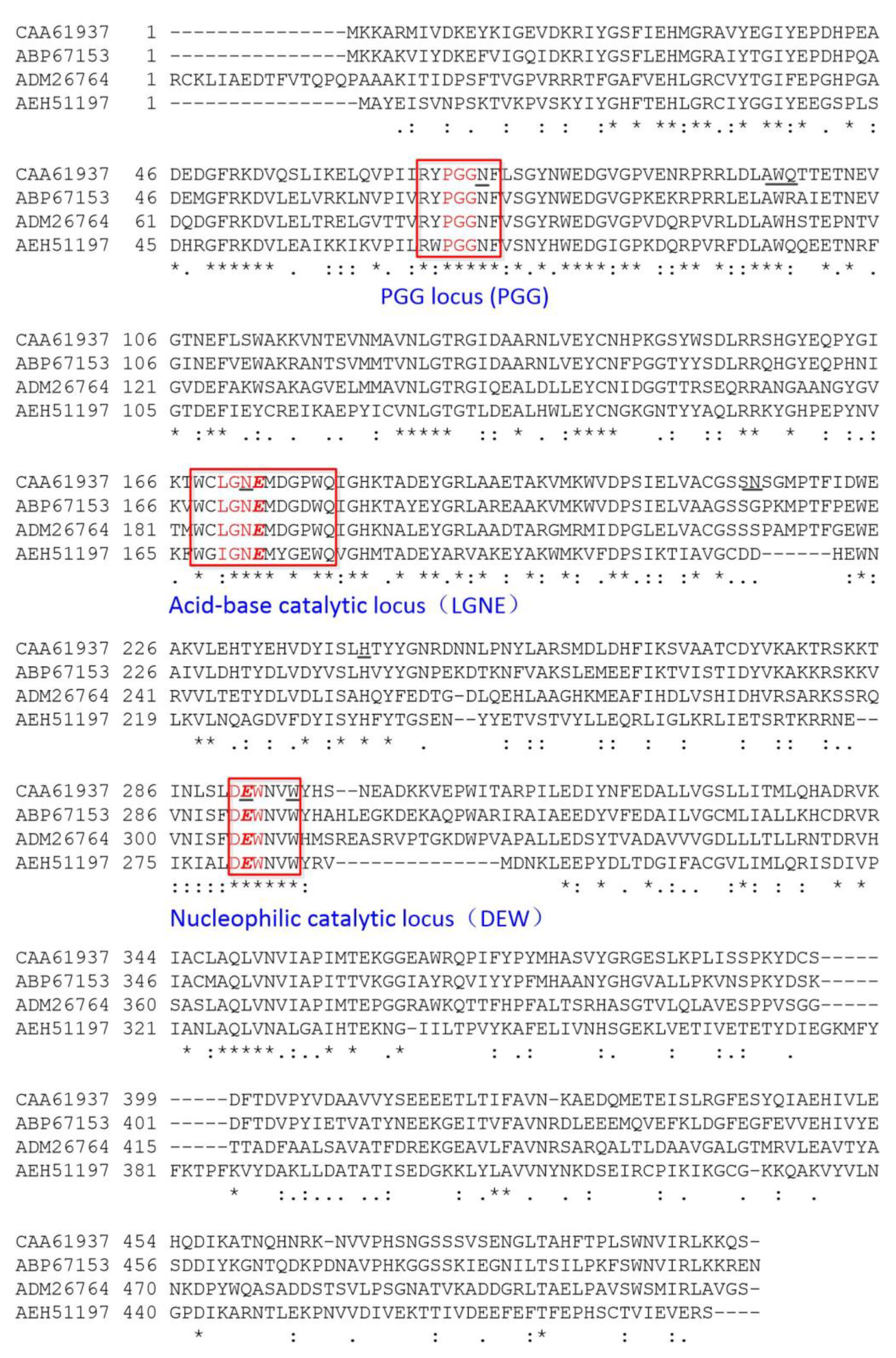
2.1. Cloning of BsAbfA Gene and Sequence Analysis

In the structural characterization of BsAbfA (GenBank accession: AL009126.3, 2938330–2939832) an alignment of the amino acid sequence of BsAbfA with several GH51 α-l-arabinofuranosidases by using ClustalX indicated the sharing of three conserved motifs. Studies have shown that arabinofuranosidases are involved in general acid-base catalysis, and two essential residues are required in the process of cleaving glycosidic bonds. In most arabinofuranosidases conservative aspartic acid and/or glutamic acid residues are available and needed [22,28]. Among the two catalytic residues, one acts as a general acid to provide proton assistance for the departure of glycosidic oxygen, and the other acts as a general base to activate a water molecule and affect the direct replacement at the anomeric center [22]. As shown in Figure 3, the first motif with a conserved RYPGG sequence is regarded as an important motif for stabilizing the structure and confers flexibility to the enzyme [29]. According to sequence similarities with BsAbfA, the motifs of WCLGNEMDGPWQ (residues 168–179) and DEWNVW (residues 291–296) are highly reserved with the GH51 α-l-arabinofuranosidases, and the residues E173 and E292 (Red bold italic letters) are regarded as typical and necessary acid-base and nucleophilic catalytic residues to hydrolyze the glycosidic bond [22,30,31], respectively. The three conserved motifs of BsAbfA are basically the same as those of AbfA (GenBank accession No. ADM26764) from R. ginsenosidimutans Gsoil 3054. This AbfA shows preferential substrate specificity for exo-polyarabinosides or oligoarabinosides, and it only hydrolyzes arabinofuranoside moieties from ginsenoside Rc and its derivatives, and no other sugar groups [15]. Between BsAbfA and the α-l-arabinofuranosidase (GenBank accession No. ABP67153) from T. thermarum DSM5069, we found only one residue difference in the three motifs. It is worth noting that they also have a high specific ability to biotransform ginsenoside Rc to Rd through the hydrolysis of the arabinoside bond [26]. Therefore, we speculate that they may share a similar catalytic mechanism.

2.2. Identification of Enzymatic Properties of Recombinant BsAbfA

The wild-type BsAbfA gene without any optimization was cloned and fused in pET-28a (+), and the results showed that expression in E. Coli BL21 (DE3) was difficult. Therefore, the codons of wild-type BsAbfA were optimized and synthesized for expression efficiency. The optimized and mutated BsAbfAs fused to His-tag were purified using Ni-NTA magnetic agarose beads from E. Coli BL21 (DE3), followed by the induction of 0.5 mM IPTG at 20 °C for 16 h. SDS-PAGE analyses revealed that the molecular masses of all the optimized and mutated BsAbfAs were similar to those of those predicted according to amino acid sequences (approximately 60 kDa, data not shown). The results indicated that the mutated BsAbfAs were correctly expressed and folded. The purified BsAbfA, with 32.6 U/mg and using p-nitrophenyl-α-l-arabinofuranoside (pNP-α-Af) as substrate, displayed a higher activity than most of the characterized α-l-arabinofuranosidases, such as α-l-arabinofuranosidase from C. saccharolyticus with 28.2 U/mg [32]. The crude enzyme and purified recombinant α-l-arabinofuranosidase from Cellulosimicrobium aquatile Lyp51 showed a lower activity of 2 U/mg and 15 U/mg, respectively [33]. The activity of T. petrophila α-l-arabinofuranosidase purified from E. Coli BL21 (DE3) toward ginsenoside Rc was only 10.3 U/mg at the optimal condition [20]. The ginsenoside-hydrolyzing α-l-arabinofuranosidase from R. ginsenosidimutans Gsoil 3054T was 14.9 U/mg [15]. To investigate the importance of the two key residues (i.e., E173 and E292) for hydrolysis of ginsenoside Rc, each one was independently substituted by an alanine, asparagine, and glutamine. It was found that the E173A or E292A mutants had no specific activity toward ginsenoside Rc and pNP-α-Af. Furthermore, the mutants of E173D, E173Q, E292D, and E292Q led to an obvious decrease of activity, and the extent of activity loss was 73.4%, 46.1%, 76.4%, and 56.1%, respectively, compared to that of BsAbfA using ginsenoside Rc as a substrate (Table 1). For investigation of the catalytic mechanism, researchers have attempted to identify the residues of α-l-arabinofuranosidase that are catalytically essential. They found that all enzymes of this superfamily possess conserved glutamates E173 and E292 (numbering for BsAbfA based on the alignment), which are catalytically essential acid-bases and nucleophiles, respectively [22,34].

2.3. Temperature and pH Dependence of Recombinant BsAbfA

To investigate the temperature and pH dependence of BsAbfA, the enzymic activity of BsAbfA and isosteric mutants E173Q and E292Q were determined using pNP-α-Af as substrate at different temperatures and pH environments. As disclosed in Figure 4A, the recombinant BsAbfA and mutants were active at 30–50 °C and relatively stable at 25–45 °C. The optimal temperature was 40 °C. When the temperature was higher than 45 °C, the activity decreased sharply. The activity loss for all the tested enzymes was more than 60%, 80%, and 90% at 50, 55, and 60 °C, respectively. It is known that many glycosidases exhibit optimal activity at mild temperature conditions. For example, the optimal temperatures of the glycosidase from Leuconostoc mesenteroides DC102 [35], β-glucosidase from Lactobacillus brevis [36], β-d-xylosidase, α-l-arabinopyranosidase, and α-l-arabinofuranosidase from Bifidobacterium breve K-110 [23,37], α-l-arabinofuranosidase from Leuconostoc sp. 22-3 [16], as well as those of soil deuteromycete Penicillium funiculosum [16] and Cellulosimicrobium aquatile Lyp51 [33] are 30–45 °C for ginsenosides biotransformation.
As shown in Figure 4B, the pH dependent curves of the activity of BsAbfA and E292Q mutants display an increase of pH sensitivity, while E173Q is characterized by a marked insensitivity to pH. Higher activities were obtained at pH 4.5‒6 for BsAbfA and E292Q mutant. In 50 mM citric acid/sodium citrate buffer, the BsAbfA and E292Q mutants showed maximal activity at pH 5, and activities of above 80% were retained at pH 6. When the pH was above 6 or below 4.5, the enzyme activity decreased significantly. Interestingly, the activity of E173Q mutant remained at a high level with the increase of pH until the pH was higher than 7. The results suggest that the mutation of E173 residue should lead to significant alteration of pH dependence. In this case, the activity of the mutant E173Q is consistent with that of a mutant enzyme that is without catalytic acid-base residue, as reported in the literature [22,38,39]. The main change of the pH dependence curve as a result of E173 mutation supports the view that just like other α-l-arabinofuranosidases, the residue is an acid-base catalyst [38,40,41]. Therefore, it is reasonable to consider that the sites of glutamate residues relative to each other, as well as to the substrate should be critical for efficient catalysis. In this study, no matter if it was pNP-α-Af or ginsenoside Rc that was used as substrate, the temperature or pH conditions for maximum enzyme activity stayed the same.
The thermal stability of BsAbfA at pH 5 versus incubation time is depicted in Figure 4C. The thermodynamic parameters confirm that BsAbfA was stable below 40 °C and it had a half-life of 225.7, 196.9, and 74.6 h at 35, 40, and 45 °C, respectively. The enzyme decreased significantly in stability above 50 °C, and the half-life was only 10.8 h. BsAbfA has a half-life comparable to those of characterized α-l-arabinofuranosidase from other species at its optimized temperature. For example, the half-life of α-l-arabinofuranosidase originating from S. solfataricus is 30 h at 85 °C [25]. Furthermore, its half-life is higher than some other β-glucosidases from Fusarium solani (159 min, 65 °C) [29] and Alteromonas sp. L82 (21 min, 40 °C) [42]. It should be emphasized that during the 3 months storage of the BsAbfA (at 4 °C), there was an activity loss of about 18%, which could be attributed to the good stability of BsAbfA during the test. It is obvious that the long half-life and appreciable thermostability of BsAbfA are properties desirable for practical applications.

2.4. Kinetic Analysis of BsAbfA

The Km, Kcat, and Kcat/Km for pNP-α-Af were determined under the optimal conditions for enzymatic reactions catalyzed by BsAbfA and mutants (Table 2). The Km, Kcat, and Kcat/Km were 0.6 mM, 108.9 s−1, and 181.5 s−1 mM−1 for BsAbfA, respectively. No activity was detected for E173A and E292A. It is noted that the Km values of the majority of other mutants, except E173A and E292A, were like those of BsAbfA, whereas the Kcat and Kcat/Km values of all the mutants decreased significantly. The E173Q mutant had a highest Kcat/Km that was only 28.8-fold lower than that of BsAbfA, and a Kcat value only 43.6-fold lower. Likewise, the Kcat values of the E173D, E292D, and E292Q mutants decrease by 217.8-, 121.1-, and 68.1-fold, while the Kcat/Km values decreased by 72.6-, 139.6-, and 78.9-fold, respectively. The Km, Kcat, and Kcat/Km values of BsAbfA for ginsenoside Rc were 0.4 mM, 79.1 s−1, and 197.8 s−1 mM−1 (data not listed). The catalytic efficiency of BsAbfA for ginsenoside Rc was higher than that of glycosidase from S. acidocaldarius for Rb1 (Kcat, 4.8 s−1 mM−1), Rc (Kcat, 4.5 s−1 mM−1), Rd (Kcat, 1 s−1 mM−1), and Rb2 (Kcat, 0.8 s−1 mM−1) [43]. The results suggest that BsAbfA is an efficient enzyme for hydrolyzing ginsenoside Rc.
The effects of metal ions and chemicals on the kinetic parameters of BsAbfA are indicated in Table 3. The BsAbfA activity was obviously enhanced by Mn2+, but significantly inhibited by Hg2+ and Cu2+. The presence of Na+, K+, Ca2+, Mg2+, Fe2+, Zn2+, Ni2+, EDTA, DDT, or SDS had no significant effect on the enzyme activity. The results demonstrate that the recombinant BsAbfA had a good catalytic activity and environmental compatibility.

2.5. Substrate Specificity of BsAbfA

The substrate specificity of BsAbfA was measured under the optimal conditions using pNP-α-Af, pNP-α-l-arabinopyranoside (pNP-α-Ap), pNP-α-l-rhamnopyranoside (pNP-α-Rp), pNP-β-d-glucopyranoside (pNP-β-Glc), Rb1, Rb2, Rc, Rd, Re, Rg1, C-Mc1, C-Mc, gentiobiose, and sophorose as substrates. As revealed in Table 4, BsAbfA displayed a high hydrolytic activity on ginsenoside Rc and pNP-α-Af, having a preference for Rc. However, BsAbfA had a low activity on C-Mc1 and C-Mc, and no activity on pNP-α-Ap, pNP-α-Rp, pNP-β-Glc, Rb1, Rb2, Rd, Re, Rg1, gentiobiose, and sophorose. Of the eight ginsenosides except for Re and Rg1, all belonged to the protopanaxadiol-type (PPD-type). In contrast, ginsenoside Rd contains only one glucopyranosyl at C20. The main difference between Rb1, Rb2, and Rc is that the sugar residues substituted at C20 of aglycone have sugar moieties (i.e., glucopyranose, arabinopyranose, and arabinofuranose) linking to the glucopyranosyl at C20 of aglycone [44]. Owing to the structure at C20 like that of Rc, the minor ginsenosides C-Mc1 and C-Mc were used as controls to test the specificity. The results suggest that BsAbfA specifically cleaves the outer glucosidic linkage at the C20 position of ginsenoside Rc, C-M, and C-M1, but does not hydrolyze the inner glucosidic linkage and glucopyranosyl at C20 of PPD-type ginsenosides. It also cannot hydrolyze any of the outer or inner sugar moieties, including glucopyranose, arabinopyranose, and rhamnopyranose at the C3 or C6 (only for the protopanaxatriol-type) position of the ginsenoside skeleton. In addition, the specific activity of BsAbfA for the ginsenosides follows the order of Rc > C-M > C-M1. Although the ginsenoside Rc, C-M, and C-M1 have the same glycosidic bond at C20 position, the recombinant BsAbfA is more active towards Rc and has specific stereo preference for C3 sugars. The C3 position of ginsenosides C-M and C-M1 contains a hydrogen or glucopyranose with small steric structures that may not benefit the formation of hydrogen bonds between Rc and BsAbfA. Therefore, BsAbfA has a high selectivity to ginsenoside Rc, and it hydrolyzes the glucoside at the C20 position in ginsenosides, whereas the enzyme does not hydrolyze the glycoside at the C3 and C6 position. That BsAbfA has the same substrate specificity to the recombinant α-l-arabinofuranosidase CaAraf51 from C. aquatile has been previously reported [33].

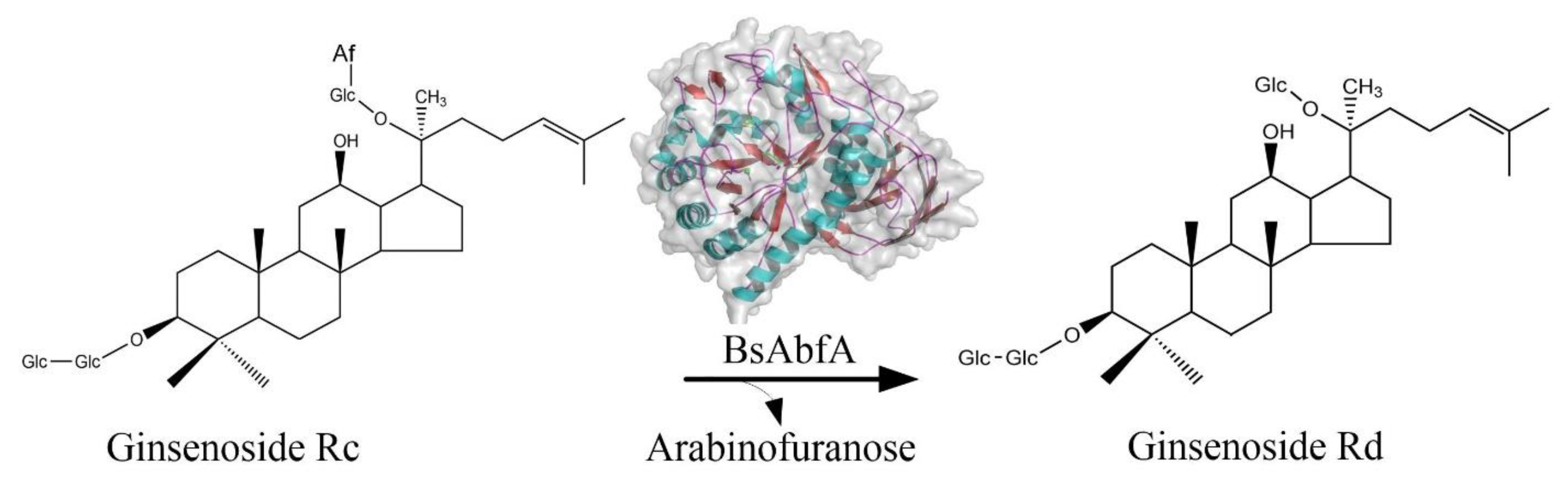
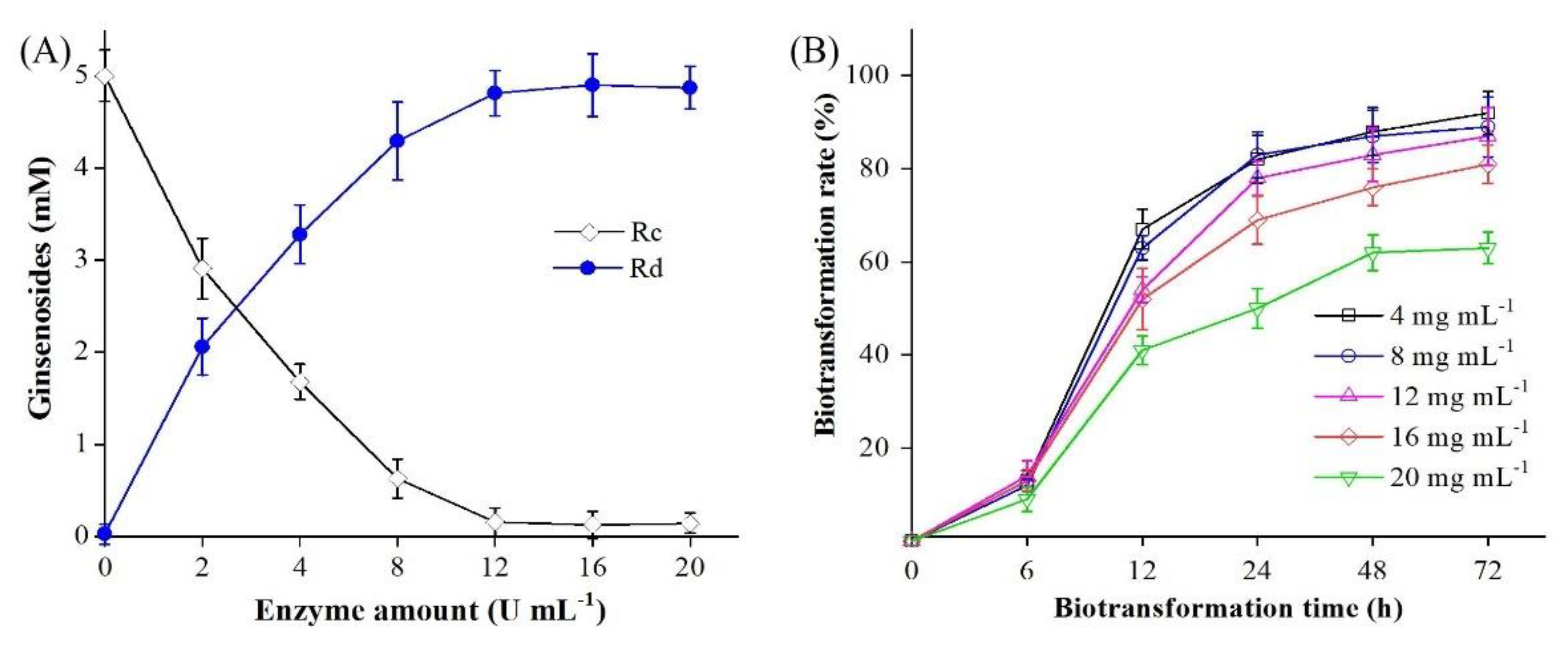
2.6. Biotransformation of Ginsenoside Rc by BsAbfA

The effect of BsAbfA on the biotransformation of ginsenoside Rc was investigated at pH 5 and 40 °C by varying the enzyme amount from 0 to 20 U/mL enzyme with 2−20 mg/mL Rc for 24 h. As shown in Figure 5A, the molar conversion rate of Rc reached 65% using 4 U/mL enzyme. At 8 U/mL enzyme, ginsenoside Rc was biotransformed to Rd with a corresponding molar conversion rate of 85%. With the increasing of enzyme activity, the productivity of ginsenoside Rd gradually increases, and at BsAbfA of 12 U/mL, the conversion rate of ginsenoside Rc reached the climax and was more than 90%. As shown in Figure 5B, 4−16 mg ginsenoside Rc/mL was efficiently converted to Rd with a more than 80% conversion rate. The conversion rate was still as high as 81% at 16 mg ginsenoside Rd/mL, but decreased obviously above 20 mg ginsenoside Rc/mL. The results indicate that BsAbfA has a good relative stability and high substrate tolerance.
To investigate the transformation mechanism, time-course analysis of the reaction under optimal conditions was performed. The biotransformed products were analyzed by LC-MS and ESI-MS/MS techniques. As shown in Figure 6A–E, there was the detection of ginsenoside Rd using UPLC after 6−48 h, and an obvious decrease of Rc and increase of Rd after 12 h. The results demonstrate that ginsenoside Rc was transformed into Rd in large amounts. At 24 h the Rd yield became much higher and Rd was the sole product of biotransformation. To further verify the transformed products, ESI-MS/MS was used for structural information (Figure 6F). The ESI-MS/MS spectrum of Rc displayed a signal from [M − H] at m/z 1077.27. For the transformed product, there was a signal at m/z 945.35 from [M − H] generated via the loss of HCOOH (46 Da) from [M + HCOO] at m/z 991.35 (Figure 6G). By comparison, the retention time and ESI-MS/MS fragment patterns of the transformed product were the same as those of the ginsenoside Rd standard. Therefore, the sole transformed product was identified as ginsenoside Rd. The BsAbfA exhibited substrate specificity for ginsenosides Rc with arabinofuranose moieties at the C20 position, indicating a specific affinity to outer C20 arabinofuranose. The proposed pathway of biotransformation ginsenoside Rc by BsAbfA was as illustrated in Figure 2.

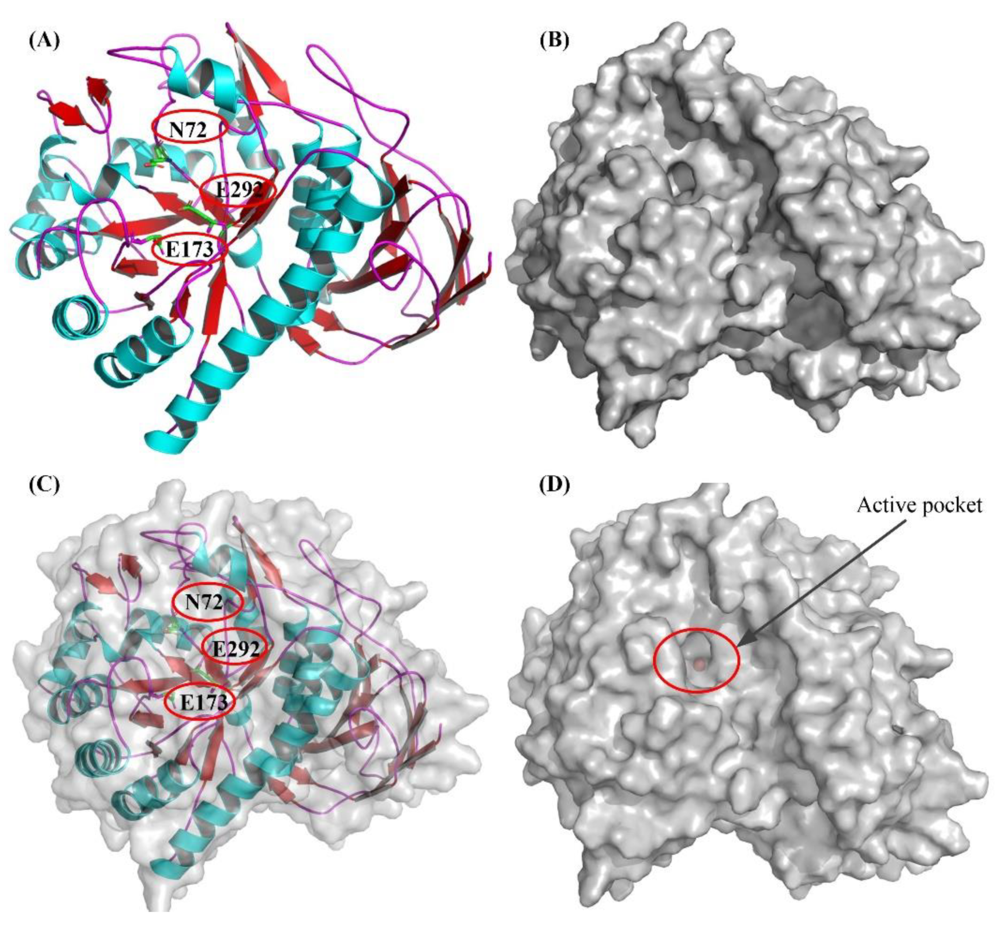
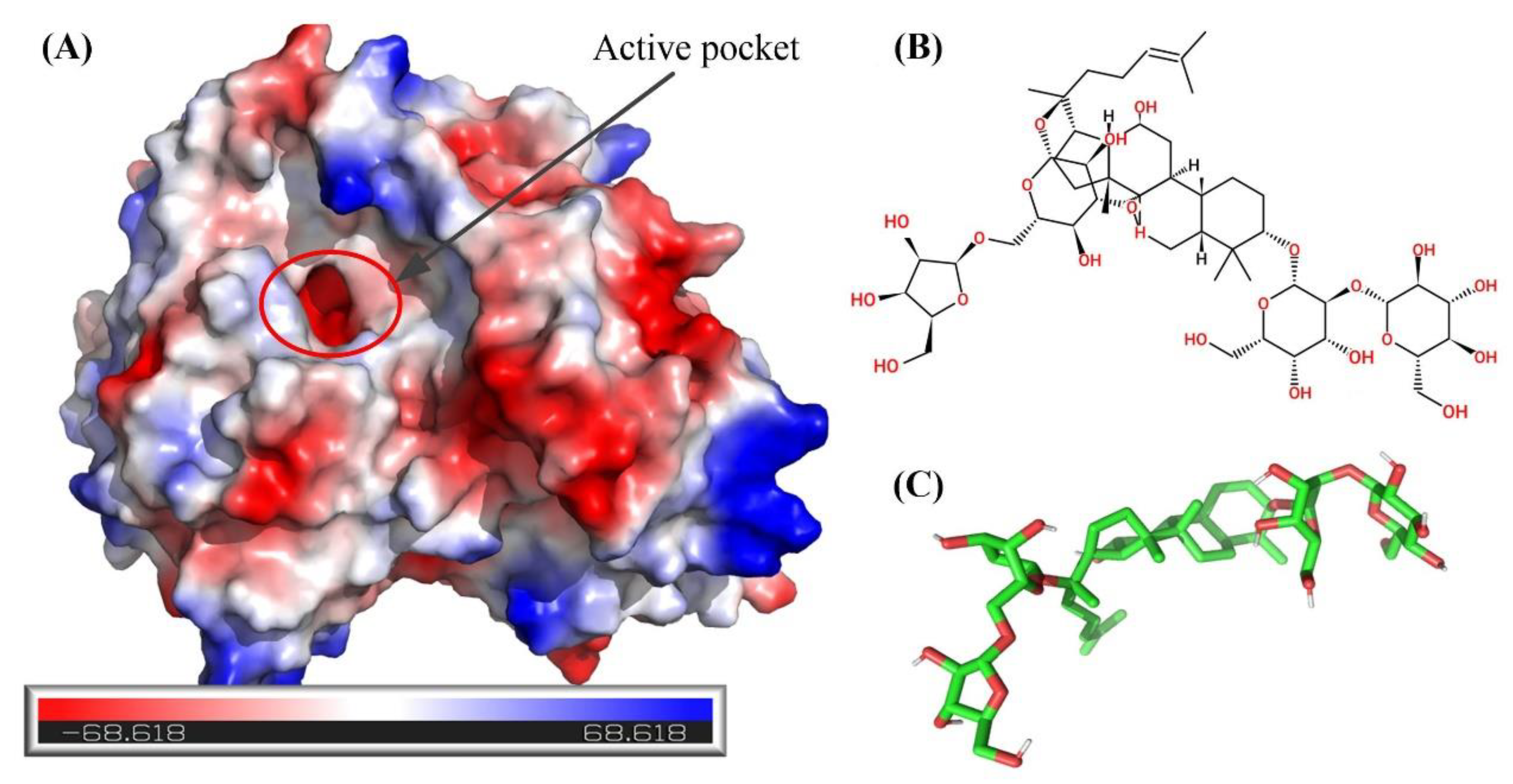
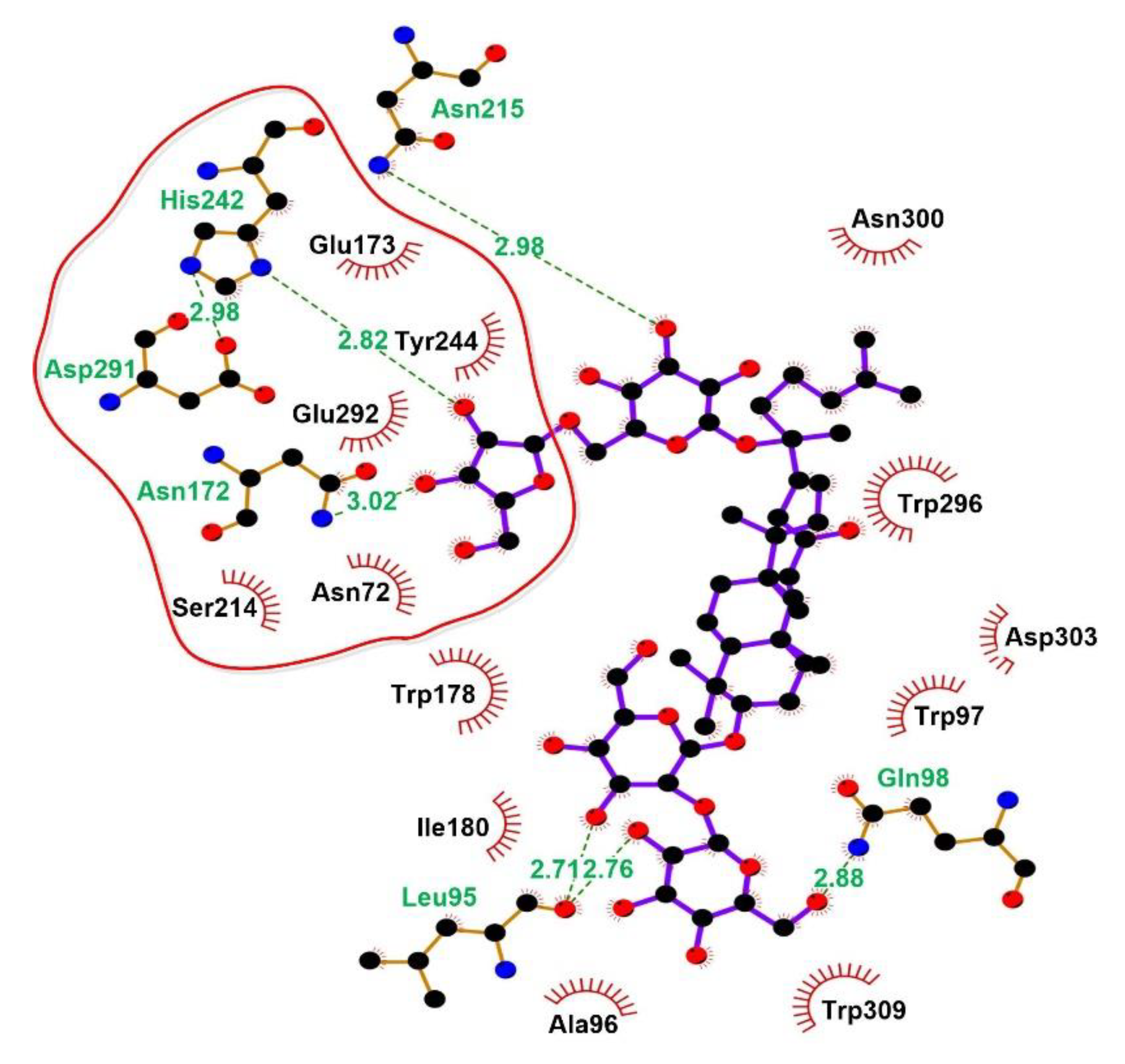
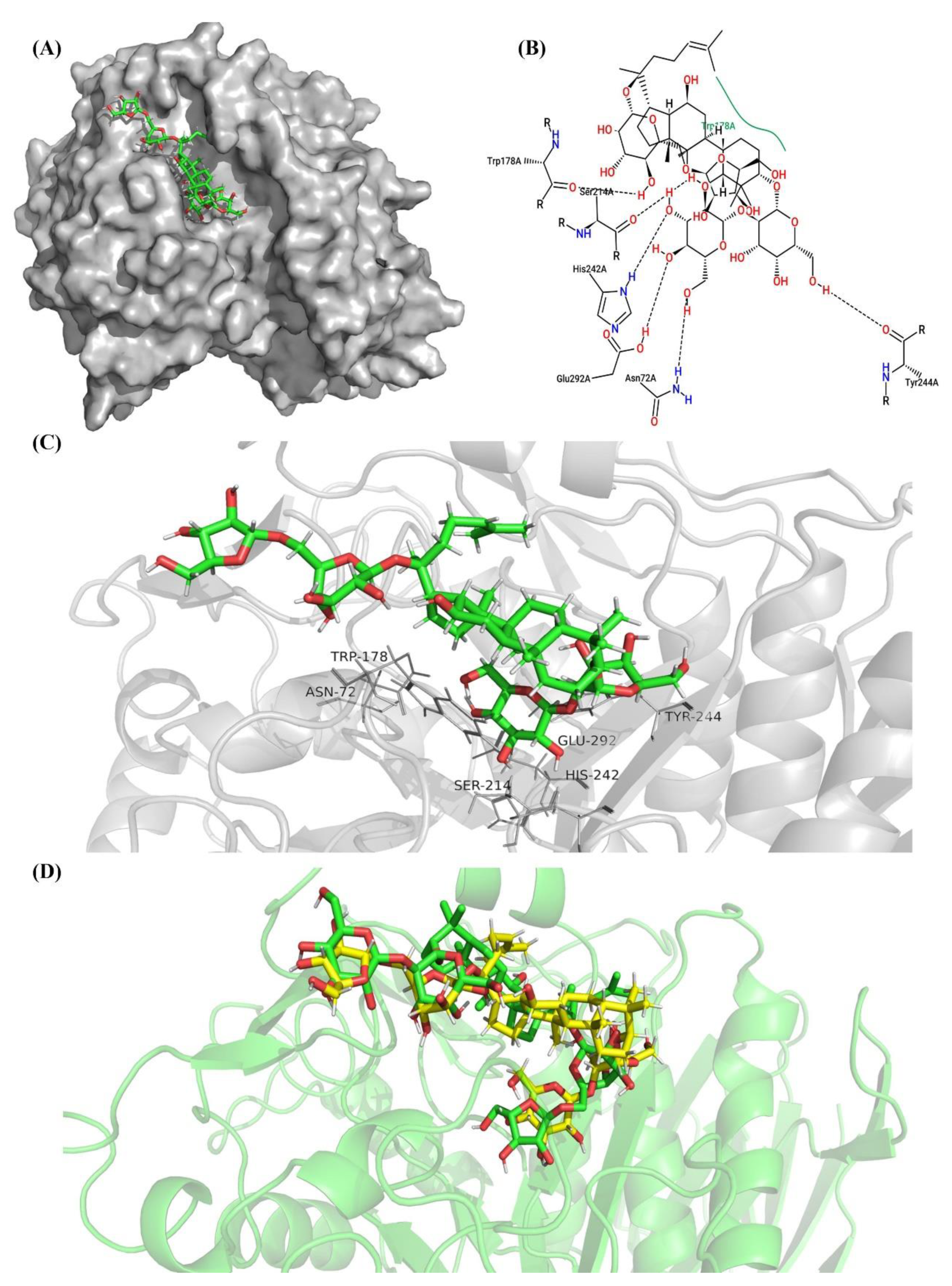
2.7. Molecular Docking and Examination of BsAbfA Active Site

From the results of the sequence analysis, it is reasonable to conclude that ginsenosides with arabinofuranose are the likely substrate of BsAbfA. Of the ginsenosides (over 100) that have been discovered, ginsenosides Rc, C-Mc1, and C-Mc contain arabinofuranoside bonds at C20 of the ginsenoside skeleton. Among them, Rc is higher in abundance, and is often used to prepare its derivatives [16,25,45]. In order to understand the molecular mechanism of the interaction between BsAbfA and ginsenoside Rc, a three-dimensional (3D) model of BsAbfA was built using alpha-l-arabinofuranosidase Ara51 (PDB code: 5O7Z) from Clostridium thermocellum [21] as a template in the SWISS-MODEL server, and the sequence identity and similarity were 64.24% and 51%, respectively. To obtain a final structural model, the collected model of BsAbfA was optimized using an energy minimization procedure followed by a short molecular dynamics simulation. As shown in Figure 7, the predicted BsAbfA structure used for docking studies with ginsenoside Rc displayed the outer part of a structural fold that contains α-helices (cyan) and loops (purple), while the inner part is almost all parallel β-sheets (red), forming a catalytically active pocket.
The surface electro-static potential plot at the active site of BsAbfA receptor (A) and the structure of ginsenoside Rc are shown in Figure 8. The electrostatic surface potential of the model was used to illustrate the potential interactions between the BsAbfA receptor and substrate ginsenoside Rc. As shown in Figure 8A, the electrostatic potential of the hydrolyzed active pocket is a deep negatively charged cavity, suggesting that the glycosidic bond and sugar ring may be attracted as a result of electrostatic interaction, which is one of the reasons for the selectivity of ginsenoside Rc (Figure 8B–C) toward the active pocket.
To understand the structural foundation of BsAbfA hydrolysis, the candidate substrate ginsenoside Rc was docked into the catalytic pocket of BsAbfA. The BsAbfA was held rigid while the substrate ginsenoside was allowed to flex during the docking process. The docking results were analyzed based on the energy and efficiency of polar and non-bonded interactions. It was found that ginsenoside Rc could easily dock into the catalytic pocket of BsAbfA and in different forms. The selected poses of ginsenoside Rc show binding energy from −8.9 to −9.8 kcal/mol, and the dissociation constant was between 6.29 × 10⁴ pM and 2.86 × 10⁵ pM (Table 5).
It is observed that the more negative the docking energy, or the smaller the dissociation constant, the better the binding ability and stability of the ligand with protein. The lowest docking score for the ligand–receptor binding indicates that the binding ability is strong. Therefore, a discussion was conducted based on the minimum and maximum docking score (cluster 1 and 5). The interactions between ginsenoside Rc and binding pocket of BsAbfA are shown in Figure 9, Figure 10 and Figure 11. As shown in Figure 9, ten important amino acid residues, viz. N72, A96, W97, Q98, N172, S214, N215, H242, E292, and W296 of BsAbfA, bind to ginsenoside Rc in the form of hydrogen bond or hydrophobic bond. Among the binding sites, five residues (i.e., N72, N172, S214, H242, and E292) form hydrogen bonds with the outer arabinofuranose moieties at C20 position, one residue (i.e., N215) binds to the inner glucopyranose moieties of C20 position, especially the hydrogen bond at N72 site is crucial to stabilizing the structure, while E173 and E292 are key residues responsible for substrate catalysis [22,29,30,31]. In this structure, N172 replaces E173 to form the hydrogen bond, while the neighbor residue E173 participates in the hydrophobic interaction with BsAbfA (Figure 10). Furthermore, the hydrophobic interaction of W97 and W296 residues could, relatively, be as important as that of hydrogen bonding. Despite the limit of space in the binding pocket for the accommodation of the fatty acid chain upon C20 insertion into the narrow hydrophilic pocket, there is binding of W97 and W296 residues to the fatty acid chain with hydrophobic bonds, squeezing the arabinofuranose into the active pocket as a consequence. There are only two residues (i.e., A96 and Q98) that bind to the glucopyranose moieties at the C3 position of ginsenoside Rc, which is at the opposite side of C20. Overall, due to the structure of the covalent glycosyl-enzyme intermediate of BsAbfA and ginsenoside Rc, the arabinofuranose cycle at C20 easily inserted into the narrow hydrophilic pocket in a stable manner. Accordingly, the C3 position of ginsenoside Rc only laid in the groove outside the binding pocket.
Further analysis of the enzyme substrate complexes by ligplot disclosed that one of the best complexes amongst the first 100 contains 19 acid residues that participate to form hydrogen bonds or hydrophobic bonds (Figure 11), and having E173 and E292 in the catalytic cleft or active site. Additionally, the different docking models and experimental results imply that the two conserved E173 and E292 residues are in close contact with the substrate to catalyze the reaction [30,31]. Site directed mutagenesis demonstrated that E173 and E292 mutations of BsAbfA reduce or disable its ability to hydrolyze ginsenoside Rc, indicating that these two amino acids are essential for recognizing hydrolysis of the outer arabinofuranosyl moieties at the C20 position. Moreover, the enzyme–substrate docking data showed conformity with the reports concerning similar Glu-based catalytic sites of α-l-arabinofuranose that emphasized the active role of E173, along with the other neighboring amino acids [22,31]. The location and function of these key residues are conservedin GH51 α-L-arabinofuranosidases [30,31], and the result is in good agreement with that of the sequence alignment in Figure 3.
In contrast, if the ginsenoside Rc docked into the BsAbfA in the manner of cluster 5 (Figure 11), there are only seven residues that can bind to ginsenoside Rc, and no bond is formed between the arabinofuranose moiety and BsAbfA. In the BsAbfA complex, the entire ginsenoside Rc can form six hydrogen bonds and one hydrophobic bond in a scattering manner. Therefore, it is less tightly bonded in comparison with the cluster 1 case. Figure 11D shows the two binding forms of ginsenoside Rc and BsAbfA, in which the green and yellow sticks stand for the binding forms of cluster 1 and cluster 5, respectively. The results indicate that arabinofuranosyl is located deeper into the active pocket than glucopyranosyl. Interestingly, according to the Kcat/Km of 197.8 s−1 mM−1, BsAbfA hydrolyzes ginsenoside Rc with higher catalytic affinity. These results suggest that the outer hexose ring at C20 position induces a steric hindrance, which makes BsAbfA protein highly selective for the recognition of pentose α-l-arabinofuranose.

3. Materials and Methods

3.1. Bacterial Strains, Plasmids, and Chemicals

Escherichia coli BL21 (DE3) used in this study was purchased from Beinuo Biotech (Shanghai, China). The plasmid pET-28a (+) used as an expression vector was purchased from GenScript (Nanjing, China). The ginsenoside standards of Rb1, Rb2, Rc, Rd, Re, Rg1, F2, C-K, C-Mc1, and C-Mc purchased from Chengdu Herbpurify (Chengdu, China) were chromatographic grade. pNP-α-Af, pNP-α-Ap, pNP-α-Rp, pNP-β-Glc, gentiobiose, and sophorose were purchased from Solarbio Science (Beijing, China). All other reagents were analytical grade.

3.2. Cloning, Site-directed Mutagenesis, Heterologous Expression, and Protein Purification

The full open reading frame (ORF) of BsAbfA gene (GenBank accession: AL009126.3, 2938330-2939832) encoding 1515 bp was synthesized by GenScript (Nanjing, China) after codon optimization [46]. The BsAbfA genes separately mutated at E173 and E292 were synthesized by GenScript using a Site-directed Mutagenesis Kit (Nanjing, China). All mutations were confirmed by DNA sequencing. The E. coli BL21 harboring BsAbfA or mutated gene was cultivated at 200 rpm in Luria–Bertani (LB) medium containing 50 μg/mL kanamycin at 37 °C for 8 h. The cultured bacterium was inoculated to the fresh LB medium to reach OD600 = 0.4–0.6. To induce BsAbfA expression, 0.5 mM IPTG, as a final concentration, was supplemented in LB medium. After the induction, the culture temperature and agitation were reduced to 20 °C and 150 rpm, respectively, and the cells were further incubated for 16 h. The induced cells were harvested by centrifugation (12,000× g, 10 min) at 4 °C and stored at 20 °C for further use. Cells were disrupted by sonication with an ultrasonic homogenizer in 50 mM citric acid/sodium citrate buffer (pH 5.5) with 1 g/L lysozyme, as well as EDTA-free protease inhibitor cocktail and 2 mg/L DNase. The debris were removed by centrifugation at 8000× g for 20 min at 4 °C. The resulting supernatants were subjected to filtration through a membrane of 0.45 μm. The filtrate was loaded onto Ni-NTA magnetic agarose beads (Qiagen, Germany) for the enrichment of the recombinant BsAbfA protein carrying His-tag. The supernatant was removed by a magnetic separator and washed at least twice with elution buffer. Purified proteins were maintained in 50 mM citric acid/sodium citrate buffer (pH 5) at 4 °C. The concentration of purified recombinant BsAbfA was assayed using Folin-phenol reagent [47]. The expression quantity and molecular weight of the protein were analyzed by SDS-PAGE.

3.3. Determination of Kinetic Parameters and Substrate Specificity

In order to determine the enzyme properties of the recombinant BsAbfA, p-Nitrophenyl α-l-arabinofuranoside (pNP-α-Af) was used as a substrate to assay the enzymatic activities in 50 mM citric acid/sodium citrate buffer (pH 5) at 40 °C. The reaction was ceased by adding 500 mM sodium carbonate with volume equal to that of the reaction. The released p-nitrophenol (pNP) was immediately measured at 405 nm. One unit (U) of the BsAbfA activity was defined as the amount of enzyme required to generate 1 μmol pNP per minute [32]. When ginsenoside Rc was used as a substrate, the reaction was ceased by adding n-butanol with a volume 2-fold that of the reaction. The products were assayed by UPLC. The kinetic parameters of BsAbfA were measured using pNP-Af and Rc as substrate at concentrations ranging from 0.1 to 5 mM. The Km, Kcat, and Vmax were calculated by fitting the activity data to a linear regression on Lineweaver–Burk double-reciprocal plots [47]. The substrate specificity of purified recombinant BsAbfA was assayed by using pNP-α-Af, pNP-α-Ap, pNP-α-Rp, pNP-β-Glc, ginsenoside Rb1, Rb2, Rc, Rd, Re, Rg1, F2, C-K, C-Mc1, C-Mc, gentiobiose, and sophorose as substrates, individually. All assays were performed in triplicate.

3.4. Effects of pH, Temperature, Metal Ions, and Chemicals on Stability

The effects of pH, temperature, metal ions, and chemicals on BsAbfA activity were investigated using pNP-Af as substrate. The buffers used were as follows: citric acid-sodium citrate buffer (50 mM, pH 3–6), sodium phosphate buffer (100 mM, pH 6–8), and glycine-NaOH (50 mM, pH 9–10). The temperature was set ranging from 25 to 70 °C. The relative activity of BsAbfA was defined as 100% at either the optimal pH or optimal temperature. The effects of metal ions and chemicals on the activity of BsAbfA were assessed in the presence of NaCl, KCl, CaCl2, MgCl2, FeCl2, MnCl2, ZnCl2, NiCl2, CuCl2, HgCl2, EDTA, DDT, and SDS at optimal pH buffer and temperature. All assays were performed in triplicate.

3.5. Biotransformation of Ginsenoside Rc

To investigate the catalytic ability of recombinant BsAbfA for the biotransformation of ginsenoside Rc, as well as to disclose the reaction pathway, ginsenoside Rc was dissolved in methanol and incubated in 50 mM citric acid/sodium citrate buffer (pH 5) containing 5 mM Rc and 12 U/mL enzyme at 40 °C. The reaction was sampled at regular intervals for a certain period and ceased by heating the mixture to 80 °C for 15 min. The biotransformed products were subsequently extracted with H2O-saturated n-butanol. After evaporation of solvents, the products were dissolved in methanol and then subjected to filtration using 0.45 μm microfiltration membranes [48]. UPLC analysis was performed with Shimadzu LC-MS 8050 with a ACQUITY UPLC BEH Shield RP18 column (1.7 μm, 2.1 mm × 50 mm). The mobile phase consisted of acetonitrile (A) and 1% formic acid (B) and the elute program was as follows: A:B (10:90) to (25:75) for 2 min; A:B (25:75) for 2‒8 min; A:B (25:75) to (45:55) for 8‒16.5 min; A:B (45:55) for 16.5‒21.5 min; A:B (45:55) to (98:2) for 21.5‒21.6 min; A:B (98:2) for 21.6‒25 min; A:B (98:2) to (10:90) for 25‒25.1 min A:B (10:90) for 25.1‒29 min. Rc, Rd, Re, F2, and C-K were used as standards. The samples were analyzed using a triple quadrupole mass spectrometer equipped with electrospray ionization source. LC-MS analyses were acquired in the negative ion mode by full scan. High purity nitrogen was used as a drying gas, the nebulizing gas flow and heating gas flow were 2 L/min and 10 L/min at 4000 V of ionspray voltage, respectively. The atomizing temperature was 300 °C [49].

3.6. Homology Modeling and Molecular Docking

3.6.1. Homology Modeling

The template crystal structure for BsAbfA was identified through BLAST [50] and downloaded from the RCSB Protein Data Bank (PDB code: 5O7Z) [51]. Homology modeling was conducted in the Swiss-model [52]. The target sequence was searched with BLAST against the primary amino acid sequence contained in the SMTL [53]. Models were built based on a target–template alignment using ProMod3 [54]. Coordinates which were conserved between the target and the template were copied from the template to the model. Insertions and deletions were remodeled using a fragment library. Side chains were then rebuilt. Finally, the geometry of the resulting model was regularized by using a force field.

3.6.2. Molecular Docking

AutoDock Vina [55] was employed for molecular docking of BsAbfA with candidate substrate ginsenoside Rc for the prediction of binding affinity and binding sites. The structure of ginsenoside Rc was acquired from PubChem [56] (ID:12855889) and converted to 3D by Avogadro [57] through energy minimization. Then, a conformation search was performed to confirm the stable geometry for the docking preprocessing. As reported in [21], the binding site was located in a beta sheet barrel surrounded by an alpha helix. The three glutamine residues were participants of the hydrolase reaction with a docking box defined at the center of a barrel (50 × 50 × 50 in size). All docked poses of ginsenoside Rc were ranked by binding energy, and the threshold of cluster analysis was set at 5 angstroms. Other parameters not mentioned were set as default.

4. Conclusions

The ginsenoside Rc-hydrolyzing α-l-arabinofuranosidase gene BsAbfA was cloned from B. subtilis, and the optimized recombinant protein was overexpressed and characterized successfully in E. coli. The enzymatic properties of BsAbfA are unique and superior to the other reported α-l-arabinofuranosidases, exhibiting higher catalytic efficiency and higher tolerance to metal ions, as well as to organic solvents and detergents. Additionally, BsAbfA shows a high selectivity to cleave the outer arabinofuranosyl moieties at C20 of ginsenoside Rc, catalyzing the conversion of ginsenoside Rc to the more pharmacologically active Rd with high productivity. The 3D structure of BsAbfA of family 51, glycosidase modeled by comparative modeling was compact and stable. The docking results revealed that the active site of BsAbfA can accommodate ginsenoside Rc very well. Site-directed mutagenesis of the E173 and E292 residues confirms that it is important to recognize ginsenoside Rc effectively and make it enter the active pocket for the hydrolysis of the outer arabinofuranose moieties at C20 position. Thus, we report the BsAbfA as a promising enzyme, and efficient for the industrial production of ginsenoside Rd.

Author Contributions

Conceptualization, R.Z.; Methodology, S.Q.T., B.L.Z., Z.Y.G. and Z.Y.L.; Validation, S.Q.T., L.Y.T. and R.Z.; Data Analysis: R.Z. and B.L.Z.; Investigation, S.Q.T. B.L.Z., Z.Y.G., L.Y.T. and P.W.; Writing—review and editing: R.Z. and B.L.Z.; Supervision: R.Z. and B.L.Z.; Funding acquisition, R.Z. and Z.Y.L.; All authors have read and agreed to the published version of the manuscript.

Funding

The study was supported by the National Natural Science Foundation of China (81874332, 81973710), the Natural Science Foundation of Hunan Province (2020JJ2012), the Scientific Research Fund of Hunan Provincial Education Department (18B387), and the Postdoctoral Science Foundation of China (2017T100601, 2016M590746).

Data Availability Statement

All data supporting the findings of this study are available in the main text.

Acknowledgments

The critical reading of the manuscript by C.T. Au, College of Materials and Chemical Engineering, Hunan Institute of Engineering is greatly appreciated.

Conflicts of Interest

The authors declare no conflict of interest.

Sample Availability

Samples of the compounds are available from the corresponding authors.

References

  1. Kim, J.H.; Oh, J.M.; Chun, S.; Park, H.Y.; Im, W.T. Enzymatic biotransformation of ginsenoside Rb2 into Rd by recombinant alpha-l-arabinopyranosidase from Blastococcus saxobsidens. J. Microbiol. Biotechnol. 2020, 30, 391–397. [Google Scholar] [CrossRef] [PubMed]
  2. Fan, S.; Zhang, Z.; Su, H.; Xu, P.; Qi, H.; Zhao, D.; Li, X. Panax ginseng clinical trials: Current status and future perspectives. Biomed. Pharmacother. 2020, 132, 110832. [Google Scholar] [CrossRef] [PubMed]
  3. Lee, J.O.; Choi, E.; Shin, K.K.; Hong, Y.H.; Kim, H.G.; Jeong, D.; Hossain, M.A.; Kim, H.S.; Yi, Y.S.; Kim, D.; et al. Compound K, a ginsenoside metabolite, plays an antiinflammatory role in macrophages by targeting the AKT1-mediated signaling pathway. J. Ginseng Res. 2019, 43, 154–160. [Google Scholar] [CrossRef] [PubMed]
  4. Xue, Q.; He, N.; Wang, Z.; Fu, X.; Aung, L.H.H.; Liu, Y.; Li, M.; Cho, J.Y.; Yang, Y.; Yu, T. Functional roles and mechanisms of ginsenosides from Panax ginseng in atherosclerosis. J. Ginseng Res. 2021, 45, 22–31. [Google Scholar] [CrossRef] [PubMed]
  5. Shin, B.K.; Kwon, S.W.; Park, J.H. Chemical diversity of ginseng saponins from Panax ginseng. J. Ginseng Res. 2015, 39, 287–298. [Google Scholar] [CrossRef] [PubMed] [Green Version]
  6. Chen, W.; Balan, P.; Popovich, D.G. Ginsenosides analysis of New Zealand-grown forest Panax ginseng by LC-QTOF-MS/MS. J. Ginseng Res. 2020, 44, 552–562. [Google Scholar] [CrossRef] [PubMed]
  7. Sun, B.S.; Gu, L.J.; Fang, Z.M.; Wang, C.Y.; Wang, Z.; Lee, M.R.; Li, Z.; Li, J.J.; Sung, C.K. Simultaneous quantification of 19 ginsenosides in black ginseng developed from Panax ginseng by HPLC-ELSD. J. Pharm. Biomed. Anal. 2009, 50, 15–22. [Google Scholar] [CrossRef]
  8. Zhang, F.; Tang, S.; Zhao, L.; Yang, X.; Yao, Y.; Hou, Z.; Xue, P. Stem-leaves of Panax as a rich and sustainable source of less-polar ginsenosides: Comparison of ginsenosides from Panax ginseng, American ginseng and Panax notoginseng prepared by heating and acid treatment. J. Ginseng Res. 2021, 45, 163–175. [Google Scholar] [CrossRef]
  9. Liu, G.M.; Lu, T.C.; Sun, M.L.; Jia, W.Y.; Ji, X.; Luo, Y.G. Ginsenoside Rd inhibits glioblastoma cell proliferation by up-regulating the expression of miR-144-5p. Biol. Pharm. Bull. 2020, 43, 1534–1541. [Google Scholar] [CrossRef]
  10. Wang, P.; Du, X.; Xiong, M.; Cui, J.; Yang, Q.; Wang, W.; Chen, Y.; Zhang, T. Ginsenoside Rd attenuates breast cancer metastasis implicating derepressing microRNA-18a-regulated Smad2 expression. Sci. Rep. 2016, 6, 33709. [Google Scholar] [CrossRef] [Green Version]
  11. Yang, N.; Liang, G.; Lin, J.; Zhang, S.; Lin, Q.; Ji, X.; Chen, H.; Li, N.; Jin, S. Ginsenoside Rd therapy improves histological and functional recovery in a rat model of inflammatory bowel disease. Phytother. Res. 2020, 34, 3019–3028. [Google Scholar] [CrossRef] [PubMed]
  12. Zhang, X.; Liu, X.; Hu, G.; Zhang, G.; Zhao, G.; Shi, M. Ginsenoside Rd attenuates blood-brain barrier damage by suppressing proteasome-mediated signaling after transient forebrain ischemia. Neuroreport 2020, 31, 466–472. [Google Scholar] [CrossRef] [PubMed]
  13. Ye, R.; Zhang, X.; Kong, X.; Han, J.; Yang, Q.; Zhang, Y.; Chen, Y.; Li, P.; Liu, J.; Shi, M.; et al. Ginsenoside Rd attenuates mitochondrial dysfunction and sequential apoptosis after transient focal ischemia. Neuroscience 2011, 178, 169–180. [Google Scholar] [CrossRef] [PubMed]
  14. Anufriev, V.P.; Malinovskaya, G.V.; Denisenko, V.A.; Uvarova, N.I.; Elyakov, G.B.; Kim, S.I.; Baek, N.I. Synthesis of ginsenoside Rg3, a minor constituent of Ginseng radix. Carbohydr. Res. 1997, 304, 179–182. [Google Scholar] [CrossRef]
  15. An, D.S.; Cui, C.H.; Sung, B.H.; Yang, H.C.; Kim, S.C.; Lee, S.T.; Im, W.T.; Kim, S.G. Characterization of a novel ginsenoside-hydrolyzing alpha-l-arabinofuranosidase, AbfA, from Rhodanobacter ginsenosidimutans Gsoil 3054T. Appl. Microbiol. Biotechnol. 2012, 94, 673–682. [Google Scholar] [CrossRef] [PubMed]
  16. Liu, Q.M.; Jung, H.M.; Cui, C.H.; Sung, B.H.; Kim, J.K.; Kim, S.G.; Lee, S.T.; Kim, S.C.; Im, W.T. Bioconversion of ginsenoside Rc into Rd by a novel alpha-l-arabinofuranosidase, Abf22-3 from Leuconostoc sp. 22-3: Cloning, expression, and enzyme characterization. Antonie Leeuwenhoek 2013, 103, 747–754. [Google Scholar]
  17. Renchinkhand, G.; Cho, S.H.; Park, Y.W.; Song, G.Y.; Nam, M.S. Biotransformation of major ginsenoside Rb1 to Rd by Dekkera anomala YAE-1 from Mongolian fermented milk (Airag). J. Microbiol. Biotechnol. 2020, 30, 1536–1542. [Google Scholar] [CrossRef]
  18. Zhong, F.L.; Ma, R.; Jiang, M.; Dong, W.W.; Jiang, J.; Wu, S.; Li, D.; Quan, L.H. Cloning and characterization of ginsenoside-hydrolyzing beta-glucosidase from Lactobacillus brevis that transforms ginsenosides Rb1 and F2 into ginsenoside Rd and compound K. J. Microbiol. Biotechnol. 2016, 26, 1661–1667. [Google Scholar] [CrossRef] [PubMed]
  19. Wang, C.Z.; Zhang, B.; Song, W.X.; Wang, A.; Ni, M.; Luo, X.; Aung, H.H.; Xie, J.T.; Tong, R.; He, T.C.; et al. Steamed American ginseng berry: Ginsenoside analyses and anticancer activities. J. Agric. Food Chem. 2006, 54, 9936–9942. [Google Scholar] [CrossRef]
  20. Kim, T.H.; Yang, E.J.; Shin, K.C.; Hwang, K.H.; Park, J.S.; Oh, D.K. Enhanced production of β-D-glycosidase and α-l-arabinofuranosidase in recombinant Escherichia coli in fed-batch culture for the biotransformation of ginseng leaf extract to ginsenoside compound K. Biotechnol. Bioproc. E 2018, 23, 183–193. [Google Scholar] [CrossRef]
  21. Taylor, E.J.; Smith, N.L.; Turkenburg, J.P.; D’Souza, S.; Gilbert, H.J.; Davies, G.J. Structural insight into the ligand specificity of a thermostable family 51 arabinofuranosidase, Araf51, from Clostridium thermocellum. Biochem. J. 2006, 395, 31–37. [Google Scholar] [CrossRef] [PubMed] [Green Version]
  22. Debeche, T.; Bliard, C.; Debeire, P.; O’Donohue, M.J. Probing the catalytically essential residues of the alpha-l-arabinofuranosidase from Thermobacillus xylanilyticus. Protein Eng. 2002, 15, 21–28. [Google Scholar] [CrossRef] [Green Version]
  23. Shin, H.Y.; Park, S.Y.; Sung, J.H.; Kim, D.H. Purification and characterization of alpha-l-arabinopyranosidase and alpha-l-arabinofuranosidase from Bifidobacterium breve K-110, a human intestinal anaerobic bacterium metabolizing ginsenoside Rb2 and Rc. Appl. Environ. Microbiol. 2003, 69, 7116–7123. [Google Scholar] [CrossRef] [PubMed] [Green Version]
  24. Shin, K.C.; Lee, G.W.; Oh, D.K. Production of ginsenoside Rd from ginsenoside Rc by alpha-l-arabinofuranosidase from Caldicellulosiruptor saccharolyticus. J. Microbiol. Biotechnol. 2013, 23, 483–488. [Google Scholar] [CrossRef] [PubMed] [Green Version]
  25. Choi, J.H.; Shin, K.C.; Oh, D.K. An L213A variant of beta-glycosidase from Sulfolobus solfataricus with increased alpha-l-arabinofuranosidase activity converts ginsenoside Rc to compound K. PLoS ONE 2018, 13, e0191018. [Google Scholar]
  26. Xie, J.; Zhao, D.; Zhao, L.; Pei, J.; Xiao, W.; Ding, G.; Wang, Z.; Xu, J. Characterization of a novel arabinose-tolerant alpha-l-arabinofuranosidase with high ginsenoside Rc to ginsenoside Rd bioconversion productivity. J. Appl. Microbiol. 2016, 120, 647–660. [Google Scholar] [CrossRef]
  27. Canakci, S.; Belduz, A.O.; Saha, B.C.; Yasar, A.; Ayaz, F.A.; Yayli, N. Purification and characterization of a highly thermostable alpha-l-arabinofuranosidase from Geobacillus caldoxylolyticus TK4. Appl. Microbiol. Biotechnol. 2007, 75, 813–820. [Google Scholar] [CrossRef]
  28. Pitson, S.M.; Voragen, A.G.; Beldman, G. Stereochemical course of hydrolysis catalyzed by arabinofuranosyl hydrolases. FEBS Lett. 1996, 398, 7–11. [Google Scholar] [CrossRef] [Green Version]
  29. Pazos, F.; Heredia, P.; Valencia, A.; de las Rivas, J. Threading structural model of the manganese-stabilizing protein PsbO reveals presence of two possible beta-sandwich domains. Protein Struct. Funct. Genet. 2001, 45, 372–381. [Google Scholar] [CrossRef]
  30. Shallom, D.; Belakhov, V.; Solomon, D.; Shoham, G.; Baasov, T.; Shoham, Y. Detailed kinetic analysis and identification of the nucleophile in alpha-l-arabinofuranosidase from Geobacillus stearothermophilus T-6, a family 51 glycoside hydrolase. J. Biol. Chem. 2002, 277, 43667–43673. [Google Scholar] [CrossRef] [PubMed] [Green Version]
  31. Shallom, D.; Belakhov, V.; Solomon, D.; Gilead-Gropper, S.; Baasov, T.; Shoham, G.; Shoham, Y. The identification of the acid-base catalyst of alpha-arabinofuranosidase from Geobacillus stearothermophilus T-6, a family 51 glycoside hydrolase. FEBS Lett. 2002, 514, 163–167. [Google Scholar] [CrossRef] [Green Version]
  32. Lim, Y.R.; Yoon, R.Y.; Seo, E.S.; Kim, Y.S.; Park, C.S.; Oh, D.K. Hydrolytic properties of a thermostable alpha-l-arabinofuranosidase from Caldicellulosiruptor saccharolyticus. J. Appl. Microbiol. 2010, 109, 1188–1197. [Google Scholar] [CrossRef]
  33. Zuo, S.S.; Wang, Y.C.; Zhu, L.; Zhao, J.Y.; Li, M.G.; Han, X.L.; Wen, M.L. Cloning and characterization of a ginsenoside-hydrolyzing alpha-l-arabinofuranosidase, CaAraf51, from Cellulosimicrobium aquatile Lyp51. Curr. Microbiol. 2020, 77, 2783–2791. [Google Scholar] [CrossRef]
  34. Zverlov, V.V.; Liebl, W.; Bachleitner, M.; Schwarz, W.H. Nucleotide sequence of arfB of Clostridium stercorarium, and prediction of catalytic residues of alpha-l-arabinofuranosidases based on local similarity with several families of glycosyl hydrolases. FEMS Microbiol. Lett. 1998, 164, 337–343. [Google Scholar]
  35. Quan, L.H.; Piao, J.Y.; Min, J.W.; Kim, H.B.; Kim, S.R.; Yang, D.U.; Yang, D.C. Biotransformation of ginsenoside Rb1 to prosapogenins, gypenoside XVII, ginsenoside Rd, ginsenoside F2, and compound K by Leuconostoc mesenteroides DC102. J. Ginseng Res. 2011, 35, 344–351. [Google Scholar] [CrossRef] [Green Version]
  36. Zhong, F.L.; Dong, W.W.; Wu, S.; Jiang, J.; Yang, D.C.; Li, D.; Quan, L.H. Biotransformation of gypenoside XVII to compound K by a recombinant beta-glucosidase. Biotechnol. Lett. 2016, 38, 1187–1193. [Google Scholar] [CrossRef]
  37. Shin, H.Y.; Lee, J.H.; Lee, J.Y.; Han, Y.O.; Han, M.J.; Kim, D.H. Purification and characterization of ginsenoside Ra-hydrolyzing beta-d-xylosidase from Bifidobacterium breve K-110, a human intestinal anaerobic bacterium. Biol. Pharm. Bull. 2003, 26, 1170–1173. [Google Scholar] [CrossRef] [PubMed] [Green Version]
  38. Zechel, D.L.; Withers, S.G. Glycosidase mechanisms: Anatomy of a finely tuned catalyst. Acc. Chem. Res. 2000, 33, 11–18. [Google Scholar] [PubMed]
  39. Moracci, M.; Capalbo, L.; Ciaramella, M.; Rossi, M. Identification of two glutamic acid residues essential for catalysis in the beta-glycosidase from the thermoacidophilic archaeon Sulfolobus solfataricus. Protein Eng. 1996, 9, 1191–1195. [Google Scholar] [CrossRef] [Green Version]
  40. Wang, Q.; Trimbur, D.; Graham, R.; Warren, R.A.; Withers, S.G. Identification of the acid/base catalyst in Agrobacterium faecalis beta-glucosidase by kinetic analysis of mutants. Biochemistry (U. S.) 1995, 34, 14554–14562. [Google Scholar] [CrossRef] [PubMed]
  41. Moracci, M.; Trincone, A.; Perugino, G.; Ciaramella, M.; Rossi, M. Restoration of the activity of active-site mutants of the hyperthermophilic beta-glycosidase from Sulfolobus solfataricus: Dependence of the mechanism on the action of external nucleophiles. Biochemistry (U. S.) 1998, 37, 17262–17270. [Google Scholar] [CrossRef]
  42. Sun, J.; Wang, W.; Yao, C.; Dai, F.; Zhu, X.; Liu, J.; Hao, J. Overexpression and characterization of a novel cold-adapted and salt-tolerant GH1 beta-glucosidase from the marine bacterium Alteromonas sp. L82. J. Microbiol. 2018, 56, 656–664. [Google Scholar] [CrossRef] [PubMed]
  43. Noh, K.H.; Oh, D.K. Production of the rare ginsenosides compound K, compound Y, and compound Mc by a thermostable beta-glycosidase from Sulfolobus acidocaldarius. Biol. Pharm. Bull. 2009, 32, 1830–1835. [Google Scholar] [CrossRef] [Green Version]
  44. Cao, H.; Nuruzzaman, M.; Xiu, H.; Huang, J.; Wu, K.; Chen, X.; Li, J.; Wang, L.; Jeong, J.H.; Park, S.J.; et al. Transcriptome analysis of methyl jasmonate-elicited Panax ginseng adventitious roots to discover putative ginsenoside biosynthesis and transport genes. Int. J. Mol. Sci. 2015, 16, 3035–3057. [Google Scholar] [CrossRef] [Green Version]
  45. Upadhyaya, J.; Yoon, M.S.; Kim, M.J.; Ryu, N.S.; Song, Y.E.; Kim, Y.H.; Kim, M.K. Purification and characterization of a novel ginsenoside Rc-hydrolyzing beta-glucosidase from Armillaria mellea mycelia. AMB Express. 2016, 6, 112. [Google Scholar] [CrossRef] [PubMed] [Green Version]
  46. Morales-Quintana, L.; Beltran, D.; Mendez-Yanez, A.; Valenzuela-Riffo, F.; Herrera, R.; Moya-Leon, M.A. Characterization of FcXTH2, a novel xyloglucan endotransglycosylase/hydrolase enzyme of Chilean strawberry with hydrolase activity. Int. J. Mol. Sci. 2020, 21, 3380. [Google Scholar] [CrossRef]
  47. Zhang, R.; Huang, X.M.; Yan, H.J.; Liu, X.Y.; Zhou, Q.; Luo, Z.Y.; Tan, X.N.; Zhang, B.L. Highly selective production of compound K from ginsenoside Rd by hydrolyzing glucose at C-3 glycoside using beta-glucosidase of Bifidobacterium breve ATCC 15700. J. Microbiol. Biotechn. 2019, 29, 410–418. [Google Scholar] [CrossRef] [Green Version]
  48. Ji, Q.C.; Harkey, M.R.; Henderson, G.L.; Gershwin, M.E.; Stern, J.S.; Hackman, R.M. Quantitative determination of ginsenosides by high-performance liquid chromatography-tandem mass spectrometry. Phytochem. Anal. 2001, 12, 320–326. [Google Scholar] [CrossRef] [PubMed]
  49. Liang, W.; Wang, S.; Yao, L.; Wang, J.; Gao, W. Quality evaluation of Panax ginseng adventitious roots based on ginsenoside constituents, functional genes, and ferric-reducing antioxidant power. J. Food Biochem. 2019, 43, e12901. [Google Scholar] [CrossRef]
  50. Camacho, C.; Coulouris, G.; Avagyan, V.; Ma, N.; Papadopoulos, J.; Bealer, K.; Madden, T.L. BLAST+: Architecture and applications. BMC Bioinform. 2009, 10, 421. [Google Scholar] [CrossRef] [PubMed] [Green Version]
  51. Berman, H.M.; Westbrook, J.; Feng, Z.; Gilliland, G.; Bhat, T.N.; Weissig, H.; Shindyalov, I.N.; Bourne, P.E. The Protein Data Bank. Nucleic Acids Res. 2000, 28, 235–242. [Google Scholar] [CrossRef] [PubMed] [Green Version]
  52. Waterhouse, A.; Bertoni, M.; Bienert, S.; Studer, G.; Tauriello, G.; Gumienny, R.; Heer, F.T.; de Beer, T.A.P.; Rempfer, C.; Bordoli, L.; et al. SWISS-MODEL: Homology modelling of protein structures and complexes. Nucleic Acids Res. 2018, 46, W296–W303. [Google Scholar] [CrossRef] [PubMed] [Green Version]
  53. Bienert, S.; Waterhouse, A.; de Beer, T.A.; Tauriello, G.; Studer, G.; Bordoli, L.; Schwede, T. The SWISS-MODEL Repository-new features and functionality. Nucleic Acids Res. 2017, 45, D313–D319. [Google Scholar] [CrossRef] [PubMed] [Green Version]
  54. Guex, N.; Peitsch, M.C.; Schwede, T. Automated comparative protein structure modeling with SWISS-MODEL and Swiss-PdbViewer: A historical perspective. Electrophoresis 2009, 30 (Suppl. S1), S162–S173. [Google Scholar] [CrossRef]
  55. Trott, O.; Olson, A.J. AutoDock Vina: Improving the speed and accuracy of docking with a new scoring function, efficient optimization, and multithreading. J. Comput. Chem. 2009, 31, 455–461. [Google Scholar] [CrossRef] [Green Version]
  56. Kim, S.; Chen, J.; Cheng, T.; Gindulyte, A.; He, J.; He, S.; Li, Q.; Shoemaker, B.A.; Thiessen, P.A.; Yu, B.; et al. PubChem 2019 update: Improved access to chemical data. Nucleic Acids Res. 2019, 47, D1102–D1109. [Google Scholar] [CrossRef] [Green Version]
  57. Hanwell, M.D.; Curtis, D.E.; Lonie, D.C.; Vandermeersch, T.; Zurek, E.; Hutchison, G.R. Avogadro: An advanced semantic chemical editor, visualization, and analysis platform. J. Cheminform. 2012, 4, 17. [Google Scholar] [CrossRef] [PubMed] [Green Version]
Figure 1. Chemical structure of ginsenoside Rb1, Rb2, Rc, Rd, Re, Rg1, F2, C-K, C-Mc, and C-Mc1. Glc, Ap, Af, and Rp are abbreviations of glucopyranosyl, arabinopyranosyl, arabinofuranosyl, and rhamnopyranosyl, respectively.
Molecules 26 01733 g001
Figure 2. Proposed biotransformation pathway of ginsenoside Rc into Rd by BsAbfA.
Molecules 26 01733 g002
Figure 3. Multiple sequence alignment of BsAbfA with α-l-arabinofuranosidases from several microorganisms: CAA61937 (BsAbfA) from B. subtilis, ADM26764 from R. ginsenosidimutans Gsoil 3054, ABP67153 from T. thermarum DSM5069, and AEH51197 from Pseudothermotoga thermarum DSM 5069. The three motifs with conserved RYPGG (residues 67–73), WCLGNEMDGPWQ (residues 168–179), and DEWNVW (residues 291–296) are highlighted in a box. The residues E173 and E292 (Red bold italic letters) are regarded as typical and necessary acid-base and nucleophilic catalytic residues for the hydrolysis of the glycosidic bond.
Molecules 26 01733 g003
Figure 4. Temperature, pH dependence, and thermal stability of enzymatic activity of BsAbfA. (A) Activity measurements catalyzed by BsAbfA, E173Q, and E292Q mutants at 25‒60 °C with pH 5. (B) Activity measurements catalyzed by BsAbfA, E173Q, and E292Q mutant at 40 °C; the buffers in a pH range of 3‒9 were citric acid-sodium citrate (50 mM, pH 3‒6), sodium phosphate (100 mM, pH 6‒8), and glycine-NaOH (50 mM, pH 9–10). (C) The thermal stabilities were tested at 35, 40, 45, and 50 °C, catalyzed by BsAbfA at pH 5. The maximum relative activity of each enzyme was defined as 100% using pNP-α-Af as substrate.
Molecules 26 01733 g004
Figure 5. Effect of enzyme amount (A) and ginsenoside Rc concentration (B) on the production of Rd by using purified BsAbfA. (A) The reaction was performed in 50 mM citric acid/sodium citrate buffer (pH 5) containing 5 mM ginsenoside Rc and 0−20 U/mL enzyme at 40 °C for 24 h. (B) The reaction was performed in 50 mM citric acid/sodium citrate buffer (pH 5) containing 2−20 mg/mL ginsenoside Rc and 12 U/mL BsAbfA at 40 °C for 0−72 h. The data represent the means of three experiments, and error bars represent standard deviation.
Molecules 26 01733 g005
Figure 6. LC/ESI-MS analysis of metabolites of ginsenoside Rc hydrolysis by BsAbfA. (A) Standard ginsenoside Rc, Rd, Re, F2, and C-K. (BE) The biotransformed products of ginsenoside Rc to Rd for 6, 12, 24, and 48 h respectively. (FG) Extracted ion chromatograms obtained from the biotransformed products of Rc by BsAbfA for 0 h and 48 h, respectively.
Molecules 26 01733 g006
Figure 7. The 3D model of BsAbfA, with location of the deduced catalytic sites indicated. The 3D model of BsAbfA in top view (A) and surface view (B) was built by comparative modeling using the crystal structure of α-l-arabinofuranosidase Ara51 (PDB code: 5O7Z) from C. thermocellum as a template in the SWISS-MODEL server. The colored segment of the skeleton structure marks the position of β-sheet (red), α-helix (cyan), and loop (purple). The red circles show the active pockets or sites. The active sites of three residues (i.e., N72, E173, and E292) are shown as a stick. (C) The top and surface view are shown in overlap form. (D) The active pocket is shown in a surface view.
Molecules 26 01733 g007
Figure 8. (A) Surface electro-static potential plot at the active site of BsAbfA receptor, (B) structure of ginsenoside Rc, and (C) geometry of ginsenoside Rc (non-polar hydrogens are implicit). Coloring represents the electrostatic surface potential (KbT/ec) with a spectrum of red (electronegative) to blue (electropositive). The red circles show the active pocket.
Molecules 26 01733 g008
Figure 9. Molecular docking of ginsenoside Rc in the cluster 1 model of BsAbfA (with the minimum docking score). (A) Stereoview of the predicted binding of BsAbfA by initial docking. (B) Key residues interacting with ginsenoside Rc. (C) The binding form and type of key residues with ginsenoside Rc. The dashed line represents hydrogen bonding, and the green line represents hydrophobic interaction.
Molecules 26 01733 g009
Figure 10. Ligplot output for H-bond and hydrophobic interactions of the amino acid residues of BsAbfA involved in binding with ginsenoside Rc. Hydrogen bonds < 3.1 Å are shown as green dotted lines.
Molecules 26 01733 g010
Figure 11. Molecular docking of ginsenoside Rc in the cluster 5 model of BsAbfA (with the maximum docking score). (A) Stereoview of the predicted binding of BsAbfA by initial docking. (B) Key residues interacting with ginsenoside Rc. (C) The binding form and type of key residues with ginsenoside Rc. The dashed line represents hydrogen bond, and the green line represents hydrophobic interaction. (D) Docking positions of cluster 5 compared to cluster 1. Ginsenoside Rc represented as yellow (cluster 5) and green (cluster 1) stick model inside a solvent excluded surface (SES) colored by elements.
Molecules 26 01733 g011
Table 1. Production of ginsenoside Rd from ginsenoside Rc by optimized and mutated BsAbfA.
EnzymesSubstrates aRelative Activity (%) b
BsAbfApNP-α-Af100.1 ± 6.2
BsAbfAGinsenoside Rc121.6 ± 4.3
E173AGinsenoside RcND c
E173DGinsenoside Rc32.3 ± 2.9
E173QGinsenoside Rc65.6 ± 2.7
E292AGinsenoside RcND
E292DGinsenoside Rc28.7 ± 3.8
E292QGinsenoside Rc53.4 ± 2.2
a Substrate concentration: 1 mM ginsenoside Rc and 10 mM pNP-α-Af. b The reaction was performed in 50 mM citric acid/sodium citrate buffer (pH 5) at 40 °C for 24 h, and the amount of enzyme was equivalent to 12 U/mL of BsAbfA. The relative activity of pNP-α-Af was defined as 100%. c ND: not detected.
Table 2. Enzymatic parameters for hydrolysis of pNP-α-Af by optimized and mutated BsAbfA a.
EnzymesKm (mM)Kcat (s−1)Kcat/Km (s−1 mM−1)
BsAbfA0.6 ± 0.08108.9 ± 8.6181.5 ± 6.9
E173AND bNDND
E173D0.2 ± 0.050.5 ± 3.12.5 ± 0.06
E173Q0.4 ± 0.072.5 ± 2.16.3 ± 0.09
E292ANDNDND
E292D0.7 ± 0.060.9 ± 0.051.3 ± 0.03
E292Q0.7 ± 0.091.6 ± 0.042.3 ± 0.05
a The reaction was performed in citric acid/sodium citrate buffer (pH 5), 12 U/mL enzyme at 40 °C for 24 h. The released pNP was assayed spectrophotometrically at 405 nm. b ND: not detected.
Table 3. Effects of metal ions and chemicals on the activity of BsAbfA.
Metal Ions or ChemicalsRelative Activity ± SD (%) a
1 mM5 mM
Na+100.4 ± 2.198.3 ± 1.7
K+99.8 ± 1.493.6 ± 1.5
Ca2+98.4 ± 2.793.1 ± 1.9
Mg2+99.6 ± 1.891.5 ± 2.3
Fe2+105.7 ± 2.295.7 ± 3.1
Mn2+119.7 ± 2.4109.4 ± 2.5
Zn2+87.1 ± 1.977.8 ± 2.4
Ni2+98.8 ± 2.891.8 ± 2.5
Cu2+36.8 ± 0.924.3 ± 1.1
Hg2+20.3 ± 1.75.3 ± 1.1
EDTA100.2 ± 2.399.7 ± 3.1
DTT98.6 ± 2.797.4 ± 2.8
SDS98.2 ± 2.581.2 ± 3.2
Control100 ± 1.9100 ± 2.6
a Relative activities of BsAbfA were assayed using 10 mM pNP-α-Af as substrate in 50 mM citric acid/sodium citrate buffer (pH 5) with 12 U/mL enzyme at 40 °C for 24 h. The relative activity of pNP-α-Af was defined as 100%.
Table 4. Substrate specificity of BsAbfA.
Substrates aRelative Activity (%) b
pNP-α-Af100 ± 3.9
pNP-α-ApND c
pNP-α-RpND
pNP-β-GlcND
Ginsenoside Rb1ND
Ginsenoside Rb2ND
Ginsenoside Rc120.6 ± 2.9
Ginsenoside RdND
Ginsenoside ReND
Ginsenoside Rg1ND
Ginsenoside F2ND
C-KND
C-Mc1106.2 ± 3.8
C-Mc108.9 ± 3.5
GentiobioseND
SophoroseND
a Substrate concentration: 10 mM pNP-α-Af, pNP-α-Ap, pNP-α-Rp, pNP-β-Glc; 1 mM Rb1, Rb2, Rc, Rd, Re, Rg1, F2, C-K, C-Mc1, C-Mc, gentiobiose, and sophorose. b The reaction was performed in 50 mM citric acid/sodium citrate buffer (pH 5), 12 U/mL enzyme at 40 °C for 24 h. The relative activity of pNP-α-Af was defined as 100%. c ND: not detected.
Table 5. Cluster analysis of the docking position (kcal/mol).
ClusterMembersEnergyDissociation Constant (pM)
13−9.8246.29 × 104
22−9.1582.19 × 105
32−9.0862.49 × 105
42−8.9912.64 × 105
51−8.9272.86 × 105
Publisher’s Note: MDPI stays neutral with regard to jurisdictional claims in published maps and institutional affiliations.

