Article

New Polyenes from the Marine-Derived Fungus Talaromyces cyanescens with Anti-Neuroinflammatory and Cytotoxic Activities

1
Marine Natural Products Chemistry Laboratory, Korea Institute of Ocean Science and Technology, 385 Haeyang-ro, Yeongdo-gu, Busan 49111, Korea
2
Department of Marine Biotechnology, University of Science and Technology (UST), 217 Gajungro, Yuseong-gu, Daejeon 34113, Korea
3
Department of Applied Life Science, Graduate school, BK21 Program, Konkuk University, Chungju 27478, Korea
4
Laboratory Animal Resource Center, Korea Research Institute of Bioscience and Biotechnology, 30 Yeongudanjiro, Cheongju 28116, Korea
5
Department of Marine Biotechnology, Nhatrang Institute of Technology Research and Application, Vietnam Academy of Science and Technology, 02 Hung Vuong, Nha Trang 650000, Vietnam
*
Author to whom correspondence should be addressed.
Molecules 2021, 26(4), 836; https://doi.org/10.3390/molecules26040836
Submission received: 24 December 2020 / Revised: 29 January 2021 / Accepted: 2 February 2021 / Published: 5 February 2021

Abstract

:
Three new polyene compounds, talacyanols A–C (13), along with two known compounds, ramulosin (4) and eurothiocin A (5), were isolated from the marine fungus Talaromyces cyanescens derived from a seaweed Caulerpa sp. Structures of 15 were established by one-dimensional and two-dimensional (1D/2D) NMR, HR-ESIMS, and the modified Mosher’s methods, as well as comparison with previously reported literature data. All the compounds (15) were tested for their in vitro cytotoxic and anti-neuroinflammatory activities. Among them, 1 showed moderate cytotoxic activity against a panel of cancer cell lines (HCT-15, NUGC-3, NCI-H23, ACHN, PC-3, and MDA-MB-231) with GI50 values ranging from 44.4 to 91.6 μM, whereas compounds 2 and 5 exhibited anti-neuroinflammatory effect without cytotoxicity against all the tested cell lines.

1. Introduction

Despite developing new therapeutic agents is a long, intricate, and costly process, the discovery and development of the new drugs are urgently needed due to the increase in the annual number of deaths caused by cancer, cardiovascular, respiratory, and neurodegenerative diseases, as well as the emergence and rapid growth of multidrug resistant pathogenic microbes [1,2].
Epiphytic and endophytic fungi have been known as potential sources of biologically active compounds, which may both directly and indirectly be used as therapeutically active substances against a wide variety of diseases [3,4,5]. Among them, the fungal genus Talaromyces has been recognized to be the prolific producers of structurally diverse and pharmacologically active secondary metabolites [6]. A large proportion of compounds, including alkaloids [7], terpenoids [8], polyketides [9], lactones [10], and quinones [11] demonstrated various bioactivities such as anticancer [12], antimicrobial [9], and antioxidant activities [13].
Cancer and Alzheimer’s disease are the top leading causes of death worldwide, and neuro-inflammation plays a crucial role in the pathogenesis of Alzheimer’s disease [14,15]. Therefore, there is an urgent and continuous need to find new classes of anticancer and anti-neuroinflammatory drugs. As a part of our continuing studies for novel marine fungal agents with potent cytotoxic and anti-neuroinflammatory effects, we isolated three new compounds possessing a polyene skeleton, talacyanols A–C (13), and two known compounds ramulosin (4) and eurothiocin A (5) from the marine-derived fungus Talaromyces sp. 168ST-51.1 (Figure 1). In this paper, we describe the isolation and structure identification of the secondary metabolites 15 and their in vitro cytotoxic and anti-neuroinflammatory activities.

2. Results and Discussion

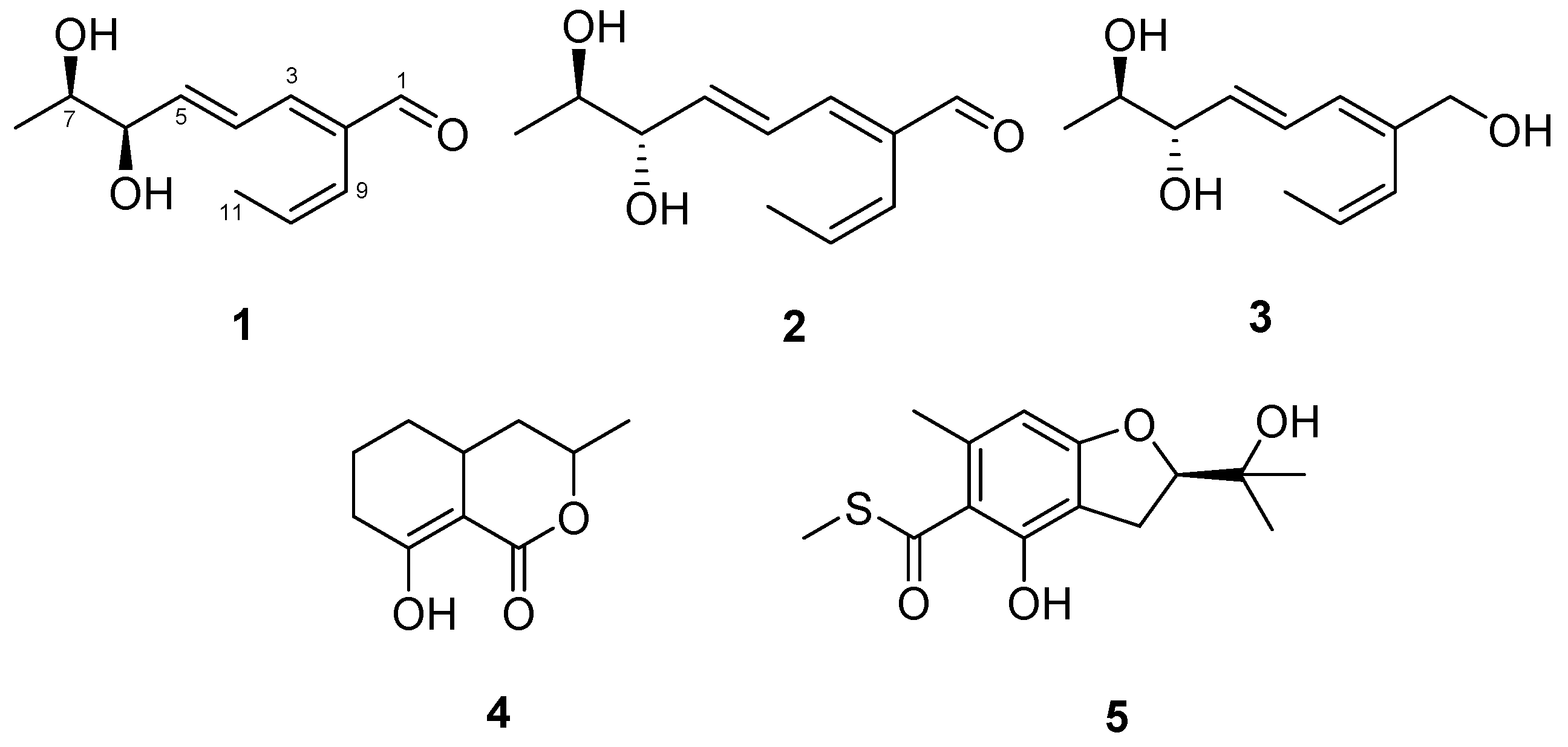
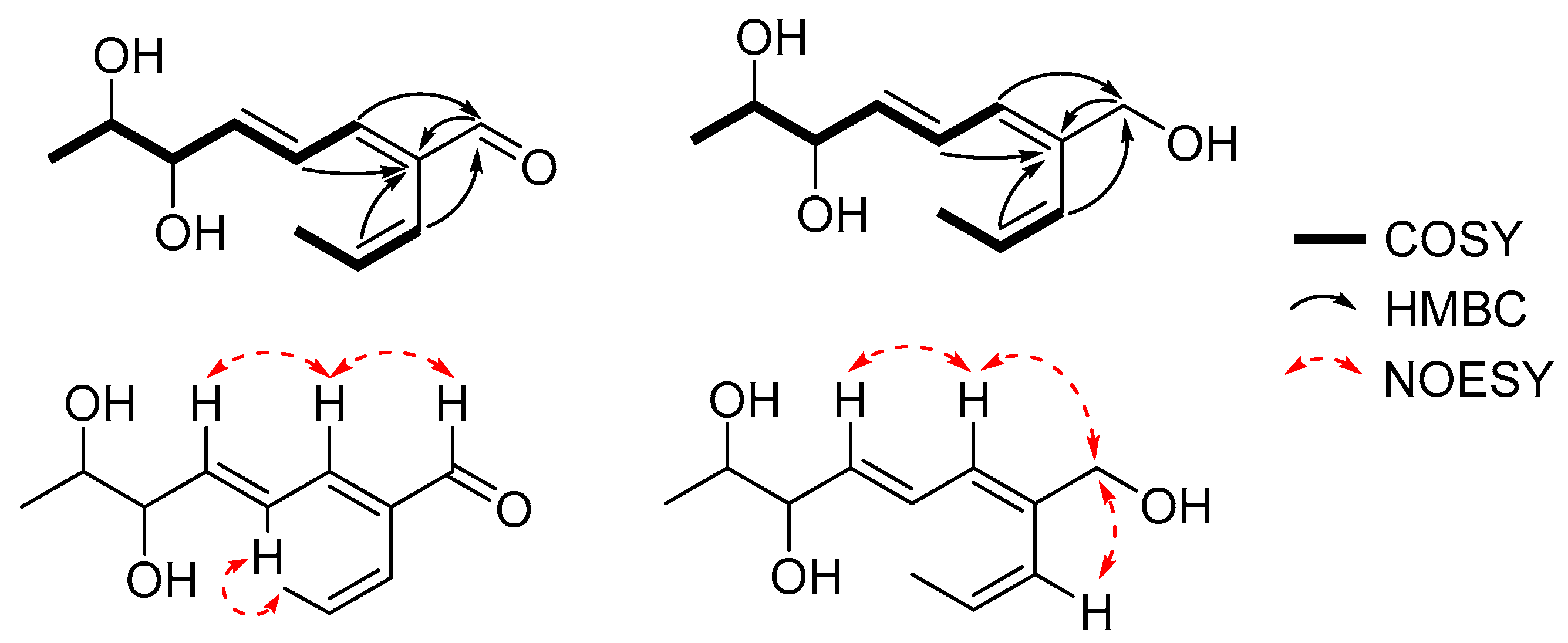
2.1. Structural Elucidation
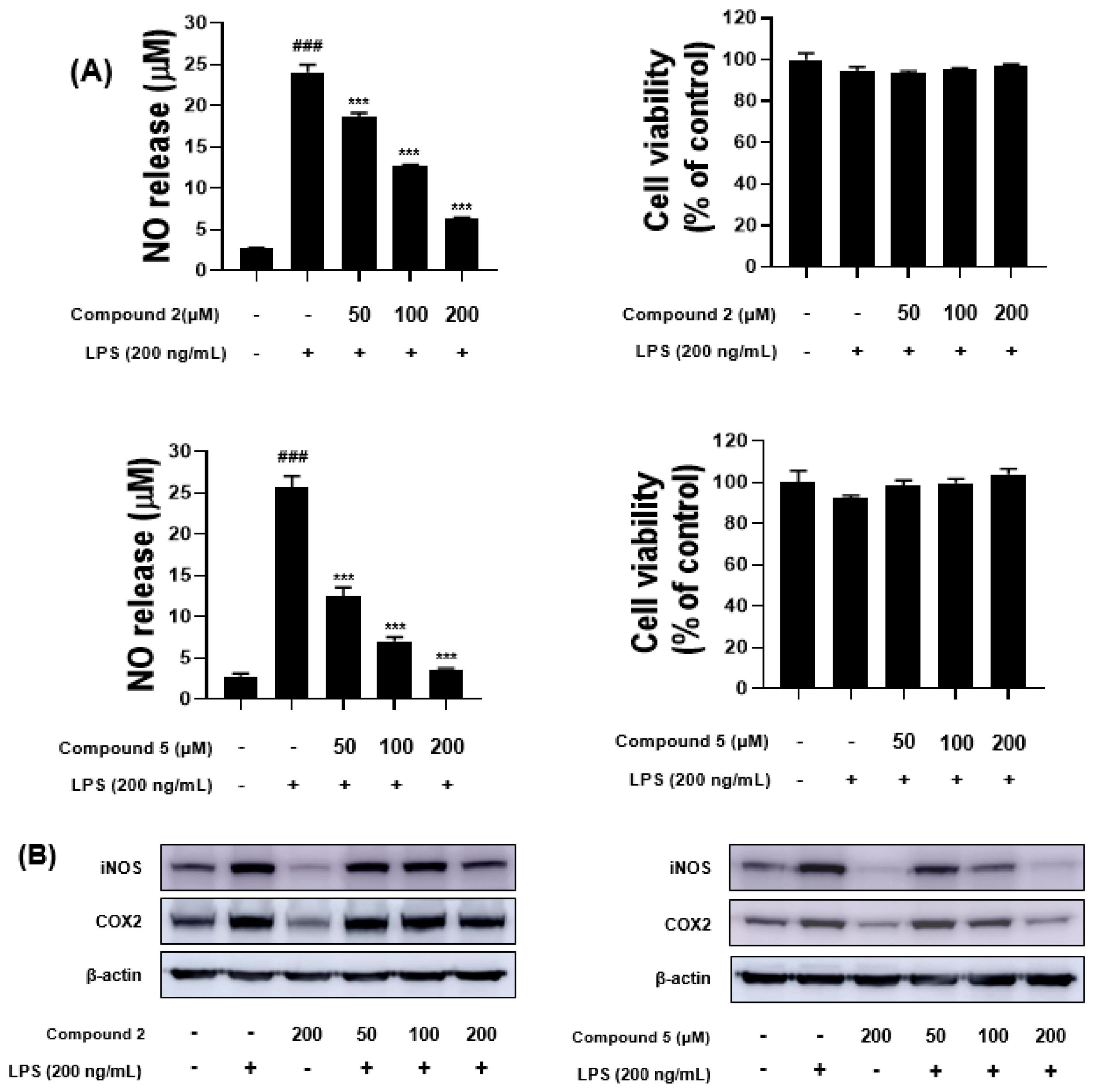
Compound 1 was isolated as a colorless oil with a molecular formula of C11H16O3, which was established from an HRESIMS peak at m/z 219.1002 [M + Na]+ (calcd. for C11H16O3Na+, 219.0992). The 1H NMR spectrum displayed signals of an aldehydic proton at δH 9.46 (H-1), five olefinic protons at δH 5.93–7.13, two oxymethine protons at δH 3.72 (H-7) and 4.09 (H-6), and two methyl groups at δH 1.14 (H3-8) and 1.54 (H3-11) (Table 1). The 13C NMR data exhibited the presence of eleven resonances, which were assigned for an aldehyde group at δC 196.0 (C-1), five protonated olefinic carbons at δC 150.2 (C-3), 146.0 (C-5), 132.5 (C-10), 128.8 (C-4), and 122.0 (C-9), a sp2 quaternary carbon at δC 139.1 (C-2), two oxygenated methines at δC 77.0 (C-6), 71.4 (C-7), and two methyls at δC 18.7 (C-8), 15.7 (C-11) (Table 1). The above data suggested that 1 possesses an acyclic skeleton with an aldehyde and three pairs of sp2 carbons, accounting for all four degrees of unsaturation in accordance with its molecular formula.
Detailed analysis of 1H-1H COSY correlations determined the partial structures of 1 including two distinct spin systems from H-3 (δH 7.13) to H3-8 (δH 1.14), and from H-9 (δH 5.98) to H3-11 (δH 1.54). Furthermore, the linkages between the partial structures were identified on the basis of the HMBC spectrum. The HMBC correlations of H-1 (δH 9.46), H-4 (δH 6.66), and H-10 (δH 5.92) to C-2 (δC 139.1), and those of H-3, H-9 to C-1 (δC 196.0) indicated that three parts of the compound were assembled via C-C bonds from C-2 to C-1, C-3, and C-9. Thus, the planar structure of 1 was determined as shown in Figure 1. The geometries of the Δ2,4,9-double bonds could be interpreted as 2E, 4E, and 9Z based on their 3JH,H coupling constants of H-4/H-5 (J = 15.3 Hz) and H-9/H-10 (J = 11.8 Hz), as well as the strong NOESY correlations of H-3/H-1, H-3/H-5, and H-4/H3-11 (Figure 2).
The absolute configurations of four possible stereoisomers of the secondary 1,2-diols could be unambiguously determined using the modified Mosher’s method (Figure 3A) by comparing the 1H NMR data (∆δS-R) of their corresponding bis-(S)- and -(R)-MTPA (α-methoxy-α-trifluoromethylphenylacetic acid) esters (Figure 3B) [16,17]. Therefore, compound 1 was treated with R- and S-α-methoxy-α-(trifluoromethyl) phenylacetyl chloride (MTPA-Cl) to give bis-S- and -R-MTPA esters (1a and 1b), respectively. As a result, the ΔδS−R values of 1a and 1b were interpreted, which were consistent with syn-1,2-diols (Figure 3A) and the absolute configurations of the stereogenic centers were determined as 6R and 7R. Thus, the structure of 1 was determined as (2E,4E,6R,7R)-6,7-dihydroxy-2-((Z)-prop-1-en-1-yl)octa-2,4-dienal and named talacyanol A.
Compound 2 was isolated as a colorless oil with the same molecular formula of C11H16O3 to that of 1, which was established from an HRESIMS ion peak at m/z 219.1003 [M + Na]+ (calcd. for C11H16O3Na+, 219.0992). The one-dimensionial (1D) and two-dimensional (2D) NMR data of 2 were almost identical to those of 1. By detailed analysis of NMR data, the planar structure of 2 was determined to be the same as that of 1 including the geometries of the double bounds (Figure 1). The only difference between 1 and 2 was the absolute stereochemistry of the secondary 1,2-diols at C-6 and C-7 according to their 1H NMR data in CDCl3 (δH-6 4.28 and δH-7 3.96 for 1; δH-6 4.04 and δH-7 3.72 for 2, Figure S23) and optical rotation values [ α ] D 25 +76.6 (c 0.2, MeOH) for 1; [ α ] D 25   −15 (c 0.2, MeOH) for 2). By comparing 1H NMR data of bis-S- and -R-MTPA esters of 2, the absolute configurations at C-6 and C-7 were assigned as 6S and 7R (Figure 3B), and the structure of 2 was determined as (2E,4E,6S,7R)-6,7-dihydroxy-2-((Z)-prop-1-en-1-yl)octa-2,4-dienal and named talacyanol B.
Compound 3 was obtained as a colorless oil with a molecular formula of C11H18O3 based on its HRESIMS data m/z 221.1155 [M + Na]+ (calcd for 221.1148, C11H18O3Na+). The 1H NMR spectrum of 3 was quite similar to that of 2, the only difference lies in the chemical shift of the singlet proton H-1 (δH 9.46 in 2, δH 4.05 in 3), indicating that the aldehyde group at C-1 in 2 was replaced by a hydroxy group in 3. Thus, the planar structure of 3 was elucidated as shown in Figure 1 based on its 1D and 2D NMR data.
A literature search revealed that the planar structure of 3 was similar to pinophol A, which was isolated from a plant endophytic fungus Talaromyces pinophilus by Zhao et al. [18]. However, the absolute configurations at C-6 and C-7 of pinophol A had not been determined yet and their relative configurations were reported as syn-1,2-diols (6R, 7R or 6S, 7S). Comparison of their optical rotation values ( [ α ] D 25 − 10 (c 0.2, MeOH) for 3 and [ α ] D 26   +32.6 (c 0.1, MeOH) for pinophol A) and 1H NMR data in CDCl3 of 3 and pinophol A (δH-6 4.12 and δH-7 3.88 for 3 and δH-6 3.87 and δH-7 3.63 for pinophol A, Figure S22) suggested that they could be a pair of diastereomers. By analyzing 1H NMR data of tri-S- and -R-MTPA esters of 3, the absolute configurations of C-6 and C-7 were determined as 6S and 7R (Figure 3B). Thus, the structure of 3 was determined as a new derivative of pinophol A, (2E,4E,6S,7R)-2-((Z)-prop-1-en-1-yl)octa-2,4-diene-1,6,7-triol, and named talacyanol C.
Aliphatic aldehydes are easily reduced to the corresponding alcohols in high yield by heterogeneous catalytic hydrogenation. To verify whether compound 3 is a true natural product or an artifact arising from compound 2 by reduction of aldehyde during the extraction process with ethyl acetate, we cultured the strain again and the culture broth was extracted successively with dichloromethane and n-butanol. Compound 3 was found in the butanol extract with a detectable concentration (Figures S31 and S32). Therefore, it could be concluded that 3 is a true natural substance.
The structures of the known compounds were identified as ramulosin (4), and eurothiocin A (5) by comparison of their spectroscopic data with those reported in the literature [19,20,21].

2.2. Bioactivities

Over the past few decades, emerging evidence has shown that many marine natural products, such as cytarabine, eribulin mesylate, brentuximab vedotin, and trabectidine exhibit beneficial effects in the prevention and treatment of cancer [22]. Furthermore, acyclic polyene polyols are a wide group of polyketides, and many of them display cytotoxicity against various cancer cell lines [17,23]. Therefore, compounds 15 were screened for in vitro cytotoxicity against six different cancer cell lines (stomach NUGC-3, colon HCT-15, lung NCI-H23, breast MDA-MB-231, prostate PC-3, and renal ACHN), the most common cancers in Korea [24]. Notably, compound 1 displayed moderate cytotoxicity against all the cancer cell lines with GI50 values ranging from 44.4 to 91.8 µM (Table 2).
Compounds 15 were also tested for their inhibitory effects on the production of nitric oxide (NO) in lipopolysaccharide (LPS)-stimulated BV-2 microglial cells. The cells were initially treated with a high concentration (200 µM) of each compound and LPS (200 ng/mL) to screen their inhibitory effect on NO production. All the compounds showed weak or strong inhibitory effects on NO production (Figure S33), and compounds 2 and 5 showed the most potent anti-inflammatory activity. Therefore, talacyanol B (2) and eurothiocin A (5) were selected for further studies to investigate NO production in BV-2 cells and LPS-induced expression levels of cyclooxygenase-2 (COX-2) and inducible nitric oxide synthase (iNOS) proteins by Western blot analysis. As shown in Figure 4A,B, the NO production and expression levels of COX-2 and iNOS proteins were suppressed by both 2 and 5 in a dose-related fashion at the concentrations of 50, 100, and 200 µM.

3. Experimental Methods

3.1. General Experimental Procedures

The 1D (1H and 13C) and 2D (COSY, HSQC, HMBC, and NOESY) NMR spectra were acquired by a Bruker 600 MHz spectrometer (Bruker BioSpin GmbH, Rheinstetten, Germany). Specific optical rotations were obtained in methanol at 25 °C on a Rudolph Research Analytical (Autopol III) polarimeter (Rudolph Research Analytical, Hackettstown, NJ, USA). UV-visible spectra were acquired by a Shimadzu UV-1650PC spectrophotometer in 1 mm quartz cells (Shimadzu Corporation, Kyoto, Japan). IR spectra were collected on a JASCO FT/IR-4100 spectrophotometer (JASCO Corporation, Tokyo, Japan). High-resolution ESIMS were recorded on a hybrid ion-trap time-of-flight mass spectrometer (Shimadzu LC/MS-IT-TOF). HPLC was conducted using a semi-prep ODS column (YMC-Triart C18, 250 × 10 mm i.d, 5 µm) and an analytical ODS column (YMC-Triart C18, 250 × 4.6 mm i.d, 5 µm) (YMC Corporation, Kyoto, Japan). All the reagents were purchased from Sigma-Aldrich (Merck KGaA, Darmstadt, Germany), and the organic solvents and water were distilled prior to use.

3.2. Fungal Material and Fermentation

The fungal strain 168ST-51.1 was isolated from the seaweed Caulerpa sp. collected in Son Tra peninsular, Da Nang, Vietnam in August 2016. The fungus was identified as Talaromyces cyanescens Stchigel & Guarro on the basis of DNA amplification and ITS gene sequencing (GenBank accession number MK 072976.1). The voucher of this strain is currently deposited in the Microbial Culture Collection, KIOST, with the name of Talaromyces sp. 168ST-51.1 under the curatorship of Hee Jae Shin.
The seed and mass cultures were performed in Bennett’s medium (1% glucose, 0.2% tryptone, 0.1% yeast extract, 0.1% beef extract, 0.5% glycerol, sea salts 32 g/L, and agar 17 g/L for agar medium). The fungus was initially cultured on Bennett’s agar medium in a Petri dish for 7 days. The actively grown mycelium was transferred aseptically into a 500 mL conical flask containing 300 mL of Bennett’s broth medium and incubated on a rotary shaker (140 rpm) at 28 °C for 4 days. An aliquot (0.1% v/v) from the seed culture was inoculated into twenty 2 L flasks each containing 1 L of the medium and grown under the same conditions as described for the seed culture for 7 days, and then harvested.

3.3. Extraction and Isolation of Metabolites

After cultivation, the culture broth was extracted with ethyl acetate (20 L × 2 times). The organic layer was evaporated under vacuum at 37 °C to yield a crude extract (3.0 g). Afterwards, the crude extract was separated into fifteen fractions (Fr. 1 to Fr. 15) by vacuum liquid chromatography on a flash ODS column (20 cm × 4.5 cm), which was stepwise eluted with 3 × 250 mL each of 20%, 40%, 60%, 80% MeOH in H2O, and 100% MeOH. Fraction 4 was purified by an analytical RP-HPLC (YMC-Pack-ODS-A, 250 × 4.6 mm i.d, 5 µm) using an isocratic condition with 14% ACN in H2O at a flow rate of 1 mL/min to afford compound 3 (3.0 mg, tR = 15 min). Fraction 5 was applied to a semi-preparative RP-HPLC (YMC-Pack-ODS-A, 250 × 10 mm i.d, 5 µm, flow rate 2.0 mL/min) using an isocratic elution with 14% ACN in H2O to yield compounds 2 (20.0 mg, tR = 38 min) and 1 (5.0 mg, tR = 42 min). Fraction 9 was recrystallized from methanol to give compound 4 (10.0 mg) as yellow needles. Finally, compound 5 (4.0 mg) was purified from fraction 10 using a semi-preparative RP-HPLC (YMC-Pack-ODS-A, 250 × 10 mm i.d, 5 µm, flow rate 2.0 mL/min) with an isocratic elution of 37% ACN in H2O for 46 min. All the purification procedure was repeated 2 times to yield sufficient amounts of 15 for structure determination and bioassays.

3.3.1. Talacyanol A (1)

Colorless oil, [ α ] D 25 +76.6 (c 0.2, MeOH). UV (MeOH) λmax (log ε) 285 (2.86), 232 (2.38) nm; IR (MeOH) νmax 3392 (br), 2975, 2933, 1671, 1632 cm−1; HRESIMS m/z 219.1002 [M + Na]+ (calcd for 219.0992, C11H16O3Na+); 1H NMR (CD3OD, 600 MHz) and 13C NMR (CD3OD, 150 MHz) see Table 1.

3.3.2. Talacyanol B (2)

Colorless oil, [ α ] D 25 −15 (c 0.2, MeOH). UV (MeOH) λmax (log ε) 285 (2.86), 232 (2.38) nm; IR (MeOH) νmax 3392 (br), 2975, 2939, 1671, 1632 cm−1; HRESIMS m/z 219.1003 [M + Na]+ (calcd for 219.0992, C11H16O3Na+); 1H NMR (CD3OD, 600 MHz) and 13C NMR (CD3OD, 150 MHz) see Table 1.

3.3.3. Talacyanol C (3)

Colorless oil, [ α ] D 25 −10 (c 0.2, MeOH). UV (MeOH) λmax (log ε) 248 (2.61), 204 (1.56) nm; IR (MeOH) νmax 3335 (br), 2971, 2943, 1056 cm−1; HRESIMS m/z 221.1155 [M + Na]+ (calcd for 221.1148, C11H18O3Na+); 1H NMR (CD3OD, 600 MHz) and 13C NMR (CD3OD, 150 MHz) see Table 1.

3.4. MTPA Esterification of Compounds 13

Compound 1 (1.0 mg for each) was dissolved in anhydrous pyridine (200 µL), and then added dimethylaminopyridine (DMAP). Afterwards, (R)-MTPA-Cl (20 μL) or (S)-MTPA-Cl (20 μL) were introduced, and reaction mixture was stirred at ambient temperature for 30 min, and then quenched with MeOH. Each mixture ((a) 1 with 1a and (b) 1 with 1b) was dried to dryness and purified by analytical reversed-phase HPLC to afford 1a and 1b. (R)- and (S)-MTPA esters of compounds 2 and 3 were prepared in the same procedure as described above for compound 1. The ΔδS-R values around the stereogenic centers of the MTPA esters were determined by 1H, HSQC, and 1H-1H COSY NMR spectra.

3.4.1. Bis-S-MTPA Ester (1a) of Talacyanol A (1)

1H NMR (600 MHz, CD3OD) δ 9.47 (s, 1H), 7.39–7.51 (m, 10H), 6.87 (d, J = 11.1, 1H), 6.54 (dd, J = 11.7, 14.9, 1H), 6.04 (dd, J = 6.3, 15.4, 1H), 5.86 (m, 2H), 5.74 (dd, J = 2.8, 6.3, 1H), 5.36 (m, 1H), 3.46 (s, 3H), 3.45 (s, 3H), 1.40 (d, J = 5.1, 3H), 1.28 (d, J = 6.5, 3H).

3.4.2. Bis-R-MTPA Ester (1b) of Talacyanol A (1)

1H NMR (600 MHz, CD3OD) δ 9.47 (s, 1H), 7.39–7.49 (m, 10H), 7.02 (d, J = 11.2, 1H), 6.52 (dd, J = 10.9, 15.1, 1H), 6.23 (dd, J = 5.9, 15.5, 1H), 5.84 (m, 2H), 5.83 (m, 1H), 5.45 (m, 1H), 3.44 (s, 3H), 3.43 (s, 3H), 1.40 (d, J = 5.4, 3H), 1.30 (d, J = 6.6, 3H).

3.4.3. Bis-S-MTPA Ester (2a) of Talacyanol B (2)

1H NMR (600 MHz, CD3OD) δ 9.47 (s, 1H), 7.32–7.49 (m, 10H), 6.98 (d, J = 11.2, 1H), 6.51 (dd, J = 11.0, 15.4, 1H), 6.12 (dd, J = 6.9, 15.4, 1H), 5.88 (m, 2H), 5.82 (dd, J = 2.5, 5.9, 1H), 5.52 (m, 1H), 3.47 (s, 3H), 3.44 (s, 3H), 1.47 (d, J = 5.2, 3H), 1.35 (d, J = 6.6, 3H).

3.4.4. Bis-R-MTPA Ester (2b) of Talacyanol B (2)

1H NMR (600 MHz, CD3OD) δ 9.49 (s, 1H), 7.33–7.48 (m, 10H), 7.08 (d, J = 11.1, 1H), 6.73 (dd, J = 11.0, 15.4, 1H), 6.32 (dd, J = 7.0, 15.5, 1H), 5.94 (m, 2H), 5.90 (dd, J = 2.7, 6.8, 1H), 5.42 (m, 1H), 3.43 (s, 3H), 3.41 (s, 3H), 1.47 (d, J = 5.1, 3H), 1.19 (d, J = 6.6, 3H).

3.4.5. Tri-S-MTPA Ester (3a) of Talacyanol C (3)

1H NMR (600 MHz, CD3OD) 7.28–7.51 (m, 15H), 6.33 (dd, J = 10.8, 15.3, 1H), 6.08 (d, J = 10.9, 1H), 5.77 (m, 2H), 5.65 (dd, J = 2.2, 7.6, 1H), 5.47 (dd, J = 7.58, 15.3, 1H), 5.44 (m, 1H), 4.88 (d, J = 10.3, 1H), 4.82 (d, J = 14.0, 1H), 3.52 (s, 3H), 3.44 (s, 3H), 3.44 (s, 3H), 1.48 (d, J = 5.3, 3H), 1.31 (d, J = 6.6, 3H).

3.4.6. Tri-R-MTPA Ester (3b) of Talacyanol C (3)

1H NMR (600 MHz, CD3OD) 7.32–7.50 (m, 15H), 6.47 (m, 1H), 6.15 (d, J = 10.9, 1H), 6.04 (dd, J = 6.3, 15.4, 1H), 5.80 (m, 2H), 5.73 (m, 2H), 5.34 (m, 1H), 4.91 (d, J = 12.8, 1H), 4.80 (d, J = 12.8, 1H), 3.51 (s, 3H), 3.42 (s, 3H), 3.39 (s, 3H), 1.44 (d, J = 5.4, 3H), 1.14 (d, J = 6.5, 3H).

3.5. Cytotoxicity Test by SRB Assay and Anti-Neuroinflammatory Test

The SRB cytotoxicity assay and anti-neuroinflammatory test for compounds 15 were performed as previously described [25,26].

4. Conclusions

Chemical examination of the ethyl acetate extract of the marine-derived fungus Talaromyces cyanescens 168ST-51.1 led to the isolation of three new compounds, talacyanol A–C (13), together with two known compounds 4 and 5. The structures of the new compounds 13 were determined by the spectroscopic and modified Mosher’s methods. The known compounds 45 were identified by comparing their spectroscopic data with those reported in literature. Talacyanol A (1) expressed in vitro cytotoxicity against various cancer cell lines. In contrast, talacyanol B (2) and eurothiocin A (5) showed anti-neuroinflammatory activity without cytotoxicity. The results demonstrated that the absolute configurations of the chiral centers may exert significant effects on biological activities of natural products. To the best of our knowledge, this is the first report on the cytotoxicity of compound 1 and the anti-neuroinflammatory effect of compounds 2 and 5.

Supplementary Materials

The following materials are available online, Figures S1–S23: HRESI-MS data, 1H NMR, 13C NMR, 1H-1H COSY, HSQC, HMBC, NOESY experimental spectra of compounds 13, Figures S24–S29: 1H NMR spectra of R- and S-MTPA esters of compounds 13.

Author Contributions

H.J.S. was the principal investigator, who proposed the ideas for the present work, managed and supervised the whole research work, prepared and corrected the manuscript, and contributed to the structure elucidation of the new and known compounds; C.V.A. achieved all experiments for compounds 15, including fermentation, isolation, and structure elucidation, and prepared the manuscript; P.T.H.T., H.-S.L., and B.-K.C. contributed to analyzing data; D.-Y.C., and D.-K.C. evaluated the anti-neuroinflammatory activity; J.S.K. performed the experiments related to cytotoxicity of 15. All authors have read and agreed to the published version of the manuscript.

Funding

This research was supported in part by the Korea Institute of Ocean Science and Technology (grant PE99852) and the Ministry of Oceans and Fisheries, Republic of Korea (grant PM59122).

Institutional Review Board Statement

Not applicable.

Informed Consent Statement

Not applicable.

Data Availability Statement

The Data presented in the article are available in the supplementary materials.

Acknowledgments

The authors express gratitude to Young Hye Kim, Korea Basic Science Institute, Ochang, Korea, for providing mass data. Authors would like to thank the Vietnam Government for allowing us to do marine microbial research.

Conflicts of Interest

The authors declare no conflict of interest.

Sample Availability

Samples of the compounds are available from the authors.

References

  1. Klemm, E.J.; Wong, V.K.; Dougan, G. Emergence of dominant multidrug-resistant bacterial clades: Lessons from history and whole-genome sequencing. Proc. Natl. Acad. Sci. USA 2018, 115, 12872–12877. [Google Scholar] [CrossRef] [PubMed] [Green Version]
  2. Mathers, C.; Stevens, G.; Hogan, D.; Mahanani, W.R.; Ho, J. Global and Regional Causes of Death: Patterns and Trends, 2000–2015. In Disease Control Priorities: Improving Health and Reducing Poverty, 3rd ed.; Jamison, D.T., Gelband, H., Horton, S., Jha, P., Laxminarayan, R., Mock, C.N., Nugent, R., Eds.; The World Bank: Washington, DC, USA, 2017; Volume 9, pp. 69–104. [Google Scholar]
  3. Newman, D.J.; Cragg, G.M. Natural products as sources of new drugs over the nearly four decades from 01/1981 to 09/2019. J. Nat. Prod. 2020, 83, 770–803. [Google Scholar] [CrossRef] [PubMed]
  4. Malve, H. Exploring the ocean for new drug developments: Marine pharmacology. J. Pharm. Bioallied. Sci. 2016, 8, 83–91. [Google Scholar] [CrossRef]
  5. Rateb, M.E.; Ebel, R. Secondary metabolites of fungi from marine habitats. Nat. Prod. Rep. 2011, 28, 290–344. [Google Scholar] [CrossRef] [PubMed]
  6. Zhai, M.M.; Li, J.; Jiang, C.X.; Shi, Y.P.; Di, D.L.; Crews, P.; Wu, Q.X. The bioactive secondary metabolites from Talaromyces species. Nat. Prod. Bioprospect. 2016, 6, 1–24. [Google Scholar] [CrossRef] [PubMed] [Green Version]
  7. Chen, S.; He, L.; Chen, D.; Cai, R.; Long, Y.; Lu, Y.; She, J. Talaramide A, an unusual alkaloid from the mangrove endophytic fungus Talaromyces sp. (HZ-YX1) as an inhibitor of mycobacterial PknG. New J. Chem. 2017, 41, 4273–4276. [Google Scholar] [CrossRef]
  8. Meng, L.H.; Li, X.M.; Zhang, F.Z.; Wang, Y.N.; Wang, B.G. Talascortenes A–G, highly oxygenated diterpenoid acids from the sea-anemone-derived endozoic fungus Talaromyces scorteus AS-242. J. Nat. Prod. 2020, 83, 2528–2536. [Google Scholar] [CrossRef] [PubMed]
  9. Wu, B.; Ohlendorf, B.; Oesker, V.; Wiese, J.; Malien, S.; Schmaljohann, R.; Imhoff, J.F. Acetylcholinesterase inhibitors from a marine fungus Talaromyces sp. Strain LF458. Mar. Biotechnol. 2015, 17, 110–119. [Google Scholar] [CrossRef] [PubMed]
  10. Küppers, L.; Ebrahim, W.; El-Neketi, M.; Özkaya, F.C.; Mándi, A.; Kurtán, T.; Orfali, R.S.; Müller, W.E.G.; Hartmann, R.; Lin, W.; et al. Lactones from the sponge-derived fungus Talaromyces rugulosus. Mar. Drugs 2017, 15, 359. [Google Scholar] [CrossRef] [PubMed] [Green Version]
  11. Noinart, J.; Buttachon, S.; Dethoup, T.; Gales, L.; Pereira, J.A.; Urbatzka, R.; Freitas, S.; Lee, M.; Silva, A.M.S.; Pinto, M.M.M.; et al. A new ergosterol analog, a new bis-anthraquinone and anti-obesity activity of anthraquinones from the marine sponge-associated fungus Talaromyces stipitatus KUFA 0207. Mar. Drugs 2017, 15, 139. [Google Scholar] [CrossRef] [PubMed] [Green Version]
  12. Li, H.; Huang, H.; Shao, C.; Huang, H.; Jiang, J.; Zhu, X.; Liu, Y.; Liu, L.; Lu, Y.; Li, M.; et al. Cytotoxic norsesquiterpene peroxides from the endophytic fungus Talaromyces flavus isolated from the mangrove plant Sonneratia apetala. J. Nat. Prod. 2011, 74, 1230–1235. [Google Scholar] [CrossRef] [PubMed]
  13. Li, H.L.; Li, X.M.; Li, X.; Wang, C.Y.; Liu, H.; Kassack, M.U.; Meng, L.H.; Wang, B.G. Antioxidant hydroanthraquinones from the marine algal-derived endophytic fungus Talaromyces islandicus EN-501. J. Nat. Prod. 2017, 80, 162–168. [Google Scholar] [CrossRef] [PubMed]
  14. WHO. The Top 10 Causes of Death. Available online: https://www.who.int/news-room/fact-sheets/detail/the-top-10-causes-of-death (accessed on 9 December 2020).
  15. Ardura-Fabregat, A.; Boddeke, E.W.G.M.; Boza-Serrano, A.; Brioschi, S.; Castro-Gomez, S.; Ceyzeriat, K.; Dansokho, C.; Dierkes, T.; Gelders, G.; Heneka, M.T.; et al. Targeting Neuroinflammation to Treat Alzheimer’s Disease. CNS Drugs 2017, 31, 1057–1082. [Google Scholar] [CrossRef] [PubMed] [Green Version]
  16. Freire, F.; Seco, J.M.; Quiñoá, E.; Riguera, R. Determining the absolute stereochemistry of secondary/secondary diols by 1H NMR: Basis and applications. J. Org. Chem. 2005, 70, 3778–3790. [Google Scholar] [CrossRef] [PubMed]
  17. Bae, M.; Kim, H.; Shin, Y.; Kim, B.Y.; Lee, S.K.; Oh, K.-B.; Shin, J.; Oh, D.-C. Separacenes A–D, novel polyene polyols from the marine Actinomycete, Streptomyces sp. Mar. Drugs 2013, 11, 2882–2893. [Google Scholar] [CrossRef] [PubMed] [Green Version]
  18. Zhao, W.T.; Shi, X.; Xian, P.J.; Feng, Z.; Yang, J.; Yang, X.L. A new fusicoccane diterpene and a new polyene from the plant endophytic fungus Talaromyces pinophilus and their antimicrobial activities. Nat. Prod. Res. 2019, 35, 1–7. [Google Scholar] [CrossRef] [PubMed]
  19. Stodola, F.H.; Cabot, C.; Benjamin, C.R. Structure of ramulosin, a metabolic product of the fungus Pestalotia ramulosa. Biochem. J. 1964, 93, 92–97. [Google Scholar] [CrossRef] [PubMed] [Green Version]
  20. Stierle, D.B.; Stierle, A.A.; Kunz, A. Dihydroramulosin from Botrytis sp. J. Nat. Prod. 1998, 61, 1277–1278. [Google Scholar] [CrossRef] [PubMed]
  21. Liu, Z.; Xia, G.; Chen, S.; Liu, Y.; Li, H.; She, Z. Eurothiocin A and B, sulfur-containing benzofurans from a soft coral-derived fungus Eurotium rubrum SH-823. Mar. Drugs 2014, 12, 3669–3680. [Google Scholar] [CrossRef] [Green Version]
  22. Khalifa, S.A.M.; Elias, N.; Farag, M.A.; Chen, L.; Saeed, A.; Hegazy, M.-E.F.; Moustafa, M.S.; Abd El-Wahed, A.; Al-Mousawi, S.M.; Musharraf, S.G.; et al. Marine Natural Products: A Source of Novel Anticancer Drugs. Mar. Drugs 2019, 17, 491. [Google Scholar] [CrossRef] [Green Version]
  23. Smyrniotopoulos, V.; Firsova, D.; Fearnhead, H.; Grauso, L.; Mangoni, A.; Tasdemir, D. Density Functional Theory (DFT)-Aided Structure Elucidation of Linear Diterpenes from the Irish Brown Seaweed Bifurcaria bifurcata. Mar. Drugs 2021, 19, 42. [Google Scholar] [CrossRef] [PubMed]
  24. Jung, K.W.; Won, Y.J.; Hong, S.; Kong, H.J.; Lee, E.S. Prediction of Cancer Incidence and Mortality in Korea. Cancer Res. Treat. 2020, 52, 351–358. [Google Scholar] [CrossRef] [PubMed]
  25. Choi, B.K.; Jo, S.H.; Choi, D.K.; Trinh, P.T.H.; Lee, H.S.; Cao, V.A.; Van, T.T.T.; Shin, H.J. Anti-neuroinflammatory agent, restricticin B, from the marine-derived fungus Penicillium janthinellum and its inhibitory activity on the no production in bv-2 microglia cells. Mar. Drugs 2020, 18, 465. [Google Scholar] [CrossRef] [PubMed]
  26. Choi, B.-K.; Lee, H.-S.; Kang, J.S.; Shin, H.J. Dokdolipids A–C, hydroxylated rhamnolipids from the marine-derived Actinomycete Actinoalloteichus hymeniacidonis. Mar. Drugs 2019, 17, 237. [Google Scholar] [CrossRef] [PubMed] [Green Version]
Figure 1. Structures of compounds 15 isolated from Talaromyces cyanescens.
Molecules 26 00836 g001
Figure 2. Key COSY, HMBC, and NOESY correlations for compounds 13.
Molecules 26 00836 g002
Figure 3. (A) Sign distribution (ΔδSR) of bis-MTPA esters of the four possible stereoisomers of 1,2-diols [16]; (B) ΔδSR values of 1a3b in CD3OD.
Molecules 26 00836 g003
Figure 4. (A) The measurements of nitrite levels in the culture media were conducted using the Griess reaction. The release of NO was measured with indomethacin as a positive control (Figure S30). Cell viability was tested using the MTT (3-(4,5-dimetylthiazol-2-yl)-2,5-diphenyltetrazol bromide) assay. Results are shown as the percentage of control samples; (B) Inhibition of inducible nitric oxide synthase (iNOS) and cyclooxygenase-2 (COX-2) protein and mRNA expression by compounds 2 and 5 in lipopolysaccharide (LPS)-stimulated BV-2 cells. The data (B) is expressed as the relative signal intensity for two independent experiments. Values are the mean ± standard error. ### p < 0.001, vs. control group and *** p < 0.001 vs. LPS-treated group.
Molecules 26 00836 g004
Table 1. 1H and 13C NMR data for 13 at 600 MHz and 150 MHz in CD3OD (δ in ppm, J in Hz), respectively.
Position123
δH (J in Hz)δCδH (J in Hz)δCδH (J in Hz)δC
19.46, s196.09.46, s196.04.05, s66.4
2 139.1 139.1 139.8
37.13 (d, 11.2)150.27.13 (d, 11.2)150.36.20 (d, 11.0)126.6
46.66 (dd, 11.3, 15.3)128.86.63 (dd, 11.2, 15.3)128.66.33 (dd, 11.0, 15.3)130.7
56.44 (dd, 5.6, 15.3)146.06.49 (dd, 5.6, 15.3)146.35.80 (dd, 7.0, 15.4)134.1
64.09 (t, 5.5)77.04.07 (t, 5.4)77.03.93 (t, 5.5)77.7
73.72, m71.43.71, m71.53.67, m71.7
81.14 (d, 6.4)18.71.17 (dd, 1.0, 6.4)18.91.13 (d, 6.4)18.6
95.98 (d, 11.8)122.05.98 (d, 11.8)122.05.90 (d, 11.5)127.1
105.92, m132.55.92, m132.45.75, m129.9
111.54 (d, 6.6)15.71.54 (d, 6.6)15.71.61 (dd, 1.8, 6.9)15.4
Table 2. Growth inhibition (GI50, μM) values of 1 against human tumor cell lines.
Cell LineGI50, μMADR a
HCT-1564.3<0.5
NUGC-362.2<0.5
NCI-H2370.9<0.5
ACHN44.4<0.5
PC-354.1<0.5
MDA-MB-23191.8<0.5
GI50 values are the concentration corresponding to 50% growth inhibition. a ADR, adriamycin as standard.
Publisher’s Note: MDPI stays neutral with regard to jurisdictional claims in published maps and institutional affiliations.

