LC-MS/MS Determination of Modified Nucleosides in The Urine of Parkinson’s Disease and Parkinsonian Syndromes Patients

 ,
,
Abstract
:1. Introduction
2. Results
3. Discussion
4. Materials and Methods
4.1. Subjects
4.2. Sample Preparation
4.3. Urinary Creatinine Determination
4.4. Analytical Methods
4.5. Validation
4.6. Statistical Analysis
5. Conclusions
Author Contributions
Funding
Conflicts of Interest
Abbreviations
| 2-dG | 2-deoxy-guanosine |
| 8-OHdG | 8-hydroxydeoxyguanosine |
| CE-LC/MS | capillary electrophoresis/liquid chromatography mass-spectrometry |
| CSF | cerebrospinal fluid |
| GC | gas chromatography |
| IQR | interquartile range |
| LC-MS | liquid chromatography-mass spectrometry |
| LLOQ | lower limit of quantification |
| LQ | lower quartile |
| Max | maximum |
| Min | minimum |
| MS | mass spectrometry |
| PD | Parkinson’s disease |
| SD | standard deviation |
| SE | standard error |
| UQ | upper quartile |
References
- Lafuente, J.V.; Requejo, C.; Carrasco, A.; Bengoetxea, H. Nanoformulation: A Useful Therapeutic Strategy for Improving Neuroprotection and the Neurorestorative Potential in Experimental Models of Parkinson’s Disease. Int. Rev. Neurobiol. 2017, 137, 99–122. [Google Scholar]
- Navarro-Sánchez, L.; Águeda-Gómez, B.; Aparicio, S.; Pérez-Tur, J. Epigenetic Study in Parkinson’s Disease: A Pilot Analysis of DNA Methylation in Candidate Genes in Brain. Cells 2018, 7, 150. [Google Scholar] [CrossRef] [Green Version]
- Thomas, B.; Beal, M.F. Parkinson’s disease. Hum. Mol. Genet. 2007, 16, R183–R194. [Google Scholar] [CrossRef] [Green Version]
- Coppede, F. Genetics and epigenetics of Parkinson’s disease. Sci. World J. 2012, 2012, 489830. [Google Scholar] [CrossRef] [Green Version]
- Bras, J.; Guerreiro, R.; Hardy, J. SnapShot: Genetics of Parkinson’s disease. Cell 2015, 160, 570. [Google Scholar] [CrossRef] [Green Version]
- Kalinderi, K.; Bostantjopoulou, S.; Fidani, L. The genetic background of Parkinson’s disease: Current progress and future prospects. Acta Neurol. Scand. 2016, 134, 314–326. [Google Scholar] [CrossRef]
- Srivanitchapoom, P.; Pitakpatapee, Y.; Suengtaworn, A. Parkinsonian syndromes: A review. Neurol India 2018, 66, 15–25. [Google Scholar] [CrossRef]
- Marsili, L.; Rizzo, G.; Colosimo, C. Diagnostic Criteria for Parkinson’s Disease: From James Parkinson to the Concept of Prodromal Disease. Front Neurol. 2018, 9, 156. [Google Scholar] [CrossRef]
- Dickson, D.W. Parkinson’s Disease and Parkinsonism: Neuropathology. Cold Spring Harb. Perspect. Med. 2012, 2, a009258. [Google Scholar] [CrossRef] [Green Version]
- Bobrowska-Korczak, B.; Gątarek, P.; Rosiak, A.; Giebultowicz, J.; Kałużna-Czaplińska, J. Reduced levels of modified nucleosides in the urine of autistic children. Preliminary studies. Anal. Biochem. 2019, 571, 62–67. [Google Scholar] [CrossRef]
- Gątarek, P.; Pawełczyk, M.; Jastrzębski, K.; Głąbiński, A.; Kałużna-Czaplińska, J. Analytical methods used in the study of Parkinson’s disease. Trends Anal. Chem. 2019, 118, 292–302. [Google Scholar] [CrossRef]
- Kovács, Z.; Juhász, G.; Palkovits, M.; Dobolyi, A.; Kékesi, K.A. Area, age and gender dependence of the nucleoside system in the brain: A review of current literature. Curr. Top. Med. Chem. 2011, 11, 1012–1033. [Google Scholar] [CrossRef]
- Struck, W.; Siluk, D.; Yumba-Mpanga, A.; Markuszewski, M.; Kaliszan, R.; Markuszewski, M.J. Liquid chromatography tandem mass spectrometry study of urinary nucleosides as potential cancer markers. J. Chromatogr. A 2013, 1283, 122–1231. [Google Scholar] [CrossRef] [PubMed]
- Keil, K.P.; Lein, P.J. DNA methylation: A mechanism linking environmental chemical exposures to risk of autism spectrum disorders? Environ. Epigenet. 2016, 2, dvv012.2. [Google Scholar] [CrossRef] [Green Version]
- Schram, K.H. Urinary nucleosides. Mass Spectrom. Rev. 1998, 17, 131–251. [Google Scholar] [CrossRef]
- Seidel, A.; Brunner, S.; Seidel, P.; Fritz, G.I.; Herbarth, O. Modified nucleosides: An accurate tumour marker for clinical diagnosis of cancer, early detection and therapy control. Br. J. Cancer. 2006, 94, 1726–1733. [Google Scholar] [CrossRef] [Green Version]
- Goris, A.; Dubois, B.; Hilven, K. Role of Genetic Factors in Pathophysiology of Multiple Sclerosi. In Multiple Sclerosis A Mechanistic View, 1st ed.; Minagar, A., Ed.; Academic Press: London, UK, 2016; pp. 153–180. [Google Scholar]
- Liebich, H.M.; Lehmann, R.; Xu, G.; Wahl, H.G.; Häring, H.U. Application of capillary electrophoresis in clinical chemistry: The clinical value of urinary modified nucleosides. J. Chromatogr. B Biomed. Sci. Appl. 2000, 745, 189–196. [Google Scholar] [CrossRef]
- Saad, A.A.; O’Connor, P.J.; Mostafa, M.H.; Metwalli, N.E.; Cooper, D.P.; Margison, G.P.; Povey, A.C. Bladder tumor contains higher N7-methylguanine levels in DNA than adjacent normal bladder epithelium. Cancer Epidemiol. Biomark. Prev. 2006, 15, 740–743. [Google Scholar] [CrossRef] [Green Version]
- Zheng, Y.F.; Kong, H.W.; Xiong, J.H.; Lv, S.; Xu, G.W. Clinical significance and prognostic value of urinary nucleosides in breast cancer patients. Clin. Biochem. 2005, 38, 24–30. [Google Scholar] [CrossRef]
- Zheng, Y.F.; Yang, J.; Zhao, X.J.; Feng, B.; Kong, H.W.; Chen, Y.J.; Lv, S.; Zheng, M.H.; Xu, G.W. Urinary nucleosides as biological markers for patients with colorectal cancer. World J. Gastroenterol. 2005, 11, 3871–3876. [Google Scholar] [CrossRef]
- Bullinger, D.; Fux, R.; Nicholson, G.; Plontke, S.; Belka, C.; Laufer, S.; Gleiter, C.H.; Kammerer, B. Identification of urinary modified nucleosides and ribosylated metabolites in humans via combined ESI-FTICR MS and ESI-IT MS analysis. J. Am. Soc. Mass Spectrom. 2008, 19, 1500–1513. [Google Scholar] [CrossRef] [Green Version]
- Wüllner, U.; Kaut, O.; deBoni, L.; Piston, D.; Schmitt, I. DNA methylation in Parkinson’s disease. J. Neurochem. 2016, 139, 108–120. [Google Scholar] [CrossRef] [PubMed] [Green Version]
- Henderson-Smith, A.; Fisch, K.M.; Hua, J.; Liu, G.; Ricciardelli, E.; Jepsen, K.; Huentelman, M.; Stalberg, G.; Edland, S.D.; Scherzer, C.R.; et al. DNA methylation changes associated with Parkinson’s disease progression: Outcomes from the first longitudinal genome-wide methylation analysis in blood. Epigenetics 2019, 14, 365–382. [Google Scholar] [CrossRef] [PubMed] [Green Version]
- Saiki, S.; Hatano, T.; Fujimaki, M.; Ishikawa, K.I.; Mori, A.; Oji, Y.; Okuzumi, A.; Fukuhara, T.; Koinuma, T.; Imamichi, Y.; et al. Decreased long-chain acylcarnitines from insufficient β-oxidation as potential early diagnostic markers for Parkinson’s disease. Sci. Rep. 2017, 7, 7328. [Google Scholar] [CrossRef]
- Havelund, J.F.; Andersen, A.D.; Binzer, M.; Blaabjerg, M.; Heegaard, N.H.H.; Stenager, E.; Faergeman, N.J.; Gramsbergen, J.B. Changes in kynurenine pathway metabolism in Parkinson patients with L-DOPA-induced dyskinesia. J. Neurochem. 2017, 142, 756–766. [Google Scholar] [CrossRef]
- Burté, F.; Houghton, D.; Lowes, H.; Pyle, A.; Nesbitt, S.; Yarnall, A.; Yu-Wai-Man, P.; Burn, D.J.; Santibanez-Koref, M.; Hudson, G. Metabolic profiling of Parkinson’s disease and mild cognitive impairment. Mov. Disord. 2017, 32, 927–932. [Google Scholar] [CrossRef]
- Roede, J.R.; Uppal, K.; Park, Y.; Lee, K.; Tran, V.; Walker, D.; Strobel, F.H.; Rhodes, S.L.; Ritz, B.; Jones, D.P. Serum metabolomics of slow vs. rapid motor progression Parkinson’s disease: A pilot study. PLoS ONE 2013, 8, e77629. [Google Scholar] [CrossRef] [Green Version]
- Wuolikainen, A.; Jonsson, P.; Ahnlund, M.; Antti, H.; Marklund, S.L.; Moritz, T.; Forsgren, L.; Andersen, P.M.; Trupp, M. Multi-platform mass spectrometry analysis of the CSF and plasma metabolomes of rigorously matched amyotrophic lateral sclerosis. Parkinson’s disease and control subjects. Mol. Biosyst. 2016, 12, 1287–1298. [Google Scholar] [CrossRef] [PubMed]
- Sankowski, B.; Księżarczyk, K.; Raćkowska, E.; Szlufik, S.; Koziorowski, D.; Giebułtowicz, J. Higher cerebrospinal fluid to plasma ratio of p-cresol sulfate and indoxyl sulfate in patients with Parkinson’s disease. Clin. Chim. Acta 2020, 501, 165–173. [Google Scholar] [CrossRef]
- Postuma, R.B.; Berg, D.; Stern, M.; Poewe, W.; Olanow, C.W.; Oertel, W.; Obeso, J.; Marek, K.; Litvan, I.; Lang, A.E.; et al. MDS clinical diagnostic criteria for Parkinson’s disease. Mov. Disord. 2015, 30, 1591–1601. [Google Scholar] [CrossRef]
- Hoehn, M.M.; Yahr, M.D. Parkinsonism: Onset, progression and mortality. Neurology 1967, 17, 427–442. [Google Scholar] [CrossRef] [PubMed] [Green Version]
- Huang, W.; Xu, Y.; Zhang, Y.; Zhang, P.; Zhang, Q.; Zhang, Z.; Xu, F. Metabolomics-driven identification of adenosine deaminase as therapeutic target in a mouse model of Parkinson’s disease. J. Neurochem. 2019, 150, 282–295. [Google Scholar] [CrossRef] [PubMed]
- Dutta, S.P.; Chheda, G.B. Metabolism of 1-methyladenosine. Biochem. Med. Metab. Biol. 1987, 38, 69–73. [Google Scholar] [CrossRef]
- Chen, X.; Yu, C.; Guo, M.; Zheng, X.; Ali, S.; Huang, H.; Zhang, L.; Wang, S.; Huang, Y.; Qie, S.; et al. Down-Regulation of m6A mRNA Methylation Is Involved in Dopaminergic Neuronal Death. Acs Chem. Neurosci. 2019, 10, 2355–2363. [Google Scholar] [CrossRef]
- Podgorska, M.; Kocbuch, K.; Pawelczyk, T. Recent advances in studies on biochemical and structural properties of equilibrative and concentrative nucleoside transporters. Acta Biochim. Pol. 2005, 52, 749–758. [Google Scholar] [CrossRef]
- Boison, D. Adenosine as a neuromodulator in neurological diseases. Curr. Opin. Pharm. 2008, 8, 2–7. [Google Scholar] [CrossRef] [Green Version]
- Schmidt, A.P.; Lara, D.R.; Souza, D.O. Proposal of a guanine-based purinergic system in the mammalian central nervous system. Pharm. Ther. 2007, 116, 401–416. [Google Scholar] [CrossRef]
- Bours, M.J.L.; Swennen, E.L.R.; Di Virgilio, F.; Cronstein, B.N.; Dagnelie, P.C. Adenosine 5′-triphosphate and adenosine as endogenous signaling molecules in immunity and inflammation. Pharm. Ther. 2006, 112, 358–404. [Google Scholar] [CrossRef]
- Kékesi, K.A.; Kovács, Z.; Szilágyi, N.; Bobest, M.; Szikra, T.; Dobolyi, A.; Juhász, G.; Palkovits, M. Concentration of nucleosides and related compounds in cerebral and cerebellar cortical areas and white matter of the human brain. Cell. Mol. Neurobiol. 2006, 26, 833–844. [Google Scholar] [CrossRef]
- Bolner, A.; Pilleri, M.; Bosello, O.; Nordera, G. Oxidative damage on nucleobases and Hoehn–Yahr stage in Parkinson’s disease. Am. J. Res. Med. Sci. 2018, 3, 36–47. [Google Scholar] [CrossRef]
- Halliwell, B.; Clement, M.V.; Long, L.H. Hydrogen peroxide in the human body. Febs Lett. 2000, 486, 10–13. [Google Scholar] [CrossRef] [Green Version]
- Mecocci, P.; Polidori, M.C.; Ingegni, T.; Cherubini, A.; Chionne, F.; Cecchetti, R.; Senin, U. Oxidative damage to DNA in lymphocytes from AD patients. Neurology 1998, 51, 1014–1017. [Google Scholar] [CrossRef]
- Bolner, A.; Pilleri, M.; De Riva, V.; Nordera, G.P. Plasma and urinary HPLC-ED determination of the ratio of 8-OHdG/2-dG in Parkinson’s disease. Clin. Lab. 2011, 57, 859–866. [Google Scholar]
- Chen, C.M.; Liu, J.L.; Wu, Y.R.; Chen, Y.C.; Cheng, H.S.; Cheng, M.L.; Chiu, D.T. Increased oxidative damage in peripheral blood correlates with severity of Parkinson’s disease. Neurobiol. Dis. 2009, 33, 429–435. [Google Scholar] [CrossRef] [PubMed]
- Hirayama, M.; Nakamura, T.; Watanabe, H.; Uchida, K.; Hama, T.; Hara, T.; Niimi, Y.; Ito, M.; Ohno, K.; Sobue, G. Urinary 8-hydroxydeoxyguanosine correlate with hallucinations rather than motor symptoms in Parkinson’s disease. Parkinsonism Relat. Disord. 2011, 17, 46–49. [Google Scholar] [CrossRef]
- Raćkowska, E.; Bobrowska-Korczak, B.; Giebułtowicz, J. Development and validation of a rapid LC-MS/MS method for determination of methylated nucleosides and nucleobases in urine. J. Chromatogr. B Anal. Technol. Biomed. Life. Sci. 2019, 1128, 121775. [Google Scholar] [CrossRef] [PubMed]


| Study Population | PD | Control |
|---|---|---|
| Participants | 22 | 30 |
| Gender | ||
| Female | 7 | 16 |
| Male | 15 | 14 |
| Age (years) | 69.7 ± 6.6 | 63.0 ± 7.2 |
| BMI | 26.2 ± 4.1 | 30.2 ± 3.8 |
| Disease duration (years) | 5.7 ± 4.3 | - |
| Hoehn and Yahr scale | 2.6 ± 1.1 | - |
| H–Y1 | 5 (23.8%) | - |
| H–Y2 | 4 (19.1%) | |
| H–Y3 | 7 (33.3%) | |
| H–Y4 | 5 (23.8%) |
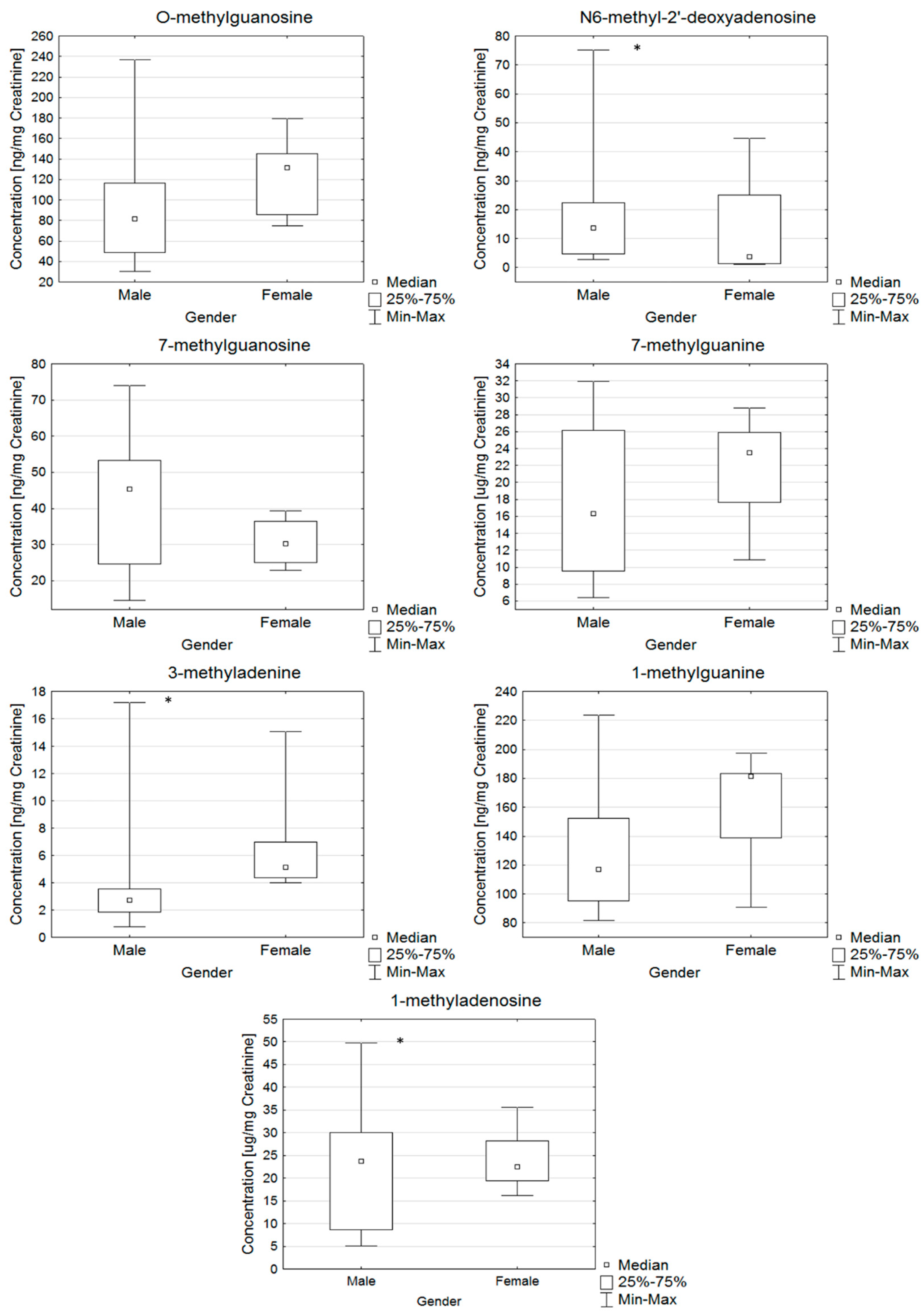
| Name of Nucleoside | Unit | Population | Mean ± SD | Median | Min | Max | LQ | UQ | SE | p-Value |
|---|---|---|---|---|---|---|---|---|---|---|
| O-methylguanosine | ng/mg Creatinine | PD | 107.3 ± 52.7 | 109.4 | 30.1 | 236.5 | 75.0 | 139.4 | 12.1 | 0.0002 |
| Control | 187.9 ± 83.1 | 169.1 | 107.5 | 478.0 | 130.7 | 213.7 | 17.7 | |||
| 3-methyladenine | ng/mg Creatinine | PD | 2.9 ± 1.3 | 2.9 | 0.8 | 5.1 | 1.9 | 4.0 | 0.3 | 0.0280 |
| Control | 5.0 ± 2.9 | 4.3 | 1.2 | 10.7 | 3.1 | 6.6 | 0.6 | |||
| 1-methylguanine | ng/mg Creatinine | PD | 139.7 ± 46.6 | 134.8 | 81.6 | 223.8 | 95.0 | 183.5 | 12.0 | 0.0257 |
| Control | 228.1 ± 142.8 | 169.5 | 104.0 | 680.0 | 133.4 | 277.3 | 30.4 | |||
| N6-methyl-2′-deoxyadenosine | ng/mg Creatinine | PD | 15.8 ± 25.1 | 5.1 | 1.0 | 75.3 | 2.2 | 17.9 | 8.9 | 0.0034 |
| Control | 48.4 ± 40.1 | 30.8 | 14.3 | 147.8 | 23.0 | 54.9 | 11.1 | |||
| 1-methyladenosine | μg/mg Creatinine | PD | 23.4 ± 12.7 | 23.0 | 5.2 | 49.7 | 15.5 | 28.2 | 2.7 | 0.7704 |
| Control | 22.5 ± 8.3 | 21.8 | 11.6 | 44.1 | 17.1 | 24.7 | 1.7 | |||
| 7-methylguanine | μg/mg Creatinine | PD | 19.3 ± 8.3 | 19.3 | 6.4 | 31.9 | 11.2 | 26.0 | 2.1 | 0.0315 |
| Control | 28.0 ± 12.6 | 24.8 | 11.5 | 63.1 | 20.0 | 36.2 | 2.5 | |||
| 7-methylguanosine | ng/mg Creatinine | PD | 38.6 ± 17.6 | 35.0 | 14.5 | 73.9 | 24.6 | 51.9 | 4.9 | 0.0616 |
| Control | 27.1 ± 14.1 | 23.6 | 12.4 | 59.1 | 16.3 | 31.2 | 3.5 |
Sample Availability: Samples of the compounds are not available from the authors. |
Publisher’s Note: MDPI stays neutral with regard to jurisdictional claims in published maps and institutional affiliations. |
© 2020 by the authors. Licensee MDPI, Basel, Switzerland. This article is an open access article distributed under the terms and conditions of the Creative Commons Attribution (CC BY) license (http://creativecommons.org/licenses/by/4.0/).
Share and Cite
Gątarek, P.; Kałużna-Czaplińska, J.; Pawełczyk, M.; Jastrzębski, K.; Giebułtowicz, J.; Głąbiński, A.; Bobrowska-Korczak, B. LC-MS/MS Determination of Modified Nucleosides in The Urine of Parkinson’s Disease and Parkinsonian Syndromes Patients. Molecules 2020, 25, 4959. https://doi.org/10.3390/molecules25214959
Gątarek P, Kałużna-Czaplińska J, Pawełczyk M, Jastrzębski K, Giebułtowicz J, Głąbiński A, Bobrowska-Korczak B. LC-MS/MS Determination of Modified Nucleosides in The Urine of Parkinson’s Disease and Parkinsonian Syndromes Patients. Molecules. 2020; 25(21):4959. https://doi.org/10.3390/molecules25214959
Chicago/Turabian StyleGątarek, Paulina, Joanna Kałużna-Czaplińska, Małgorzata Pawełczyk, Karol Jastrzębski, Joanna Giebułtowicz, Andrzej Głąbiński, and Barbara Bobrowska-Korczak. 2020. "LC-MS/MS Determination of Modified Nucleosides in The Urine of Parkinson’s Disease and Parkinsonian Syndromes Patients" Molecules 25, no. 21: 4959. https://doi.org/10.3390/molecules25214959