
Phytochemical Investigation and Anti-Inflammatory Activity of the Leaves of Machilus japonica var. kusanoi

 , ,
, ,
Abstract
:
1. Introduction
2. Results
3. Discussion
4. Materials and Methods
4.1. General Experiment Procedures
4.2. Plant Material
4.3. Extraction and Isolation
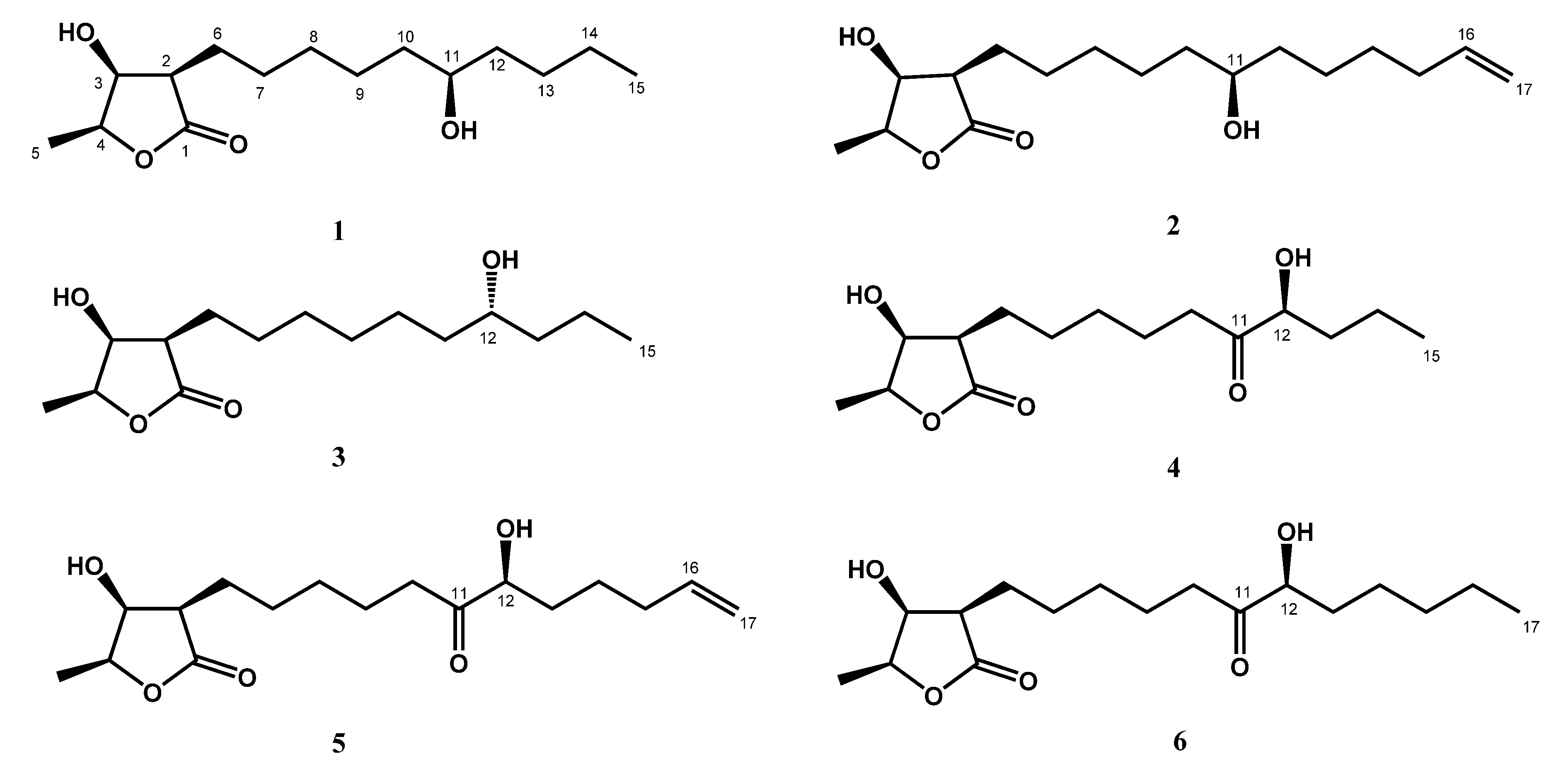
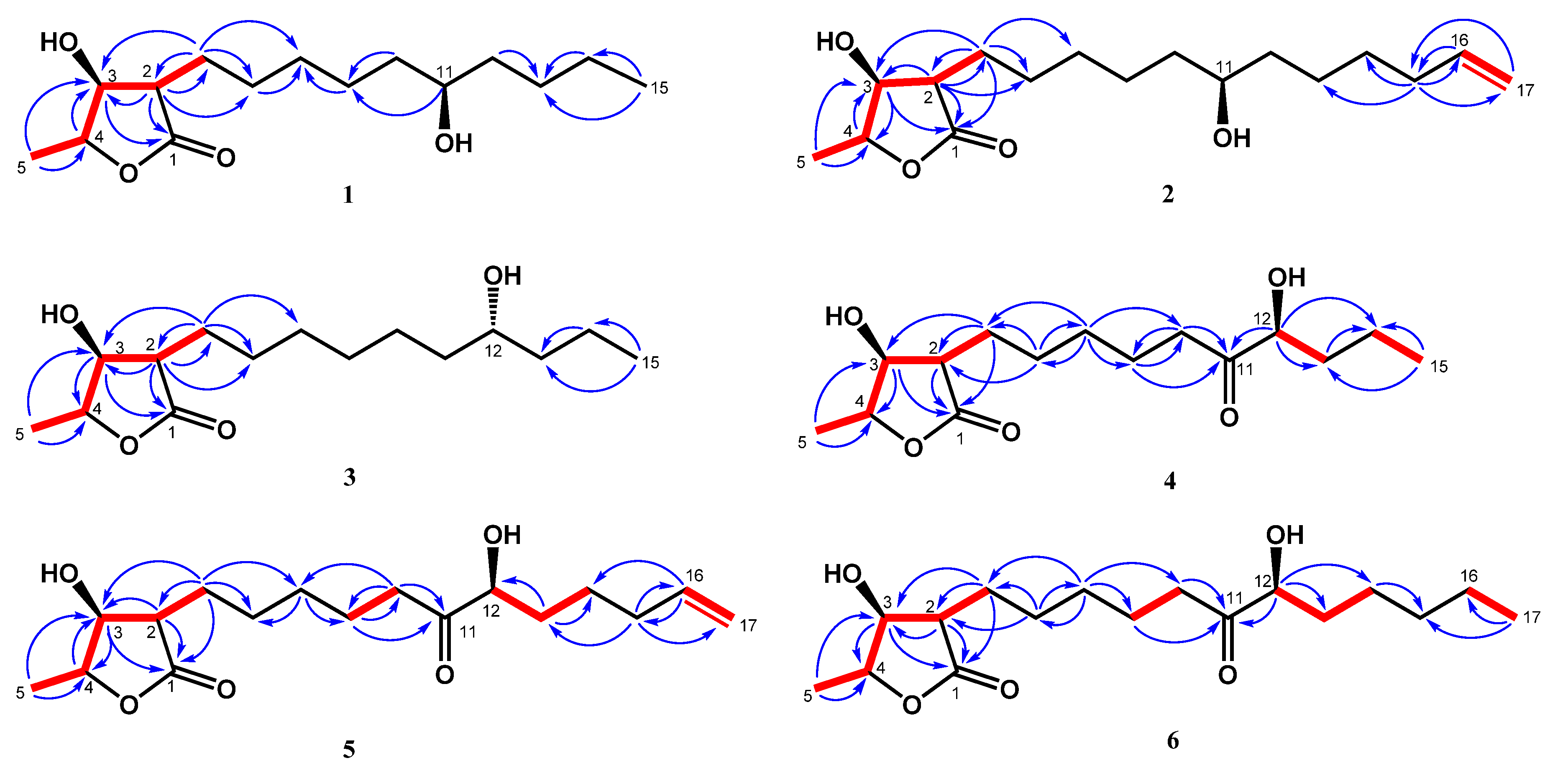
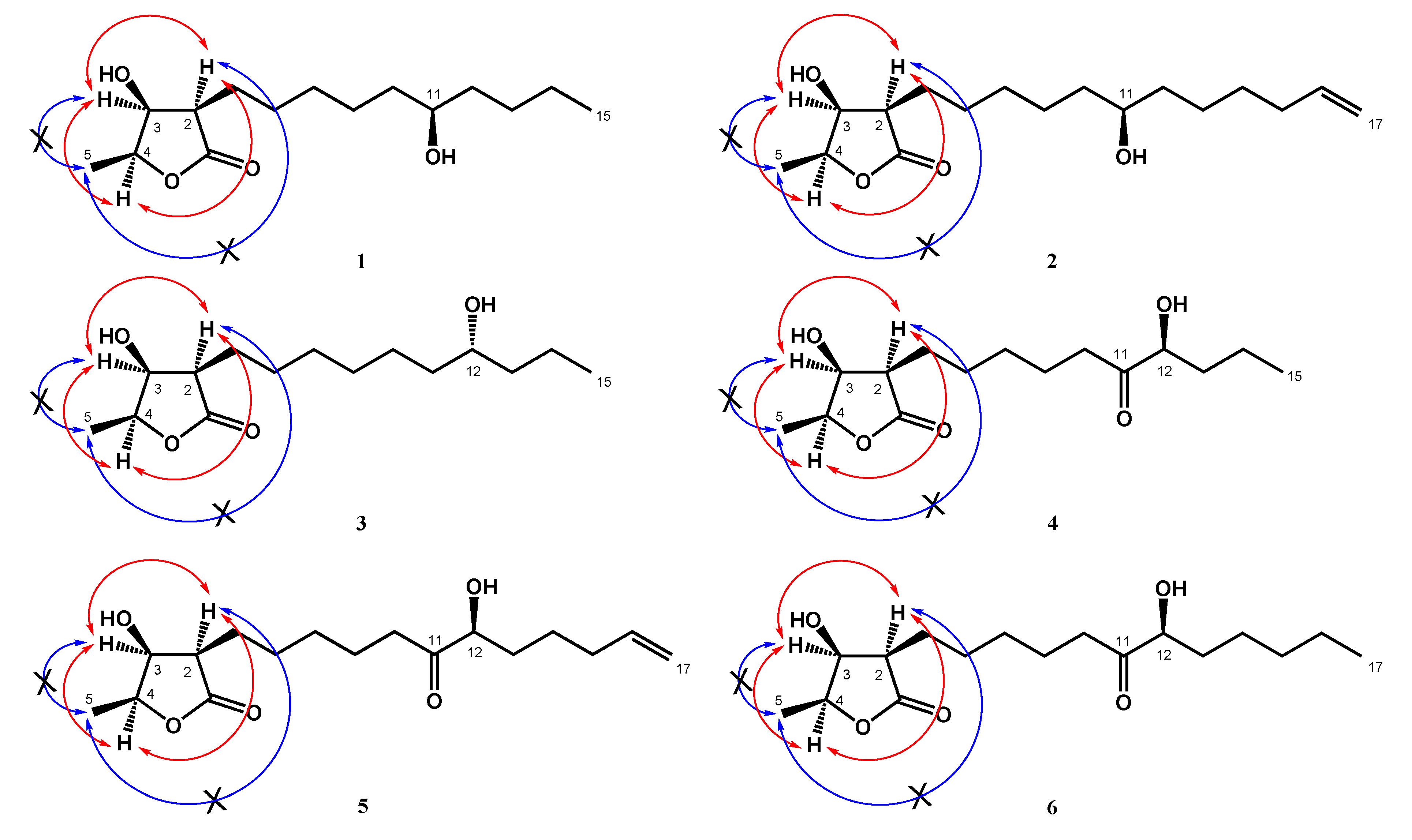
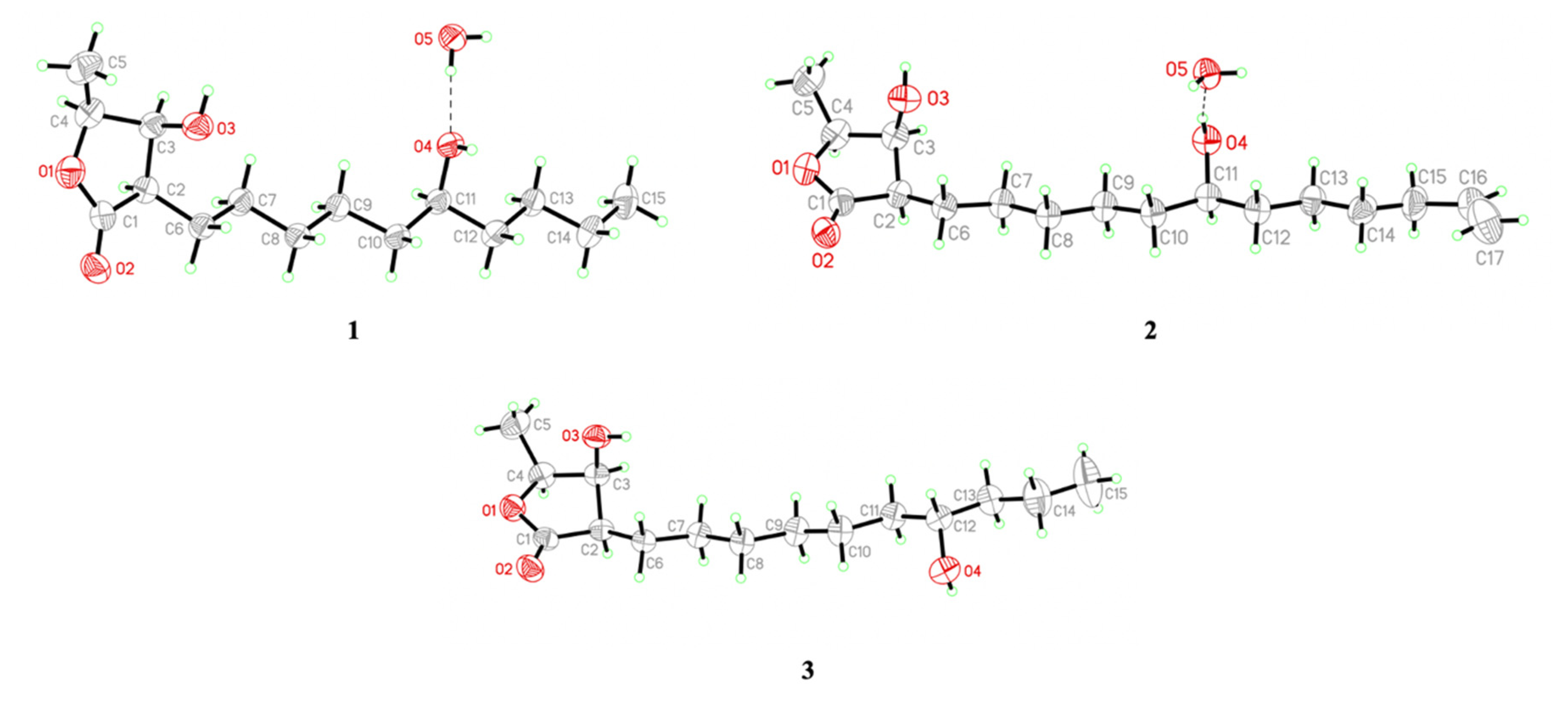
4.3.1. Machinolide A (1)
4.3.2. Machinolide B (2)
4.3.3. Machinolide C (3)
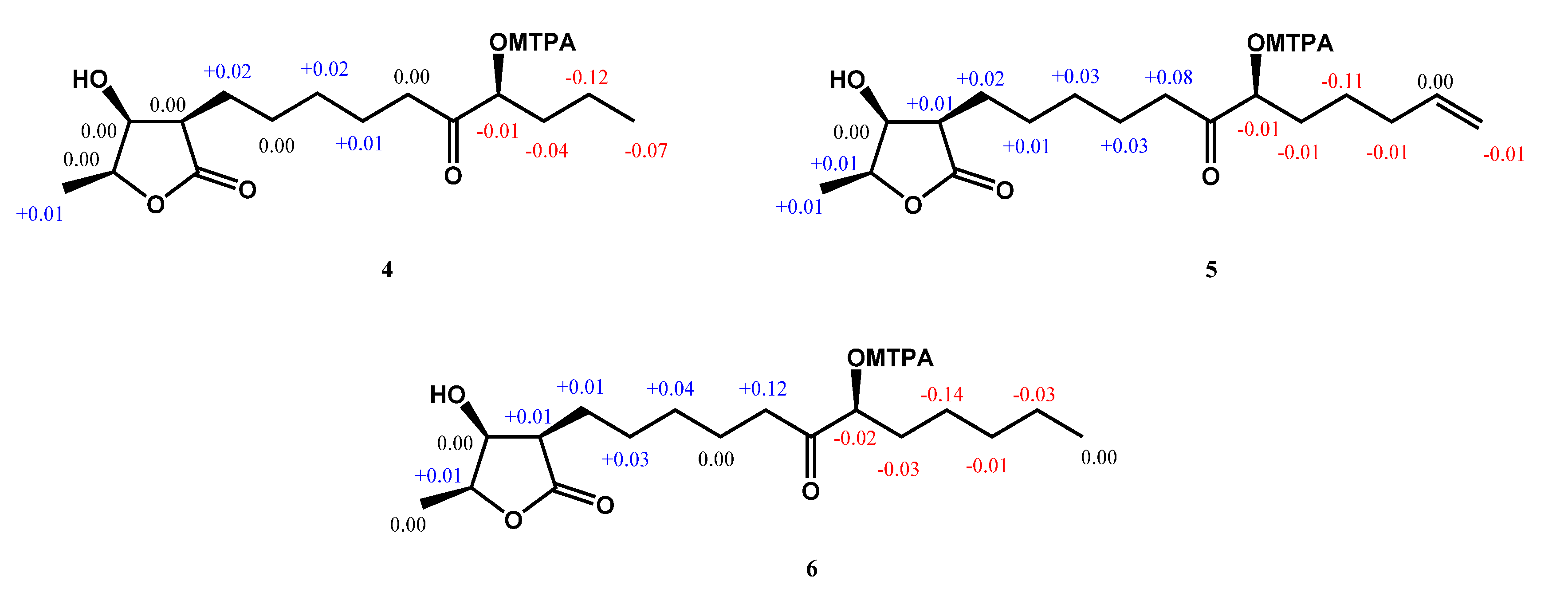
4.3.4. Machinolide D (4)
4.3.5. Machinolide E (5)
4.3.6. Machinolide F (6)
4.4. X-Ray Crystallographic Data for Machinolide A (1), Machinolide B (2), and Machinolide C (3)
4.5. Preparation of (S)-MTPA and (R)-MTPA Esters of 4a, 4b, 5a, 5b, 6a, and 6b from 4, 5, and 6
4.6. Superoxide Anion and Elastase Release Assays
5. Conclusions
Supplementary Materials
Author Contributions
Funding
Acknowledgments
Conflicts of Interest
References
- Chiang, C.-C.; Cheng, W.-J.; Korinek, M.; Lin, C.-Y.; Hwang, T.-L. Neutrophils in Psoriasis. Front. Immunol. 2019, 10, 2376. [Google Scholar] [CrossRef]
- Hwang, T.-L.; Su, Y.-C.; Chang, H.-L.; Leu, Y.-L.; Chung, P.-J.; Kuo, L.-M.; Chang, Y.-J. Suppression of superoxide anion and elastase release by C18 unsaturated fatty acids in human neutrophils. J. Lipid Res. 2009, 50, 1395–1408. [Google Scholar] [CrossRef] [PubMed] [Green Version]
- Korkmaz, B.; Horwitz, M.; Jenne, D.E.; Gauthier, F. Neutrophil Elastase, Proteinase 3, and Cathepsin G as Therapeutic Targets in Human Diseases. Pharmacol. Rev. 2010, 62, 726–759. [Google Scholar] [CrossRef] [PubMed] [Green Version]
- Chang, H.-S.; Chen, I.-S. Chemical constituents and bioactivity of Formosan lauraceous plants. J. Food Drug Anal. 2016, 24, 247–263. [Google Scholar] [CrossRef] [PubMed] [Green Version]
- Liao, J.C. Lauraceae in Flora of Taiwanm, 2nd ed.; Editorial Committee of the Flora of Taiwan: Taipei, Taiwan, 1996; Volume 2, pp. 1433–1499. [Google Scholar]
- Masao, T.; Yang, T.H.; Teh, L.S. Studies on the alkaloids of Formosan Lauraceous plants. I. Alkaloids of Machilus kusanoi Hayata. (1). The isolation of L-(-)-N-norarmepavine. Yakugaku Zasshi 1963, 83, 15–18. [Google Scholar] [CrossRef]
- Teh, L.S. Studies on the alkaloids of Formosan Louraceous plants. II. Alkaloids of Machilus Kusanoi Hayata. (2). The isolation of dl-coclaurine. Yakugaku Zasshi 1963, 83, 19–21. [Google Scholar] [CrossRef]
- Lee, S.-S.; Lin, Y.-S.; Chen, C.-K. Three Adducts of Butenolide and Apigenin Glycoside from the Leaves of Machilus Japonica. J. Nat. Prod. 2009, 72, 1249–1252. [Google Scholar] [CrossRef]
- Ho, C.-L.; Hsu, K.-P.; Tseng, Y.-H.; Wang, E.I.-C.; Liao, P.-C.; Chou, J.-C.; Lin, C.-N.; Su, Y.-C. Composition and antimicrobial activities of the leaf essential oil of Machilus kusanoi from Taiwan. Nat. Prod. Commun. 2011, 6. [Google Scholar] [CrossRef] [Green Version]
- Du, Y.; Abedi, A.K.; Valenciano, A.L.; Fernaández-Murga, M.L.; Cassera, M.B.; Rasamison, V.E.; Applequist, W.L.; Miller, J.S.; Kingston, D.G.I. Isolation of the New Antiplasmodial Butanolide, Malleastrumolide A, from Malleastrum sp. (Meliaceae) from Madagascar. Chem. Biodivers. 2017, 14, e1700331. [Google Scholar] [CrossRef]
- Lorenzo, M.; Brito, I.; Cueto, M.; D’Croz, L.; Darias, J. 13C NMR-Based Empirical Rules to Determine the Configuration of Fatty Acid Butanolides. Novel γ-Dilactones from Pterogorgia spp. Org. Lett. 2006, 8, 5001–5004. [Google Scholar] [CrossRef]
- Hoye, T.R.; Jeffrey, C.S.; Shao, F. Mosher ester analysis for the determination of absolute configuration of stereogenic (chiral) carbinol carbons. Nat. Protoc. 2007, 2, 2451–2458. [Google Scholar] [CrossRef]
- Pardede, A.; Adfa, M.; Kusnanda, A.J.; Ninomiya, M.; Koketsu, M. Flavonoid rutinosides from Cinnamomum parthenoxylon leaves and their hepatoprotective and antioxidant activity. Med. Chem. Res. 2017, 26, 2074–2079. [Google Scholar] [CrossRef]
- Chang, C.-W.; Chang, H.-S.; Cheng, M.-J.; Peng, C.-F.; Chen, I.-S. Identification of Five New Minor Constituents from the Whole Plant of Amischotolype hispida. Helvetica Chim. Acta 2015, 98, 347–358. [Google Scholar] [CrossRef]
- Liu, C.-M.; Kao, C.-L.; Wu, H.-M.; Li, W.-J.; Huang, C.-T.; Li, H.-T.; Chen, C.-Y. Antioxidant and Anticancer Aporphine Alkaloids from the Leaves of Nelumbo nucifera Gaertn. cv. Rosa-plena. Molecules 2014, 19, 17829–17838. [Google Scholar] [CrossRef]
- Wu, M.-D.; Cheng, M.-J.; Lin, R.-J.; Chan, H.-Y.; Hsieh, S.-Y.; Chang, H.-S.; Lin, C.-L.; Chen, J.-J. Chemical Constituents of the Fungus Biscogniauxia cylindrospora. Chem. Nat. Compd. 2019, 55, 924–926. [Google Scholar] [CrossRef]
- Zhang, W.; Wang, Y.; Geng, Z.; Guo, S.; Cao, J.; Zhang, Z.; Pang, X.; Chen, Z.; Du, S.S.; Deng, Z. Antifeedant Activities of Lignans from Stem Bark of Zanthoxylum armatum DC. against Tribolium castaneum. Molecules 2018, 23, 617. [Google Scholar] [CrossRef] [Green Version]
- Holzbach, J.C.; Lopes, L.M.X. Aristolactams and Alkamides of Aristolochia gigantea. Molecules 2010, 15, 9462–9472. [Google Scholar] [CrossRef]
- Lee, S.Y.; Woo, K.W.; Kim, C.S.; Lee, D.U.; Lee, K.R. New Lignans from the Aerial Parts of Rudbeckia laciniata. Helvetica Chim. Acta 2013, 96, 320–325. [Google Scholar] [CrossRef] [Green Version]
- Sribuhom, T.; Sriphana, U.; Thongsri, Y.; Yenjai, C. Chemical constituents from the stems of Alyxia schlechteri. Phytochem. Lett. 2015, 11, 80–84. [Google Scholar] [CrossRef]
- Park, C.H.; Kim, K.H.; Lee, I.K.; Lee, S.Y.; Choi, S.U.; Lee, J.H.; Lee, K.R. Phenolic constituents of Acorus gramineus. Arch. Pharmacal Res. 2011, 34, 1289–1296. [Google Scholar] [CrossRef]
- Rye, C.E.; Barker, D. Asymmetric Synthesis of (+)-Galbelgin, (−)-Kadangustin J, (−)-Cyclogalgravin and (−)-Pycnanthulignenes A and B, Three Structurally Distinct Lignan Classes, Using a Common Chiral Precursor. J. Org. Chem. 2011, 76, 6636–6648. [Google Scholar] [CrossRef] [PubMed]
- You, C.-X.; Yang, K.; Wang, C.-F.; Zhang, W.; Wang, Y.; Han, J.; Fan, L.; Du, S.S.; Geng, Z.; Deng, Z. Cytotoxic Compounds Isolated from Murraya tetramera Huang. Molecules 2014, 19, 13225–13234. [Google Scholar] [CrossRef] [PubMed] [Green Version]
- Collado, I.G.; Hanson, J.R.; Macías-Sánchez, A.J.; Mobbs, D. The Biotransformation of Some Clovanes by Botrytis cinerea. J. Nat. Prod. 1998, 61, 1348–1351. [Google Scholar] [CrossRef] [PubMed]
- Ashour, A.; Amer, M.; Marzouk, A.M.; Shimizu, K.; Kondo, R.; El-Sharkawy, S. Corncobs as a Potential Source of Functional Chemicals. Molecules 2013, 18, 13823–13830. [Google Scholar] [CrossRef]
- Lopes, N.P.; Silva, D.H.S.; Kato, M.J.; Yoshida, M. Butanolides as a common feature of Iryanthera lancifolia and Virola surinamensis. Phytochemistry 1998, 49, 1405–1410. [Google Scholar] [CrossRef]
- Franco, C.M.M.; Borde, U.P.; Vijayakumar, E.K.S.; Chatterjee, S.; Blumbach, J.; Ganguli, B.N. Butalactin, a new butanolide antibiotic. Taxonomy, fermentation, isolation and biological activity. J. Antibiot. 1991, 44, 225–231. [Google Scholar] [CrossRef]
- Nihira, T.; Shimizu, Y.; Kim, H.S.; Yamada, Y. Structure-activity relationships of virginiae butanolide C, an inducer of virginiamycin production in Streptomyces virginiae. J. Antibiot. 1988, 41, 1828–1837. [Google Scholar] [CrossRef] [Green Version]
- Kim, H.S.; Tada, H.; Nihira, T.; Yamada, Y. Purification and characterization of virginiae butanolide C-binding protein, a possible pleiotropic signal-transducer in Streptomyces virginiae. J. Antibiot. 1990, 43, 692–706. [Google Scholar] [CrossRef] [Green Version]
- Hoshino, S.; Wakimoto, T.; Onaka, H.; Abe, I. Chojalactones A–C, Cytotoxic Butanolides Isolated from Streptomyces sp. Cultivated with Mycolic Acid Containing Bacterium. Org. Lett. 2015, 17, 1501–1504. [Google Scholar] [CrossRef]
- Li, F.; Chen, D.; Lu, S.; Yang, G.; Zhang, X.; Chen, Z.; Fan, S.; Wu, S.-H.; He, J. Anti-Influenza A Viral Butenolide from Streptomyces sp. Smu03 Inhabiting the Intestine of Elephas maximus. Viruses 2018, 10, 356. [Google Scholar] [CrossRef] [Green Version]
- Cheng, M.-J.; Tsai, I.-L.; Lee, S.-J.; Jayaprakasam, B.; Chen, I.-S. Steryl epoxide, secobutanolide and butanolides from the stem wood of Machilus zuihoensis. Phytochemical 2005, 66, 1180–1185. [Google Scholar] [CrossRef] [PubMed]
- Liu, M.-T.; Lin, S.; Gan, M.; Liu, B.; Zi, J.; Song, W.-X.; Zhang, Y.-L.; Fan, X.-N.; Liu, Y.; Tan, W.; et al. Butanolide derivatives from the bark of Machilus yaoshansis. J. Asian Nat. Prod. Res. 2012, 14, 713–720. [Google Scholar] [CrossRef]
- Kim, W.; Lyu, H.-N.; Kwon, H.-S.; Kim, Y.S.; Lee, K.-H.; Kim, -Y.; Chakraborty, G.; Choi, K.Y.; Yoon, H.S.; Kim, K.-T. Obtusilactone B from Machilus Thunbergii Targets Barrier-to-Autointegration Factor to Treat Cancer. Mol. Pharmacol. 2012, 83, 367–376. [Google Scholar] [CrossRef] [PubMed]
- Yang, C.-P.; Huang, G.-J.; Huang, H.-C.; Chen, Y.-C.; Chang, C.-I.; Wang, S.-Y.; Chen, I.-S.; Tseng, Y.-H.; Chien, S.-C.; Kuo, Y.-H. A New Butanolide Compound from the Aerial Part of Lindera akoensis with Anti-inflammatory Activity. Molecules 2012, 17, 6585–6592. [Google Scholar] [CrossRef] [PubMed]
- Tsai, I.-L.; Hung, C.-H.; Duh, C.-Y.; Chen, I.-S. Cytotoxic Butanolides and Secobutanolides from the Stem Wood of Formosan Lindera communis. Planta Medica 2002, 68, 142–145. [Google Scholar] [CrossRef]
- Cheng, M.-J.; Wang, T.-A.; Lee, S.-J.; Chen, I.-S. A new butanolide and a new secobutanolide from Litsea lii var. nunkao-tahangensis. Nat. Prod. Res. 2010, 24, 647–656. [Google Scholar] [CrossRef]
- Shen, K.-H.; Lin, E.-S.; Kuo, P.-L.; Chen, C.-Y.; Hsu, Y.-L. Isolinderanolide B, a Butanolide Extracted From the Stems of Cinnamomum subavenium, Inhibits Proliferation of T24 Human Bladder Cancer Cells by Blocking Cell Cycle Progression and Inducing Apoptosis. Integr. Cancer Ther. 2011, 10, 350–358. [Google Scholar] [CrossRef]
- Yang, S.-Y.; Wang, H.-M.; Wu, T.-W.; Chen, Y.-J.; Shieh, J.-J.; Lin, J.-H.; Ho, T.-F.; Luo, R.-J.; Chen, C.-Y.; Chang, C.-C. Subamolide B Isolated from Medicinal Plant Cinnamomum subavenium Induces Cytotoxicity in Human Cutaneous Squamous Cell Carcinoma Cells through Mitochondrial and CHOP-Dependent Cell Death Pathways. Evid. Based Complement Alternat. Med. 2013, 2013, 1–13. [Google Scholar] [CrossRef] [Green Version]
- Chen, C.-Y.; Hsu, Y.-L.; Chen, Y.-Y.; Hung, J.-Y.; Huang, M.-S.; Kuo, P.-L. Isokotomolide A, a new butanolide extracted from the leaves of Cinnamomum kotoense, arrests cell cycle progression and induces apoptosis through the induction of p53/p21 and the initiation of mitochondrial system in human non-small cell lung cancer A549 cells. Eur. J. Pharmacol. 2007, 574, 94–102. [Google Scholar] [CrossRef]
- Le Dang, Q.; Kwon, H.R.; Choi, Y.H.; Choi, G.J.; Jang, K.S.; Park, M.S.; Lim, C.H.; Ngoc, L.H.; Kim, J.-C. Nematicidal activity against Bursaphelenchus xylophilus of isoobtusilactone A isolated from Persea americana. Nematology 2010, 12, 247–253. [Google Scholar] [CrossRef]
- Kuo, P.; Hung, H.-Y.; Nian, C.-W.; Hwang, T.-L.; Cheng, J.-C.; Kuo, D.-H.; Lee, E.-J.; Tai, S.-H.; Wu, T.-S. Chemical Constituents and Anti-inflammatory Principles from the Fruits of Forsythia suspensa. J. Nat. Prod. 2017, 80, 1055–1064. [Google Scholar] [CrossRef] [PubMed]
- Hwang, T.-L.; Li, G.-L.; Lan, Y.-H.; Chia, Y.-C.; Hsieh, P.-W.; Wu, Y.-H.; Wu, Y.-C. Potent inhibition of superoxide anion production in activated human neutrophils by isopedicin, a bioactive component of the Chinese medicinal herb Fissistigma oldhamii. Free. Radic. Boil. Med. 2009, 46, 520–528. [Google Scholar] [CrossRef] [PubMed]
Sample Availability: Samples of all compounds are available from the authors. |


| Position | 1 a | 2 b | 3 b | |||
|---|---|---|---|---|---|---|
| δH (m, J in Hz) | δC | δH (m, J in Hz) | δC | δH (m, J in Hz) | δC | |
| 1 | 177.5 | 177.4 | 177.4 | |||
| 2 | 2.57, dt (9.8, 4.8) | 47.6 | 2.57, dt (10.2, 4.6) | 47.6 | 2.57, dt (8.7, 4.8) | 47.6 |
| 3 | 4.31, dd (4.8, 3.2) | 71.2 | 4.31, dd (4.6, 3.2) | 71.3 | 4.31, dd (4.8, 3.2) | 71.3 |
| 4 | 4.45, qd, (6.6, 3.2) | 78.8 | 4.45, qd (6.6, 3.2) | 78.7 | 4.45, qd (6.6, 3.2) | 78.7 |
| 5 | 1.43, d (6.6) | 13.7 | 1.44, d (6.6) | 13.7 | 1.44, d (6.6) | 13.7 |
| 6 | 1.82, m 1.66, m | 23.1 | 1.84, m 1.67, m | 23.2 | 1.84, m 1.65, m | 23.3 |
| 7 | 1.26~1.47, m | 27.5 | 1.32~1.51, m | 27.6 | 1.30~1.49, m | 27.5 |
| 8 | 1.26~1.47, m | 29.4 | 1.32~1.51, m | 29.4 | 1.30~1.49, m | 29.3 c |
| 9 | 1.26~1.47, m | 25.1 | 1.32~1.51, m | 25.1 c | 1.30~1.49, m | 29.4 c |
| 10 | 1.26~1.47, m | 37.16 c | 1.32~1.51, m | 37.2 d | 1.30~1.49, m | 25.5 |
| 11 | 3.59, m | 71.9 | 3.59, m | 71.9 | 1.30~1.49, m | 37.4 |
| 12 | 1.26~1.47, m | 37.19 c | 1.32~1.51, m | 37.4 d | 3.60, m | 71.8 |
| 13 | 1.26~1.47, m | 27.8 | 1.32~1.51, m | 25.2 c | 1.30~1.49, m | 39.7 |
| 14 | 1.26~1.47, m | 22.7 | 1.37, m | 28.9 | 1.30~1.49, m | 18.8 |
| 15 | 0.91, t (7.0) | 14.0 | 2.07, m | 33.7 | 0.93, t (7.2) | 14.1 |
| 16 | 5.81, ddt (17.1, 10.2, 6.6) | 138.9 | ||||
| 17 | 5.00, ddt (17.1, 3.3, 1.5) 4.94, ddt (10.2, 3.3, 1.5) | 114.4 | ||||
| Position | 4 a | 5 b | 6 b | |||
|---|---|---|---|---|---|---|
| δH (m, J in Hz) | δC | δH (m, J in Hz) | δC | δH (m, J in Hz) | δC | |
| 1 | 177.4 | 177.3 | 177.7 | |||
| 2 | 2.55, dt (9.9, 5.0) | 47.5 | 2.56, dt (9.8, 4.5) | 47.5 | 2.54, dt (10.0, 5.0) | 47.5 |
| 3 | 4.31, dd (5.0, 3.0) | 71.3 | 4.30, br t (4.5) | 71.3 | 4.30, dd (5.0, 3.1) | 71.1 |
| 4 | 4.45, qd (6.5, 3.0) | 78.8 | 4.54, qd (6.4, 2.9) | 78.7 | 4.44, qd (6.0, 3.1) | 79.0 |
| 5 | 1.44, d (6.5) | 13.7 | 1.44, d (6.4) | 13.7 | 1.42, d (6.0) | 13.7 |
| 6 | 1.80, m 1.67, m | 23.1 | 1.84, m 1.67, m | 23.1 | 1.79, m 1.64, m | 23.1 |
| 7 | 1.35~1.55, m | 27.4 | 1.33~1.58, m | 27.4 | 1.24~1.52, m | 27.3 |
| 8 | 1.35~1.55, m | 29.0 | 1.33~1.58, m | 29.0 | 1.24~1.52, m | 29.0 |
| 9 | 1.67, m | 23.2 | 1.67, m | 23.2 | 1.64, m | 23.2 |
| 10 | 2.40~2.54, m | 37.7 | 2.46, m | 37.7 | 2.46, m | 37.7 |
| 11 | 212.6 | 212.4 | 212.7 | |||
| 12 | 4.17, dd (7.5, 3.9) | 76.3 | 4.17, dd (7.2, 3.6) | 76.3 | 4.15, dd (7.4, 3.8) | 76.5 |
| 13 | 1.35~1.55, m | 35.9 | 1.84, m 1.33~1.58, m | 33.1 | 1.79, m 1.24~1.52, m | 33.7 |
| 14 | 1.35~1.55, m | 18.2 | 1.33~1.58, m | 24.0 | 1.24~1.52, m | 24.5 |
| 15 | 0.95, t (6.9) | 13.9 | 2.09, m | 33.3 | 1.24~1.52, m | 31.6 |
| 16 | 5.78, ddt (17.2, 10.4, 6.8) | 138.1 | 1.24~1.52, m | 22.5 | ||
| 17 | 5.03, m 4.98, m | 115.1 | 0.88, t (6.8) | 14.0 | ||
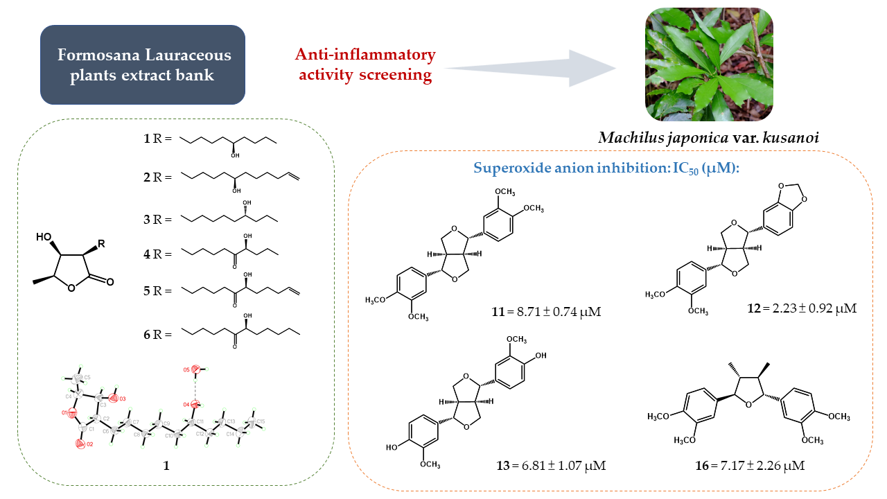
| Compound | Superoxide Anion | Elastase Release |
|---|---|---|
| IC50 (μM) a | IC50 (μM) a | |
| machinolide A (1) | >10 | >10 |
| machinolide B (2) | >10 | >10 |
| machinolide C (3) | >10 | >10 |
| machinolide F (6) | >10 | >10 |
| (+)-eudesmin (11) | 8.71 ± 0.74 | >10 |
| (+)-methylpiperitol (12) | 2.23 ± 0.92 | >10 |
| (+)-pinoresinol (13) | 6.81 ± 1.07 | >10 |
| (+)-galbelgin (16) | 7.15 ± 2.26 | >10 |
| LY294002 b | 2.17 ± 0.53 | 6.38 ± 1.72 |
© 2020 by the authors. Licensee MDPI, Basel, Switzerland. This article is an open access article distributed under the terms and conditions of the Creative Commons Attribution (CC BY) license (http://creativecommons.org/licenses/by/4.0/).
Share and Cite
Li, S.-L.; Wu, H.-C.; Hwang, T.-L.; Lin, C.-H.; Yang, S.-S.; Chang, H.-S. Phytochemical Investigation and Anti-Inflammatory Activity of the Leaves of Machilus japonica var. kusanoi. Molecules 2020, 25, 4149. https://doi.org/10.3390/molecules25184149
Li S-L, Wu H-C, Hwang T-L, Lin C-H, Yang S-S, Chang H-S. Phytochemical Investigation and Anti-Inflammatory Activity of the Leaves of Machilus japonica var. kusanoi. Molecules. 2020; 25(18):4149. https://doi.org/10.3390/molecules25184149
Chicago/Turabian StyleLi, Shiou-Ling, Ho-Cheng Wu, Tsong-Long Hwang, Chu-Hung Lin, Shuen-Shin Yang, and Hsun-Shuo Chang. 2020. "Phytochemical Investigation and Anti-Inflammatory Activity of the Leaves of Machilus japonica var. kusanoi" Molecules 25, no. 18: 4149. https://doi.org/10.3390/molecules25184149