
Role of Photoactive Phytocompounds in Photodynamic Therapy of Cancer


1
Laser Research Centre, Faculty of Health Sciences, University of Johannesburg, 17011, Doornfontein 2028, South Africa
2
Bioprospecting Laboratory, Department of Botany, School of Life Sciences, Bharathiar University, Coimbatore, Tamil Nadu 641046, India
*
Author to whom correspondence should be addressed.
Molecules 2020, 25(18), 4102; https://doi.org/10.3390/molecules25184102
Submission received: 30 July 2020
/
Revised: 26 August 2020
/
Accepted: 4 September 2020
/
Published: 8 September 2020
(This article belongs to the Special Issue Anticancer Properties of Natural and Derivative Products)
Abstract
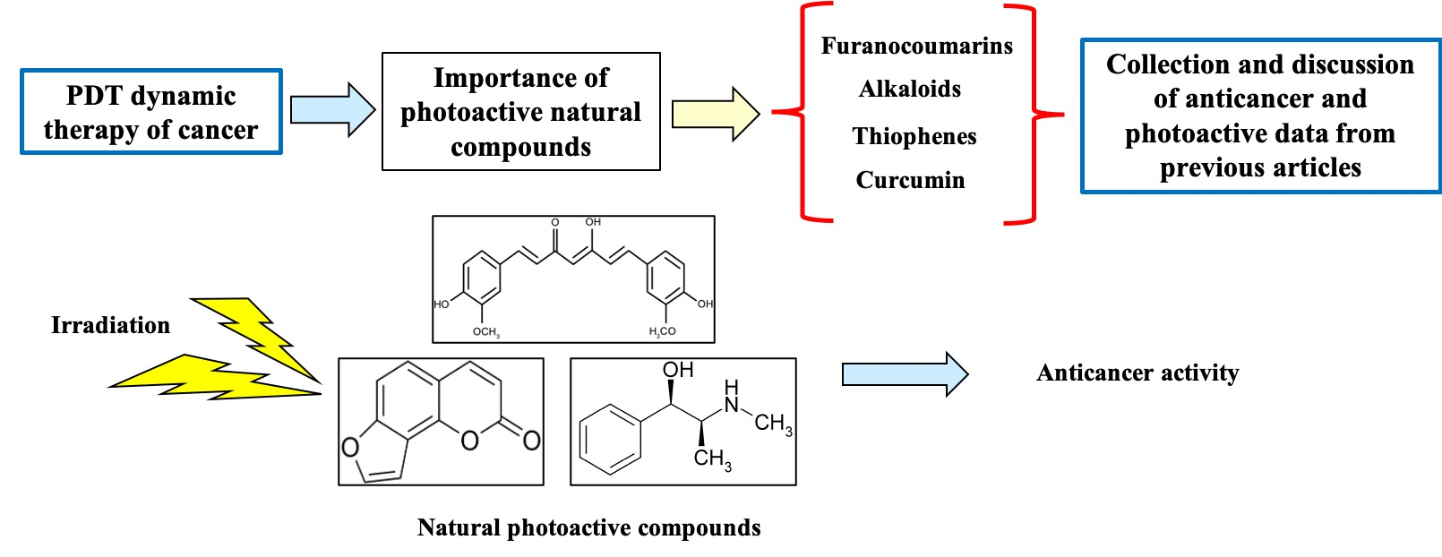
:Cancer is one of the greatest life-threatening diseases conventionally treated using chemo- and radio-therapy. Photodynamic therapy (PDT) is a promising approach to eradicate different types of cancers. PDT requires the administration of photosensitisers (PSs) and photoactivation using a specific wavelength of light in the presence of molecular oxygen. This photoactivation exerts an anticancer effect via apoptosis, necrosis, and autophagy of cancer cells. Recently, various natural compounds that exhibit photosensitising potentials have been identified. Photoactive substances derived from medicinal plants have been found to be safe in comparison with synthetic compounds. Many articles have focused on PDT mechanisms and types of PSs, but limited attention has been paid to the phototoxic activities of phytocompounds. The reduced toxicity and side effects of natural compounds inspire the researchers to identify and use plant extracts or phytocompounds as a potent natural PS candidate for PDT. This review focusses on the importance of common photoactive groups (furanocoumarins, polyacetylenes, thiophenes, curcumins, alkaloids, and anthraquinones), their phototoxic effects, anticancer activity and use as a potent PS for an effective PDT outcome in the treatment of various cancers.

1. Introduction
Cancer is one of the deadliest diseases reported in developed as well as developing countries [1]. It is mainly characterised by the uncontrolled cell growth and development of normal cells due to genetic alterations or exposure to the carcinogenic substances. The mutation of normal cells leads to abnormal cellular proliferation and develops into tumour [2]; this can be either benign, premalignant (non-cancerous) or malignant (cancerous) [3,4]. Presently surgery, radiotherapy, and chemotherapy either as monotherapy or as combined treatments are used in the treatment of cancer. However, these treatments frequently stimulate redundant side effects [2]. Many of the current chemotherapeutic drugs are of low molecular weight with high pharmacokinetic profiles [4]. Hence, in order to achieve the bioavailability and cytotoxicity induction, the drugs are administrated in high concentrations. In photodynamic therapy (PDT), photoactive drugs are generally administered systemically, but because of the precise application of light from the laser source, the cytotoxicity is attained in the tumour location. Due to the lesser drug specificity and toxicity to healthy cells, the chemotherapeutic drugs used in cancer treatments need to be improved.
Photodynamic therapy (PDT) is a promising minimally invasive therapy for the treatment of cancer. This involves the administration of photosensitiser (PS) and subsequent excitation of PS by light irradiation at a specific wavelength. The excited PS then reacts with cellular oxygen and produces reactive oxygen species (ROS). This reaction results in oxidising the cellular macromolecules surrounding tumour cells [5]. This remedial method has been developed over the last few years [6,7], and has not only been utilised in cancer treatment, but also in dermatological [8] and ophthalmic [9] conditions, including psoriasis and age-related diseases [10,11,12]. The use of photodynamic therapy to treat cancers has gained attention around the world [13,14]. The mechanism of PDT is based on various photocatalytic reactions that induce the destruction of cancer cells, and it has been clinically used for the treatment of cancer for over a decade [5]. In the first clinical PDT study reported by Granelli et al. [15], hematoporphyrin was used as a potent photosensitiser (PS) against glioma cancer cells. PDT destroys cancer cells through three fundamentally different pathways, namely, by damaging cancer cells over time, damaging vascular tissues that supply oxygen to cells, and finally by activating host immune response systems [13,16]. Combining PDT with chemotherapy, radiotherapy, and herbal therapy could be an emerging future methodology in cancer treatment. The combination therapy has more of a tendency to reduce the side effects when compared to monotherapy regimes and can significantly lower cancer cell proliferation by improving the drug uptake [17].
Since ancient times, herbal medicine from natural products has been utilised for treating various human ailments [18]. Most current medicines are derived from various medicinal plants, and it is evident that herbal extracts and their compounds should be examined as possible active lead components in cancer drug discoveries [19,20]. Nature is a valuable reserve for medicinal plants, and many of the pharmaceutically active compounds isolated from medicinal plants have not been tested for photoactive properties. There have been few studies attempting to identify new chemical compounds with photoactivity from plant extracts that can be used as potent natural PSs [21,22,23,24,25]. Hypericin (isolated from Hypericum perforatum) is a recognised plant-based PS used in PDT. The in vitro and in vivo studies reported that hypericin PS activated at 594 nm could destroy cancer cell proliferation effectively. The researchers already demonstrated that the effect of herbal extracts combined with illumination could significantly reduce cancer development by prohibiting metabolic viability and proliferation cancer cells [26,27,28,29].
Due to the low or no adverse side effects, herbal products have been used for the treatment of many more ailments than synthetic drugs. Studies have shown that plant-based compounds could be used in the treatment of various cancers [30]. Many phototoxic substances were subsequently reported in various plant species that are equally efficient as of conventional PSs [31]. These studies recommend that natural compounds with photosensitising abilities can be isolated from plants and used as alternatives for conventional PSs used in PDT. In this review, the underlying principles of PDT, PSs and plant-based photoactive compounds were addressed. This review mainly focused on the anticancer activity of furanocoumarins, polyacetylenes, thiophenes, curcumins, alkaloids and anthraquinones in relation to the light-absorbing properties.
2. Basic Principles of Photodynamic Therapy
Photodynamic therapy involves coordination with three individual factors, namely, the photosensitiser, oxygen, and light [7]. These components are not toxic to cells individually, but when irradiated, these can initiate a photochemical reaction that generates highly reactive singlet oxygen (1O2) and cause significant toxicity, leading to cell death. PDT is normally described in two stages, the first is administration of the PS and the second stage is the irradiation. Generally, the effect of PDT is affected by the PS type, dosage, light fluence, as well as exposure time. PDT can be used either before or after chemotherapy, radiotherapy, or surgery without compromise. The clinically approved PS should not accumulate in the body and does not develop resistant cancer cells. Pain during administration and continuous photosensitisation are the major drawbacks of PDT treatment. There are three types of lights ranges from 600 to 800 nm that are commonly used in PDT, namely, blue, red, and infrared lights. Among them, blue light penetrates the tissue the least when compared to red and infrared lights. The wavelengths below 800 nm are mostly used in PDT than higher wavelengths (above 800 nm) due to their lack of photodynamic reactions. The choice of light source is commonly based on PS nature, absorption spectra of PS, location, and size and characteristics of the infected tissue [7,32].
More than 300 chemical compounds have already been identified as potential candidates to be used as PSs. Amongst these, a few were authorised for clinical application in PDT, and others were medically evaluated, whereas some are still under examination [33,34]. We have tabulated some PSs which are used in various cancer treatments in Table 1. Photosensitisers are naturally or chemically produced compound conjugated with a visible light-absorbing chromophore group with a strong chemical absorbance. Choosing the correct PS is the most important phase in PDT for a successful outcome [33,34]. The purity and the presence of a tetrapyrrole structure with good storage stability are the preferable properties of most PSs used in PDT. The potent and effective PS should have the ability to initiate a photodynamic reaction after irradiation with 600–800 nm lights and should not cause any toxicity under dark conditions. It should be easily distinguishable from the body with no or minimum phototoxic side effects [35]. The better diffusion of PS through the cells after long administration might contribute to the effectiveness of PDT [36]. The production of a significant amount of ROS after irradiation that induces apoptosis with less inflammation is most likely to be a suitable PS for PDT application [37,38].
When a PS is subjected to a particular wavelength light, the electron of the outermost orbital will be shifted from the ground state (S0) to the first excited state (S1). Subsequently, the electromagnetic propulsion switches the molecule to an excited triplet state (T1) with a longer life span (Figure 1). In each of these excited states, PSs are quite unstable and lose their energy in the form of fluorescence, phosphorescence, and internal heat conversion. PSs in the T1 state may react photochemically in any of the two pathways. In the type 1 pathway, the excited PS reacts through an electron transfer process with the surrounding oxygen, which ultimately leads to generation of reactive oxygen species (ROS). Such free radicals communicate readily with the biomolecules (lipids, peptides, proteins, and nucleic acids) and destroy them [39,40]. In contrast, in the type 2 pathway, the energy is directly transferred from the T1 state of the PS to the S0-state oxygen. This results in the ground-state PS transformation and excited-state singlet reactive oxygen. The disruption caused by PDT is local because both singlet oxygen as well as free radicals have a short half-life between 10–300 nanoseconds and a small diffusion distance of 10–55 nm [41].

Table 1.
List of photosensitisers used in photodynamic therapy of various cancers.
| Photosensitiser | Commercial Name | λ max (nm) | Structure | Type of Cancer | Reference |
|---|---|---|---|---|---|
| First-Generation Photosensitiser | |||||
| Hematoporphyrin derivatives | Photofrin Photoheme | 630 |  | Lung, bladder, skin, cervical, breast cancer. | [17,42] |
| Second-Generation Photosensitisers | |||||
| 5-Aminolevulinic acid | Levulan Alasens | 635 |  | Bladder, skin, lung, ovary and gastrointestinal cancer. | [43,44,45] |
| Meta-tetra(hydroxyphenyl) chlorin | Foscan | 652 |  | Approved drug for the treatment of bronchial and oesophageal cancers. | [46,47,48] |
| Chlorin e6 | MACEDACEPhotoditazine | 664 |  | Gynaecological diseases, prostate cancer, fibrosarcoma, Liver, brain, lung, and oral cancers. | [49,50,51] |
| Benzoporphyrin | Visudyne | 690 |  | Prostate and skin cancer. | [52,53] |
| Texaphyrins | Lutrin, Antrin, Optrin, Xcytrin | 720–760 |  | Hepatocellular cancer, leukaemia, nasopharyngeal carcinoma, colon, prostate, bronchial and oesophageal cancers. | [54,55,56,57,58] |
| Phthalocyanines | Photosense | 640–690 |  | Breast, cervical, skin, lung, liver, colon and gastrointestinal cancers. | [17,59,60,61] |
| Purpurins | Purlytin | 660 |  | Breast cancer, prostate cancer and Kaposi’s sarcoma. | [62,63,64] |
3. PDT’s Cancer Cell Death Mechanism
PDT’s cancer cell death mechanism starts after the activation of administrated PS by a specific wavelength of light. The PS’s hydrophilic, hydrophobic, and ionic charge-related interaction nature plays an important role in the targeting of particular cancer cell receptor (globulins and Low-Density Lipoprotein (LDL) receptors) [34]. After the activation of PSs, the cancer cell death mechanism might occur in three main pathways (Figure 1), namely, apoptosis, necrosis, and autophagy [33,65,66]. However, the level of cell death induced by PDT may be affected by various aspects, including subcellular localisation, bioavailability, the physicochemical nature of the PS, the cellular oxygen concentration, as well as the applied light intensity and wavelength [67]. In general, the light-absorbed PS interacts with cellular oxygen and highly produce ROS (hydroperoxides, superoxide, or hydroxyl radicals) as well as singlet oxygen (1O2). These produced ROS can induce cancerous cell death via the above-mentioned mechanisms. Both type 1 and 2 reactions may occur separately or in combination, but type 1 (generation of ROS followed by the apoptotic cell death mechanism) is commonly exhibited by most approved PSs [67].
4. PS from Natural Resources
The effectiveness of PDT is mainly based on the PS; it should possess all the properties of the PS as previously explained. The PS can be divided into first- and second-generation types. Hematoporphyrin and its derivative Photofrin®® were classified as first-generation PSs. After extensive studies, new and improved second-generation PSs, such as Levulan®®, Alasens®®, and Foscan have been introduced for PDT application (Table 1). Although these are widely used for various cancer treatments, their clinical usage is limited by various drawbacks such as lack of chemical purity, a longer half-life, accumulation in tissues and poor ability in relation to depth of tissue penetration [31,32,33,34,35,36,37,38,39].
Subsequently, there are some research reports on PSs with potent pharmaceutical properties to overcome the shortcomings of first- (Porphyrin based sensitisers) and second-generation (non-porphyrin derivatives) PSs [35,36,37]. These drawbacks of current PSs specifically imply the need for new PSs as anticancer agents from natural resources. The discovery of new PS compounds with anticipated pharmacological properties and clinical application is an inspiring task. Recently, a greater number of plant-based compounds have been reported for their anticancer activity, and these compounds are pharmaceutically very important for the development of potent drugs. The use of light to activate the bioactivities of natural products is generally called photopharmacology (a combination of photophysics and photochemistry). The absorption of lights (λ < 350 nm) by a molecule mainly depends on the chromophore compound attached (Figure 2) [36,37]. This review presents an overview of natural photoactive compounds as potent third-generation photosensitisers in the improvement of PSs in relation to their prospective application in cancer treatments.
5. Natural Photoactive Compounds from Plants
The search for the natural compounds as efficient PSs has been progressively moving forward because of the side effects caused by current synthetic drugs. The advanced isolation, identification and characterisation techniques improved the extraction of desirable compounds from plants. Recently, using these advanced techniques, the isolation of natural photoactive compounds has become easy. Although there have been few studies attempting to identify new chemical compounds with photoactivity from plant extracts, this review discusses the photoactivity as well as the anticancer activity of some plant-based compounds such as furanocoumarins, polyacetylenes, thiophenes, curcumins, alkaloids and anthraquinones (Table 2).
5.1. Furanocoumarins
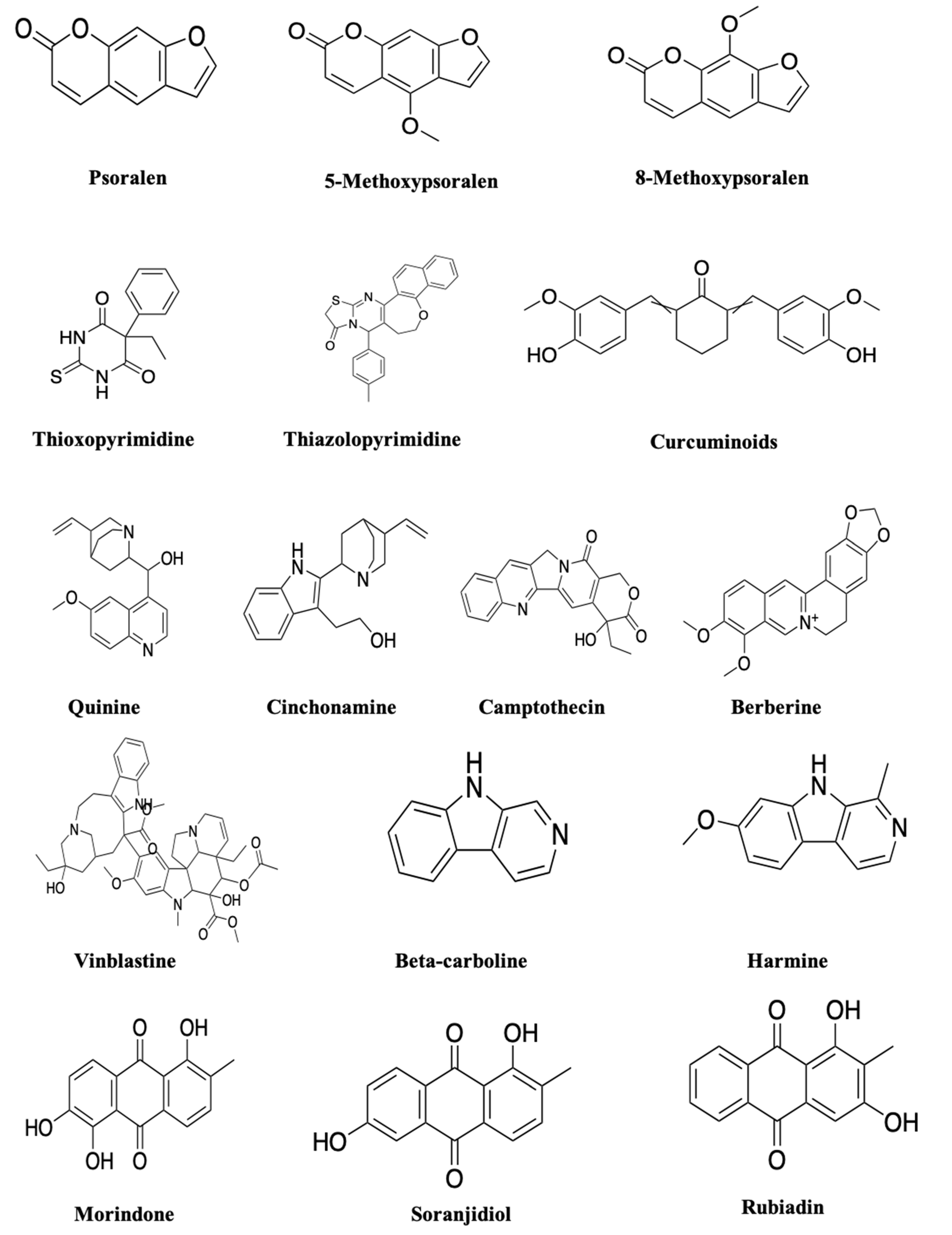
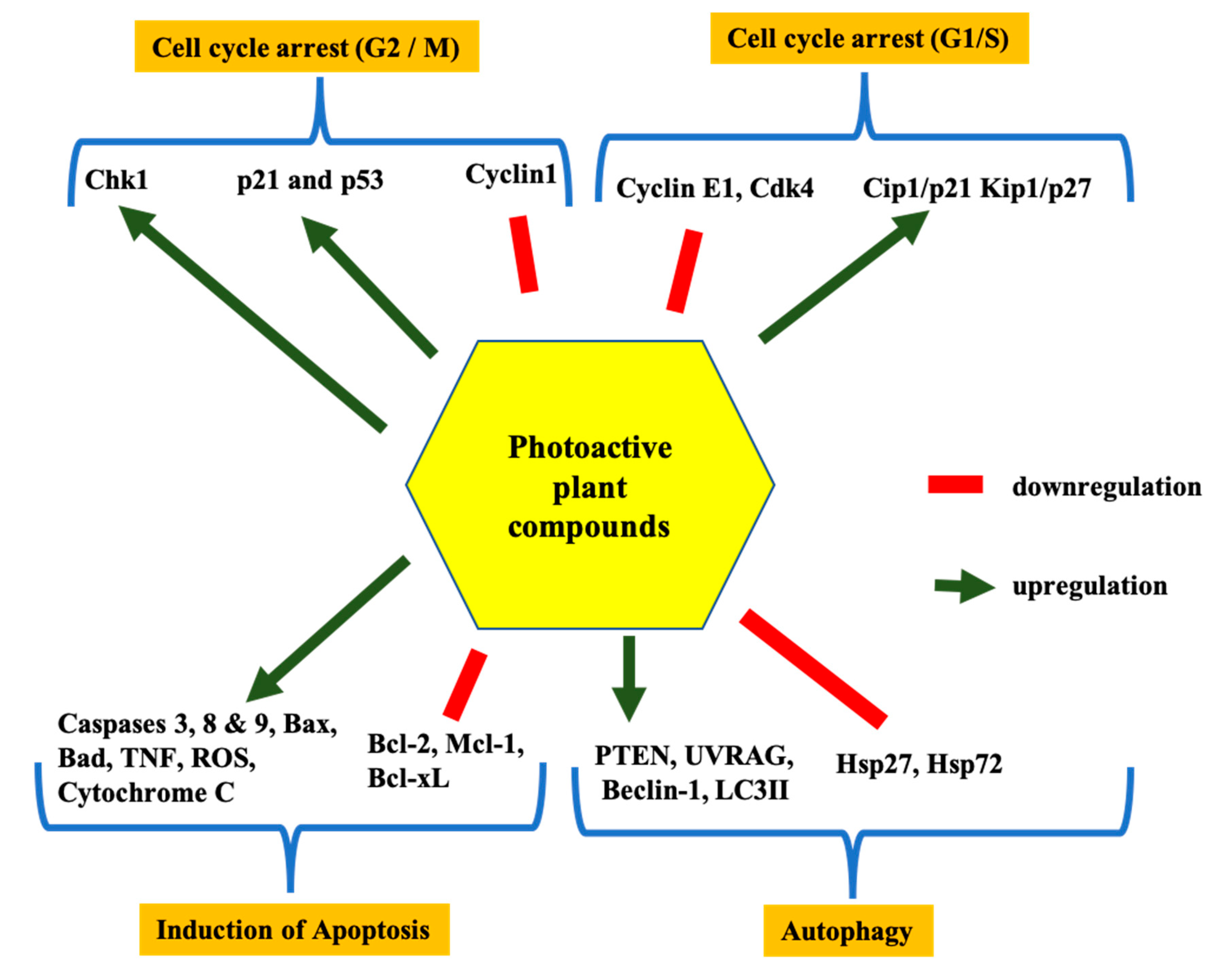
The secondary metabolites, furanocoumarins (FC; Figure 3), are mostly present in higher plants. The photoactive furanocoumarins were mainly composed of a linear core, and the biological distribution, photochemistry and phototoxicity mechanisms of FC after PUVA (psoralen and long-wave ultraviolet radiation) irradiation were reported in previous study [92]. In terms of phototherapy, psoralen is activated in the wavelength range of 300–400 nm ultraviolet radiation to treat psoriasis, dermatitis, eczema, and other skin problems [92]. Over a few years, many researchers reported anticancer activity of FCs against various types of cancer such as breast, skin, and leukaemia. FCs modulate several pathways inducing cancer cell death by inhibiting signal transducer and activator of transcription 3 (STAT3), nuclear factor-κB (NF-κB), phosphatidylinositol-3-kinase and AKT protein expression (Figure 4). These pathways play a key role in tumour development through regular activation of several inflammatory genes. Studies show that FC displayed potent activity against breast cancer development by inhibiting STAT3 protein expression [93]. Panno et al. [92,93], demonstrated inhibition of breast cancer cell growth in a dose-dependent manner through activation of p53 and Bax, leading to the cleavage of caspase 9. In contrast, in leukaemia cells, FC inactivated the JAK (Janus-activated kinase), protein c-Src, and STAT3, and downregulated Bcl-xl and Bcl-2 proteins which are responsible for apoptosis [93,94,95,96]. The enhanced activity against the malignant melanoma cell line (A375) after UV irradiation of plant extracts containing FC also supported the possible photoactive nature of FCs [93,94,95,96]. The linear forms of furanocoumarins like psoralen and its derivatives 5-methoxypsoralen (5-MOP) and 8-methoxypsoralen (8-MOP) are reported to increase the cytotoxicity after irradiation by ultraviolet light in the 320–400 nm wavelength range against cutaneous T-cell lymphoma [97,98], and photoactivated psoralens induce apoptosis by forming adducts with DNA. This leads to the activation of p21waf/Cip and p53 and subsequently leads to cell death by the release of mitochondrial cytochrome c. The photoactivation of psoralen can also cause cell death by blocking oncogenic receptor tyrosine kinase signalling and the PI3K pathway by interfering with efficient recruitment of effector Akt kinase to the activated plasma membrane [92,93,94,95,96,97,98,99,100,101,102,103,104]. PUVA treatment was found effectively against B16F10 murine melanoma cells by cell cycle arrest in G2/M phases [93,94,95,96].
5.2. Polyacetylene and Thiophenes
These group compounds are characterised by a triple bond carbon–carbon molecule [75] and thiophenes compounds. Generally, the aliphatic compounds conjugated with three or more acetylenic bonds are considered phototoxic in nature. Among these, polyacetylenes compounds can produce 1O2 under irradiation and thiophenes can provide high photo yield, leading to type 2 PDT reaction yields [105]. The polyacetylene and thiophenes compounds were reported to be activated or excited at a wavelength range of 314–350 nm absorbance maximum for the relevant photobiological effects [75]. The derivatives of these compounds were reported with a variety of potent biological activities, including analgesic, anti-inflammatory, antitumour, and antimicrobial activities. Some of the derivatives substituted by pyrimidines show antimicrobial, anti-inflammatory, and antitumour activities. A few numbers of thiophenes were reported for their cytotoxic effects against human cancer cell lines. Echinops grijisii root-derived thiophenes exhibited cytotoxicity against HL-60, K562 and MCF-7 cells [106]. Notably, derivatives such as thioxopyrimidine and thiazolopyrimidine were reported to possess anticancer activities against MCF-7 (breast adenocarcinoma), NCI-H460 (non-small cell lung cancer), and SF-268 (CNS cancer) cells. These acetylenic compounds and derivatives when combined with PDT might improve the efficacy of various cancer treatments [107,108,109,110,111,112]. The UV irradiation of some thiophenes also showed increased cytotoxic activities [113]; this might be due to their instable nature under UV radiation. Hence, the UV irradiation of polyacetylene and thiophene compounds can form free radicals that would induce cell death. Due to the ability to produce ROS after irradiation, these compounds can be used as an alternative PS from natural sources.
5.3. Curcumins
Curcumin (CU) is a plant-based therapeutic compound isolated from rhizome of Curcuma longa of the Zingiberaceae family. Curcuminoid is one of the most extensively studied plant-derived bioactive compounds [114]. Since the 1980s, the photobiological potential of CU was of great interest [115,116], and studies described CU as a desirable, highly promising photosensitiser [115,117,118,119,120,121]. The foremost property of CU is that it is biologically safe even at higher doses, and it can be easily produced on a large scale [117,122]. The photobleaching analysis reported the degradation profile of curcumin derivatives and its ability to produce singlet oxygen species [123]. CU was characterised by an absorption spectrum of 300–500 nm with a high extinction coefficient. This suggests that CU can induce a strong phototoxic reaction even at lower concentrations [124,125,126]. Curcumin is considered as a potential anticancer agent and inhibits cancer cell proliferation in breast, lung, colon, kidney, ovary, and liver cancers [114]. The in vitro and in vivo anticancer activity of curcumin has been proved by inhibition of various transcription factors such as NF-κB, AP-1, VEGF, iNOS, COX-2, 5-LOX, MMP-2, MMP-9 and IL-8, which are mainly responsible for angiogenesis and tumour growth [127,128]. The administration of curcumin significantly reduced the expression of the CDK4/cylin D1 complex by inhibiting p53 expression and causing the apoptotic process by inducing ROS generation. Furthermore, the enhanced antitumour activity was noted after UVB irradiation of curcumin by caspase activation on HaCaT (human keratinocyte cell) cells. It was also efficient against MCF-7 breast cancer cells at 30 J/cm2 [77,78]. These data suggested that curcumin may act as a potent anticancer agent by preventing cancer progression, migration and invasion [129,130,131]. Dovigo et al. [132] found the light absorption ability of CU in the range of 300 and 500 nm with a maximum absorption at 430 nm, which might support its usage as a PS. The ROS-inducing and anticancer ability of CU makes it a potent candidate as a natural PS [133]. Nevertheless, Chan and Wu [134] observed that the photoactive nature of CU on human epidermal A431 carcinoma cells and the higher amount of CU also affect the irradiation penetration [132,134,135]. The irradiation of CU under a 290–320 nm UVB light source with the fluence of 100 mJ/cm2 induced apoptosis in HaCaT keratinocyte cells [78]. Based on the above reports, CU can be used as a natural PS, and it can achieve high efficacy at a low concentration when combined with PDT. The existing PDT and photoactive reports on CU suggest that CU can be used as a potential and promising natural PS in PDT. In conclusion, CU can be a potent photosensitiser in the treatment of cancer and skin infections. Therefore, investigating the photodynamic potential of CU derivatives in terms of higher absorption and extinction coefficient will contribute to the increased efficacy of photodynamic toxicity.
5.4. Alkaloids
Alkaloids, a diverse secondary metabolites group from higher plants, contain a heterocyclic structure with a nitrogen atom in the ring [136]. Nitrogen-containing alkaloids are normally photoactive in nature, e.g., quinine and cinchonamine. The alkaloids were reported for many significant properties, such as analgesic and anticancer activity [136,137,138,139]. The alkaloids camptothecin and vinblastine are few alkaloids were successfully utilised as chemotherapeutic drugs [138,140]. The anticancer activity of alkaloids was proved by different studies by means of disturbing tumour progression by induction of cell cycle arrest at the G1 or G2/M phases, regulating cyclin-dependent kinase (CDK) and promoting apoptosis as well as autophagy in tumour cells. Furthermore, these compounds induce apoptosis by regulating Bax, Bcl-2, Bcl-xL, NF-κB and various caspase proteins [140,141,142,143]. In addition, the combination of alkaloids with chemotherapeutic drugs and irradiation also enhanced the biological activities [144,145]. Furthermore, alkaloids induce the formation of intracellular ROS in cancer cells, which leads to the destruction of cancer cell metabolism [140,141,142,143]. The photochemically best-known alkaloid is berberine; Luiza Andreazza et al. [146], Bhattacharyya et al. [147] and Inbaraj et al. [81] reported the antitumour activity of berberine upon UV and blue light irradiation. The irradiation of berberine at 410 nm proved to be effective in controlling brain cancer cell growth [146]. Beta-carboline and harmine are also a noticeable alkaloid with a photoactive nature and are reported to produce a significant amount of ROS after irradiation [148], which is considered an important feature of potent PSs. The photoactivity of harmine was proved by the UVA (long-wave ultraviolet radiation) irradiation against tumour cell lines [148]. Berberine was extensively investigated as a potential photosensitising agent for PDT [149,150,151]. The fluorescent active nature of berberine is indicated for its efficiency in PDT [149]; thus, berberine and its associated alkaloids can be used as a new candidate for photodynamic therapy [150]. Different studies have proved the photosensitising as well as ROS generation ability of alkaloids in the presence of a light source [151]. Therefore, berberine can be studied as a natural photosensitiser in PDT applications with minimal side effects.
5.5. Anthraquinones (AQ)
Anthraquinone are the largest group among natural quinones from higher plants, which, including naphthoquinones and benzoquinones, includes over 700 compounds, including emodin, physcion, catenarin and rhein [152,153]. The hydroxylation pattern, however, dictates the possibility of AQs’ photopharmacological properties. Notably, AQs’ aminoanthraquinone derivatives were studied extensively for their photoactive properties among the plant compounds due to their UV/vis absorption and photosensitising nature [154,155]. The AQs were reported as kinase and tyrosinase inhibitors as well as cytotoxicity agents. The M. elliptica AQs such as morindone, soranjidiol and rubiadin were also reported for their antitumour activity against lymphocytic leukaemia (P-388) cells [156]. The anthraquinones isolated from H. pustulata leaves and stem exhibited photosensitising properties by generation of singlet oxygen and/or superoxide anion radicals [157]. Comini et al. [158] reported that irradiation of AQs (soranjidiol and rubiadin) under visible radiation of 380–480 nm can promote the anti-proliferative effect on MCF-7 breast cancer cells. In addition, Montoya et al. [157] and Vittar et al. [159] also reported photosensitisation effects of AQs in Balb/c mice and their leukocyte-inhibiting ability in a dose-dependent manner by inducing apoptosis, necrosis, or autophagy. These study results show the photoactive nature of AQs to inhibit the proliferation of cancerous cells. Based on the previous studies and the above data, molecular targets responsible for the anticancer activity of AQs and major phytocompounds are summarised in Figure 4.
6. Theorical Studies for Assessing the Photoactivity of Natural Compounds
The development of various antitumor compounds with different molecular targets initiated an exciting field of investigation with recently developed theoretical studies. The theoretical studies including density functional theory (DFT) and time-dependent density functional theory (TD-DFT) were used to assess a series of photophysical properties, including absorption spectra, excitation energies (singlet and triplet) and spin–orbit matrix elements. All the reported compounds are potential UVA chemotherapeutic agents which require the lowest triplet-state energy for producing highly cytotoxic ROS [160,161].
7. Advantages and Scope of Natural PSs
The anticancer property of many plant extracts and bioactive compounds have been analysed, but not so much in terms of as sources of photosensitisers. Selecting proper PSs is the first step in PDT, and, to date, only a few PSs are clinically approved, such as Photofrin, Foscan and Levulan. The present study explored the common photoactive nature of various phytocompounds. Many of the natural photoactive compounds were reported for their non-toxicity against normal cells and toxicity towards cancer cells. The important property of a PS is the nontoxic nature during the absence of light. The increasing activity of extracts or phytocompounds after irradiation by light makes them good photosensitising candidates for PDT. Another important feature that makes photoactive plant compounds suitable photosensitisers is their absorption maxima at 400–700 nm, which is biologically compatible. The selective nature of these compounds is important in clinical PDT to overcome side effects. Future studies are warranted to isolate and evaluate these specific photoactive compounds from plants to be used as a potent PSs for PDT for cancer and related disorders [162,163].
8. Conclusions and Future Perspectives
As discussed in this review, plant-based photoactive compounds can be used as a natural PSs in PDT application. There are wide range of unknown natural compounds with different photoactive and phototoxic properties. This review summarises and encourages researchers to identify and elucidate natural photoactive plant-based compounds and to use them as alternatives for the synthesis PSs for a better PDT outcome. Furthermore, discovering natural phototoxic agents as PSs will be helpful to reduce toxicity and side effects and improve selectivity. In conclusion, use the plant-based PSs in PDT typically causes less and minimal adverse effects than other treatments that are commonly used in cancer therapies.
Author Contributions
Conceptualisation and writing, K.M.; review and editing, K.M., B.G., T.P. and H.A.; supervision, B.G., T.P. and H.A. The final version of the submitted manuscript was read and agreed by all the authors. All authors have read and agreed to the published version of the manuscript.
Funding
This work is supported by Science and Engineering Research Board (SERB), Department of Science and Technology (DST), Government of India in the form of SERB—Overseas Visiting Doctoral Fellowship (ODF/2018/000072). This work is also based on the research supported by the South African Research Chairs Initiative of the Department of Science and Technology and National Research Foundation of South Africa (Grant No. 98337).
Acknowledgments
The authors sincerely thank the Science and Engineering Research Board (SERB), Department of Science and Technology (DST), Government of India and the Laser research centre, University of Johannesburg, South Africa for their support.
Conflicts of Interest
The authors declare no conflict of interest.
References
- El-Hussein, A.; Harith, M.; Abrahamse, H. Assessment of DNA Damage after Photodynamic Therapy Using a Metallophthalocyanine Photosensitizer. Int. J. Photoenergy 2012, 2012, 1–10. [Google Scholar] [CrossRef] [Green Version]
- Klug, W.S.; Cummings, M.R.; Spencer, C.A. Concepts of Genetics, 8th ed.; Pearson Education International: Upper Saddle River, NJ, USA, 2006. [Google Scholar]
- Matés, J.M.; Segura, J.A.; Alonso, F.J.; Márquez, J.D. Intracellular redox status and oxidative stress: Implications for cell proliferation, apoptosis, and carcinogenesis. Arch. Toxicol. 2008, 82, 273–299. [Google Scholar] [CrossRef] [PubMed]
- Santiago-Montero, R.; Sossa-Azuela, H.; Gutiérrez-Hernández, D.; Zamudio, V.; Hernández-Bautista, I.; Valadez-Godínez, S. Novel Mathematical Model of Breast Cancer Diagnostics Using an Associative Pattern Classification. Diagnostics 2020, 10, 136. [Google Scholar] [CrossRef] [PubMed] [Green Version]
- Zhou, Z.; Song, J.; Nie, L.; Chen, X.S. Reactive oxygen species generating systems meeting challenges of photodynamic cancer therapy. Chem. Soc. Rev. 2016, 45, 6597–6626. [Google Scholar] [CrossRef] [PubMed] [Green Version]
- Baskaran, R.; Lee, J.; Yang, S.-G. Clinical development of photodynamic agents and therapeutic applications. Biomater. Res. 2018, 22, 25. [Google Scholar] [CrossRef] [PubMed]
- De Almeida, D.R.Q.; Terra, L.F.; Labriola, L.; Dos Santos, A.F.; Baptista, M.S. Photodynamic therapy in cancer treatment—An update review. J. Cancer Metastasis Treat. 2019, 2019, 10–20517. [Google Scholar] [CrossRef] [Green Version]
- Nguyen, K.; Khachemoune, A. An update on topical photodynamic therapy for clinical dermatologists. J. Dermatol. Treat. 2019, 30, 732–744. [Google Scholar] [CrossRef]
- Blasi, M.A.; Pagliara, M.M.; Lanza, A.; Sammarco, M.G.; Caputo, C.G.; Grimaldi, G.; Scupola, A. Photodynamic Therapy in Ocular Oncology. Biomedicines 2018, 6, 17. [Google Scholar] [CrossRef] [Green Version]
- Choi, Y.M.; Adelzadeh, L.; Wu, J.J. Photodynamic therapy for psoriasis. J. Dermatol. Treat. 2014, 26, 202–207. [Google Scholar] [CrossRef]
- Silva, A.M.; Siopa, J.R.; Martins-Gomes, C.; Teixeira, M.D.C.; Santos, D.J.; Pires, M.D.A.; Andreani, T. New strategies for the treatment of autoimmune diseases using nanotechnologies. Emerg. Nanotechnol. Immunol. 2018, 135–163. [Google Scholar] [CrossRef]
- Hatz, K.; Schneider, U.; Henrich, P.B.; Braun, B.; Sacu, S. Ranibizumab plus Verteporfin Photodynamic Therapy in Neovascular Age-Related Macular Degeneration: 12 Months of Retreatment and Vision Outcomes from a Randomized Study. Ophthalmologia 2014, 233, 66–73. [Google Scholar] [CrossRef] [PubMed]
- Oniszczuk, A.; Wojtunik-Kulesza, K.A.; Oniszczuk, T.; Kasprzak, K. The potential of photodynamic therapy (PDT)—Experimental investigations and clinical use. Biomed. Pharmacother. 2016, 83, 912–929. [Google Scholar] [CrossRef] [PubMed]
- Zhang, J.; Jiang, C.; Longo, J.P.F.; Azevedo, R.B.; Zhang, H.; Muehlmann, L.A. An updated overview on the development of new photosensitizers for anticancer photodynamic therapy. Acta Pharm. Sin. B 2017, 8, 137–146. [Google Scholar] [CrossRef] [PubMed]
- Granelli, S.G.; Diamond, I.; McDonagh, A.F.; Wilson, C.B.; Nielsen, S.L. Photochemotherapy of glioma cells by visible light and hematoporphyrin. Cancer Res. 1975, 35, 2567–2570. [Google Scholar]
- Abrahamse, H.; Hamblin, M.R. Photomedicine and Stem Cells: The Janus Face of Photodynamic Therapy (PDT) to Kill Cancer Stem Cells, and Photobiomodulation (PBM) to Stimulate Normal Stem Cells; Morgan & Claypool Publishers: Bristol, UK, 2017. [Google Scholar]
- Moreira, L.M.; Dos Santos, F.V.; Lyon, J.P.; Maftoum-Costa, M.; Soares, C.P.; Da Silva, N.S. Photodynamic Therapy: Porphyrins and Phthalocyanines as Photosensitizers. Aust. J. Chem. 2008, 61, 741–754. [Google Scholar] [CrossRef] [Green Version]
- Mohammadi, A.; Mansoori, B.; Baradaran, B. Regulation of miRNAs by herbal medicine: An emerging field in cancer therapies. Biomed. Pharmacother. 2017, 86, 262–270. [Google Scholar] [CrossRef]
- Mohammadi, A.; Mansoori, B.; Aghapour, M.; Baradaran, B. Urtica dioica dichloromethane extract induce apoptosis from intrinsic pathway on human prostate cancer cells (PC3). Cell. Mol. Boil. 2016, 62, 78–83. [Google Scholar]
- Mohammadi, A.; Mansoori, B.; Goldar, S.; Shanehbandi, D.; Khaze, V.; Mohammadnejad, L.; Baghbani, E.; Baradaran, B. Effects of Urtica dioica dichloromethane extract on cell apoptosis and related gene expression in human breast cancer cell line (MDA-MB-468). Cell. Mol. Boil. 2016, 62, 62–67. [Google Scholar]
- Alali, F.Q.; Tawaha, K. Dereplication of bioactive constituents of the genus hypericum using LC-(+,−)-ESI-MS and LC-PDA techniques: Hypericum triquterifolium as a case study. Saudi Pharm. J. 2009, 17, 269–274. [Google Scholar] [CrossRef] [Green Version]
- Bailly, C. Ready for a comeback of natural products in oncology. Biochem. Pharmacol. 2009, 77, 1447–1457. [Google Scholar] [CrossRef] [Green Version]
- Mishra, B.B.; Tiwari, V.K. Natural products: An evolving role in future drug discovery. Eur. J. Med. Chem. 2011, 46, 4769–4807. [Google Scholar] [CrossRef] [PubMed]
- Rodrigues, M.C. Photodynamic Therapy Based on Arrabidaea chica (Crajiru) Extract Nanoemulsion: In vitro Activity against Monolayers and Spheroids of Human Mammary Adenocarcinoma MCF-7 Cells. J. Nanomed. Nanotechnol. 2015, 6, 1–6. [Google Scholar] [CrossRef]
- Tan, P.J.; Appleton, D.R.; Mustafa, M.R.; Lee, H.B. Rapid Identification of Cyclic Tetrapyrrolic Photosensitisers for Photodynamic Therapy Using On-line Hyphenated LC-PDA-MS Coupled with Photo-cytotoxicity Assay. Phytochem. Anal. 2011, 23, 52–59. [Google Scholar] [CrossRef]
- Skalkos, D.; Gioti, E.; Stalikas, C.; Meyer, H.; Papazoglou, T.; Filippidis, G.; Papazoglou, T.G. Photophysical properties of Hypericum perforatum L. extracts—Novel photosensitizers for PDT. J. Photochem. Photobiol. B Boil. 2006, 82, 146–151. [Google Scholar] [CrossRef] [PubMed]
- Zeisser-Labouèbe, M.; Lange, N.; Gurny, R.; Delie, F. Hypericin-loaded nanoparticles for the photodynamic treatment of ovarian cancer. Int. J. Pharm. 2006, 326, 174–181. [Google Scholar] [CrossRef] [PubMed]
- Mirmalek, S.A.; Azizi, M.A.; Jangholi, E.; Yadollah-Damavandi, S.; Javidi, M.A.; Parsa, Y.; Parsa, T.; Salimi-Tabatabaee, S.A.; Kolagar, H.G.; Alizadeh-Navaei, R. Cytotoxic and apoptogenic effect of hypericin, the bioactive component of Hypericum perforatum on the MCF-7 human breast cancer cell line. Cancer Cell Int. 2016, 16, 3. [Google Scholar] [CrossRef] [Green Version]
- Yonar, D.; Süloğlu, A.K.; Selmanoğlu, G.; Sünnetçioğlu, M.M. An Electron paramagnetic resonance (EPR) spin labeling study in HT-29 Colon adenocarcinoma cells after Hypericin-mediated photodynamic therapy. BMC Mol. Cell Boil. 2019, 20, 16. [Google Scholar] [CrossRef] [Green Version]
- Aggarwal, B.B.; Ichikawa, H.; Garodia, P.; Weerasinghe, P.; Sethi, G.; Bhatt, I.D.; Pandey, M.K.; Shishodia, S.; Nair, M.G. From traditional Ayurvedic medicine to modern medicine: Identification of therapeutic targets for suppression of inflammation and cancer. Expert Opin. Ther. Targets 2006, 10, 87–118. [Google Scholar] [CrossRef]
- Chaturvedi, D.; Singh, K.; Singh, V.K. Therapeutic and pharmacological aspects of photodynamic product chlorophyllin. Eur. J. Biol. Res. 2019, 9, 64–76. [Google Scholar]
- Juzeniene, A.; Nielsen, K.P.; Moan, J. Biophysical Aspects of Photodynamic Therapy. J. Environ. Pathol. Toxicol. Oncol. 2006, 25, 7–28. [Google Scholar] [CrossRef]
- George, B.P.; Abrahamse, H. A Review on Novel Breast Cancer Therapies: Photodynamic Therapy and Plant Derived Agent Induced Cell Death Mechanisms. Anti-Cancer Agents Med. Chem. 2016, 15, 1. [Google Scholar] [CrossRef]
- Aniogo, E.C.; George, B.P.; Abrahamse, H. The role of photodynamic therapy on multidrug resistant breast cancer. Cancer Cell Int. 2019, 19, 91. [Google Scholar] [CrossRef] [PubMed]
- Allison, R.R.; Sibata, C.H. Oncologic photodynamic therapy photosensitizers: A clinical review. Photodiagn. Photodyn. Ther. 2010, 7, 61–75. [Google Scholar] [CrossRef] [PubMed]
- Chen, B.; Roskams, T.; De Witte, P.A.M. Antivascular tumor eradication by hypericin-mediated photodynamic therapy. Photochem. Photobiol. 2002, 76, 509. [Google Scholar] [CrossRef]
- Ascencio, M.; Collinet, P.; Farine, M.; Mordon, S. Protoporphyrin IX fluorescence photobleaching is a useful tool to predict the response of rat ovarian cancer following hexaminolevulinate photodynamic therapy. Lasers Surg. Med. 2008, 40, 332–341. [Google Scholar] [CrossRef]
- Garg, A.D.; Nowis, D.; Golab, J.; Vandenabeele, P.; Krysko, D.V.; Agostinis, P. Immunogenic cell death, DAMPs and anticancer therapeutics: An emerging amalgamation. Biochim. Biophys. Acta (BBA) Rev. Cancer 2010, 1805, 53–71. [Google Scholar] [CrossRef]
- Castano, A.P.; Demidova, T.N.; Hamblin, M.R. Mechanisms in photodynamic therapy: Part three-Photosensitizer pharmacokinetics, biodistribution, tumor localization and modes of tumor destruction. Photodiagn. Photodyn. Ther. 2005, 2, 91–106. [Google Scholar] [CrossRef] [Green Version]
- Ogilby, P.R. Singlet oxygen: There is indeed something new under the sun. Chem. Soc. Rev. 2010, 39, 3181. [Google Scholar] [CrossRef]
- Oseroff, A.R.; Blumenson, L.R.; Wilson, B.D.; Mang, T.S.; Bellnier, D.A.; Parsons, J.C.; Frawley, N.; Cooper, M.; Zeitouni, N.; Dougherty, T.J. A dose ranging study of photodynamic therapy with porfimer sodium (Photofrin®) for treatment of basal cell carcinoma. Lasers Surg. Med. 2006, 38, 417–426. [Google Scholar] [CrossRef]
- Juzeniene, A.; Juzenas, P.; Ma, L.-W.; Iani, V.; Moan, J. Effectiveness of different light sources for 5-aminolevulinic acid photodynamic therapy. Lasers Med. Sci. 2004, 19, 139–149. [Google Scholar] [CrossRef]
- Lang, P. Methyl aminolaevulinate–photodynamic therapy: A review of clinical trials in the treatment of actinic keratoses and nonmelanoma skin cancer. Yearb. Dermatol. Dermatol. Surg. 2008, 2008, 322–323. [Google Scholar] [CrossRef]
- Jeffes, E.W.; McCullough, J.L.; Weinstein, G.D.; Kaplan, R.; Glazer, S.D.; Taylor, J. Photodynamic therapy of actinic keratoses with topical aminolevulinic acid hydrochloride and fluorescent blue light. J. Am. Acad. Dermatol. 2001, 45, 96–104. [Google Scholar] [CrossRef] [PubMed]
- Lu, K.; He, C.; Lin, W. A Chlorin-Based Nanoscale Metal–Organic Framework for Photodynamic Therapy of Colon Cancers. J. Am. Chem. Soc. 2015, 137, 7600–7603. [Google Scholar] [CrossRef] [PubMed] [Green Version]
- Moore, C.M.; Nathan, T.; Lees, W.; Mosse, C.; Freeman, A.; Emberton, M.; Bown, S. Photodynamic therapy using meso tetra hydroxy phenyl chlorin (mTHPC) in early prostate cancer. Lasers Surg. Med. 2006, 38, 356–363. [Google Scholar] [CrossRef]
- Grosjean, P.; Savary, J.-F.; Wagnières, G.; Mizeret, J.; Woodtli, A.; Theumann, J.-F.; Fontolliet, C.; Bergh, H.V.D.; Monnier, P. Tetra(m-hydroxyphenyl)chlorin clinical photodynamic therapy of early bronchial and oesophageal cancers. Lasers Med. Sci. 1996, 11, 227–235. [Google Scholar] [CrossRef]
- Kessel, D. Pharmacokinetics of N-aspartyl chlorin e6 in cancer patients. J. Photochem. Photobiol. B Boil. 1997, 39, 81–83. [Google Scholar] [CrossRef]
- Taber, S.W.; Fingar, V.H.; Coots, C.T.; Wieman, T.J. Photodynamic therapy using mono-L-aspartyl chlorin e6 (Npe6) for the treatment of cutaneous disease: A Phase I clinical study. Clin. Cancer Res. 1998, 4, 2741–2746. [Google Scholar]
- Lagudaev, D.M. Sorokatyĭ Photodynamic therapy of prostatic adenoma. Urologiia 2007, 4, 34–37. [Google Scholar]
- Momma, T.; Hamblin, M.R.; Wu, H.C.; Hasan, T. Photodynamic therapy of orthotopic prostate cancer with benzoporphyrin derivative: Local control and distant metastasis. Cancer Res. 1998, 58, 5425–5431. [Google Scholar]
- Levy, J.G.; Waterfield, E.; Richter, A.M.; Smits, C.; Lui, H.; Hruza, L.; Anderson, R.R.; Salvatori, V. Photodynamic therapy of malignancies with benzoporphyrin derivative monoacid ring A. Europto Biomedical Optics ’93 1994, 2078, 91–101. [Google Scholar] [CrossRef]
- Young, S.W.; Woodburn, K.W.; Wright, M.; Mody, T.D.; Fan, Q.; Sessler, J.L.; Dow, W.C.; Miller, R.A. Lutetium Texaphyrin (PCI-0123): A Near-Infrared, Water-Soluble Photosensitizer. Photochem. Photobiol. 1996, 63, 892–897. [Google Scholar] [CrossRef]
- Sessler, J.L.; Miller, R.A. Texaphyrins: New drugs with diverse clinical applications in radiation and photodynamic therapy. Biochem. Pharmacol. 2000, 59, 733–739. [Google Scholar] [CrossRef]
- Du, K.; Mick, R.; Busch, T.; Zhu, T.C.; Finlay, J.; Yu, G.; Yodh, A.; Malkowicz, S.; Smith, D.; Whittington, R.; et al. Preliminary results of interstitial motexafin lutetium-mediated PDT for prostate cancer. Lasers Surg. Med. 2006, 38, 427–434. [Google Scholar] [CrossRef] [PubMed]
- Rockson, S.G.; Lorenz, D.P.; Cheong, W.-F.; Woodburn, K.W. Photoangioplasty: An emerging clinical cardiovascular role for photodynamic therapy. Circulation 2000, 102, 591–596. [Google Scholar] [CrossRef] [PubMed] [Green Version]
- Kogias, E.; Vougioukas, V.; Hubbe, U.; Halatsch, M.-E. Minimally Invasive Approach for the Treatment of Lateral Lumbar Disc Herniations. Technique and Results. Minim. Invasive Neurosurg. 2007, 50, 160–162. [Google Scholar] [CrossRef] [Green Version]
- Wood, S.R.; Holroyd, J.A.; Brown, S.B. The Subcellular Localization of Zn(ll) Phthalocyanines and Their Redistribution on Exposure to Light. Photochem. Photobiol. 1997, 65, 397–402. [Google Scholar] [CrossRef] [PubMed]
- Stuchinskaya, T.; Moreno, M.; Cook, M.J.; Edwards, D.R.; Russell, D.A. Targeted photodynamic therapy of breast cancer cells using antibody–phthalocyanine–gold nanoparticle conjugates. Photochem. Photobiol. Sci. 2011, 10, 822. [Google Scholar] [CrossRef] [Green Version]
- Sekhejane, P.R.; Houreld, N.N.; Abrahamse, H. Multiorganelle Localization of Metallated Phthalocyanine Photosensitizer in Colorectal Cancer Cells (DLD-1 and CaCo-2) Enhances Efficacy of Photodynamic Therapy. Int. J. Photoenergy 2014, 2014, 1–10. [Google Scholar] [CrossRef] [Green Version]
- Hunt, D.W.C. Rostaporfin (Miravant Medical Technologies). IDrugs 2002, 5, 180–186. [Google Scholar]
- Kaplan, M.; Somers, R.H.; Greenburg, R.; Ackler, J. Photodynamic therapy in the management of metastatic cutaneous adenocarcinomas: Case reports from phase 1/2 studies using tin ethyl etiopurpurin (SnET2). J. Surg. Oncol. 1998, 67, 121–125. [Google Scholar] [CrossRef]
- Selman, S.H.; Keck, R.W.; Hampton, J.A. Transperineal Photodynamic Ablation of the Canine Prostate. J. Urol. 1996, 156, 258–260. [Google Scholar] [CrossRef]
- Agostinis, P.; Berg, K.; Cengel, K.A.; Foster, T.H.; Girotti, A.W.; Gollnick, S.O.; Hahn, S.M.; Hamblin, M.R.; Juzeniene, A.; Kessel, D.; et al. Photodynamic therapy of cancer: An update. CA Cancer J. Clin. 2011, 61, 250–281. [Google Scholar] [CrossRef] [PubMed]
- Reiners, J.J.; Agostinis, P.; Berg, K.; Oleinick, N.L.; Kessel, D. Assessing autophagy in the context of photodynamic therapy. Autophagy 2010, 6, 7–18. [Google Scholar] [CrossRef] [PubMed]
- Kruger, C.; Abrahamse, H. Utilisation of Targeted Nanoparticle Photosensitiser Drug Delivery Systems for the Enhancement of Photodynamic Therapy. Molecules 2018, 23, 2628. [Google Scholar] [CrossRef] [Green Version]
- Redmond, R.W.; Gamlin, J.N. A compilation of singlet oxygen yields from biologically relevant molecules. Photochem. Photobiol. 1999, 70, 391–475. [Google Scholar] [CrossRef]
- Kitamura, N.; Kohtani, S.; Nakagaki, R. Molecular aspects of furocoumarin reactions: Photophysics, photochemistry, photobiology, and structural analysis. J. Photochem. Photobiol. C Photochem. Rev. 2005, 6, 168–185. [Google Scholar] [CrossRef]
- Fracarolli, L.; Rodrigues, G.B.; Pereira, A.C.; Júnior, N.S.M.; Silva-Junior, G.J.; Bachmann, L.; Wainwright, M.; Bastos, J.K.; Braga, G.U. Inactivation of plant-pathogenic fungus Colletotrichum acutatum with natural plant-produced photosensitizers under solar radiation. J. Photochem. Photobiol. B Boil. 2016, 162, 402–411. [Google Scholar] [CrossRef]
- Chobot, V.; Vytlačilová, J.; Kubicová, L.; Opletal, L.; Jahodář, L.; Laakso, I.; Vuorela, P. Phototoxic activity of a thiophene polyacetylene from Leuzea carthamoides. Fitoterapia 2006, 77, 194–198. [Google Scholar] [CrossRef]
- Lima, B.; Agüero, M.B.; Zygadlo, J.; Tapia, A.; Solís, C.; De Arias, A.R.; Yaluff, G.; Zacchino, S.; Feresin, G.E.; Schmeda-Hirschmann, G. Antimicrobial activity of extracts, essential oil and metabolites obtained from tagetes mendocina. J. Chil. Chem. Soc. 2009, 54, 68–72. [Google Scholar] [CrossRef] [Green Version]
- Jin, Q.; Lee, J.W.; Jang, H.; Choi, J.E.; Kim, H.S.; Lee, N.; Hong, J.T.; Lee, M.K.; Hwang, B.Y. Dimeric sesquiterpene and thiophenes from the roots of Echinops latifolius. Bioorg. Med. Chem. Lett. 2016, 26, 5995–5998. [Google Scholar] [CrossRef]
- Postigo, A.; Funes, M.; Petenatti, E.; Bottai, H.; Pacciaroni, A.; Sortino, M. Antifungal photosensitive activity of Porophyllum obscurum (Spreng.) DC.: Correlation of the chemical composition of the hexane extract with the bioactivity. Photodiagn. Photodyn. Ther. 2017, 20, 263–272. [Google Scholar] [CrossRef] [PubMed]
- Ibrahim, S.R.M.; Abdallah, H.M.; El Halawany, A.M.; Mohamed, G.A. Naturally occurring thiophenes: Isolation, purification, structural elucidation, and evaluation of bioactivities. Phytochem. Rev. 2015, 15, 197–220. [Google Scholar] [CrossRef]
- Park, K.; Lee, J.-H. Photosensitizer effect of curcumin on UVB-irradiated HaCaT cells through activation of caspase pathways. Oncol. Rep. 2007, 17, 537–540. [Google Scholar] [CrossRef] [PubMed]
- Lin, H.-Y.; Lin, J.-N.; Ma, J.-W.; Yang, N.-S.; Ho, C.-T.; Kuo, S.-C.; Way, T.-D. Demethoxycurcumin induces autophagic and apoptotic responses on breast cancer cells in photodynamic therapy. J. Funct. Foods 2015, 12, 439–449. [Google Scholar] [CrossRef]
- Randazzo, W.; Aznar, R.; Sánchez, G. Curcumin-Mediated Photodynamic Inactivation of Norovirus Surrogates. Food Environ. Virol. 2016, 8, 244–250. [Google Scholar] [CrossRef]
- Lee, H.-J.; Kang, S.-M.; Jeong, S.-H.; Chung, K.-H.; Kim, B.-I. Antibacterial photodynamic therapy with curcumin and Curcuma xanthorrhiza extract against Streptococcus mutans. Photodiagnosis Photodyn. Ther. 2017, 20, 116–119. [Google Scholar] [CrossRef]
- Bhavya, M.; Hebbar, H.U. Efficacy of blue LED in microbial inactivation: Effect of photosensitization and process parameters. Int. J. Food Microbiol. 2019, 290, 296–304. [Google Scholar] [CrossRef]
- Morten, A.G.; Martinez, L.J.; Holt, N.; Sik, R.H.; Reszka, K.; Chignell, C.F.; Tonnesen, H.H.; Roberts, J.E. Photophysical Studies on Antimalariai Drugs. Photochem. Photobiol. 2008, 69, 282–287. [Google Scholar] [CrossRef]
- Inbaraj, J.J.; Kukielczak, B.M.; Bilski, P.; Sandvik, S.L.; Chignell, C.F. Photochemistry and Photocytotoxicity of Alkaloids from Goldenseal (Hydrastis canadensis L.) 1. Berberine. Chem. Res. Toxicol. 2001, 14, 1529–1534. [Google Scholar] [CrossRef]
- Flors, C.; Prat, C.; Suau, R.; Najera, F.; Nonell, S. Photochemistry of Phytoalexins Containing Phenalenone-like Chromophores: Photophysics and Singlet Oxygen Photosensitizing Properties of the Plant Oxoaporphine Alkaloid Oxoglaucine. Photochem. Photobiol. 2005, 81, 120. [Google Scholar] [CrossRef]
- Lorente, C.; Thomas, A.H. Photophysics and photochemistry of pterins in aqueous solution. Acc. Chem. Res. 2006, 39, 395–402. [Google Scholar] [CrossRef] [PubMed]
- Phillipson, J.D.; Roberts, M.F.; Zenk, M.H. The Chemistry and Biology of Isoquinoline Alkaloids; Springer Science & Business Media: Berlin, Germany, 2012. [Google Scholar]
- Vignoni, M.; Erra-Balsells, R.; Epe, B.; Cabrerizo, F.M. Intra- and extra-cellular DNA damage by harmine and 9-methyl-harmine. J. Photochem. Photobiol. B Boil. 2014, 132, 66–71. [Google Scholar] [CrossRef] [PubMed]
- Reid, L.O.; Roman, E.A.; Thomas, A.H.; Dántola, M.L. Photooxidation of Tryptophan and Tyrosine Residues in Human Serum Albumin Sensitized by Pterin: A Model for Globular Protein Photodamage in Skin. Biochemistry 2016, 55, 4777–4786. [Google Scholar] [CrossRef] [PubMed]
- Yañuk, J.G.; Denofrio, M.P.; Rasse-Suriani, F.A.O.; Villarruel, F.D.; Fassetta, F.; Einschlag, F.S.G.; Erra-Balsells, R.; Epe, B.; Cabrerizo, F.M. DNA damage photo-induced by chloroharmine isomers: Hydrolysis versus oxidation of nucleobases. Org. Biomol. Chem. 2018, 16, 2170–2184. [Google Scholar] [CrossRef]
- Daub, M.E.; Herrero, S.; Chung, K.-R. Photoactivated perylenequinone toxins in fungal pathogenesis of plants. FEMS Microbiol. Lett. 2005, 252, 197–206. [Google Scholar] [CrossRef] [Green Version]
- Montoya, S.C.N.; Comini, L.R.; Sarmiento, M.; Becerra, C.; Albesa, I.; Argüello, G.A.; Cabrera, J.L. Natural anthraquinones probed as Type I and Type II photosensitizers: Singlet oxygen and superoxide anion production. J. Photochem. Photobiol. B Boil. 2005, 78, 77–83. [Google Scholar] [CrossRef]
- Comini, L.R.; Montoya, S.C.N.; Sarmiento, M.; Cabrera, J.L.; Argüello, G.A. Characterizing some photophysical, photochemical and photobiological properties of photosensitizing anthraquinones. J. Photochem. Photobiol. A Chem. 2007, 188, 185–191. [Google Scholar] [CrossRef]
- Mastrangelopoulou, M.; Grigalavicius, M.; Berg, K.; Ménard, M.; Theodossiou, T.A. Cytotoxic and Photocytotoxic Effects of Cercosporin on Human Tumor Cell Lines. Photochem. Photobiol. 2018, 95, 387–396. [Google Scholar] [CrossRef] [Green Version]
- Panno, M.L.; Giordano, F.; Palma, M.G.; Bartella, V.; Rago, V.; Maggiolini, M.; Sisci, D.; Lanzino, M.; De Amicis, F.; Ando, S. Evidence that bergapten, independently of its photoactivation, enhances p53 gene expression and induces apoptosis in human breast cancer cells. Curr. Cancer Drug Targets 2009, 9, 469–481. [Google Scholar] [CrossRef]
- Panno, M.L.; Giordano, F.; Rizza, P.; Pellegrino, M.; Zito, D.; Giordano, C.; Mauro, L.; Catalano, S.; Aquila, S.; Sisci, D.; et al. Bergapten induces ER depletion in breast cancer cells through SMAD4-mediated ubiquitination. Breast Cancer Res. Treat. 2012, 136, 443–455. [Google Scholar] [CrossRef]
- Kim, S.-M.; Lee, J.H.; Sethi, G.; Kim, C.; Baek, S.H.; Nam, D.; Chung, W.-S.; Shim, B.S.; Ahn, K.S.; Kim, S.-H. Bergamottin, a natural furanocoumarin obtained from grapefruit juice induces chemosensitization and apoptosis through the inhibition of STAT3 signaling pathway in tumor cells. Cancer Lett. 2014, 354, 153–163. [Google Scholar] [CrossRef] [PubMed]
- Kim, S.-M.; Lee, E.-J.; Lee, J.H.; Yang, W.M.; Nam, D.; Lee, J.H.; Lee, S.-G.; Um, J.-Y.; Shim, B.S.; Ahn, K.S. Simvastatin in combination with bergamottin potentiates TNF-induced apoptosis through modulation of NF-κB signalling pathway in human chronic myelogenous leukaemia. Pharm. Boil. 2016, 54, 1–11. [Google Scholar] [CrossRef] [PubMed] [Green Version]
- Ge, Z.-C.; Qu, X.; Yu, H.-F.; Zhang, H.-M.; Wang, Z.-H.; Zhang, Z.-T. Antitumor and apoptotic effects of bergaptol are mediated via mitochondrial death pathway and cell cycle arrest in human breast carcinoma cells. Bangladesh J. Pharmacol. 2016, 11, 489. [Google Scholar] [CrossRef] [Green Version]
- Nagatani, T.; Matsuzaki, T.; Kim, S.; Baba, N.; Ichiyama, S.; Miyamoto, H.; Nakajima, H. Treatment of cutaneous T-cell lymphoma (CTCL) by extracorporeal photochemotherapy. J. Dermatol. Sci. 1990, 1, 226. [Google Scholar] [CrossRef]
- Bethea, D.; Fullmer, B.; Syed, S.; Seltzer, G.; Tiano, J.; Rischko, C.; Gillespie, L.; Brown, D.; Gasparro, F.P. Psoralen photobiology and photochemotherapy: 50 years of science and medicine. J. Dermatol. Sci. 1999, 19, 78–88. [Google Scholar] [CrossRef]
- McKenna, K.E. PUVA, Psoralens and Skin Cancer. Skin Cancer UV Radiat. 1997, 416–424. [Google Scholar]
- El-Domyati, M.; Moftah, N.H.; Nasif, G.A.; Abdel-Wahab, H.M.; Barakat, M.T.; Abdel-Aziz, R.T. Evaluation of apoptosis regulatory proteins in response to PUVA therapy for psoriasis. Photodermatol. Photoimmunol. Photomed. 2013, 29, 18–26. [Google Scholar] [CrossRef]
- Holtick, U.; Wang, X.N.; Marshall, S.R.; Von Bergwelt-Baildon, M.; Scheid, C.; Dickinson, A.M. In Vitro PUVA Treatment Preferentially Induces Apoptosis in Alloactivated T Cells. Transplantation 2012, 94, e31–e34. [Google Scholar] [CrossRef]
- Schmitt, I.M.; Chimenti, S.; Gasparro, F.P. Psoralen-protein photochemistry—A forgotten field. J. Photochem. Photobiol. B Boil. 1995, 27, 101–107. [Google Scholar] [CrossRef]
- Van Aelst, B.; Devloo, R.; Zachee, P.; T’Kindt, R.; Sandra, K.; Vandekerckhove, P.; Compernolle, V.; Feys, H.B. Psoralen and Ultraviolet A Light Treatment Directly Affects Phosphatidylinositol 3-Kinase Signal Transduction by Altering Plasma Membrane Packing. J. Boil. Chem. 2016, 291, 24364–24376. [Google Scholar] [CrossRef] [Green Version]
- Xia, W.; Gooden, D.; Liu, L.; Zhao, S.; Soderblom, E.J.; Toone, E.J.; Beyer, W.F.; Walder, H.; Spector, N. Photo-Activated Psoralen Binds the ErbB2 Catalytic Kinase Domain, Blocking ErbB2 Signaling and Triggering Tumor Cell Apoptosis. PLoS ONE 2014, 9, e88983. [Google Scholar] [CrossRef] [PubMed] [Green Version]
- Ghosh, G.; Colón, K.L.; Fuller, A.; Sainuddin, T.; Bradner, E.; McCain, J.; Monro, S.M.A.; Yin, H.; Hetu, M.W.; Cameron, C.G.; et al. Cyclometalated Ruthenium(II) Complexes Derived from α-Oligothiophenes as Highly Selective Cytotoxic or Photocytotoxic Agents. Inorg. Chem. 2018, 57, 7694–7712. [Google Scholar] [CrossRef] [PubMed]
- Zhang, P.; Jin, W.-R.; Shi, Q.; He, H.; Ma, Z.; Qu, H.-B. Two novel thiophenes from Echinops grijissi Hance. J. Asian Nat. Prod. Res. 2008, 10, 977–981. [Google Scholar] [CrossRef] [PubMed]
- Galushko, S.; Shishkina, I.; Alekseeva, I. Relationship between retention parameters in reversed-phase high-performance liquid chromatography and antitumour activity of some pyrimidine bases and nucleosides. J. Chromatogr. A 1991, 547, 161–166. [Google Scholar] [CrossRef]
- Wang, Y.D.; Johnson, S.; Powell, D.; McGinnis, J.P.; Miranda, M.; Rabindran, S.K. Inhibition of tumor cell proliferation by thieno[2,3-d]pyrimidin-4(1H)-one-based analogs. Bioorg. Med. Chem. Lett. 2005, 15, 3763–3766. [Google Scholar] [CrossRef]
- Alagarsamy, V.; Meena, S.; Ramseshu, K.; Solomon, V.; Thirumurugan, K.; Dhanabal, K.; Murugan, M. Synthesis, analgesic, anti-inflammatory, ulcerogenic index and antibacterial activities of novel 2-methylthio-3-substituted-5,6,7,8-tetrahydrobenzo (b) thieno[2,3-d]pyrimidin-4(3H)-ones. Eur. J. Med. Chem. 2006, 41, 1293–1300. [Google Scholar] [CrossRef]
- Starcevic, K.; Kralj, M.; Piantanida, I.; Šuman, L.; Pavelic, K.; Karminski-Zamola, G. Synthesis, photochemical synthesis, DNA binding and antitumor evaluation of novel cyano- and amidino-substituted derivatives of naphtho-furans, naphtho-thiophenes, thieno-benzofurans, benzo-dithiophenes and their acyclic precursors. Eur. J. Med. Chem. 2006, 41, 925–939. [Google Scholar] [CrossRef]
- Galindo, M.A.; Romero, M.A.; Navarro, J.A. Cyclic assemblies formed by metal ions, pyrimidines and isogeometrical heterocycles: DNA binding properties and antitumour activity. Inorg. Chim. Acta 2009, 362, 1027–1030. [Google Scholar] [CrossRef]
- Prachayasittikul, S.; Worachartcheewan, A.; Nantasenamat, C.; Chinworrungsee, M.; Sornsongkhram, N.; Ruchirawat, S.; Prachayasittikul, V. Synthesis and structure–activity relationship of 2-thiopyrimidine-4-one analogs as antimicrobial and anticancer agents. Eur. J. Med. Chem. 2011, 46, 738–742. [Google Scholar] [CrossRef]
- Jin, W.; Shi, Q.; Hong, C.; Cheng, Y.; Ma, Z.; Qu, H. Cytotoxic properties of thiophenes from Echinops grijissi Hance. Phytomedicine 2008, 15, 768–774. [Google Scholar] [CrossRef]
- Karunagaran, D.; Rashmi, R.; Kumar, T.R.S. Induction of Apoptosis by Curcumin and Its Implications for Cancer Therapy. Curr. Cancer Drug Targets 2005, 5, 117–129. [Google Scholar] [CrossRef] [PubMed]
- Tønnesen, H.H.; De Vries, H.; Karlsen, J.; Van Henegouwen, G.B. Studies on Curcumin and Curcuminoids IX: Investigation of the Photobiological Activity of Curcumin Using Bacterial Indicator Systems. J. Pharm. Sci. 1987, 76, 371–373. [Google Scholar] [CrossRef]
- Dahl, T.A.; McGowan, W.M.; Shand, M.A.; Srinivasan, V.S. Photokilling of bacteria by the natural dye curcumin. Arch. Microbiol. 1989, 151, 183–185. [Google Scholar] [CrossRef] [PubMed]
- Haukvik, T.; Bruzell, E.; Kristensen, S.; Tønnesen, H.H. Photokilling of bacteria by curcumin in selected polyethylene glycol 400 (PEG 400) preparations. Studies on curcumin and curcuminoids, XLI. Die Pharm. 2010, 65, 600–606. [Google Scholar]
- Nardo, L.; Andreoni, A.; Másson, M.; Haukvik, T.; Tønnesen, H.H. Studies on Curcumin and Curcuminoids. XXXIX. Photophysical Properties of Bisdemethoxycurcumin. J. Fluoresc. 2010, 21, 627–635. [Google Scholar] [CrossRef] [PubMed] [Green Version]
- Bruzell, E.M.; Morisbak, E.; Tønnesen, H.H. Studies on curcumin and curcuminoids. XXIX. Photoinduced cytotoxicity of curcumin in selected aqueous preparations. Photochem. Photobiol. Sci. 2005, 4, 523. [Google Scholar] [CrossRef]
- Nardo, L.; Andreoni, A.; Bondani, M.; Másson, M.; Haukvik, T.; Tønnesen, H.H. Studies on Curcumin and Curcuminoids. XLVI. Photophysical Properties of Dimethoxycurcumin and Bis-dehydroxycurcumin. J. Fluoresc. 2011, 22, 597–608. [Google Scholar] [CrossRef]
- Nardo, L.; Andreoni, A.; Bondani, M.; Másson, M.; Tønnesen, H.H. Studies on curcumin and curcuminoids. XXXIV. Photophysical properties of a symmetrical, non-substituted curcumin analogue. J. Photochem. Photobiol. B Boil. 2009, 97, 77–86. [Google Scholar] [CrossRef]
- Araújo, N.C.; Fontana, C.R.; Gerbi, M.E.M.; Bagnato, V.S. Overall-Mouth Disinfection by Photodynamic Therapy Using Curcumin. Photomed. Laser Surg. 2012, 30, 96–101. [Google Scholar] [CrossRef]
- Rego-Filho, F.D.A.; De Araujo, M.T.; De Oliveira, K.T.; Bagnato, V.S. Validation of Photodynamic Action via Photobleaching of a New Curcumin-Based Composite with Enhanced Water Solubility. J. Fluoresc. 2014, 24, 1407–1413. [Google Scholar] [CrossRef]
- Sreedhar, A.; Sarkar, I.; Rajan, P.; Pai, J.; Malagi, S.; Kamath, V.; Barmappa, R. Comparative evaluation of the efficacy of curcumin gel with and without photo activation as an adjunct to scaling and root planing in the treatment of chronic periodontitis: A split mouth clinical and microbiological study. J. Nat. Sci. Boil. Med. 2015, 6, 102–S109. [Google Scholar] [CrossRef] [PubMed] [Green Version]
- MacRobert, A.J.; Komerik, N. Photodynamic Therapy as an Alternative Antimicrobial Modality for Oral Infections. J. Environ. Pathol. Toxicol. Oncol. 2006, 25, 487–504. [Google Scholar] [CrossRef] [PubMed]
- Priyadarsini, K.I. Photophysics, photochemistry and photobiology of curcumin: Studies from organic solutions, bio-mimetics and living cells. J. Photochem. Photobiol. C Photochem. Rev. 2009, 10, 81–95. [Google Scholar] [CrossRef]
- Maheshwari, R.K.; Singh, A.K.; Gaddipati, J.; Srimal, R.C. Multiple biological activities of curcumin: A short review. Life Sci. 2006, 78, 2081–2087. [Google Scholar] [CrossRef] [PubMed]
- Yance, D.R.; Sagar, S.M. Targeting Angiogenesis with Integrative Cancer Therapies. Integr. Cancer Ther. 2006, 5, 9–29. [Google Scholar] [CrossRef] [PubMed] [Green Version]
- Salvioli, S.; Sikora, E.; Cooper, E.L.; Franceschi, C. Curcumin in Cell Death Processes: A Challenge for CAM of Age-Related Pathologies. Evidence-Based Complement Altern. Med. 2007, 4, 181–190. [Google Scholar] [CrossRef]
- Vallianou, N.; Evangelopoulos, A.; Schizas, N.; Kazazis, C. Potential anticancer properties and mechanisms of action of curcumin. Anticancer Res. 2015, 35, 645–651. [Google Scholar]
- Yang, J.-Y.; Zhong, X.; Yum, H.-W.; Lee, H.-J.; Kundu, J.K.; Na, H.-K.; Surh, Y.-J. Curcumin Inhibits STAT3 Signaling in the Colon of Dextran Sulfate Sodium-treated Mice. J. Cancer Prev. 2013, 18, 186–191. [Google Scholar] [CrossRef]
- Dovigo, L.N.; Pavarina, A.C.; Carmello, J.C.; Machado, A.L.; Brunetti, I.L.; Bagnato, V.S. Susceptibility of clinical isolates of Candida to photodynamic effects of curcumin. Lasers Surg. Med. 2011, 43, 927–934. [Google Scholar] [CrossRef]
- Bernd, A. Visible light and/or UVA offer a strong amplification of the anti-tumor effect of curcumin. Phytochem. Rev. 2013, 13, 183–189. [Google Scholar] [CrossRef] [Green Version]
- Chan, W.-H.; Wu, H.-J. Anti-apoptotic effects of curcumin on photosensitized human epidermal carcinoma A431 cells. J. Cell. Biochem. 2004, 92, 200–212. [Google Scholar] [CrossRef] [PubMed]
- Araújo, N.C.; Fontana, C.R.; Bagnato, V.S.; Gerbi, M.E.M. Photodynamic Effects of Curcumin Against Cariogenic Pathogens. Photomed. Laser Surg. 2012, 30, 393–399. [Google Scholar] [CrossRef] [Green Version]
- Benyhe, S. Morphine: New aspects in the study of an ancient compound. Life Sci. 1994, 55, 969–979. [Google Scholar] [CrossRef]
- Li, W.; Shao, Y.; Hu, L.; Zhang, X.; Chen, Y.; Tong, L.; Li, C.; Shen, X.; Ding, J. BM6, a new semi-synthetic vinca alkaloid, exhibits its potent in vivo anti-tumor activities via its high binding affinity for tubulin and improved pharmacokinetic profiles. Cancer Boil. Ther. 2007, 6, 787–794. [Google Scholar] [CrossRef] [PubMed] [Green Version]
- Huang, M.; Gao, H.; Chen, Y.; Zhu, H.; Cai, Y.; Zhang, X.; Miao, Z.; Jiang, H.; Zhang, J.; Shen, H.; et al. Chimmitecan, a Novel 9-Substituted Camptothecin, with Improved Anticancer Pharmacologic Profiles In vitro and In vivo. Clin. Cancer Res. 2007, 13, 1298–1307. [Google Scholar] [CrossRef] [PubMed] [Green Version]
- Sun, Y.; Xun, K.; Wang, Y.; Chen, X. A systematic review of the anticancer properties of berberine, a natural product from Chinese herbs. Anti-Cancer Drugs 2009, 20, 757–769. [Google Scholar] [CrossRef] [Green Version]
- Eom, K.S.; Kim, H.-J.; So, H.-S.; Park, R.; Kim, T.Y. Berberine-induced apoptosis in human glioblastoma T98G cells is mediated by endoplasmic reticulum stress accompanying reactive oxygen species and mitochondrial dysfunction. Boil. Pharm. Bull. 2010, 33, 1644–1649. [Google Scholar] [CrossRef] [Green Version]
- Diogo, C.V.; Machado, N.G.; Barbosa, I.A.; Serafim, T.L.; Burgeiro, A.; Oliveira, P.J. Berberine as a Promising Safe Anti-Cancer Agent- Is there a Role for Mitochondria? Curr. Drug Targets 2011, 12, 850–859. [Google Scholar] [CrossRef]
- Tan, W.; Lu, J.-J.; Huang, M.; Li, Y.; Chen, M.; Wu, G.; Gong, J.; Zhong, Z.; Xu, Z.; Dang, Y.; et al. Anti-cancer natural products isolated from chinese medicinal herbs. Chin. Med. 2011, 6, 27. [Google Scholar] [CrossRef] [Green Version]
- Burgeiro, A.A.C.; Gajate, C.; Dakir, E.H.; Villa-Pulgarin, J.A.; Oliveira, P.J.; Mollinedo, F. Involvement of mitochondrial and B-RAF/ERK signaling pathways in berberine-induced apoptosis in human melanoma cells. Anti-Cancer Drugs 2011, 22, 507–518. [Google Scholar] [CrossRef]
- Youn, M.-J.; So, H.-S.; Cho, H.-J.; Kim, H.-J.; Kim, Y.; Lee, J.-H.; Sohn, J.S.; Kim, Y.K.; Chung, S.-Y.; Park, R. Berberine, a Natural Product, Combined with Cisplatin Enhanced Apoptosis through a Mitochondria/Caspase-Mediated Pathway in HeLa Cells. Boil. Pharm. Bull. 2008, 31, 789–795. [Google Scholar] [CrossRef] [PubMed] [Green Version]
- Hur, J.-M.; Hyun, M.-S.; Lim, S.; Lee, W.-Y.; Kim, N. The combination of berberine and irradiation enhances anti-cancer effects via activation of p38 MAPK pathway and ROS generation in human hepatoma cells. J. Cell. Biochem. 2009, 107, 955–964. [Google Scholar] [CrossRef] [PubMed]
- Andreazza, N.L.; Vevert-Bizet, C.; Bourg-Heckly, G.; Sureau, F.; Salvador, M.J.; Bonneau, S. Berberine as a Photosensitizing Agent for Antitumoral Photodynamic Therapy: Insights into its Association to Low Density Lipoproteins. Int. J. Pharm. 2016, 510, 240–249. [Google Scholar] [CrossRef] [PubMed] [Green Version]
- Bhattacharyya, R.; Gupta, P.; Bandyopadhyay, S.K.; Patro, B.S.; Chattopadhyay, S. Coralyne, a protoberberine alkaloid, causes robust photosenstization of cancer cells through ATR-p38 MAPK-BAX and JAK2-STAT1-BAX pathways. Chem. Interact. 2018, 285, 27–39. [Google Scholar] [CrossRef] [PubMed]
- Martín, J.P.; Labrador, V.; Freire, P.F.; Molero, M.L.; Hazen, M. Ultrastructural changes induced in HeLa cells after phototoxic treatment with harmine. J. Appl. Toxicol. 2004, 24, 197–201. [Google Scholar] [CrossRef] [PubMed]
- Arnason, J.T.; Towers, G.H.N.; Abramowski, Z.; Campos, F.; Champagne, D.; McLachlan, D.; Philogène, B.J.R. Berberine: A naturally occurring phototoxic alkaloid. J. Chem. Ecol. 1984, 10, 115–123. [Google Scholar] [CrossRef]
- Cheng, L.-L.; Wang, M.; Zhu, H.; Li, K.; Zhu, R.-R.; Sun, X.-Y.; Yao, S.-D.; Wu, Q.-S.; Wang, S.-L. Characterization of the transient species generated by the photoionization of Berberine: A laser flash photolysis study. Spectrochim. Acta Part A Mol. Biomol. Spectrosc. 2009, 73, 955–959. [Google Scholar] [CrossRef]
- Jantova, S.; Letašiová, S.; Brezová, V.; Cipak, L.; Lábaj, J. Photochemical and phototoxic activity of berberine on murine fibroblast NIH-3T3 and Ehrlich ascites carcinoma cells. J. Photochem. Photobiol. B Boil. 2006, 85, 163–176. [Google Scholar] [CrossRef]
- Dave, H.; Ledwani, L. A review on anthraquinones isolated from Cassia species and their applications. Indian J. Nat. Prod. Resour. 2012, 3, 291–319. [Google Scholar]
- Seigler, D.S. Plant Secondary Metabolism; Springer Science & Business Media: Berlin, Germany, 1998. [Google Scholar]
- Gutiérrez, I.; Bertolotti, S.G.; Biasutti, M.; Soltermann, A.T.; García, N.A. Quinones and hydroxyquinones as generators and quenchers of singlet molecular oxygen. Can. J. Chem. 1997, 75, 423–428. [Google Scholar] [CrossRef]
- Pawłowska, J.; Tarasiuk, J.; Wolf, C.R.; Paine, M.J.I.; Borowski, E. Differential Ability of Cytostatics From Anthraquinone Group to Generate Free Radicals in Three Enzymatic Systems: NADH Dehydrogenase, NADPH Cytochrome P450 Reductase, and Xanthine Oxidase. Oncol. Res. Featur. Preclin. Clin. Cancer Ther. 2003, 13, 245–252. [Google Scholar] [CrossRef] [PubMed]
- Singh, A. Herbal Drugs as Therapeutic Agents; CRC Press: Boca Raton, FL, USA, 2014. [Google Scholar]
- Montoya, S.C.N.; Comini, L.; Vittar, B.R.; Fernández, I.M.; Rivarola, V.A.; Cabrera, J.L. Phototoxic effects of Heterophyllaea pustulata (Rubiaceae). Toxicon 2008, 51, 1409–1415. [Google Scholar] [CrossRef] [PubMed]
- Comini, L.; Fernandez, I.; Vittar, N.R.; Montoya, S.N.; Cabrera, J.L.; Rivarola, V.A. Photodynamic activity of anthraquinones isolated from Heterophyllaea pustulata Hook f. (Rubiaceae) on MCF-7c3 breast cancer cells. Phytomedicine 2011, 18, 1093–1095. [Google Scholar] [CrossRef] [PubMed]
- Vittar, N.B.R.; Awruch, J.; Azizuddin, K.; Rivarola, V.A. Caspase-independent apoptosis, in human MCF-7c3 breast cancer cells, following photodynamic therapy, with a novel water-soluble phthalocyanine. Int. J. Biochem. Cell Boil. 2010, 42, 1123–1131. [Google Scholar] [CrossRef]
- Pirillo, J.; De Simone, B.C.; Russo, N. Photophysical properties prediction of selenium- and tellurium-substituted thymidine as potential UVA chemotherapeutic agents. Theor. Chem. Accounts 2015, 135, 8. [Google Scholar] [CrossRef]
- Mazzone, G.; Alberto, M.E.; De Simone, B.C.; Marino, T.; Russo, N. Can Expanded Bacteriochlorins Act as Photosensitizers in Photodynamic Therapy? Good News from Density Functional Theory Computations. Molecules 2016, 21, 288. [Google Scholar] [CrossRef] [PubMed] [Green Version]
- Mansoori, B.; Mohammadi, A.; Doustvandi, M.A.; Mohammadnejad, F.; Kamari, F.; Gjerstorff, M.F.; Baradaran, B.; Hamblin, M.R. Photodynamic therapy for cancer: Role of natural products. Photodiagnosis Photodyn. Ther. 2019, 26, 395–404. [Google Scholar] [CrossRef] [PubMed]
- Pandey, R.K.; Goswami, L.N.; Chen, Y.; Gryshuk, A.; Missert, J.R.; Oseroff, A.; Dougherty, T.J. Nature: A rich source for developing multifunctional agents. tumor-imaging and photodynamic therapy. Lasers Surg. Med. 2006, 38, 445–467. [Google Scholar] [CrossRef]
Figure 1.
The general mechanism of photodynamic therapy.

Figure 2.
Phototherapeutic window of natural compounds.

Figure 3.
Natural photoactive compounds presented in this review.

Figure 4.
Common molecular targets of major photoactive compounds.

Table 2.
List of plant-based natural photoactive compounds with known photoactivity.
| Name | Absorption Maxima | Chemical Property and Groups | Natural Sources | Possible Mode of Action | Reference |
|---|---|---|---|---|---|
| Furanocoumarins | 333 nm | Aromatic compounds possessing a furan ring. | Angelicae dahuricae, Tetradium daniellii, Glehnia littoralis, Heracleum persicum, Syzygium Sps, Ruta graveolens, Ficus sps. | DNA intercalation under dark type 2 PDT reaction. Crosslinking and adduct formation with DNA and RNA. Cell membrane damage. | [68,69,70] |
| Polyacetylenes and Thiophenes | 488 nm | Furanoacetylenes thiarubrines, thiophenes, polyacetylene (aliphatic compounds with more than three conjugated triple bonds), thiophenes (aromatic acetylenes; e.g., phenylheptatriyne). | Asteraceae spp, Heliopsisa, Rudbeckia spp, Arnica, Centaurea scabiosa, Tagetes erecta, Porophyllum obscurum, Echinops, Bidens, Ambrosia chamissonis, T. minuta, E. latifolius, E. sgrijissi, Rhaponticum uniflorum. | Membrane damage or erythrocyte leakage; type 1 and type 2 PDT reaction, as well as type 1 and 2 PDT mixed reaction. | [71,72,73,74,75] |
| Curcumins | 420–480 nm | Dicinnamoylmethane, curcumin, curcuminoids, demethoxycurcumin, bisdemethoxycurcumin. | Curcuma longa. | Cell membrane is the primary target of curcuminoids. Induction of caspase-mediated cell death. | [76,77,78] |
| Alkaloids | 360 nm | Chinolin alkaloids, pterins, benzylisoquinolines, beta-carbolines, harmine. | Guatteria blepharophylla, Berberis vulgaris, Sanguinaria Canadensis, Mahonia aquifolium Peganum harmala, Indigofera tinctoria. | Photo-oxidises histidine and tryptophan, resulting in DNA crosslinking. Photooxidation, type 1 PDT mechanism and targets mitochondria. | [79,80,81,82,83,84,85,86,87] |
| Anthraquinones | 437 nm | Hydroxyanthraquinones, rhein, physcion, emodin, rubiadin, damnacanthol, soranjidiol, alizarin, purpurin, rubiadin, aloe-emodin, 1,5-dihydroxy przewalsquinone B, ziganein, uredinorubellins, caeruleoramularin, hypericin, cercosporin, elsinochromes A-C pleichrome, hypocrellin. | Polygonum cuspidatum, Heterophyllaea pustulata, H. lycioides Aloe vera, Rheum palmatum, Rumex crispus Polyathia suberosa, Dactylopius coccus, Xanthoria parietina, Drechslera avenae, Ramularia collo-cygni. H. perforatum, Fagopyrum esculentum. | Type 1 and 2 PDT action. | [88,89,90,91] |
© 2020 by the authors. Licensee MDPI, Basel, Switzerland. This article is an open access article distributed under the terms and conditions of the Creative Commons Attribution (CC BY) license (http://creativecommons.org/licenses/by/4.0/).
Share and Cite
MDPI and ACS Style
Muniyandi, K.; George, B.; Parimelazhagan, T.; Abrahamse, H. Role of Photoactive Phytocompounds in Photodynamic Therapy of Cancer. Molecules 2020, 25, 4102. https://doi.org/10.3390/molecules25184102
AMA Style
Muniyandi K, George B, Parimelazhagan T, Abrahamse H. Role of Photoactive Phytocompounds in Photodynamic Therapy of Cancer. Molecules. 2020; 25(18):4102. https://doi.org/10.3390/molecules25184102
Chicago/Turabian StyleMuniyandi, Kasipandi, Blassan George, Thangaraj Parimelazhagan, and Heidi Abrahamse. 2020. "Role of Photoactive Phytocompounds in Photodynamic Therapy of Cancer" Molecules 25, no. 18: 4102. https://doi.org/10.3390/molecules25184102