Characterization of Anti-Inflammatory and Antioxidant Constituents from Scutellaria baicalensis Using LC-MS Coupled with a Bioassay Method

 ,
,
Abstract
:1. Introduction
2. Results and Discussion
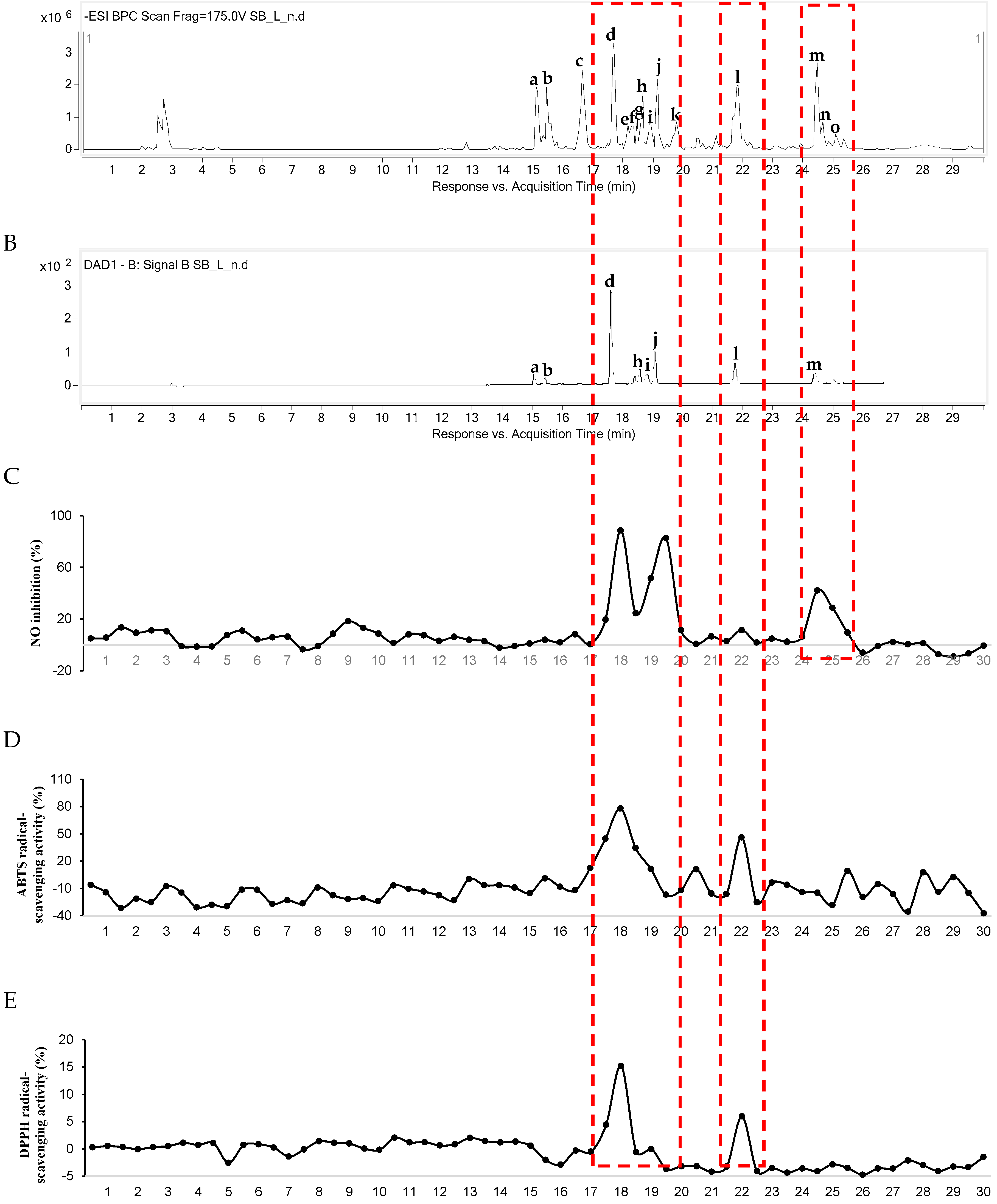
2.1. LC-Quadrupole Time of Flight (QTOF) MS/MS Coupled with Bioassay
2.2. Anti-Inflammatory and Antioxidant Activities of Target Components
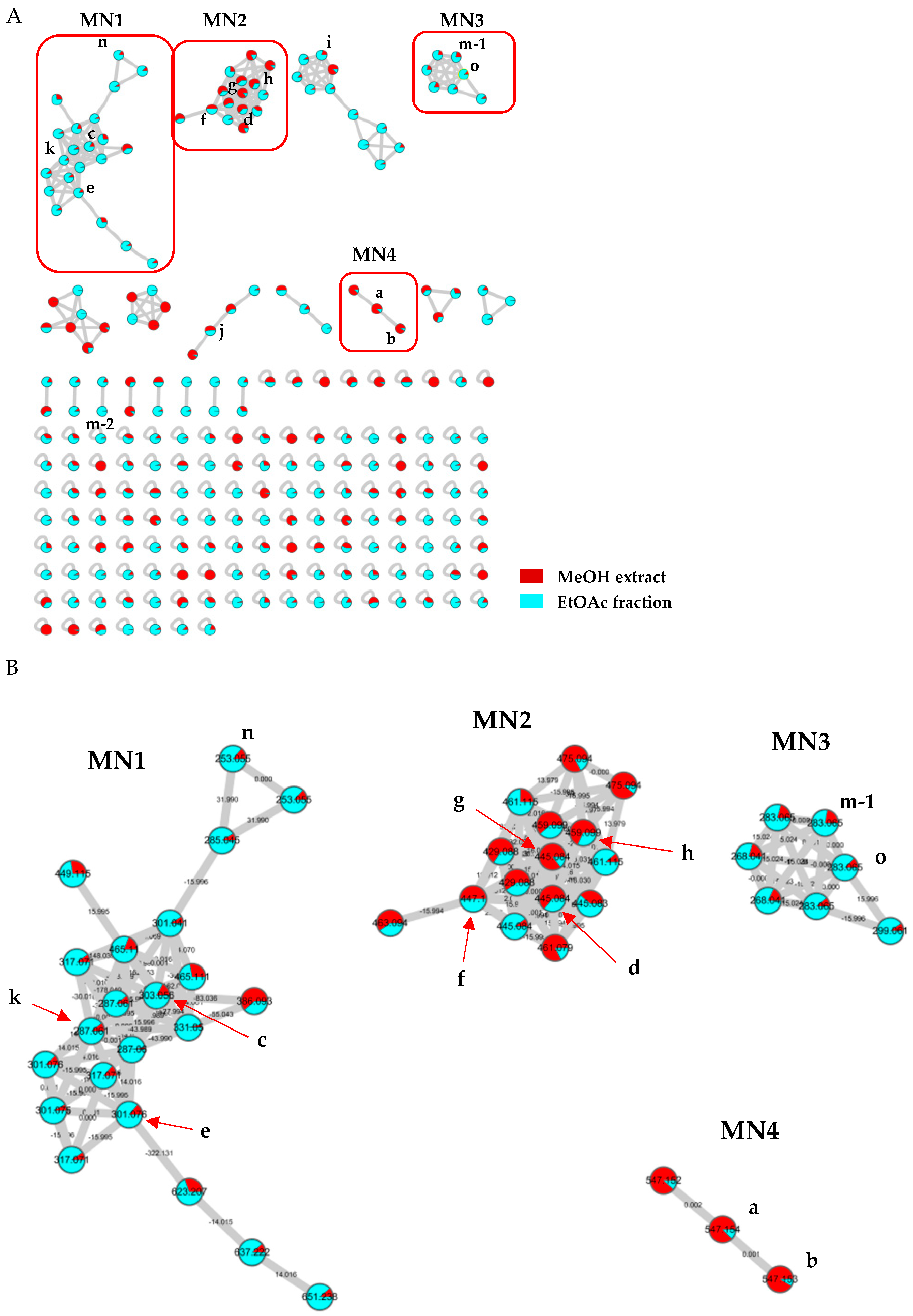
2.3. Dereplication through Molecular Networking
3. Materials and Methods
3.1. General Experimental Procedures
3.2. Plant Material
3.3. Extraction and Isolation
3.4. LC-QTOF MS/MS Coupled with Bioassay
3.4.1. NO Production Inhibitory Assay and Cell Viability
3.4.2. DPPH and ABTS Free Radical-Scavenging Assay
3.5. Molecular Networking
4. Conclusions
Supplementary Materials
Author Contributions
Funding
Conflicts of Interest
References
- Tabudravu, J.N.; Pellissier, L.; Smith, A.J.; Subko, K.; Autreau, C.; Feussner, K.; Hardy, D.; Butler, D.; Kidd, R.; Milton, E.J.; et al. LC-HRMS-database screening metrics for rapid prioritization of samples to accelerate the discovery of structurally new natural products. J. Nat. Prod. 2019, 82, 211–220. [Google Scholar] [CrossRef] [PubMed]
- Nothias, L.F.; Boutet-Mercey, S.; Cachet, X.; De La Torre, E.; Laboureur, L.; Gallard, J.F.; Retailleau, P.; Brunelle, A.; Dorrestein, P.C.; Costa, J.; et al. Environmentally friendly procedure based on supercritical fluid chromatography and tandem mass spectrometry molecular networking for the discovery of potent antiviral compounds from Euphorbia semiperfoliata. J. Nat. Prod. 2017, 80, 2620–2629. [Google Scholar] [CrossRef]
- Kang, K.B.; Park, E.J.; da Silva, R.R.; Kim, H.W.; Dorrestein, P.C.; Sung, S.H. Targeted isolation of neuroprotective dicoumaroyl neolignans and lignans from Sageretia theezans using in Silico molecular network annotation propagation-based dereplication. J. Nat. Prod. 2018, 81, 1819–1828. [Google Scholar] [CrossRef] [PubMed]
- Woo, S.; Kang, K.B.; Kim, J.; Sung, S.H. Molecular networking reveals the chemical diversity of selaginellin derivatives, natural phosphodiesterase-4 inhibitors from Selaginella tamariscina. J. Nat. Prod. 2019, 82, 1820–1830. [Google Scholar] [CrossRef] [PubMed]
- Olivon, F.; Remy, S.; Grelier, G.; Apel, C.; Eydoux, C.; Guillemot, J.C.; Neyts, J.; Delang, L.; Touboul, D.; Roussi, F.; et al. Antiviral compounds from Codiaeum peltatum targeted by a multi-informative molecular networks approach. J. Nat. Prod. 2019, 82, 330–340. [Google Scholar] [CrossRef] [PubMed]
- Wang, M.; Carver, J.J.; Phelan, V.V.; Sanchez, L.M.; Garg, N.; Peng, Y.; Nguyen, D.D.; Watrous, J.; Kapono, C.A.; Luzzatto-Knaan, T.; et al. Sharing and community curation of mass spectrometry data with global natural products social molecular networking. Nat. Biotechnol. 2016, 34, 828–837. [Google Scholar] [CrossRef] [Green Version]
- Nothias-Esposito, M.; Nothias, L.F.; Da Silva, R.R.; Retailleau, P.; Zhang, Z.; Leyssen, P.; Roussi, F.; Touboul, D.; Paolini, J.; Dorrestein, P.C.; et al. Investigation of premyrsinane and myrsinane esters in Euphorbia cupanii and Euphobia pithyusa with MS2LDA and combinatorial molecular network annotation propagation. J. Nat. Prod. 2019, 82, 1459–1470. [Google Scholar] [CrossRef]
- Quinn, R.A.; Nothias, L.F.; Vining, O.; Meehan, M.; Esquenazi, E.; Dorrestein, P.C. Molecular Networking As a Drug Discovery, Drug Metabolism, and Precision Medicine Strategy. Trends Pharmacol. Sci. 2017, 38, 143–154. [Google Scholar] [CrossRef]
- Nothias, L.F.; Nothias-Esposito, M.; da Silva, R.; Wang, M.; Protsyuk, I.; Zhang, Z.; Sarvepalli, A.; Leyssen, P.; Touboul, D.; Costa, J.; et al. Bioactivity-Based Molecular Networking for the Discovery of Drug Leads in Natural Product Bioassay-Guided Fractionation. J. Nat. Prod. 2018, 81, 758–767. [Google Scholar] [CrossRef] [Green Version]
- Liu, G.; Rajesh, N.; Wang, X.; Zhang, M.; Wu, Q.; Li, S.; Chen, B.; Yao, S. Identification of flavonoids in the stems and leaves of Scutellaria baicalensis Georgi. J. Chromatogr. B Analyt. Technol. Biomed. Life Sci. 2011, 879, 1023–1028. [Google Scholar] [CrossRef]
- Zhi, H.; Jin, X.; Zhu, H.; Li, H.; Zhang, Y.; Lu, Y.; Chen, D. Exploring the effective materials of flavonoids-enriched extract from Scutellaria baicalensis roots based on the metabolic activation in influenza A virus induced acute lung injury. J. Pharm. Biomed. Anal. 2020, 177, 112876. [Google Scholar] [CrossRef] [PubMed]
- Orzechowska, B.U.; Wrobel, G.; Turlej, E.; Jatczak, B.; Sochocka, M.; Chaber, R. Antitumor effect of baicalin from the Scutellaria baicalensis radix extract in B-acute lymphoblastic leukemia with different chromosomal rearrangements. Int. Immunopharmacol. 2020, 79, 106114. [Google Scholar] [CrossRef] [PubMed]
- Dong, Q.; Chu, F.; Wu, C.; Huo, Q.; Gan, H.; Li, X.; Liu, H. Scutellaria baicalensis Georgi extract protects against alcoholinduced acute liver injury in mice and affects the mechanism of ER stress. Mol. Med. Rep. 2016, 13, 3052–3062. [Google Scholar] [CrossRef] [PubMed] [Green Version]
- Yoon, S.B.; Lee, Y.J.; Park, S.K.; Kim, H.C.; Bae, H.; Kim, H.M.; Ko, S.G.; Choi, H.Y.; Oh, M.S.; Park, W. Anti-inflammatory effects of Scutellaria baicalensis water extract on LPS-activated RAW 264.7 macrophages. J. Ethnopharmacol. 2009, 125, 286–290. [Google Scholar] [CrossRef]
- Cole, I.B.; Cao, J.; Alan, A.R.; Saxena, P.K.; Murch, S.J. Comparisons of Scutellaria baicalensis, Scutellaria lateriflora and Scutellaria racemosa: Genome size, antioxidant potential and phytochemistry. Planta Med. 2008, 74, 474–481. [Google Scholar] [CrossRef]
- Shen, J.; Li, P.; He, C.-N.; Liu, H.-T.; Liu, Y.-Z.; Sun, X.-B.; Xu, R.; Xiao, P.-G. Simultaneous determination of 15 flavonoids from different parts of Scutellaria baicalensis and its chemometrics analysis. Chin. Herb. Med. 2019, 11, 20–27. [Google Scholar] [CrossRef]
- Park, S.; Shin, H.; Park, Y.; Choi, I.; Park, B.; Lee, K.Y. Characterization of inhibitory constituents of NO production from Catalpa ovata using LC-MS coupled with a cell-based assay. Bioorg. Chem. 2018, 80, 57–63. [Google Scholar] [CrossRef]
- Min, B.-S. Revision of structures of flavanoids from Scutellaria indica and their protein tyrosine phosphatase 1B inhibitory activity. Nat. Prod. Sci. 2006, 12, 205–209. [Google Scholar]
- Zhang, Q.; Guo, W.J.; Fu, C.L.; Ma, S.; Zhu, M.Q. Chemical constituents from an endophyte, Cercosporella sp. Chem. Nat. Compd. 2013, 49, 117–118. [Google Scholar] [CrossRef]
- Zahra, T.-N.; Javad, A.; Heydar, P.; Seyed, H.M.; Naser, V.M.; Alireza, M.; Seyed, A.E. Wogonin and neobaicalein from Scutellaria litwinowii roots are apoptotic for HeLa cells. Rev. Bras. Farmacogn. Braz. J. Pharmacogn. 2012, 22, 268–276. [Google Scholar] [CrossRef]
- Kimura, Y.; Okuda, H.; Tani, T.; Arichi, S. Studies on Scutellariae radix. IV. Effects on lipid peroxidation in rat liver. Chem. Pharm. Bull. 1982, 30, 1792–1795. [Google Scholar] [CrossRef] [Green Version]
- Dinda, B.; Mohanta, B.C.; Arima, S.; Sato, N.; Harigaya, Y. Flavonoids from the stem-bark of Oroxylum indicum. Nat. Prod. Sci. 2007, 13, 190–194. [Google Scholar]
- Zhang, Y.-Y.; Guo, Y.-Z.; Onda, M.; Hashimoto, K.; Ikeya, Y.; Okada, M.; Maruno, M. Four flavonoids from Scutellaria baicalensis. Phytochemistry 1994, 35, 511–514. [Google Scholar] [CrossRef]
- Jiang, W.J.; Ishiuchi, K.; Furukawa, M.; Takamiya, T.; Kitanaka, S.; Iijima, H. Stereospecific inhibition of nitric oxide production in macrophage cells by flavanonols: Synthesis and the structure-activity relationship. Bioorg. Med. Chem. 2015, 23, 6922–6929. [Google Scholar] [CrossRef] [PubMed]
- Wu, S.; Sun, A.; Liu, R. Separation and purification of baicalin and wogonoside from the Chinese medicinal plant Scutellaria baicalensis Georgi by high-speed counter-current chromatography. J. Chromatogr. A 2005, 1066, 243–247. [Google Scholar] [CrossRef] [PubMed]
- Dinda, B.; Dinda, S.; DasSharma, S.; Banik, R.; Chakraborty, A.; Dinda, M. Therapeutic potentials of baicalin and its aglycone, baicalein against inflammatory disorders. Eur. J. Med. Chem. 2017, 131, 68–80. [Google Scholar] [CrossRef] [PubMed]
Sample Availability: Samples of the compounds are not available from the authors. |


| Peak No. | Expected Compounds | tR (min) | Observed m/z | Calculated m/z | Molecular Formula [M − H]− | MS/MS Fragments (m/z) | UV (λ max, nm) | Isolated Compounds |
|---|---|---|---|---|---|---|---|---|
| a | Chrysin-6-C-ara-8-C-glu | 15.096 | 547.1565 | 547.1457 | C26H27O13 | 337 [M-C11H14O4-H]− | 273, 314 | |
| b | Chrysin-6-C-glu-8-C-ara | 15.471 | 547.1559 | 547.1457 | C26H27O13 | 337 [M-C11H14O4-H]− | 273, 314 | |
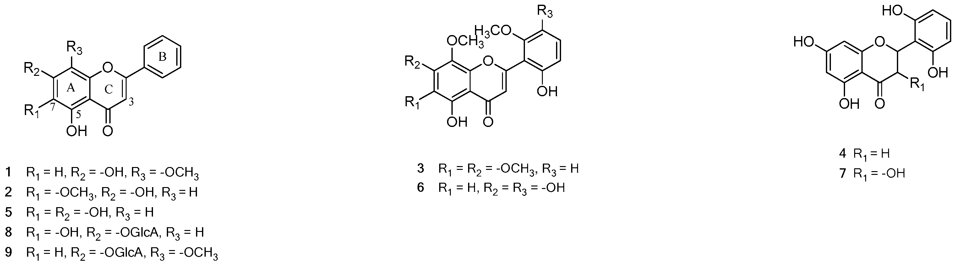
| c | 2′,3,5,6′,7-Pentahydroxyflavanone | 16.595 | 303.0572 | 303.0510 | C15H11O7 | 125 [M-C9H6O4-H]− | 285 | 7 |
| d | Baicalin | 17.657 | 445.0859 | 445.0776 | C21H17O11 | 269 [M-C6H8O6-H]− | 278, 315 | 8 |
| e | Unidentified flavonoid | 18.094 | 301.0774 | 301.0718 | C16H13O6 | 139 [M-C9H6O3-H]C− | - | |
| f | Dihydrobaicalin | 18.281 | 447.1014 | 447.0933 | C21H19O11 | 271 [M-C6H8O6-H]− | 285 | |
| g | Baicalein-6-glucuronide | 18.469 | 445.0851 | 445.0776 | C21H17O11 | 269 [M-C6H8O6-H]− | 280 | |
| h | Oroxylin A-7-glucuronide | 18.593 | 459.1016 | 459.0933 | C22H19O11 | 283 [M-C6H8O6-H]− | 273, 314 | |
| i | Viscidulin Ⅲ | 18.843 | 345.0680 | 345.0616 | C17H13O8 | 315 [M-CH2O-H]− | 265 | 6 |
| j | Wogonoside | 19.093 | 459.1013 | 459.0933 | C22H19O11 | 283 [M-C6H8O6-H]− | 274 | 9 |
| k | 2′,5,6′,7-Tetrahydroflavanone | 19.718 | 287.0617 | 287.0561 | C15H11O6 | 125 [M-C9H6O3-H]− | - | 4 |
| l | Baicalein | 21.779 | 269.0511 | 269.0455 | C15H9O5 | 195 [M-C6H2-H]− | 275, 323 | 5 |
| m | Wogonin/Skullcapflavone Ⅱ | 24.402 | 283.0670/373.1001 | 283.0612/373.0929 | C16H11O5/C19H17O8 | 268 [M-CH3-H]− /343 [M-CH2O-H]− | 275 | 1/3 |
| n | Chrysin | 24.590 | 253.0561 | 253.0506 | C15H9O4 | 143 [M-C6H6O2-H]− | - | |
| o | Oroxylin A | 25.027 | 283.0673 | 283.0612 | C16H11O5 | 268 [M-CH3-H]− | - | 2 |
| Compounds | Concentration (mM) | Relative NO Inhibition (%) 1 | Viability (%) |
|---|---|---|---|
| Control | 100.0 ± 0.0 ** | 100.0 ± 1.6 | |
| LPS | 100 ng/mL | 0.0 ± 1.4 | 102.4 ± 1.9 |
| Dexamethasone 2 | 10 | 57.4 ± 1.7 *** | 98.4 ± 0.5 |
| 1 | 0.1 | 67.5 ± 0.4 ** | 106.4 ± 0.4 |
| 1 | 72.5 ± 1.8 * | 111.7 ± 0.1 | |
| 5 | 91.4 ± 0.1 ** | 110.6 ± 2.3 | |
| 2 | 0.1 | 60.5 ± 4.2 * | 112.2 ± 3.3 |
| 1 | 58.7 ± 3.6 ** | 111.0 ± 3.2 | |
| 5 | 69.7 ± 1.0 ** | 101.1 ± 3.2 | |
| 3 | 0.1 | 57.4 ± 1.0 ** | 115.7 ± 3.1 |
| 1 | 56.7 ± 0.7 ** | 108.0 ± 1.3 | |
| 5 | 70.4 ± 0.3 ** | 116.4 ± 0.2 | |
| 4 | 0.1 | 59.5 ± 0.8 ** | 105.5 ± 0.8 |
| 1 | 61.8 ± 1.8 ** | 103.5 ± 1.1 | |
| 5 | 66.9 ± 0.9 ** | 104.1 ± 1.8 | |
| 5 | 0.1 | 61.6 ± 0.1 ** | 99.2 ± 0.4 |
| 1 | 65.6 ± 2.3 ** | 97.0 ± 2.3 | |
| 5 | 71.3 ± 0.3 ** | 100.4 ± 0.2 | |
| 6 | 0.1 | 63.1 ± 0.4 ** | 102.4 ± 1.6 |
| 1 | 60.0 ± 1.8 ** | 102.2 ± 0.1 | |
| 5 | 68.8 ± 0.2 ** | 101.8 ± 0.3 | |
| 7 | 0.1 | 62.3 ± 3.2 * | 113.9 ± 1.3 |
| 1 | 60.7 ± 1.6 ** | 112.4 ± 1.9 | |
| 5 | 62.5 ± 1.0 * | 111.4 ± 3.6 | |
| 8 | 0.1 | 62.5 ± 0.4 ** | 114.8 ± 2.0 |
| 1 | 63.0 ± 0.0 ** | 116.3 ± 2.2 | |
| 5 | 82.2 ± 1.0 ** | 109.3 ± 1.6 | |
| 9 | 0.1 | 63.9 ± 2.4 * | 117.7 ± 1.6 |
| 1 | 66.6 ± 1.0 ** | 117.8 ± 0.4 | |
| 5 | 83.2 ± 0.9 ** | 108.9 ± 0.7 |
| Compounds | DPPH | ABTS |
|---|---|---|
| IC50 1 (μM) | ||
| 1 | >50 | >50 |
| 2 | >50 | >50 |
| 3 | >50 | >50 |
| 4 | >50 | >50 |
| 5 | 17.0 ± 1.7 | 16.5 ± 0.5 |
| 6 | 16.4 ± 1.6 | 15.3 ± 0.5 |
| 7 | >50 | >50 |
| 8 | 15.1 ± 0.8 | 10.8 ± 0.8 |
| 9 | >50 | >50 |
| Trolox 2 | 38.1 ± 1.0 | 12.8 ± 0.3 |
© 2020 by the authors. Licensee MDPI, Basel, Switzerland. This article is an open access article distributed under the terms and conditions of the Creative Commons Attribution (CC BY) license (http://creativecommons.org/licenses/by/4.0/).
Share and Cite
Han, Y.K.; Kim, H.; Shin, H.; Song, J.; Lee, M.K.; Park, B.; Lee, K.Y. Characterization of Anti-Inflammatory and Antioxidant Constituents from Scutellaria baicalensis Using LC-MS Coupled with a Bioassay Method. Molecules 2020, 25, 3617. https://doi.org/10.3390/molecules25163617
Han YK, Kim H, Shin H, Song J, Lee MK, Park B, Lee KY. Characterization of Anti-Inflammatory and Antioxidant Constituents from Scutellaria baicalensis Using LC-MS Coupled with a Bioassay Method. Molecules. 2020; 25(16):3617. https://doi.org/10.3390/molecules25163617
Chicago/Turabian StyleHan, Yoo Kyong, Hyunwoo Kim, Hyeji Shin, Jiyeon Song, Mi Kyeong Lee, Byoungduck Park, and Ki Yong Lee. 2020. "Characterization of Anti-Inflammatory and Antioxidant Constituents from Scutellaria baicalensis Using LC-MS Coupled with a Bioassay Method" Molecules 25, no. 16: 3617. https://doi.org/10.3390/molecules25163617