Ursolic Acid and Its Derivatives as Bioactive Agents


1
Department of Chemistry, Faculty of Science and Agriculture, University of Fort Hare, Private Bag X1314, Alice 5700, South Africa
2
Department of Chemical and Physical Sciences, Faculty of Natural Sciences, Walter Sisulu University, Private Bag X1, Mthatha 5117, South Africa
3
Department of Human Biology, Faculty of Health Sciences, Walter Sisulu University, Private Bag X1, Mthatha 5117, South Africa
*
Author to whom correspondence should be addressed.
Molecules 2019, 24(15), 2751; https://doi.org/10.3390/molecules24152751
Submission received: 20 June 2019
/
Revised: 24 July 2019
/
Accepted: 25 July 2019
/
Published: 29 July 2019
(This article belongs to the Special Issue Discovery of Active Ingredients from Natural Products)
Abstract
:Non-communicable diseases (NCDs) such as cancer, diabetes, and chronic respiratory and cardiovascular diseases continue to be threatening and deadly to human kind. Resistance to and side effects of known drugs for treatment further increase the threat, while at the same time leaving scientists to search for alternative sources from nature, especially from plants. Pentacyclic triterpenoids (PT) from medicinal plants have been identified as one class of secondary metabolites that could play a critical role in the treatment and management of several NCDs. One of such PT is ursolic acid (UA, 3 β-hydroxy-urs-12-en-28-oic acid), which possesses important biological effects, including anti-inflammatory, anticancer, antidiabetic, antioxidant and antibacterial effects, but its bioavailability and solubility limits its clinical application. Mimusops caffra, Ilex paraguarieni, and Glechoma hederacea, have been reported as major sources of UA. The chemistry of UA has been studied extensively based on the literature, with modifications mostly having been made at positions C-3 (hydroxyl), C12-C13 (double bonds) and C-28 (carboxylic acid), leading to several UA derivatives (esters, amides, oxadiazole quinolone, etc.) with enhanced potency, bioavailability and water solubility. This article comprehensively reviews the information that has become available over the last decade with respect to the sources, chemistry, biological potency and clinical trials of UA and its derivatives as potential therapeutic agents, with a focus on addressing NCDs.
1. Introduction
Non-communicable diseases (NCDs), including cancers, cardiovascular diseases, pulmonary illnesses, and diabetes, account for nearly 71 percent of all fatalities globally, as reported by the World Health Organization. The rapid increase of NCDs has been influenced by a series of flexible behavioural threat variables, including unhealthy eating habits, physical inactivity, cigarette smoke exposure and damaging use of alcoholic drinks, as well as natural air pollutants, metabolic (hypertension, obesity, hyperglycaemia, hypercholesterolemia) and occupational (gases, carcinogens, fumes, particulates) risks [1].
On the other hand, natural products have been recognized in therapeutic drugs for many years as an essential source of active substances. Therapeutic agent complexity depends on synthetically prepared chemical combinations. Therapeutic drugs are regarded to be natural, synthetic or semi-synthetic, depending on the source from which they have been derived. Natural therapeutic agents are made from naturally occurring compounds that contain active ingredients extracted from sources such as plants [2]. One group of compounds from plants is called pentacyclic triterpenoids (PT). These are defined as a group of 30 carbon structural compounds with interesting biological activities and great diversity, with more than 100 chemical skeletons of carbon. PT, which have been studied for over two decades, with a perceptible increase in their pharmacological significance, are described to have strong health benefits, including hepatoprotective, wound healing, antibacterial, anti-inflammatory, antitumoral, and antiviral properties, together with their low toxicity [3]. PT are used in treatment of several cancers without damaging normal cells through toxicity [4]. These PT have also revealed therapeutic and chemical preventive characteristics in skin, lung, liver and lung disease [5]. The consumption of vegetables and fruit by animals is thought to have a reduced incidence of cancer and several illnesses. PT have been separated and elucidated from various plants [6]. Oleanolic acid (OA), ursolic acid (UA) (1), betulinic acid (BA), and lanosterol are examples of PT [7,8]. Triterpene groups, along with their classified chemical compounds and their characteristics, are shown in Table 1. For interesting chemical structures of pentacyclic triterpenoids such as UA, their reactivity and interactions with a disease are essential for searching through effective drug sites. Many triterpenes can be used as bioactive compounds immediately or modified to enhance their potency and selectivity [9]. Therefore, this review provides an overview of the structural development of UA and its derivatives, along with their advantages as anti-cancer, anti-inflammatory, antibacterial, anti-diabetic and neuroprotective therapeutic agents, as well as their herbicidal activities.
2. Chemistry of UA
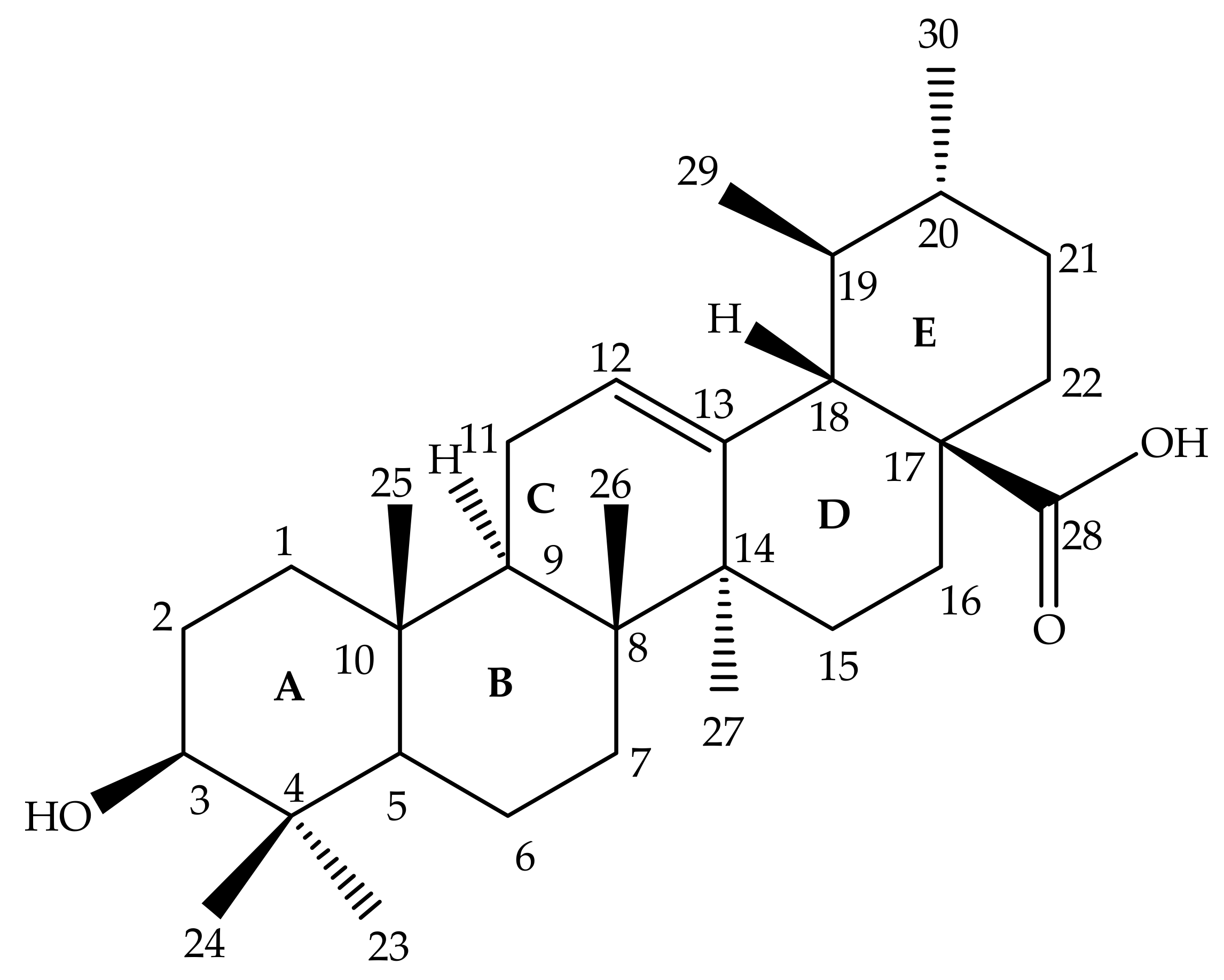
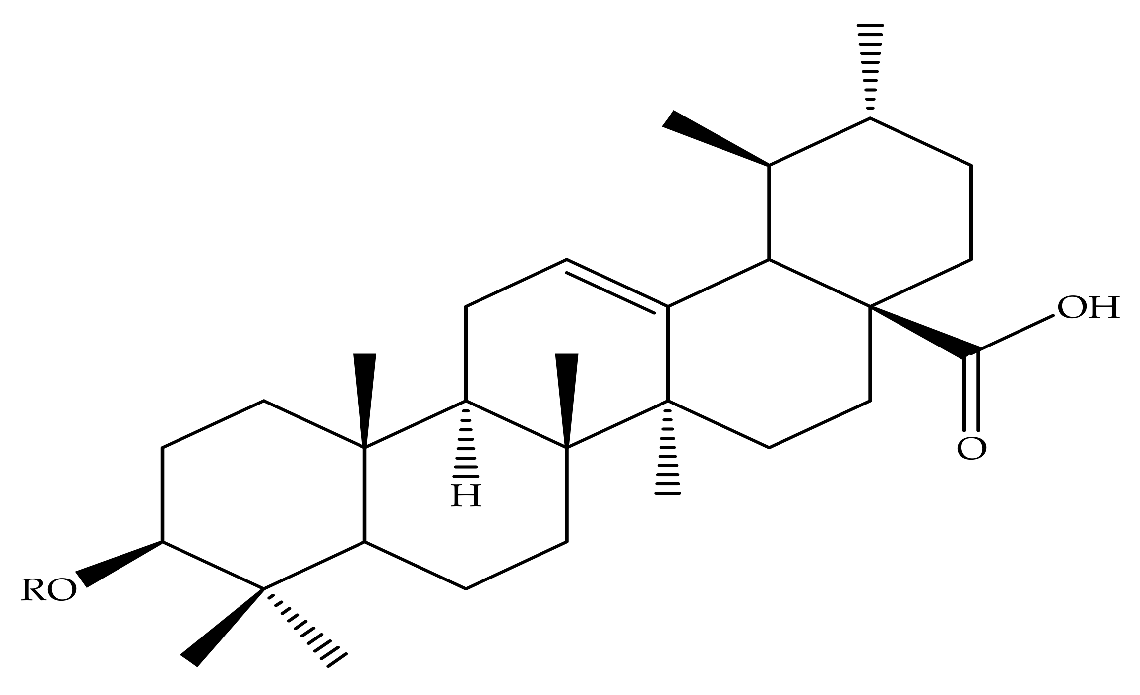
Ursolic acid (3 β-hydroxy-urs-12-en-28-oic acid) is a pentacyclic triterpenoid composed of a C-30 chemical structure built from isoprenoid units with A, B, C, D and E rings (Figure 1) [51]. UA may occur as an aglycone or free acid of saponins [52,53]. UA has a low solubility in water, but high solubility in alcoholic sodium hydroxide (NaOH) and glacial acetic acid [54,55,56,57]. The UA chemical formula is C30H48O3, with a melting point of 283–285 °C. The properties of the UA structure indicate that it has a molecular weight of 456.7g/mol [20,52,53,58]. UA is mainly biosynthesized by folding and cycling squalene from a dammarenyl cation, which makes the third UA ring by expanding the ring and creating an added ring [58]. The biosynthesis of UA found in plant cells originates from the cyclical (3S)-oxidosqualene cycling. The predominant (3S)-oxidosqualene predecessor is converted into a cationic dammarenyl structure undergoing chain growth and other cyclic changes in order to form this third distinct ring, which is present in the α-amyrin skeleton, a nucleus in the UA [51].
It is well known that low aqueous solubility of a drug may seriously affect its medication effectiveness. It has also been documented that certain treatments have a low solubility and few side effects. Ursolic acid, however, has low water solubility, which limits bioavailability in the human body [59]. The limited solubility, poor bioavailability and rapid metabolism of UA have limited their additional clinical applications for therapeutic effect in numerous diseases. Hence, many researchers in recent years have reported various modifications in an attempt to improve UA pentacyclic triterpenoids’ potency, solubility and bioavailability in water [59,60]. Hence, this review focuses on some of the recent modifications to UA aiming at their use as bioactive agents with promising therapeutic effects of non-communicable diseases.
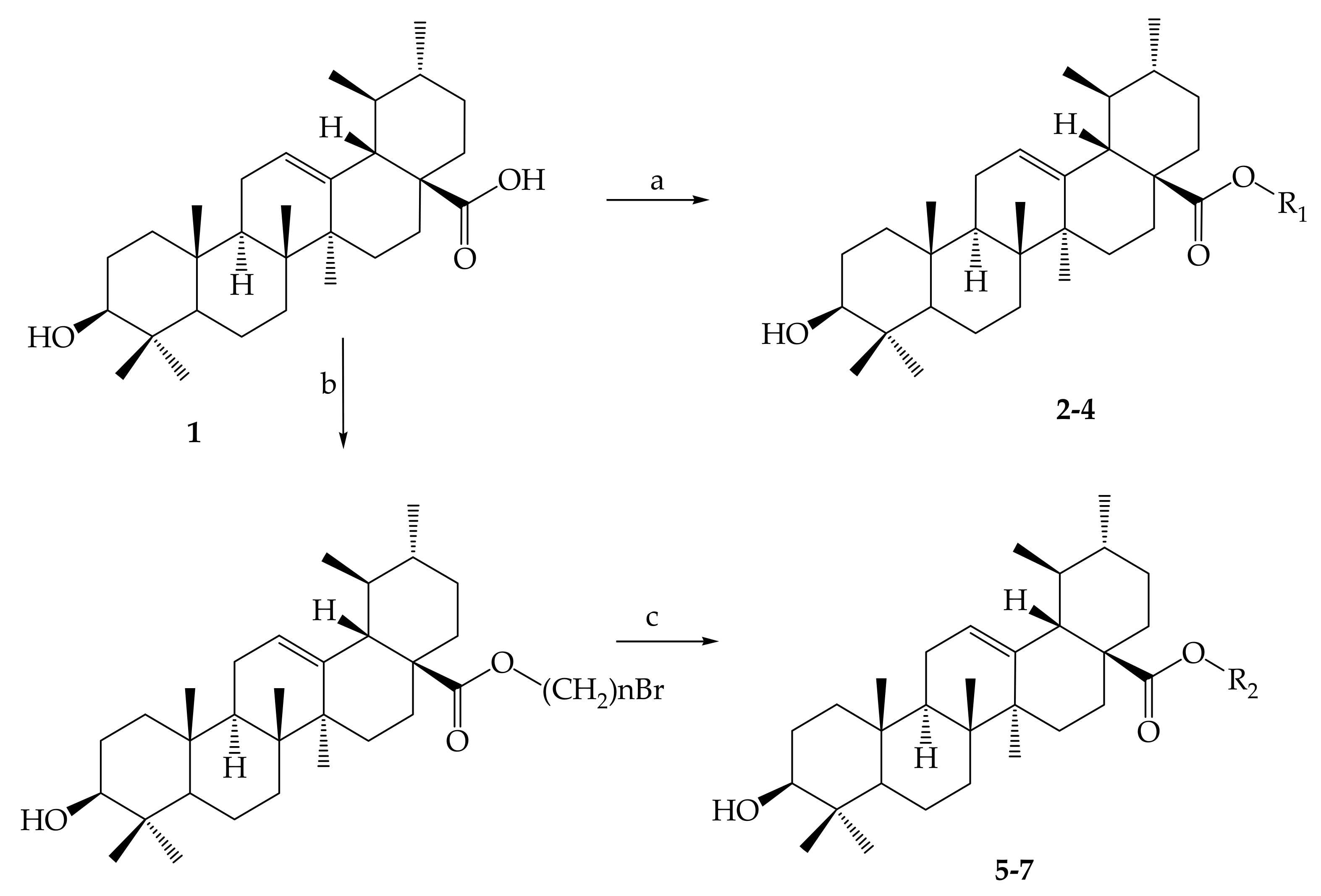
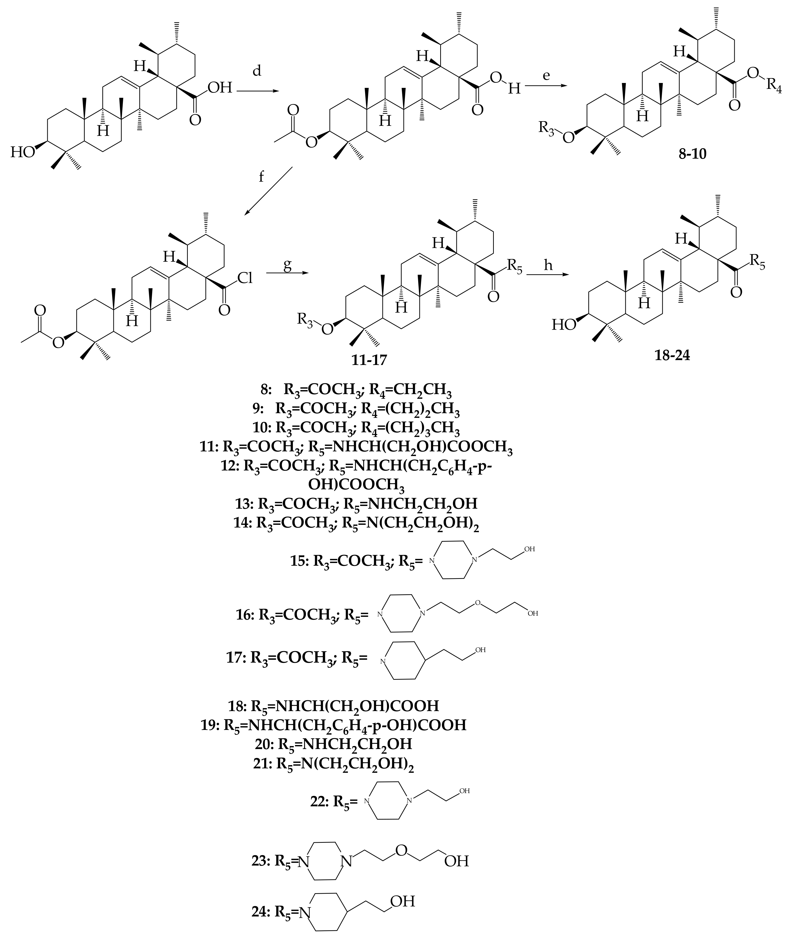
The chemical modifications to date have focused mainly on the hydroxyl group at position C-3, the unsaturated double bond at position C12-C13, and the carboxylic acid at position C-28 at rings A, C and E, respectively, as displayed in Figure 1 [61]. Mostly, structural modifications were made to above-mentioned positions in an effort to enhance the potency and bioavailability of UA and its derivatives, and study their structure-activity relationships and mechanisms [62]. For this reason, researchers started the search for new novel derivatives of through modifications of chemical groups in UA. Shao and colleagues (2011) in their UA (1) structure-activity relationship synthesized a total of twenty-three analogues through modification at position C-3 and C-28. 3-O-acetylursolic acid was obtained from acetylation of UA, which was later treated with bromo-diolefine to yield fatty esters as shown in Figure 2. They further synthesized UA amides and ester derivatives (Figure 3) [60].
Batra and Sastry (2013) reported isolation of UA (1) from Ocimum sanctum, which led to the preparation of three derivatives, namely, 3β-acetoxy-urs-12-en-28-oic acid (25), [N-(3β-acetoxy-urs-11-oxo-12-en-28-acyl) aniline] (26) and [Methyl N-(3β-butyryloxyl-urs-12-en-28-oyl)-2-amine acetate] (27) as shown in Figure 4 [63].
Nascimento and colleagues (2014) semi-synthesized two analogues of UA isolated from the Sambucus australis plant. Modifications to the UA structure were made at C-3 (hydroxyl group) in order to yield 3β-formyloxy-urs-12-en-28-oic acid (19) and 3β-acetoxy-urs-12-en-28-oic acid (20) [64].
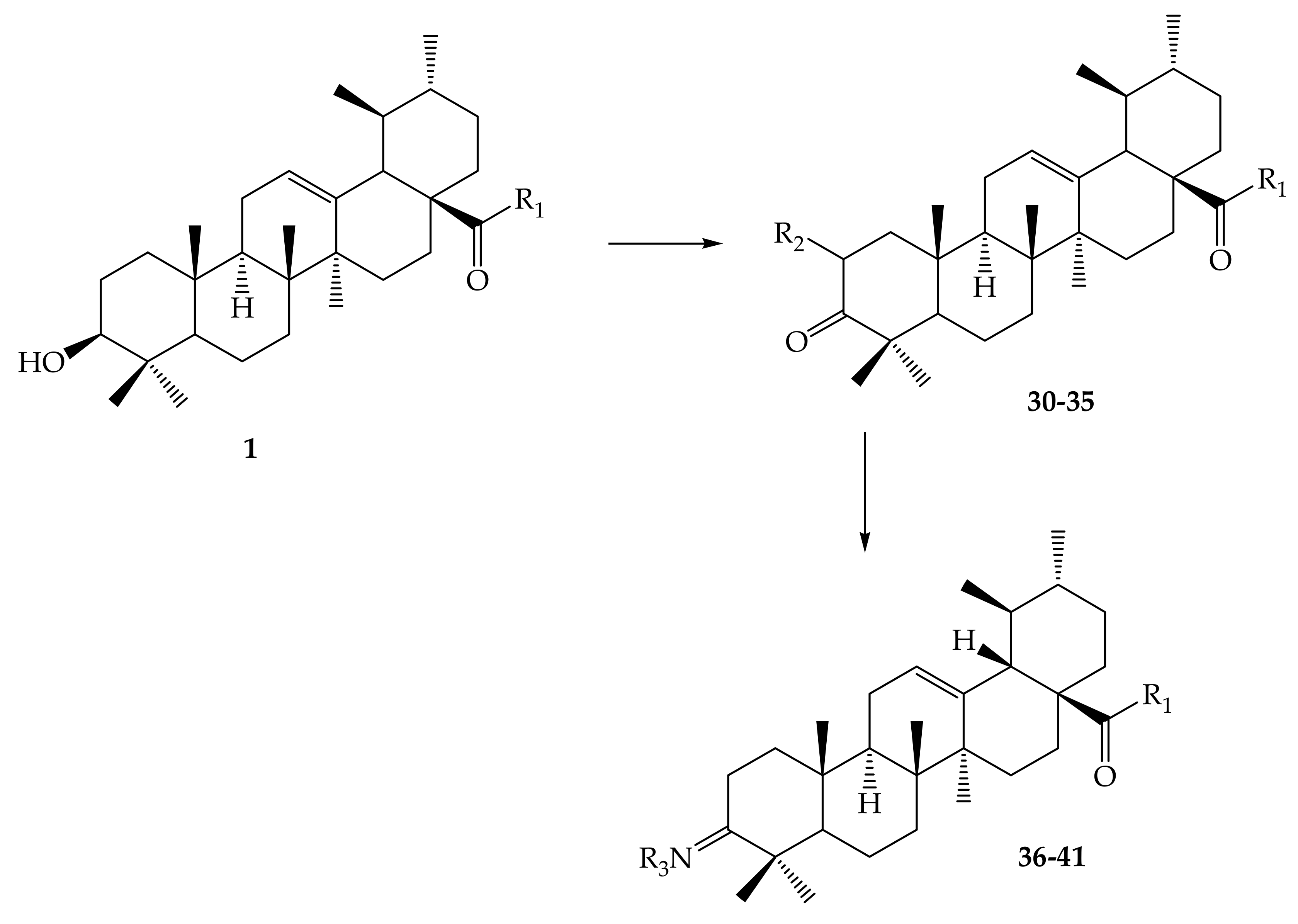
In order to examine the effective locations, Meng and collaborators (2017) discovered a number of exciting ursolic UA derivatives. They developed and evaluated 11 derivatives (30–41) (Figure 5, Figure 6, Table 2). They found that the inclusion of an acetyl group at C-3 and an amino alkyl at C28 increased biological potential [65].
Sahni and others (2016) reported the isolation of UA from acetone extract of a Eucalyptus hybrid. In an attempt to increase the potency of UA, these authors synthesized a total of 6 compounds (esters and amide) at positions C-3 and C-28 (Figure 7). In the preparation of methyl (42), ethyl (43) and propyl esters (44), UA was treated with acetic anhydride, butyril chloride, and propyl chloride at position C-3 respectively in the presence of DMAP and THF catalysts. C-17 methyl ester (45) was developed by treating UA with methyl iodide and modifying the C28 carboxylic acid group. A C-17 propyl amine (46) variant was prepared by the oxalyl chloride treatment of UA in the presence of CH2Cl2 and then propyl amine [66].
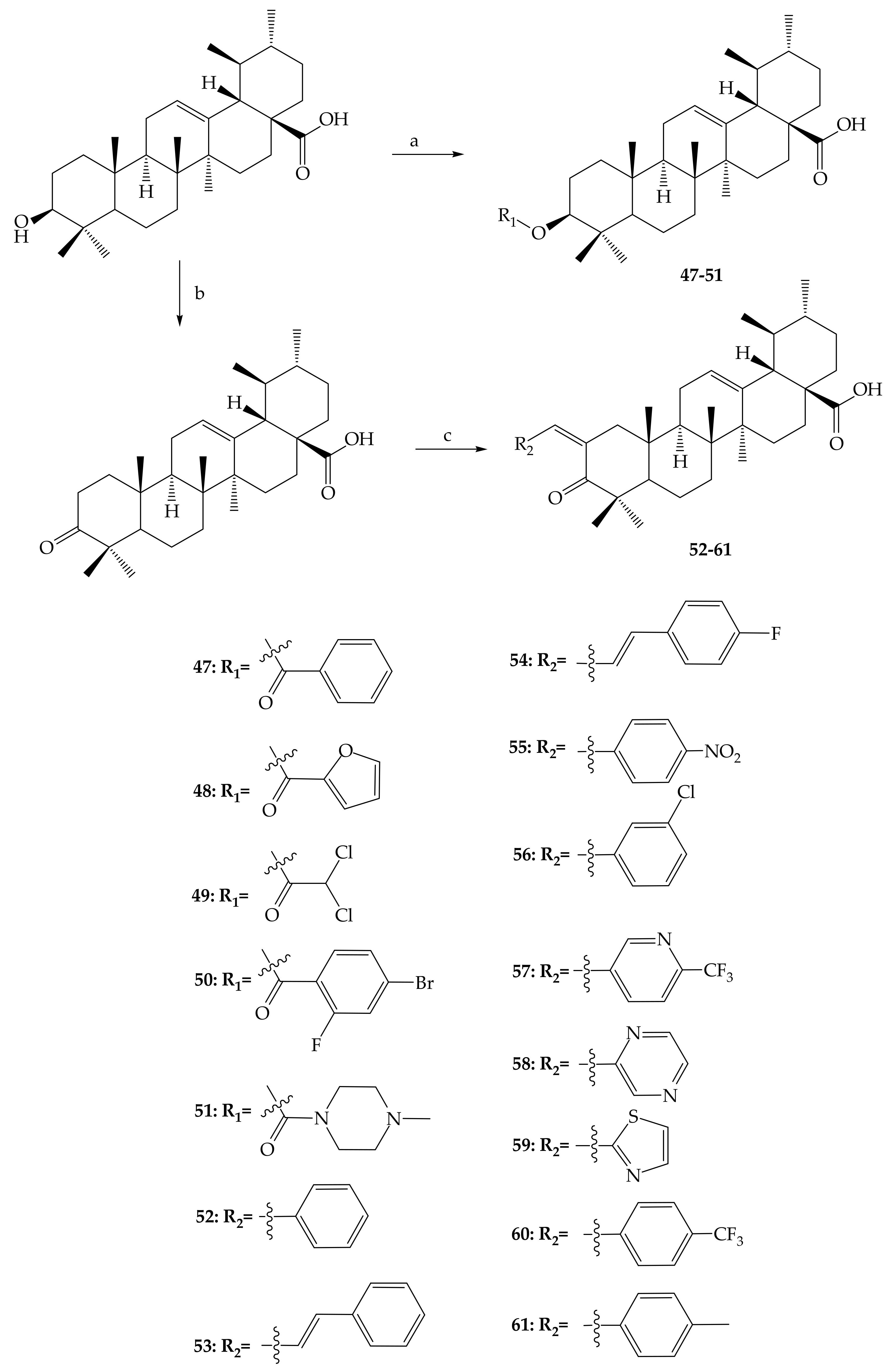
Wu and colleagues (2017) produced several UA derivatives (47–61) in an effort to study the greater power and bioavailability of the UA by adding an acyl group at position C-3 to make the most of the structure-activity relationship. They further synthesized ester derivatives by a process of esterification with suitable acid chlorides in the presence of a DMAP catalyst. In addition, Claisen Schmidt condensation and Jones oxidation steps at positions C-2 and C-3, respectively, were used for bioactive UA derivatives (Figure 8) [62].
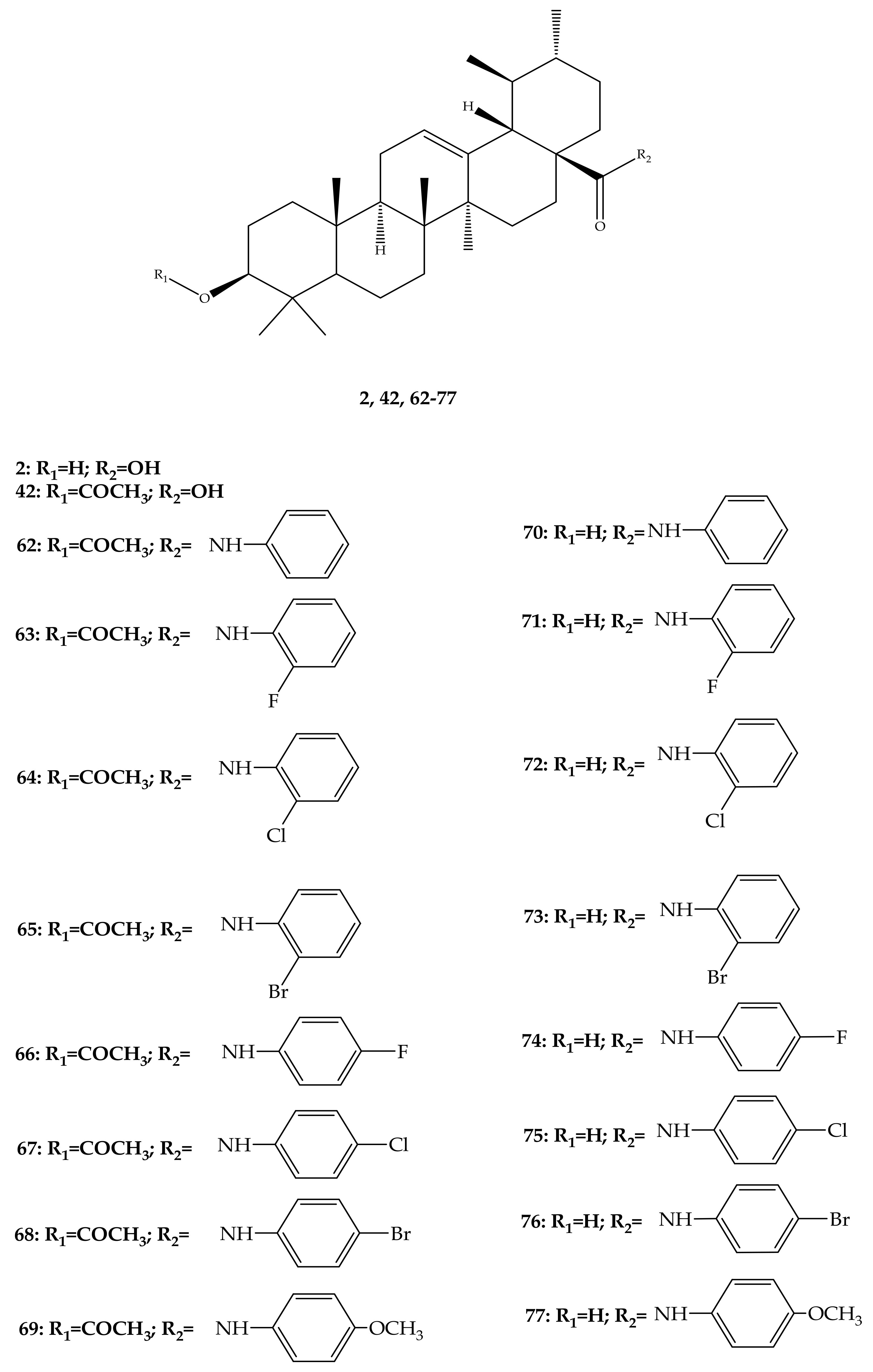
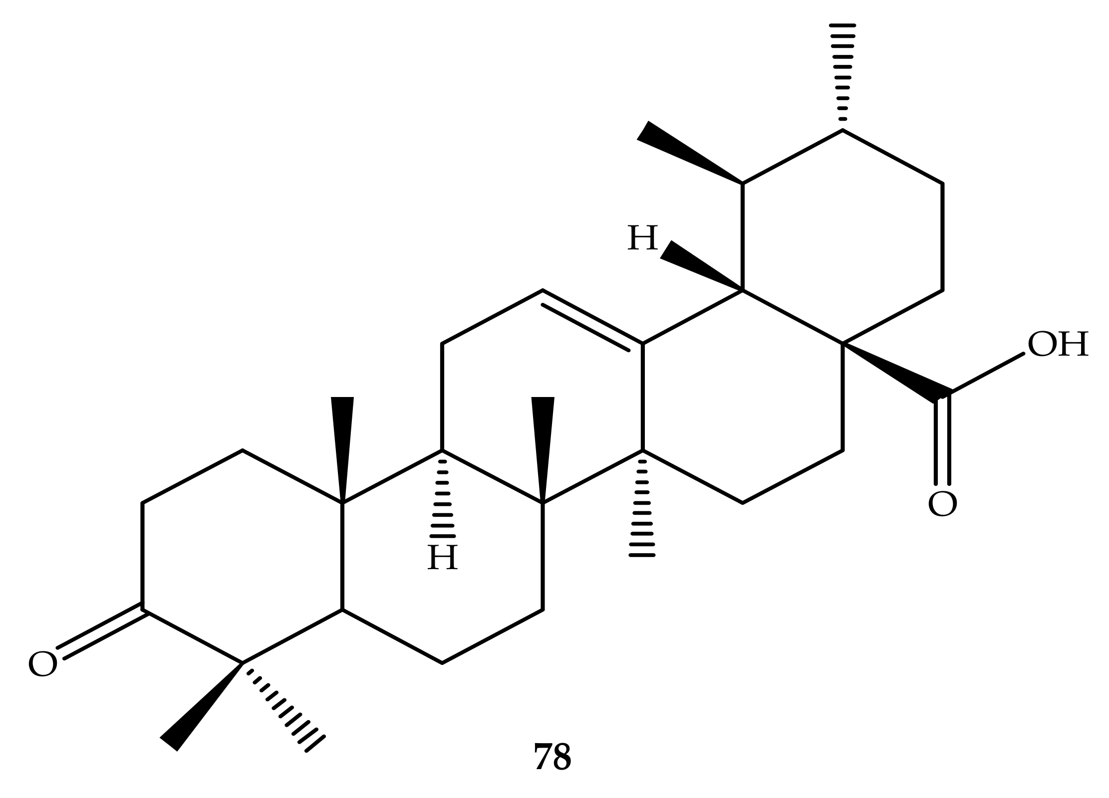
In a separate study, Wu and colleagues (2015) synthesized and elucidated several UA derivatives (62–78) as displayed in Figure 9. These researchers explored the structure-activity relationship of the synthesized bioactive analogues. They mainly focused their modifications on hydroxyl position C-3 and carboxylic group C-17 of the UA parent compound to produce a series of derivatives. In the first step, 3-O-acetate derivative was developed by the addition of acetic anhydride in anhydrous pyridine, which was later treated with oxalyl chloride to yield the 28-acyl-chloride intermediate. They further dissolved the intermediate in dichloromethane to give a series of amino compounds including aminobenzene, p-fluoroaniline, p-chloroaniline, p-bromobenzenamine, p-methoxylaniline o-fluoroaniline, o-chloroaniline and o-bromobenzenamine through condensation in the presence of triethylamine. After this, saponification analogues (62–69) were produced, which were later hydrolysed to give more derivatives (70–77). Lastly, the 3-oxo anlogue (78) was developed through oxidation with pyridinium chlorochromate. All the structure of these UA derivatives were elucidated by a series of spectroscopic techniques, such as nuclear magnetic resonance (NMR) including 13C-NMR and 1H-NMR with melting point, high resolution mass spectrometry, and electrospray ionization mass spectrometry (ESI-MS) [67].
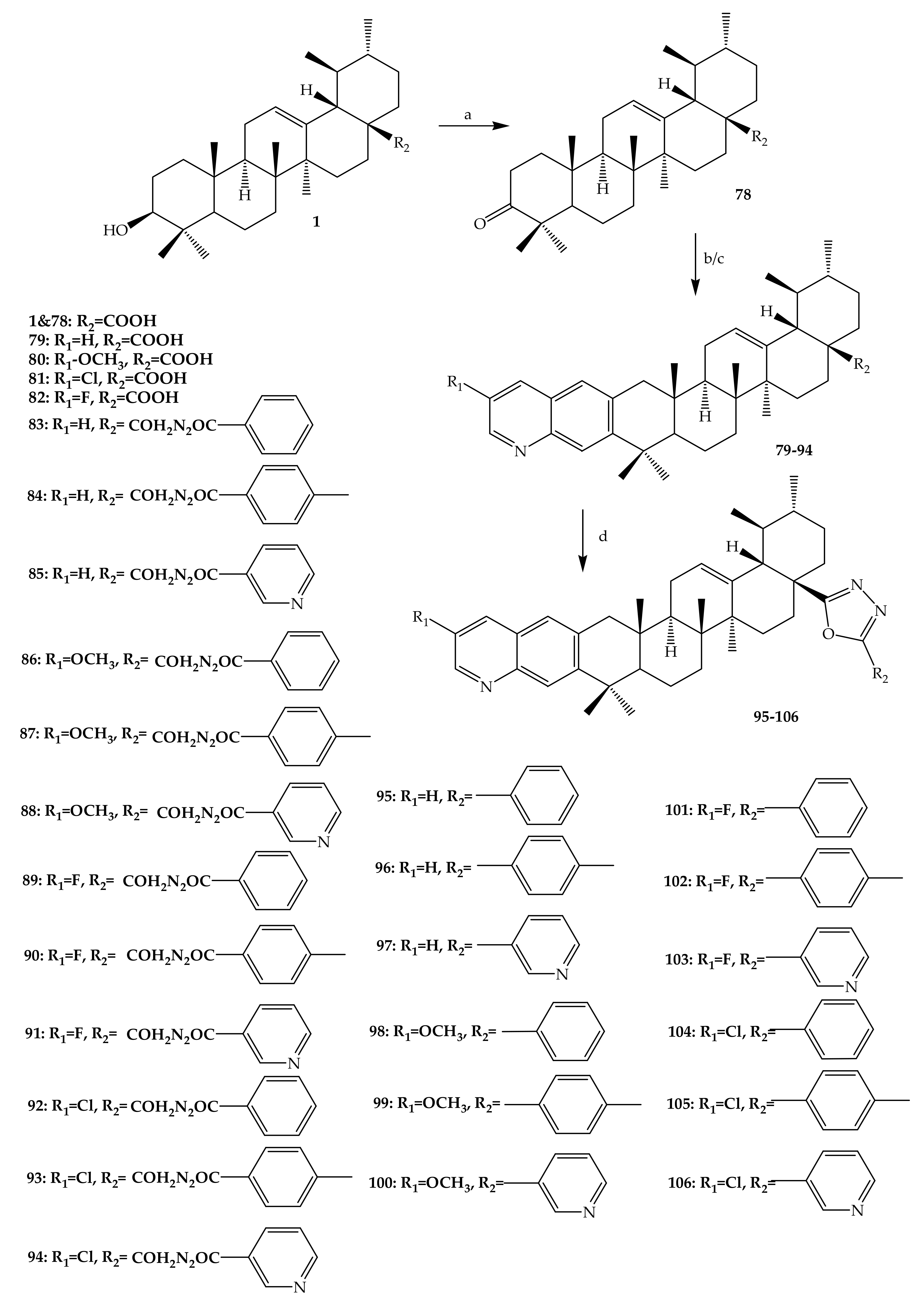
Several UA-derived compounds have been developed with the aim of improving their potency and selectivity. Gu and others (2017) synthesized an interesting series of UA derivatives containing oxadiazole and quinoline chemical groups. One analogue is 3-oxo-ursolic acid (78) (78% yield), which was prepared by dissolving UA in acetone solvent and later oxidized using Jones reagent, focussing on position C-3. More derivatives (79–82) (62–68% yield) were synthesized by reacting 3-oxo-ursolic acid with corresponding o-aminobenzaldehyde under nitrogen (N2) molecule atmospheric conditions, as shown in Figure 10. Compound (79) was synthesized by reacting 3-oxo-ursolic acid with o-aminobenzaldehyde according the Friedlander reaction. Afterwards, 28-acylchloride derivatives were prepared by treating compound (79–82) with thionyl chloride. Furthermore, 28-acylchloride derivatives were treated with aryl hydrazine in the presence of trimethylamine (EtN3) to yield 83–94 (47–48% yield). Meanwhile, the dehydration condensation of 83–94 yielded oxadiazole derivatives (95–106) (56–74% yield), as shown in Figure 10. All of these compounds were purified through column chromatography and elucidated by different spectroscopic techniques including infrared (IR), 13C-NMR, 1H-NMR, ESI-MS and elementary analysis [68].
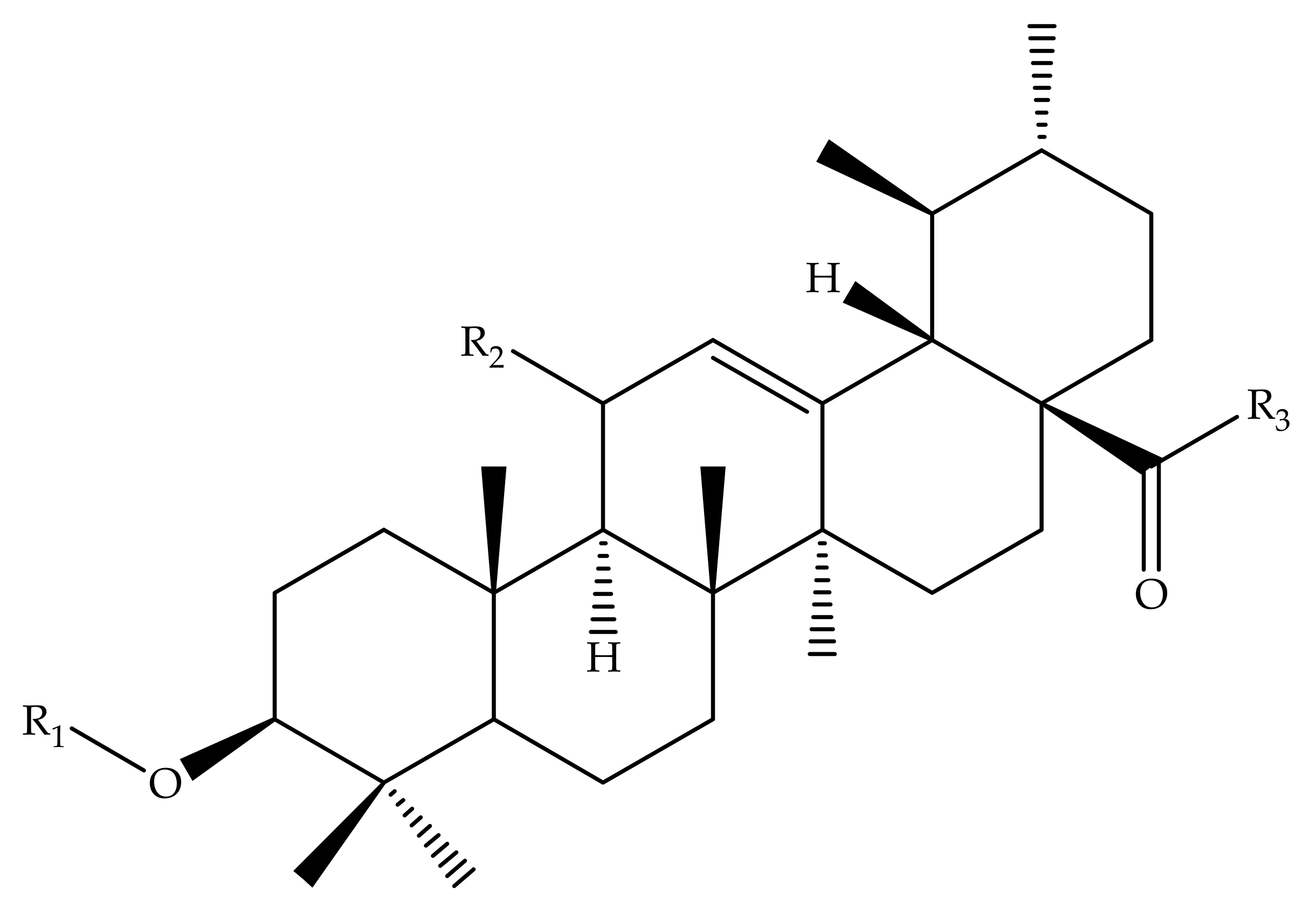
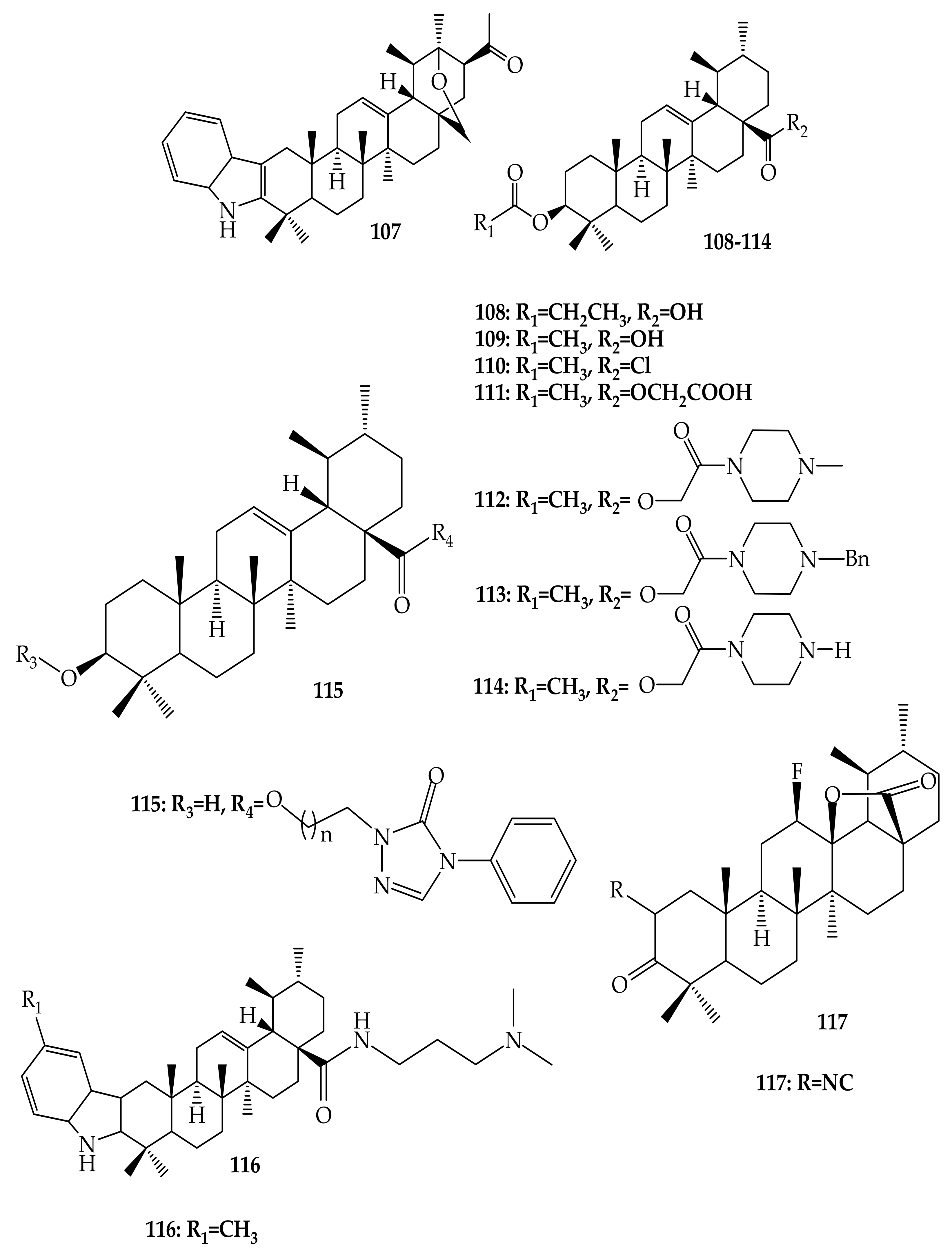
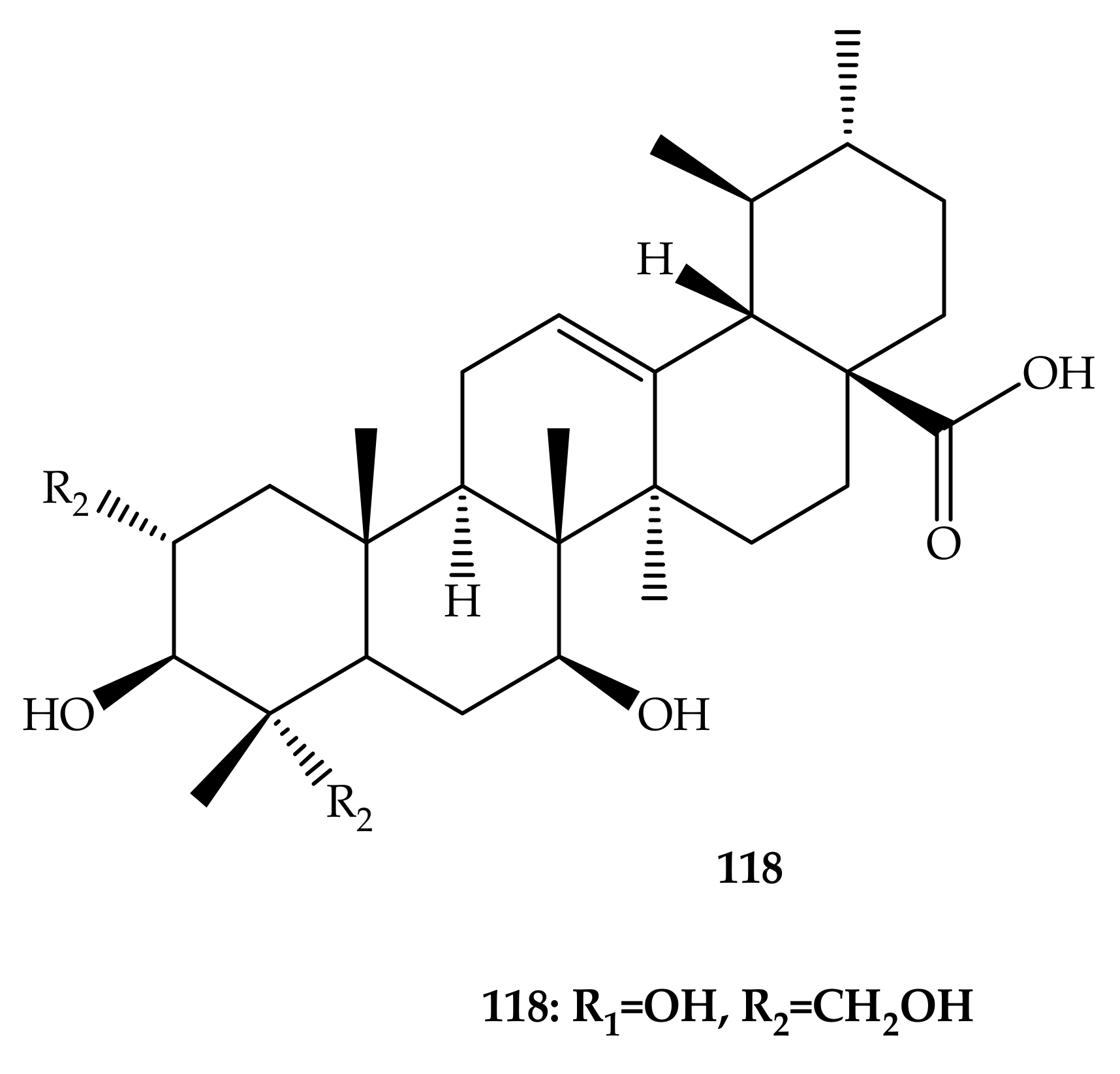
Many researchers, in search of more bioactive compounds, have reported different UA derivatives. Herewith are some of UA analogues with interesting mechanisms related to currently existing non-communicable diseases utilizing positions C-2, C-3, C-20 and C-28 (Figure 11) [69,70,71,72,73]. One UA derivative compound, 118 (2α,3β,7β,23-tetrahydroxyurs-12-ene-28-oic acid) (Figure 11), is a naturally occurring compound isolated from Castanea crenata Sieb. et Zucc. This compound was elucidated using several spectroscopic techniques, including IR, 1H-NMR, 13C-NMR, and HR-ESI MS [74].
3. Sources of UA and Its Biological Potency
Ursolic acid is a five-membered ring extensively distributed in food, medicinal herbs, fruit, vegetables, etc. [19,75,76,77,78,79,80,81,82]. UA triterpenoid is well documented to be available in fruits such as cranberries (Vaccinium macrocarpon) [53,83], blueberry (Vaccinium spp.) [77], basil (Ocimum basilicum) [78,84], olive (Olea europaea) [20], heather flower (Calluna vulgaris) [79], pear (Pyrus pyrifolia), Labrador tea (Ledum groenlandicum Retzius) [77], rosemary (Rosmarinus officinalis) [65] and apple peels, which possess important health benefits. UA is also distributed among higher plants, such as Mimusops caffra, Ilex paraguarieni, Glechoma hederaceaes, Ichnocarpus frutescens, and Syzygium claviflorum [19,77,80]. More plants sources, quantities of UA and its biological applications are hereby reported in Table 3.
UA has been investigated and is reported to possess many health benefits, including anti-apoptotic, anti-carcinogenic, anti-inflammatory, antioxidant, antirheumatic, antiviral, antitumoral, trypanocidal, etc. [61,62,78]. UA also has health benefits, and there are many reports of its anticancer activity in several cancers, including breast, skin, lung, prostate and pancreatic cancers [19,20,63]. Due to its low toxicity, anticancer activities, and commercial availability with various structural modifications, UA is regarded as a pillar through organic semi-synthesis, and this has attracted more researchers to studying and discovering various ursolic acid derivatives [61]. UA is popular because of its antiproliferative properties, inducement of cancer apoptosis, prevention of tumorigenesis, and/or blocking of the cell cycle in cancer cells. This was evident in the observation from one apoptosis mechanism which showed UA to have the ability to prevent Nuclear Factor κB (NF-κB) pathway by p65 phosphorylation suppression, resulting in mandatory decrease in various downstream oncogenes such as B-cell lymphoma-extra-large (Bcl-XL) and B-cell lymphoma-2 (Bcl2). Nonetheless, the antitumor potency of UA is poor because of its lower solubility, which decreases the drug absorption in the human system, leading to challenges in obtaining its full benefits. Therefore, it is necessary to construct its derivatives through semi-synthetic modifications in order to improve its antitumor activity. Mostly, the modifications of ursolic acid occur at sites C-3, C12-C13, and at position C-28 [59,63,64,72]. Jiménez-Arellanes and colleagues (2013) reported antimicrobial activity of pentacyclic triterpenoid compounds, namely, UA against Mycobacterium tuberculosis H37Rv [85]. Ursolic acid has been investigated in different stages of clinical trials for its therapeutic effects and selectivity against a diversity of diseases [86].
4. Biological Effects and Clinical Trials of UA and Some Derivatives
4.1. Anti-Inflammatory
Pathogenesis and homeostasis are part of inflammation. The inflammatory response is initiated after harm and/or microbial invasion to recover homeostatic tissue balance between composition and physiological function. Persistent inflammation may lead to damage of tissues, resulting in non-functioning organs [93]. Inflammation is a complicated occurrence linked to the development of different diseases, such as cardiovascular and neurodegenerative diseases, and cancer [55]. Rudolf Virchow proposed the link between inflammation and cancer as early as 1863. Currently, acute inflammation, with concomitant cytokine activity and enhanced output of reactive oxygen species, is identified as a cancer-promoting disease [3,16]. Zerin and co-authors (2016) examined the capacity of UA to decrease the production of TNF-α in RAW 267.4 and A549 cells infected with Mycobacterium tuberculosis and Con A-stimulated mouse splenocytes to detect anti-inflammatory activity. TNF-α is pivotal to inflammation and its impact on IL-1β and IL-6 decrease has been examined under comparable circumstances of therapy. These authors also studied UA activity with the aim of decreasing the levels of inflammatory intercessor, cyclooxygenase 2 and NO synthase found in stimulated cells. UA exhibited significant inhibitory effects on cytokine expression levels, immunomodulatory mediators, and release of NO. It is suggested that this compound can be used for tuberculosis and antibiotic therapy because of the UA anti-inflammatory potency in the mentioned cells [94]. Huang and others (2016) reported a similar study in which in vitro inhibition of cyclooxygenase-2 activity was due to active compounds (UA, cis-hydroxycinnamoyl ursolic acid and trans-hydroxycinnamoyl ursolic acid) and cranberry extracts [73]. Forbes and co-workers (2009) synthesized eight derivatives, with 4 showing promising anti-inflammatory and antioxidant activity (50% lipid peroxidation inhibition at 25 µg/mL). The anti-inflammatory activity was tested through the use of enzyme inhibitory assays referred to as in vitro cyclooxegenase-1 (COX-1) and cyclooxegenase-2 (COX-2) [95].
Wei and collaborators (2018) recorded and assessed for anti-inflammatory action the synthesis of 20 UA derivatives comprising oxadiazole, triazolone, and piperazine moieties. Compound (115) (Figure 11) displayed the most potent ear inflammation efficacy of all the synthetic compounds (69.76%), which was greater than ibuprofen (25.17%) and indomethacin (26.83%) at 100 mg/kg (i.p.), and was 1- and 2-fold more powerful than conventional medicines. The MTT assay evaluated the cytotoxicity of the compounds and, in contrast to UA, no compounds were reported to show any significant cytotoxic behaviour (IC50 > 100 µmol/L). In addition, the molecular docking findings stated that the UA derivatives showed elevated attraction for effective COX-2 location, potentially exhibiting anti-inflammatory potency through COX-2 inhibition. The findings from this study provide information about UA and its derivatives as anti-inflammatory agents, which could lead to the development of potentially new and safe COX-2 inhibitors [96].
4.2. Anticancer Activity
In recent memory, cancer has been ranked as the top deadly diseases and is responsible for many deaths worldwide, being ranked second in economically developed countries. With several forms of cancer being poorly controlled through treatments that have serious side effects themselves and the inevitable limitations of cancer screening programs, chemoprevention and its potential have generated great hope and interest over the past decades [97]. Batra and Sastry (2013) reported isolation of UA (1) (Figure 1) from Ocimum sanctum, which led to the preparation of three novel derivatives from 3β-acetoxy-urs-12-en-28-oic acid [N-(3β-acetoxy-urs-11-oxo-12-en-28-acyl) aniline and [Methyl N-(3β-butyryloxyl-urs-12-en-28-oyl)-2-amine acetate] with high in vitro anticancer potency when compared to the ursolic acid parent compound. The modifications of compound 1 occurred at hydroxyl C3, C-11, and carboxylic C-28, as shown in Figure 4. Compounds 2, 3 and 4 showed potent anti-proliferation activity in cancer cells, with compound 3 displaying higher significance [63].
Meng and colleagues (2017) reported interesting ursolic acid novel derivatives. These authors prepared and synthesized eleven derivatives, which were later assessed for their antitumor inhibition and cytotoxicity against numerous cell lines developed by cancer such as HepG2, cervical carcinoma (HeLa) and BGC-823 using MTT assay. The modifications occurred in positions C-2, C-3 and C-28 to yield 12 compounds (5–17), as shown in Figure 5. Compound 15 [IC50 = 9.25 (HeLa), 21.2 (HepG2) and 8.06 µmol/L (BGC-823)] and compound 16 [IC50 = 13.8 (HeLa), 23.7 (HepG2) and 9.15 µmol/L (BGC-823)] were more significant because of their evident high antitumor inhibition against cancer cells. This led to a belief that the antitumor properties of the mentioned compounds could be attributed to the substitution made during the synthesis modification of UA; electron withdrawing, alkanoyloxy imino chain at position C-2, and alkyl side chains at position C-3 [65].
In a different investigation, Shao and co-authors (2011) explored the in vivo and in vitro anticancer activity of a number of synthesized UA derivatives. The synthesized compounds were evaluated against various cancer cells of BGC-823, SH-SY5Y, HELF, HeLa and HepG2 by the assay called MTT. N-[3β-acetoxy-urs-12-en-28-oyl]-2-aminodiethanol, N-[3b-acetoxy-urs-12-en-28-oyl]-amino-N-(2-hydroxyethyl) piperazine, N-[3b-acetoxy-urs-12-en-28-oyl]-amino-1-hydroxyethylethoxy piperazine and N-[3b-acetoxy-urs-12-en-28-oyl]-amino-4-piperidineethanol exhibited better in vitro antiproliferative activity. The N-[3β-acetoxy-urs-12-en-28-oyl]-2-aminodiethanol derivative was reported to have anticancer activity (45 ± 4.3%) on Kunming mice compared to the control group [60]. Rashid and others (2013) produced several derivatives of ursolic acid-triazolyl. The antitumor potency of these compounds was assessed against a number of individual cancer cells, including leukaemia (THP-1), heart (MCF-7), colon (HCT-116), pulmonary (A-549) and ordinary adult epithelial cells (FR-2) using sulforhodamine-B testing. The antitumor potencies of four UA-derivatives against the above-mentioned cells were observed [98]. More authors have reported on ursolic acid and its derivatives as anticancer agents.
In an attempt to develop new anticancer agents, Gu and co-authors (2017) proposed a series of quinoline and oxidiazole derivatives of UA. These researchers evaluated the cytotoxicity of synthesized derivatives against cancer cell lines such as human breast (MDA-MB-231), heptacarcinoma (SMMC-7721), HeLa and normal hepatocyte cell lines (QSG-7701) using MTT assay. The cancer drug atoposide was used as a positive control. The results from this particular study showed interesting significant effects of UA derivatives (79–82, 91, 97) against at least one of the cancer cell lines (MDA-MB-231, SMMC-7721 and HeLa) (IC50 < 10µM). Compound 79 showed a higher potency compared to the positive control, with IC50 values of 12.49 ± 0.08, 0.36 ± 0.05, and 0.61 ± 0.07 µM against SMMC-7721, HeLa and MDA-MB-231 cell lines, respectively. The bioactive derivatives (79–82, 91, 97) did not show any significant cytotoxicity against QSG-7701 (IC50 > 40µM) [68].
Ursolic acid caused bax up-regulation and down-regulation of Bcl-2 and discharge from mitochondria of cytochrome C to the cytosol. In addition, UA cleaved caspase-9 and reduced mitochondrial membrane ability. Thus, UA induces apoptosis in MDA-MB-231 cells through both the mitochondrial death pathway and a mechanism dependent on the extrinsic death receptor. Therefore, ursolic acid could be used as a promising anti-cancer drug in treatment of breast cancer [99]. UA (1) suppressed the proliferation of androgen-independent DU145 and androgen-dependent human prostate cancer cells (LNCaP) through inhibition of NF-κB and STAT3 activation. Tumour growth was suppressed significantly when four-week-old athymic BALB/c male nude mice inoculated with DU145 cells were treated with (+)-UA (i.p., 200mg/kg, twice a week) for 6 weeks. No significant effects on body weight were observed in mice [100]. Moreover, UA displayed an inhibitor effect through the DNA binding capacity of STAT3, constitutive and inducible STAT3 phosphorylation. In fact, UA therapy also displayed protein expression of cycline D1 and Bcl-2 in a dose-dependent way. In the G1 phase, UA quickly collected in the cell population at 50 mM, sucked by flow cytometry, confirming that ursolic acid caused G1 in the regulation of the cell cycle [101].
Many clinical trials using chemical, subcutaneous, orthotopic human xenograft and randomly transgenic tumour development designs have provided extensive proof that naturally existing and synthetic UA products have chemopreventive and therapeutic effects. Liposomal ursolic acid (LUA) was used as a fresh drug in normal young volunteers and in individuals with developed solid tumours in order to determine the highest permitted level (MTD), dose-limiting toxicities (DLTs), and UA pharmacokinetics. A total sample of LUA (11, 22, 37, 56, 74, 98 and 130 mg/m2) was given to 63 subjects (4 patients, 35 healthy participants and 24 adults). For the first season, clinical information revealed that LUA had manageable 98 mg/m2 MTD toxicity. The DLTs were predominantly diarrhoea and hepatotoxicity. Furthermore, a linear pharmacokinetic profile was found in the ursolic acid liposomal model [52]. UA is a promising antitumor agent. It can induce apoptosis in tumour cells, on one hand, and stop ordinary cell transformation, on the other. It also interferes with countless proteins, including those that directly serve the structure of DNA. It can prevent the development of many tumour cell types and cause apoptosis. It has been shown that this compound acts at different phases of tumour growth. It efficiently prevents angiogenesis, tumour cell intrusion, and metastasis. It is comparatively non-toxic and can be used in clinical practice as a chemopreventive/chemoprotective product [102]. Therefore, it is evident that UA and its analogues are promising therapeutic agents against several types of cancers.
4.3. Antibacterial
Public health is faced with many challenges, including the resistance of many already-available antibiotics. There is hope in the use of naturally occurring products as a substitute that can produce a better and more promising therapeutic effect through the discovery of new compounds and derivatives against bacterial pathogens. Nascimento and colleagues (2014) semi-synthesized two analogues of UA isolated from the Sambucus australis plant. Modifications of the UA structure were made at C-3 (hydroxyl group) to yield 3β-formyloxy-urs-12-en-28-oic acid (19) and 3β-acetoxy-urs-12-en-28-oic acid (20), as shown in Figure 5. Ursolic acid and its analogues showed significant antibacterial and antioxidant properties. These authors tested the minimal inhibitory concentration (MIC) of both ursolic acid and its derivatives against several microbial pathogen strains (Staphylococcus aureus, Bacillus cereus, Shigella flexneri, two strains of Escherichia coli, Aeromonas caveae, Pseudomonas aeruginosa, Klebsiella pneumonia, and Vibrio colareae) using the microdilution method. Compound 19 (64 μg/mL) when combined with kanamycin showed significant activity towards Escherichia coli compared to the multidrug resistance observed from sputum, which reduced MIC from 128 μg/mL to 8 μg/mL [64]. This shows that the antibacterial efficiency of UA improves with modification.
Wang and co-authors (2016), in their investigation, explored the biological antibacterial properties of UA against Staphylococcus aureus (MRSA), which is resistant to methicillin. Researchers in this research noted that with no haemolytic consequences, UA produced a reduction in staphylococcal membrane integrity. UA-treated protein type relative to ordinary MRSA cells showed that UA impacted translation efficiency, chaperon subunits, ribonuclease, oxidative reactions, and glycolysis of a variety of proteins engaged in the transformation method [8]. Zhao and others (2018) isolated ursolic acid from the leaves of Ilex hainanensis Merr. These authors assessed seven UA derivatives against Fusobacterium nucleatum (Gram-ve) and Streptococcus mutans (Gram + ve) for their antibacterial activity. Only three derivatives displayed important activity against Gram positive at distinct levels and displayed substantial activity against Gram negative at a minimum inhibitor level (MIC) of 625 μg/mL [103]. Park and other researchers (2015) recorded the action of ursolic acid (UA) against bacterial (Streptococcus mutans) development in their quest for powerful antimicrobial drugs [104].
4.4. Anti-Diabetes
Diabetes mellitus is a chronic disease that is triggered by insulin resistance or deficiency. This disease is one of the major risks to human well-being worldwide [105]. Long-term use of oral hypoglycaemic agents may reduce their pharmacological activity. Due to an utter glucose insufficiency, type 1 diabetes mellitus (DM1) results in hyperglycaemia. This results in numerous problems, including microvascular and macrovascular modifications in pathology, retinopathy, neuropathy, diabetic nephropathy, diabetic osteopenia, and osteoporosis. Recent clinical studies have shown that DM1 increases knee, vertebral, proximal humerus, tibia, shoulder and knee fracture hazards independent of bone mineral thickness (BMD) [71]. Consequently, there is a growing interest in scientific knowledge regarding, and clinical approval for the search for and use of, antidiabetic drugs from natural sources. Ursolic acid is known to have a beneficial impact on lowering blood glucose concentrations and on the healing of diabetic problems in diabetic mice. On the other hand, derivatives of UA reduce protein tyrosine phosphatase 1B, improve phosphorylation of insulin receptors, and stimulate glucose absorption [69].
Wu and colleagues (2014) produced a series of UA derivatives and evaluated their potency as an antidiabetic agent against α-glucosidase. Most of the derivatives showed activity, except for one compound with four derivatives showing significant inhibition at IC50 of 2.66 ± 0.84, 1.01 ± 0.44, 3.26 ± 0.21 and 3.24 ± 0.21 µM [106]. Wu and co-authors (2015) explored the antidiabetic activity of several UA analogues against the α-glucosidase. These authors discovered that most of these analogues exhibited significant inhibition activity, with the two highest potencies being IC50 = 1.27 ± 0.27 and IC50 = 1.28 ± 0.27 µM, compared to the other derivatives and the positive control [67]. Khusnutdinova and others (2015) explored UA derivatives with respect to their medicinal effects (in vitro inhibition) against α-glucosidase. 2,3-Indole UA derivative (107) showed a greater efficiency against α-glucosidase with an IC50 value of 115.1 μM; 3,5 times more potent than the standard drug (acarbose) [69].
Yu and others (2015) reported the antidiabetic effects of UA derivatives in bone deteriorations (BMD) of diabetic mice (6 weeks old) induced by streptozotocin (STZ). In this research study, UA derivative (108) was used as hypoglycaemic agents to evaluate their therapeutic effects against non-obese type 2 diabetic mice for two weeks. In this study, biomarkers in serum and urine were measured. In addition, protein expression, gene, and histomorphology analysis were measured from mice tibias. Moreover, femurs were taken for bone Ca measurement and trabecular bone three-dimensional architecture. These authors revealed a reduced testosterone level in the STZ serum of mice. UA analogues showed increased bone Ca, BMD, significantly increased Fibroblast growth factor 23 (FGF-23) and osteocalcin (OCN), and reduced diabetic mice parathyroid hormone (PTH) levels and crosslaps (CTX). UAD reversed the trabecular deleterious effects caused by STZ and stimulated remodelling of the bone. Treatment with UAD for the STZ group significantly increased the osteoprotegerin (OPG)/nuclear factor (NF)-κB ligand (RANKL) ratio. It has been shown that UA derivatives can improve STZ-induced bone deterioration by improving the dysfunction of mesenchymal stem cells [71].
Chinese hamster ovary (CHO-K1) neurons with the TGR5 gene were transfected by Lo and others (2017) to test the antidiabetic effects of UA. Using a fluorescent marker, the features of the transfected cells were verified through glucose uptake. In addition, NCI-H716 cells that secreted incretin were also explored, and ELISA sets were used to quantify the glucagon-like protein (GLP-1) concentrations. In fact, Type 1-like diabetic rats induced by STZ were used to define the impact of in vitro ursolic acid. The level of UA dependently enhanced glucose uptake in TGR5 producing CHO-K1 cells. UA caused a concentration-dependent increase in GLP-1 secretion of NCI-H716 cells, which was inhibited by triamterene at efficient levels to inhibit TGR5. Ursolic acid also improved the amount of GLP-1 plasma by activating TGR5, which was further described in vitro with diabetic rats of type 1 [107].
4.5. Neuroprotective Activity
Neurological disorders include anxiety, depression, stroke and Alzheimer’s disease, among others [64]. Long-term neurological outcomes cannot be improved by subarachnoid haemorrhage (SAH) therapies that reduce the incidence of cerebral vasospasm, indicating that their importance in patient outcome has been misinterpreted. More recently, early brain injury (EBI) was seen as the primary cause of negative results in patients with SAH, rather than cerebral vasospasm. UA remains an important and popular antioxidant reagent, with many reports suggesting that it has brain protective effects against ischemic stroke [108]. Oxidative stress was identified as being among the complicated causes associated with neuronal death after SAH. Reactive oxygen species (ROS) or reactive nitrogen species (RNS), such as peroxynitrite (ONOO−), nitric oxide (NOO), superoxide anion (•O2), hydroxyl radical (•OH), and hydrogen peroxide (H2O2), play a crucial role after SAH [109]. Several researchers have shown that redox-sensitive Nrf2 activation has a central position in improving the endogenous defence mechanism through which the brain protects itself against ischemic harm and recovers from the stroke [110].
Sahni and co-workers (2016) reported that UA derivatives of C-6 (C-17 propyl amide) and C-2 (C-3 methyl ester) displayed important neuroprotective activity in an in vivo model of D-galactose-induced neurotoxicity in rats. Thus, C-2 and C-6 could be advantageous in cognitive disorder therapy, for instance, in the treatment of Alzheimer’s disease and dementia [66]. Li and colleagues (2012) reported an interesting piece of research on the in vivo neuroprotective effect of UA on cerebral dischemia. These researchers showed important and necessary anti-inflammatory and antioxidative effects of UA in the brains of the mice after middle cerebral occlusion (MCAO) at 24 h [110]. Moreover, Zhang and colleagues (2014) proposed in vivo effects of UA against EBI (neurological deficiency, blood–brain barrier disruption, neural cell apoptosis, and brain edema) after SAH on Sprague Dawley rat models. The effects of UA in reducing EBI could be explained by alleviation of oxidative stress on SD rats. Therefore, UA might be regarded as a potential therapeutic agent for neurological disorders [108].
Another disease, known as Parkinson’s disease (PD), is a chronic progressive neurodegenerative disorder that is defined both by motor and nonmotor characteristics. Through its progressive degenerative impacts on mobility and muscle control, it has a major clinical effect on patients, families, and caregivers [111]. PD is the second most common age-related progressive neurodegenerative disease, following Alzheimer’s, and characterized by dopaminergic (DA) neuron reduction, α-synuclein protein accumulation, and neuroinflammation [112,113,114]. Rai and others (2015) explored the neuroprotective efficiency of UA in 1-methyl-4-phenyl-1,2,3,6-tetrahydropyridine (MPTP)-induced PD mouse models (5 mg/kg, 25 mg/kg, and 50 mg/kg body weight). Immunostaining of substantia nigra dopaminergic cells was also conducted, as well as HPLC quantification of dopamine and its 3,4-dihydroxyphenylacetic acid (DOPAC) and homovanilic acid (HVA) metabolites. In addition, these authors discovered that UA increases cognitive deficits, restores modified dopamine levels (p < 0.001), and protects the MPTP-intoxicated mouse’s dopaminergic neurons (p < 0.01). Among three distinct doses, the most efficient dose for PD was 25 mg/kg body wt. [113]. The literature suggests that UA and its derivatives can be considered to be potential therapeutic agents for neurological disorders.
4.6. Herbicidal Activity
Compounds with allelophatic potency are denoted as allelochemicals. Allelochemicals can cause inhibition of photosynthesis, decreases in chlorophyll content, inhibition of enzymatic activity, and disruption of the membrane and structure of cells. These effects can also be beneficial for host plants, inhibiting the development of pathogenic fungus organisms as a result of their antibacterial, antifungal and growth-inhibiting activities [115]. Phenolic compounds and their derivatives, particularly simple phenols such as phenolic acids, affect the permeability of the membrane, inhibiting the growth of the aerial part and the elongation of the roots [89]. Specifically, the IC50 values of UA range between 75 and 700 μM for drastic growth of Lactuca sativa (lettuce) [89,116]. Tuyen et al. [74] explored an interesting isolation and elucidation of a naturally occurring UA derivative called 2_,3_,7_,23-tetrahydroxyurs-12-ene-28-oic acid (118) from Castanea crenata (Japanese chestnut). These authors reported the herbicidal effects of compound 118 in root and shoot growth of Echinochloa crus-galli (barnyard grass), L. sativa, and Raphanus sativus (radish) p-hydroxybenzoic acid. Compound 118 inhibition (IC50 = 2.62 and 0.41 mM) was >5 times higher than p-hydroxybenzoic acid (IC50 = 15.33 and 2.11 mM) in the Echinochloa crus-galli shoot and root growth, respectively. These results indicate that the isolated compound 118 has the ability to create natural herbicides for Echinochloa crus-galli management [74].
The elder Sambucus nigra L. is a major berry crop plant globally that has been commonly converted into jams, jellies and drinks commonly used in gastronomy, and which has previously been used for its prospective health advantages [117,118]. Basas-Jaumandreu et al. [118] detected ursolic acid in the leaves and inflorescences of S. nigra by means of Gas Chromatography with Electron Impact Mass Spectrometry (GC-EIMS). UA (allelochemical) was most abundant in the leaves and flowers of S. nigra (57 and 67%, respectively) [118]. In addition, Saidi et al. [115] studied the herbicidal activity of triterpenoids isolated and elucidated UA from ethyl acetate flower extract of Citharexylum spinosum L. including UA in germination and seedlings growth of plants such as R. sativus, L. sativa and Phalaris canariensis (canary grass). The roots were the most adversely affected; UA (1) was the most phytotoxic and caused 91.74 ± 0.24% and 89.55 ± 0.31% inhibition of roots and shoots, respectively [115].
UA was isolated and characterized from the Salvia syriaca L. methanol extract. Significant growth inhibition (GI) of wheat seedlings was shown by UA, decreasing shoot lengths and Triticum aestivum cv (wheat) seedling roots when evaluated at 0.2 mg/L (200 ppm) as reported by Abu-Irmaileh and Abu-Zarga (2015). These authors reported UA GI (p = 0.05) on seed germination, root length and shoot height of T. aestivum at 70%, 9.1 and 6 cm, respectively [119].
5. Conclusions
It is evident that the therapeutic activity of ursolic acid improves with modifications when compared with various current used standard drugs. A majority of reports described increases in the solubility, bioavailability, and potency of UA through modifications at different positions. Many authors report modifications at the hydroxyl position C-3, hydrogen C-2, and carboxylic acid position C-28. Although several synthetic ursolic acid inhibitors have already been submitted, fresh derivatives still need to be developed and synthesized to further enhance their medicinal impacts on non-communicable diseases. Therefore, UA and its derivatives provide hope as potential therapeutic agents for non-communicable diseases through in vivo, in vitro, preclinical and clinical trials. Moreover, UA and its derivatives possess herbicidal effects in the growth of various plants.
Author Contributions
All the authors contributed in preparation draft and approval for submission of this manuscript for individual; writing—original draft preparation, S.M.; writing—review and editing, A.O.O.; visualization in biological reports, M.G.; supervision, O.O.O.
Funding
This research was funded by NATIONAL RESEARCH FOUNDATION (NRF), grant number 101942
Acknowledgments
This study is supported by National Research Foundation (NRF), Sasol Inzalo Foundation (SIF), Research Directorate of Walter Sisulu University and Govan Mbeki Research and Development Centre (GMRDC).
Conflicts of Interest
The authors declare no conflict of interest.
References
- The Global Health Observation (GHO). Is WHO’s Portal Providing Access to Data and Analyses for Monitoring the Global Health Situation. Available online: https://www.who.int/gho/ncd/en/ (accessed on 28 March 2018).
- Mathur, S.; Hoskins, C. Drug development: Lessons from nature. Biomed. Rep. 2017, 6, 612–614. [Google Scholar] [CrossRef] [PubMed] [Green Version]
- Jesus, J.A.; Lago, J.H.G.; Laurenti, M.D.; Yamamoto, E.S.; Passero, L.F.D. Antimicrobial activity of oleanolic and ursolic acids: An update. Evid.-Based Complementary Altern. Med. 2015, 2015, 1–14. [Google Scholar] [CrossRef] [PubMed]
- Chudzik, M.; Korzonek-Szlacheta, I.; Król, W. Triterpenes as Potentially Cytotoxic Compounds. Molecules 2015, 20, 1610–1625. [Google Scholar] [CrossRef] [PubMed] [Green Version]
- Thoppil, R.J.; Bishayee, A. Terpenoids as potential chemopreventive and therapeutic agents in liver cancer. World J. Hepatol. 2011, 3, 228–249. [Google Scholar] [CrossRef]
- Sandjo, L.P.; Kuete, V. Triterpenes and Steroids from the Medicinal Plants of Africa; Elsevier: Amsterdam, The Netherlands, 2013; pp. 135–202. [Google Scholar]
- Babalola, I.T.; Shode, F.O. Ubiquitous ursolic acid: A potential pentacyclic triterpene natural product. J. Pharmacogn. Phytochem. 2013, 2, 214–222. [Google Scholar]
- Wang, C.-M.; Chen, H.-T.; Wu, Z.-Y.; Jhan, Y.-L.; Shyu, C.-L.; Chou, C.-H. Antibacterial and Synergistic Activity of Pentacyclic Triterpenoids Isolated from Alstonia scholaris. Molecules 2016, 21, 139. [Google Scholar] [CrossRef]
- Phillipson, J. Phytochemistry and medicinal plants. Phytochemistry 2001, 56, 237–243. [Google Scholar] [CrossRef]
- Mazumder, K.; Tanaka, K.; Fukase, K. Cytotoxic Activity of Ursolic Acid Derivatives Obtained by Isolation and Oxidative Derivatization. Molecules 2013, 18, 8929–8944. [Google Scholar] [CrossRef] [PubMed] [Green Version]
- Xu, C.; Liao, Y.; Fang, C.; Tsunoda, M.; Zhang, Y.; Song, Y.; Deng, S. Simultaneous analysis of ursolic acid and oleanolic acid in guava leaves using QuEChERS-based extraction followed by high-performance liquid chromatography. J. Anal. Methods Chem. 2017, 2017, 1–7. [Google Scholar] [CrossRef]
- Meng, Y.; Song, Y.; Yan, Z.; Xia, Y. Synthesis and in vitro Cytotoxicity of Novel Ursolic Acid Derivatives. Molecules 2010, 15, 4033–4040. [Google Scholar] [CrossRef] [Green Version]
- Kwon, T.H.; Lee, B.M.; Chung, S.H.; Kim, D.H.; Lee, Y.S. Synthesis and NO production inhibitory activities of ursolic acid and oleanolic acid derivatives. Bull. Korean Chem. Soc. 2009, 30, 119–123. [Google Scholar] [CrossRef]
- Basir, D.; Julinar, J.; Agustriana, E.; Untari, B. Oxidation and Acetylation of Ursolic and Oleanolic Acids Isolated from Fragraea fragrans fruits; Antiproliferation of P388 Leukemia Cells. Indones. J. Chem. 2014, 14, 269–276. [Google Scholar] [CrossRef]
- Wang, Y.; He, Z.; Deng, S. Ursolic acid reduces the metalloprotease/anti-metalloprotease imbalance in cerebral ischemia and reperfusion injury. Drug Des. Devel. Ther. 2016, 10, 1663–1674. [Google Scholar] [CrossRef] [PubMed] [Green Version]
- Jäger, S.; Trojan, H.; Kopp, T.; Laszczyk, M.N.; Scheffler, A. Pentacyclic Triterpene Distribution in Various Plants—Rich Sources for a New Group of Multi-Potent Plant Extracts. Molecules 2009, 14, 2016–2031. [Google Scholar] [CrossRef] [PubMed]
- Neto, C.C. Ursolic acid and other pentacyclic triterpenoids: Anticancer activities and occurrence in berries. In Berries and Cancer Prevention; Seeram, N., Stoner, G., Eds.; Springer: New York, NY, USA, 2011; pp. 41–49. [Google Scholar]
- Woźniak, Ł.; Skąpska, S.; Marszałek, K. Ursolic Acid—A Pentacyclic Triterpenoid with a Wide Spectrum of Pharmacological Activities. Molecules 2015, 20, 20614–20641. [Google Scholar] [CrossRef] [PubMed]
- Vazquez, A.M.; Aimar, M.L.; Demmel, G.I.; Criado, S.G.; Ruiz, G.M.; Cantero, J.J.; Rossi, L.I.; Velasco, M.I. Determination of volatile organic compounds of Tagetes argentina Cabrera (Asteraceae) using HS-SPME analysis. Bol. Latinoam. Caribe Plantas Med. Aromat. 2011, 10, 463–469. [Google Scholar]
- Stiti, N.; Hartmann, M.-A. Nonsterol Triterpenoids as Major Constituents of Olea europaea. J. Lipids 2012, 2012, 1–13. [Google Scholar] [CrossRef] [PubMed]
- Rali, S.; Oyedeji, O.O.; Aremu, O.O.; Oyedeji, A.O.; Nkeh-Chungag, B.N. Semisynthesis of derivatives of oleanolic acid from Syzygium aromaticum and their antinociceptive and anti-inflammatory properties. Mediators Inflamm. 2016, 2016, 1–9. [Google Scholar] [CrossRef]
- Garcıa-Granados, A.; López, P.E.; Melguizo, E.; Parra, A.; Simeo, Y. Partial synthesis of C-ring derivatives from oleanolic and maslinic acids. Formation of several triene systems by chemical and photochemical isomerization processes. Tetrahedron 2004, 60, 1491–1503. [Google Scholar] [CrossRef]
- Mallavadhani, U.V.; Vanga, N.R.; Jeengar, M.K.; Naidu, V.G.M. Synthesis of novel ring-A fused hybrids of oleanolic acid with capabilities to arrest cell cycle and induce apoptosis in breast cancer cells. Eur. J. Med. Chem. 2014, 74, 398–404. [Google Scholar] [CrossRef]
- Yap, W.H.; Lim, Y.M. Mechanistic Perspectives of Maslinic Acid in Targeting Inflammation. Biochem. Res. Int. 2015, 2015, 1–9. [Google Scholar] [CrossRef] [PubMed] [Green Version]
- Zheng, X.; Xu, H.; Ma, X.; Zhan, R.; Chen, W. Triterpenoid Saponin Biosynthetic Pathway Profiling and Candidate Gene Mining of the Ilex asprella Root Using RNA-Seq. Int. J. Mol. Sci. 2014, 15, 5970–5987. [Google Scholar] [CrossRef] [PubMed]
- Fadipe, V.O.; Mongalo, N.I.; Opoku, A.R.; Dikhoba, P.M.; Makhafola, T.J. Isolation of anti-mycobacterial compounds from Curtisia dentata (Burm. f.) CA Sm (Curtisiaceae). BMC Complement. Altern. Med. 2017, 17, 306. [Google Scholar] [CrossRef] [PubMed]
- Saleem, M. Lupeol, a novel anti-inflammatory and anti-cancer dietary triterpene. Cancer Lett. 2010, 285, 109–115. [Google Scholar] [CrossRef] [PubMed]
- Saleem, M.; Kaur, S.; Kweon, M.-H.; Adhami, V.M.; Afaq, F.; Mukhtar, H. Lupeol, a fruit and vegetable based triterpene, induces apoptotic death of human pancreatic adenocarcinoma cells via inhibition of Ras signaling pathway. Carcinogenesis 2005, 26, 1956–1964. [Google Scholar] [CrossRef] [PubMed] [Green Version]
- Gallo, M.B.; Sarachine, M.J. Biological activities of lupeol. Int. J. Biomed. Pharm. Sci. 2009, 3, 46–66. [Google Scholar]
- Saratha, V.; Pillai, S.I.; Subramanian, S. Isolation and characterization of lupeol, a triterpenoid from Calotropis gigantea latex. Int. J. Pharm. Sci. Rev. Res. 2011, 10, 54–57. [Google Scholar]
- Wal, A.; Srivastava, R.S.; Wal, P.; Rai, A.; Sharma, S. Lupeol as a magical drug. Pharm. Biol. Eval. 2015, 2, 142–151. [Google Scholar]
- Manjula, K.; Rajendran, K.; Eevera, T.; Kumaran, S. Quantitative Estimation of Lupeol and Stigmasterol in Costus Igneus by High-Performance Thin-Layer Chromatography. J. Liq. Chromatogr. Relat. Technol. 2013, 36, 197–212. [Google Scholar] [CrossRef]
- Laghari, A.H.; Memon, S.; Nelofar, A.; Khan, K.M. Alhagi maurorum: A convenient source of lupeol. Ind. Crop. Prod. 2011, 34, 1141–1145. [Google Scholar] [CrossRef]
- Siddique, H.R.; Saleem, M. Beneficial health effects of lupeol triterpene: A review of preclinical studies. Life Sci. 2011, 88, 285–293. [Google Scholar] [CrossRef]
- Innocente, A.M.; Silva, G.N.S.; Cruz, L.N.; Moraes, M.S.; Nakabashi, M.; Sonnet, P.; Gosmann, G.; Garcia, C.R.S.; Gnoatto, S.C.B. Synthesis and Antiplasmodial Activity of Betulinic Acid and Ursolic Acid Analogues. Molecules 2012, 17, 12003–12014. [Google Scholar] [CrossRef] [Green Version]
- Tadesse, G.; Reneela, P.; Dekebo, A. Isolation and characterization of natural products from Helinus mystachnus (Rhamnaceae). J. Chem. Pharm. Res. 2012, 4, 1756–1762. [Google Scholar]
- Dewir, Y.H.; Singh, N.; Mngomezulu, S.; Omar, A.M.K. Micropropagation and detection of important triterpenes in in vitro and field grown plants of Syzygium cordatum. J. Med. Plants Res. 2011, 5, 3078–3083. [Google Scholar]
- Hill, R.A.; Connolly, J.D. Triterpenoids. Nat. Prod. Rep. 2015, 32, 273–327. [Google Scholar] [CrossRef]
- Rambabu, P.; Ramana, K.V.; Ganapaty, S. Dammarane and ceanothane triterpenes from Zizyphus xylopyra. Int. J. Chem. Sci. 2010, 8, 1231–1239. [Google Scholar]
- Ruan, J.; Zheng, C.; Qu, L.; Liu, Y.; Han, L.; Yu, H.; Zhang, Y.; Wang, T. Plant Resources, 13C-NMR Spectral Characteristic and Pharmacological Activities of Dammarane-Type Triterpenoids. Molecules 2016, 21, 1047. [Google Scholar] [CrossRef]
- Ganapaty, S.; Thomas, P.S.; Ramana, K.V.; Karagianis, G.; Waterman, P.G. Dammarane and Ceanothane Triterpenes from Zizyphus glabrata. Zeitschrift für Naturforschung B 2006, 61, 87–92. [Google Scholar] [CrossRef]
- Hill, R.A.; Connolly, J.D. Triterpenoids. Nat. Prod. Rep. 2013, 30, 1028–1065. [Google Scholar] [CrossRef] [Green Version]
- Melo, M.N.; Ingólfsson, H.I.; Marrink, S.J. Parameters for Martini sterols and hopanoids based on a virtual-site description. J. Chem. Phys. 2015, 143, 243152. [Google Scholar] [CrossRef]
- Belin, B.J.; Busset, N.; Giraud, E.; Molinaro, A.; Silipo, A.; Newman, D.K. Hopanoid lipids: From membranes to plant–bacteria interactions. Nat. Rev. Genet. 2018, 16, 304–315. [Google Scholar] [CrossRef]
- Simonin, P.; Tindall, B.; Rohmer, M. Structure elucidation and biosynthesis of 31-methylhopanoids from Acetobacter europaeus. Studies on a new series of bacterial triterpenoids. JBIC J. Boil. Inorg. Chem. 1994, 225, 765–771. [Google Scholar] [CrossRef]
- Saenz, J.P.; Grosser, D.; Bradley, A.S.; Lagny, T.J.; Lavrynenko, O.; Broda, M.; Simons, K. Hopanoids as functional analogues of cholesterol in bacterial membranes. Proc. Natl. Acad. Sci. USA 2015, 112, 11971–11976. [Google Scholar] [CrossRef] [Green Version]
- Kamboj, A.; Saluja, A.K. Isolation of stigmasterol and β-sitosterol from petroleum ether extract of aerial parts of Ageratum conyzoides (Asteraceae). Int. J. Pharm. Pharm. Sci. 2011, 3, 94–96. [Google Scholar]
- Nirmal, S.A.; Pal, S.C.; Mandal, S.C.; Patil, A.N. Analgesic and anti-inflammatory activity of β-sitosterol isolated from Nyctanthes arbortristis leaves. Inflammopharmacology 2012, 20, 219–224. [Google Scholar] [CrossRef]
- Chaturvedula, V.S.P.; Prakash, I. Isolation of Stigmasterol and β-Sitosterol from the dichloromethane extract of Rubus suavissimus. Int. Curr. Pharm. J. 2012, 1, 239–242. [Google Scholar] [CrossRef]
- Dighe, S.B.; Kuchekar, B.S.; Wankhede, S.B. Analgesic and anti-inflammatory activity of β-sitosterol isolated from leaves of Oxalis corniculata. Int. J. Pharmacol. Res. 2016, 6, 109–113. [Google Scholar]
- Pironi, A.M.; de Araújo, P.R.; Fernandes, M.A.; Salgado, H.R.N.; Chorilli, M. Characteristics, biological properties and analytical methods of ursolic acid: A review. Crit. Rev. Anal. Chem. 2018, 48, 86–93. [Google Scholar] [CrossRef]
- Shanmugam, M.K.; Dai, X.; Kumar, A.P.; Tan, B.K.; Sethi, G.; Bishayee, A. Ursolic acid in cancer prevention and treatment: Molecular targets, pharmacokinetics and clinical studies. Biochem. Pharmacol. 2013, 85, 1579–1587. [Google Scholar] [CrossRef] [Green Version]
- López-Hortas, L.; Pérez-Larrán, P.; González-Muñoz, M.J.; Falqué, E.; Domínguez, H. Recent developments on the extraction and application of ursolic acid. A review. Food Res. Int. 2018, 103, 130–149. [Google Scholar] [CrossRef]
- Navin, R.; Kim, S.M. Therapeutic interventions using ursolic acid for cancer treatment. Med. Chem. 2016, 6, 339–344. [Google Scholar] [CrossRef]
- Kashyap, D.; Tuli, H.S.; Sharma, A.K. Ursolic acid (UA): A metabolite with promising therapeutic potential. Life Sci. 2016, 146, 201–213. [Google Scholar] [CrossRef]
- Zacchigna, M.; Cateni, F.; Drioli, S.; Procida, G.; Altieri, T. PEG–Ursolic Acid Conjugate: Synthesis and In Vitro Release Studies. Sci. Pharm. 2014, 82, 411–421. [Google Scholar] [CrossRef]
- Hussain, H.; Green, I.R.; Ali, I.; Khan, I.A.; Ali, Z.; Al-Sadi, A.M.; Ahmed, I. Ursolic acid derivatives for pharmaceutical use: A patent review (2012–2016). Expert Opin. Ther. Pat. 2017, 27, 1–38. [Google Scholar] [CrossRef]
- Seo, D.Y.; Lee, S.R.; Heo, J.W.; No, M.H.; Rhee, B.D.; Ko, K.S.; Kwak, H.B.; Han, J. Ursolic acid in health and disease. Korean J. Physiol. Pharmacol. 2018, 22, 235–248. [Google Scholar] [CrossRef] [Green Version]
- Chen, H.; Gao, Y.; Wang, A.; Zhou, X.; Zheng, Y.; Zhou, J. Evolution in Medicinal Chemistry of Ursolic Acid Derivatives as Anticancer Agents. Eur. J. Med. Chem. 2015, 92, 648–655. [Google Scholar] [CrossRef]
- Shao, J.W.; Dai, Y.C.; Xue, J.P.; Wang, J.C.; Lin, F.P.; Guo, Y.H. In vitro and in vivo anticancer activity evaluation of ursolic acid derivatives. Eur. J. Med. Chem. 2011, 46, 2652–2661. [Google Scholar] [CrossRef]
- Mendes, V.I.S.; Bartholomeusz, G.A.; Ayres, M.; Gandhi, V.; Salvador, J.A.R. Synthesis and cytotoxic activity of novel A-ring cleaved ursolic acid derivatives in human non-small cell lung cancer cells. Eur. J. Med. Chem. 2016, 123, 317–331. [Google Scholar] [CrossRef] [Green Version]
- Wu, P.P.; Zhang, B.J.; Cui, X.P.; Yang, Y.; Jiang, Z.Y.; Zhou, Z.H.; Zhong, Y.Y.; Mai, Y.Y.; Ouyang, Z.; Chen, H.-S.; et al. Synthesis and biological evaluation of novel ursolic acid analogues as potential α-glucosidase inhibitors. Sci. Rep. 2017, 7, 45578. [Google Scholar] [CrossRef]
- Batra, A.; Sastry, V.G. Extraction of ursolic acid from Ocimum sanctum and synthesis of its novel derivatives: Effects on extracellular homocysteine, dihydrofolate reductase activity and proliferation of HepG2 human hepatoma cells. Pteridines 2013, 24, 191–199. [Google Scholar] [CrossRef]
- Nascimento, P.G.D.; Lemos, T.L.; Bizerra, A.M.; Arriaga, A.M.; Ferreira, D.A.; Santiago, G.M.; Filho, R.B.; Costa, J.G.M. Antibacterial and Antioxidant Activities of Ursolic Acid and Derivatives. Molecules 2014, 19, 1317–1327. [Google Scholar] [CrossRef]
- Meng, Y.Q.; Zhao, Y.W.; Kuai, Z.Y.; Liu, L.W.; Li, W. Synthesis and antitumor activity evaluation of novel oleanolic acid derivatives. J. Asian Nat. Prod. Res. 2017, 19, 1000–1010. [Google Scholar] [CrossRef]
- Sahni, R.; Parcha, V.; Dobhal, Y.; Maithani, A. Isolation, characterization of ursolic acid and its synthetic modification as new neuro-protective agent for prevention of cognition defects and oxidative damage. Pharm. Biol. Eval. 2016, 3, 126–134. [Google Scholar]
- Wu, P.; Huang, T.; Li, D.; Hu, Q.; Cheng, A.; Jiang, Z.; Jiao, L.; Zhao, S.; Zhang, K. Synthesis and Evaluation of Novel Triterpene Analogues of Ursolic Acid as Potential Antidiabetic Agent. PLoS ONE 2015, 10, e0138767. [Google Scholar] [CrossRef]
- Gu, W.; Jin, X.Y.; Li, D.D.; Wang, S.F.; Tao, X.B.; Chen, H. Bioorganic and Medicinal Chemistry Letters Design, synthesis and in vitro anticancer activity of novel quinoline and oxadiazole derivatives of ursolic acid. Bioorg. Med. Chem. Lett. 2017, 27, 4128–4132. [Google Scholar] [CrossRef]
- Khusnutdinova, E.F.; Smirnova, I.E.; Giniyatullina, G.V.; Medvedeva, N.I.; Yamansarov, E.Y.; Kazakov, D.V.; Kazakova, O.B.; Linh, P.T.; Viet, D.Q.; Huong, D.T. Inhibition of Alpha-Glucosidase by Synthetic Derivatives of Lupane, Oleanane, Ursane and Dammarane Triterpenoids. Nat. Prod. Commun. 2016, 11, 33–35. [Google Scholar] [CrossRef]
- Kazakova, O.B.; Gul’nara, V.G.; Yamansarov, E.Y.; Tolstikov, G.A. Bioorganic and medicinal chemistry letters betulin and ursolic acid synthetic derivatives as inhibitors of Papilloma virus. Bioorg. Med. Chem. Lett. 2010, 20, 4088–4090. [Google Scholar] [CrossRef]
- Yu, S.-G.; Zhang, C.-J.; Xu, X.-E.; Sun, J.-H.; Zhang, L.; Yu, P.-F. Ursolic acid derivative ameliorates streptozotocin-induced diabestic bone deleterious effects in mice. Int. J. Clin. Exp. Pathol. 2015, 8, 3681–3690. [Google Scholar]
- Tian, T.; Liu, X.; Jingyang, E.L. Synthesis of novel oleanolic acid and ursolic acid in C-28 position derivatives as potential anticancer agents. Arch. Pharm. Res. 2017, 40, 458–468. [Google Scholar] [CrossRef]
- Huang, Q.; Chen, H.; Ren, Y.; Wang, Z.; Zeng, P.; Li, X.; Wang, J.; Zheng, X. Anti-hepatocellular carcinoma activity and mechanism of chemopreventive compounds: Ursolic acid derivatives. Pharm. Boil. 2016, 54, 3189–3196. [Google Scholar] [CrossRef]
- Tuyen, P.; Xuan, T.; Tu Anh, T.; Mai Van, T.; Ahmad, A.; Elzaawely, A.; Khanh, T. Weed suppressing potential and isolation of potent plant growth inhibitors from Castanea crenata Sieb. et Zucc. Molecules 2018, 23, 345. [Google Scholar] [CrossRef]
- Gupta, A.; Maheta, P.; Chauhan, R.; Pandey, S.; Yadav, J.S.; Shah, S. Simultaneous Quantification of Bioactive Triterpene acids (Ursolic acid and Oleanolic acid) in Different Extracts of Eucalyptus globulus (L) by HPTLC Method. Pharmacogn. J. 2018, 10, 179–185. [Google Scholar] [CrossRef]
- Lawal, H.O.; Etatuvie, S.O.; Fawehinmi, A.B. Ethnomedicinal and pharmacological properties of Morinda lucida. J. Nat. Prod. 2012, 5, 93–99. [Google Scholar]
- Ikeda, Y.; Murakami, A.; Ohigashi, H. Ursolic acid: An anti-and pro-inflammatory triterpenoid. Mol. Nutr. Food Res. 2008, 52, 26–42. [Google Scholar] [CrossRef]
- Rout, K.K.; Singh, R.K.; Barik, D.P.; Mishra, S.K. Thin-Layer Chromatographic separation and validated HPTLC Method for Quantification of Ursolic Acid in Various Ocimum Species. J. Food Drug Anal. 2012, 20, 865–871. [Google Scholar]
- Yamaguchi, H.; Noshita, T.; Kidachi, Y.; Umetsu, H.; Hayashi, M.; Komiyama, K.; Funayama, S.; Ryoyama, K. Isolation of Ursolic Acid from Apple Peels and Its Specific Efficacy as a Potent Antitumor Agent. J. Heal. Sci. 2008, 54, 654–660. [Google Scholar] [CrossRef] [Green Version]
- Leal, A.S.; Wang, R.; Salvador, J.A.R.; Jing, Y. Synthesis of novel ursolic acid heterocyclic derivatives with improved abilities of antiproliferation and induction of p53, p21waf1 and NOXA in pancreatic cancer cells. Bioorg. Med. Chem. 2012, 20, 5774–5786. [Google Scholar] [CrossRef]
- Mngomezulu., S.T.; Oyedeji, A.O.; Shode, F.O.; Oyedeji, O.O.; Opoku, A.R.S. The cytotoxicity of Mimusops caffra-derived ursolic acid and its three triterpenoid semi-synthesized derivatives on HEK293 and HepG2 cells. In Chemistry for Clean and Healthy Planet; Ramasami, P., Gupta Bhowon, M., Jhaumeer Laulloo, S., Li Kam Wah, H., Eds.; Springer: New York, NY, USA, 2019; accepted. [Google Scholar]
- Ma, J.-Q.; Ding, J.; Zhang, L.; Liu, C.-M. Ursolic acid protects mouse liver against CCl4-induced oxidative stress and inflammation by the MAPK/NF-κB pathway. Environ. Toxicol. Pharmacol. 2014, 37, 975–983. [Google Scholar] [CrossRef]
- Yang, G.; Yang, T.; Zhang, W.; Lu, M.; Ma, X.; Xiang, G. In vitro and in vivo antitumor effects of folate-targeted ursolic acid stealth liposome. J. Agric. Food Chem. 2014, 62, 2207–2215. [Google Scholar] [CrossRef]
- Silva, M.G.V.; Vieira, I.; Mendes, F.N.P.; Albuquerque, I.L.; Dos Santos, R.N.; Silva, F.O.; Morais, S.M. Variation of Ursolic Acid Content in Eight Ocimum Species from Northeastern Brazil. Molecules 2008, 13, 2482–2487. [Google Scholar] [CrossRef]
- Jiménez-Arellanes, A.; Luna-Herrera, J.; Cornejo-Garrido, J.; López-García, S.; Castro-Mussot, M.E.; Meckes-Fischer, M.; Mata-Espinosa, D.; Marquina, B.; Torres, J.; Hernández-Pando, R. Ursolic and oleanolic acids as antimicrobial and immunomodulatory compounds for tuberculosis treatment. BMC Complement. Altern. Med. 2013, 13, 258. [Google Scholar] [CrossRef]
- Kataev, V.E.; Khaybullin, R.N.; Garifullin, B.F.; Sharipova, R.R. New Targets for Growth Inhibition of Mycobacterium tuberculosis: Why Do Natural Terpenoids Exhibit Antitubercular Activity? Russ. J. Bioorganic Chem. 2018, 44, 438–452. [Google Scholar] [CrossRef]
- Vetal, M.D.; Lade, V.G.; Rathod, V.K. Extraction of ursolic acid from Ocimum sanctum leaves: Kinetics and modeling. Food Bioprod. Process. 2012, 90, 793–798. [Google Scholar] [CrossRef]
- Bulus, T.; Atawodi, S.E.; Mamman, M. Acute toxicity effect of the aqueous extract of Terminalia avicennioides on white albino rats. Sci. World J. 2011, 6, 1–4. [Google Scholar]
- Pereira, S.R.; Fonseca, D.R.; Matias, R.; Correa, B.O.; Pedrinho, D.R. Phytochemistry and Allelophatic Potential of Torelliodora Eucalyptus Leaves on Germination and Initial Growth of Mutambo. Planta Daninha 2018, 36, 1–13. [Google Scholar] [CrossRef]
- Abu-Gharbieh, E.; Shehab, N.G.; Almasri, I.M.; Bustanji, Y. Antihyperuricemic and xanthine oxidase inhibitory activities of Tribulus arabicus and its isolated compound, ursolic acid: In vitro and in vivo investigation and docking simulations. PLoS ONE 2018, 13, e0202572. [Google Scholar] [CrossRef]
- Somova, L.; Nadar, A.; Rammanan, P.; Shode, F.; Shode, F. Cardiovascular, antihyperlipidemic and antioxidant effects of oleanolic and ursolic acids in experimental hypertension. Phytomedicine 2003, 10, 115–121. [Google Scholar] [CrossRef]
- Rodríguez-López, V.; Figueroa-Suárez, M.F.M.Z.; González Christen, J.; Cardoso-Taketa, A.T. Anti-inflammatory and antihistaminic activity of triterpenoids isolated from Bursera cuneata (Schldl.) Engl. J. Ethnopharmacol. 2019, 238, 111786. [Google Scholar]
- Costa, J.F.O.; Barbosa-Filho, J.M.; de Azevedo Maia, G.L.; Guimarães, E.T.; Meira, C.S.; Ribeiro-dos-Santos, R.; de Carvalho, L.C.P.; Soares, M.B.P. Potent anti-inflammatory activity of betulinic acid treatment in a model of lethal endotoxemia. Int. Immunopharmacol. 2014, 23, 469–474. [Google Scholar] [CrossRef] [Green Version]
- Zerin, T.; Lee, M.; Jang, W.S.; Nam, K.W.; Song, H.Y. Anti-inflammatory potential of ursolic acid in Mycobacterium tuberculosis-sensitized and Concanavalin A-stimulated cells. Mol. Med. Rep 2016, 13, 2736–2744. [Google Scholar] [CrossRef] [Green Version]
- Bowen-Forbes, C.S.; Mulabagal, V.; Liu, Y.; Nair, M.G. Ursolic acid analogues: Non-phenolic functional food components in Jamaican raspberry fruits. Food Chem. 2009, 116, 633–637. [Google Scholar] [CrossRef]
- Wei, Z.Y.; Chi, K.Q.; Wang, K.S.; Wu, J.; Liu, L.P.; Piao, H.R. Design, synthesis, evaluation, and molecular docking of ursolic acid derivatives containing a nitrogen heterocycle as anti-inflammatory agents. Bioorg. Med. Chem. Lett. 2018, 28, 1797–1803. [Google Scholar] [CrossRef]
- Benetou, V.; Lagiou, A.; Lagiou, P. Chemoprevention of cancer: Current evidence and future prospects. F1000Research 2015, 4. [Google Scholar] [CrossRef]
- Rashid, S.; Dar, B.A.; Majeed, R.; Hamid, A.; Bhat, B.A. Synthesis and biological evaluation of ursolic acid-triazolyl derivatives as potential anti-cancer agents. Eur. J. Med. Chem. 2013, 66, 238–245. [Google Scholar] [CrossRef]
- Sultana, N. Triterpenes and Triterpenoids Clinically Useful with Multiple Targets in Cancer, Malaria and More Treatment: Focus on Potential Therapeutic Value. Int. J. Biochem. Res. Rev. 2017, 16, 1–35. [Google Scholar] [CrossRef]
- Ren, Y.; Kinghorn, A.D. Natural Product Triterpenoids and Their Semi-Synthetic Derivatives with Potential Anticancer Activity. Planta Medica 2019. [Google Scholar] [CrossRef]
- Yin, R.; Li, T.; Tian, J.X.; Xi, P.; Liu, R.H. Ursolic acid, a potential anticancer compound for breast cancer therapy. Crit. Rev. Food Sci. Nutr. 2018, 58, 568–574. [Google Scholar] [CrossRef]
- Sultana, N. Clinically useful anticancer, antitumor, and antiwrinkle agent, ursolic acid and related derivatives as medicinally important natural product. J. Enzym. Inhib. Med. Chem. 2011, 26, 616–642. [Google Scholar] [CrossRef]
- Zhao, W.W.; Zan, K.; Wu, J.Y.; Gao, W.; Yang, J.; Ba, Y.Y.; Wu, X.; Chen, X.Q. Antibacterial triterpenoids from the leaves of Ilex hainanensis Merr. Nat. Prod. Res. 2018, 33, 2435–2439. [Google Scholar] [CrossRef]
- Park, S.-N.; Ahn, S.-J.; Kook, J.-K. Oleanolic acid and ursolic acid inhibit peptidoglycan biosynthesis in Streptococcus mutans UA159. Braz. J. Microbiol. 2015, 46, 613–617. [Google Scholar]
- Wang, L.-J.; Jiang, B.; Wu, N.; Shi, D.-Y. Natural and semisynthetic protein tyrosine phosphatase 1B (PTP1B) inhibitors as anti-diabetic agents. RSC Adv. 2015, 5, 48822–48834. [Google Scholar] [CrossRef]
- Wu, P.-P.; Zhang, K.; Lu, Y.-J.; He, P.; Zhao, S.-Q. In vitro and in vivo evaluation of the antidiabetic activity of ursolic acid derivatives. Eur. J. Med. Chem. 2014, 80, 502–508. [Google Scholar] [CrossRef]
- Lo, S.-H.; Li, Y.; Niu, C.-S.; Cheng, K.C. Ursolic acid activates the TGR5 receptor to enhance GLP-1 secretion in type 1-like diabetic rats. Naunyn-Schmiedebergs Arch. Pharmacol. 2017, 390, 1097–1104. [Google Scholar] [CrossRef]
- Zhang, T.; Su, J.; Wang, K.; Zhu, T.; Li, X. Ursolic acid reduces oxidative stress to alleviate early brain injury following experimental subarachnoid hemorrhage. Neurosci. Lett. 2014, 579, 12–17. [Google Scholar] [CrossRef]
- Zhuang, Z.; Zhou, M.-L.; You, W.-C.; Zhu, L.; Ma, C.-Y.; Sun, X.-J.; Shi, J.-X. Hydrogen-rich saline alleviates early brain injury via reducing oxidative stress and brain edema following experimental subarachnoid hemorrhage in rabbits. BMC Neurosci. 2012, 13, 47. [Google Scholar] [CrossRef]
- Li, L.; Zhang, X.; Cui, L.; Wang, L.; Liu, H.; Ji, H.; Du, Y. Ursolic acid promotes the neuroprotection by activating Nrf2 pathway after cerebral ischemia in mice. Brain Res. 2013, 1497, 32–39. [Google Scholar] [CrossRef]
- DeMaagd, G.; Philip, A. Parkinson’s disease and its management: Part 1: Disease entity, risk factors, pathophysiology, clinical presentation, and diagnosis. Pharm. Ther. 2015, 40, 504–532. [Google Scholar]
- Huang, Y.; Nikolic, D.; Pendland, S.; Doyle, B.J.; Locklear, T.D.; Mahady, G.B. NIH Public Access. Pharm. Biol. 2010, 47, 18–25. [Google Scholar] [CrossRef]
- Rai, S.N.; Yadav, S.K.; Singh, D.; Singh, S.P. Ursolic acid attenuates oxidative stress in nigrostriatal tissue and improves neurobehavioral activity in MPTP-induced Parkinsonian mouse model. J. Chem. Neuroanat. 2016, 71, 41–49. [Google Scholar] [CrossRef]
- Maiti, P.; Manna, J.; Dunbar, G.L. Current understanding of the molecular mechanisms in Parkinson’s disease: Targets for potential treatments. Transl. Neurodegener. 2017, 6, 1–35. [Google Scholar] [CrossRef]
- Saidi, I.; El Ayeb-Zakhama, A.; Harzallah-Skhiri, F.; Ben Jannet, H. Phytotoxicity of pentacyclic triterpene acids from Citharexylum spinosum L. to radish, lettuce and canary grass. Allelopath. J. 2018, 45, 243–254. [Google Scholar] [CrossRef]
- Macías, F.A.; Mejías, F.J.R.; Molinillo, J.M.G. Recent advances in allelopathy for weed control: From knowledge to applications. Pest Manag. Sci. 2019. [Google Scholar] [CrossRef]
- Schmitzer, V.; Veberic, R.; Stampar, F. European elderberry (Sambucus nigra L.) and American Elderberry (Sambucus canadensis L.): Botanical, chemical and health properties of flowers, berries and their products. Berries Prop. Consum. Nutr. 2012, 2012, 127–144. [Google Scholar]
- Basas-Jaumandreu, J.; de las Heras, F.X.C. Allelochemicals and esters from leaves and inflorescences of Sambucus nigra L. Phytochem. Lett. 2019, 30, 107–115. [Google Scholar] [CrossRef]
- Abu-Irmaileh, B.E.; Abu-Zarga, M.H. Selective growth inhibitory compounds isolated from shoots of Salvia syriaca L. Crop Res. 2015, 49, 86–90. [Google Scholar]
Figure 1.
Ursolic acid chemical structure.

Figure 2.
Preparation of UA ester derivatives. Reagents and conditions: (a) CH3(CH2)nBr, potassium carbonate (K2CO3), dimethylformamide ((CH₃)₂NCH); (b) Br(CH2)nBr, K2CO3, (CH₃)₂NCH; (c) Acetonitrile CH3CN/silver nitrate (AgNO3), COO(CH2)nBr. 2: R1 = CH2CH3; 3: R1 = (CH2)2CH3; 4: R1 = (CH2)3CH3; 5: R2 = (CH2)2ONO2; 6: R2 = (CH2)3ONO2; 7: R2 = (CH2)4ONO2.

Figure 3.
Modification of UA yielded amides and ester derivatives. Reagents and conditions: (d) anhydride/Pyridine/N, N-dimethyl-4-aminopyridine (DMAP); (e) CH3(CH2)nBr, K2CO3, (CH₃)₂NCH; (f) rhodium carbonyl chloride ((CO)2Cl), dichloromethane (CH2Cl2); (g) CH2Cl2, triethylamine (Et3N), HR; (h) NaOH, methanol (CH3OH)/tetrahydrofuran (THF). All the reactions were carried out at room temperature.

Figure 4.
Some of UA derivatives. 1, 25–27; 1: R1 = H; R2 = H; R3 = OH; 25: R1 = OCCH3; R2 = H; R3=OH; 26: R1 = OCCH3; R2 = O; R3 = NHC6H5; 27: R1 = OCH2CH2CH3; R2 = H; R3 = NHCH2CO2CH3.
Figure 4.
Some of UA derivatives. 1, 25–27; 1: R1 = H; R2 = H; R3 = OH; 25: R1 = OCCH3; R2 = H; R3=OH; 26: R1 = OCCH3; R2 = O; R3 = NHC6H5; 27: R1 = OCH2CH2CH3; R2 = H; R3 = NHCH2CO2CH3.

Figure 5.
UA analogues. 28–29; 28: R = COH; 29: R = COCH3.

Figure 6.
Derivatives synthesized from UA.

Figure 7.
Synthesized UA derivatives. 42–46; 42: R1 = COCH3; R2=OH; 43: R1 = COCH2CH3; R2 = OH; 44: R1 = COCH2CH2CH3; R2 = OH; 5: R1 = H; R2 = OCH3; 46: R1 = H; R2 = NHCH2CH2CH3.
Figure 7.
Synthesized UA derivatives. 42–46; 42: R1 = COCH3; R2=OH; 43: R1 = COCH2CH3; R2 = OH; 44: R1 = COCH2CH2CH3; R2 = OH; 5: R1 = H; R2 = OCH3; 46: R1 = H; R2 = NHCH2CH2CH3.

Figure 8.
Synthesis of UA derivatives; reagents and conditions: (a) acid chloride or anhydride, DMAP, pyridine, reflux; (b) chromium (III) oxide (CrO3), sulphuric acid (H2SO4), acetone, 0 °C, 1 h; (c) R1-CHO, potassium hydroxide (KOH), ethanol, room temperature.
Figure 8.
Synthesis of UA derivatives; reagents and conditions: (a) acid chloride or anhydride, DMAP, pyridine, reflux; (b) chromium (III) oxide (CrO3), sulphuric acid (H2SO4), acetone, 0 °C, 1 h; (c) R1-CHO, potassium hydroxide (KOH), ethanol, room temperature.

Figure 9.
Some reported UA analogues.


Figure 10.
Summary of reaction scheme to yield UA analogues (78–106). Reagents and conditions: (a) Jones reagent, acetone at 0 °C for 5 h; (b) ethanol, substituted oaminobenzaldehyde, potassium hydroxide (KOH), reflux under N2 atmospheric conditions for 24 h; (c) Thionyl chloride (SOCl2), benzene, reflux for 3 h; RCONHNH2, trimethylamine (Et3N), dichloromethane (CH2Cl2)/ether at room temperature for 8–12 h; (d) p-Toluenesulfonic acid (TsOH), toluene, reflux for 6 h.

Figure 11.
Some derivatives prepared by modification of UA with interesting potential as therapeutic agents.
Figure 11.
Some derivatives prepared by modification of UA with interesting potential as therapeutic agents.


Table 1.
Pentacyclic triterpene compounds from plant sources with their chemical characteristics.
| Pentacyclic Triterpenes | Compound | Chemical Formula | Molecular Mass (g/mol) | Reference(s) |
|---|---|---|---|---|
| ursane | ||||
| ursolic acid | C30H48O3 | 456.71 | [7,10,11,12,13,14,15] | |
| uvaol | C30H50O2 | 442.72 | [16,17,18] | |
| α-amyrin | C30H50O | 426.70 | [17,18,19,20] | |
| oleanane | oleanolic acid | C30H48O3 | 456.71 | [13,14,15,20,21,22,23] |
| maslinic acid | C30H48O4 | 472.70 | [20,22,24] | |
| β-amyrin | C30H50O | 442.70 | [18,20,25] | |
| erythrodiol | C30H50O2 | 442.72 | [17,18,20] | |
| lupane | lupeol | C30H50O | 426.70 | [15,20,26,27,28,29,30,31,32,33,34] |
| betulin | C30H50O2 | 442.72 | [15,17] | |
| betulinic acid | C30H48O3 | 456.71 | [15,17,26,35,36,37] | |
| dammarane | dammarane | C30H54 | 414.75 | [38,39,40] |
| pseudojujubogenin-3-o-β-d-glucopyranoside | C36H58O10 | 650.85 | [41] | |
| hopane | C30H52 | 412.75 | [38,42] | |
| diploptene | C30H50 | 410.73 | [43,44] | |
| bacteriohopanetetrol | C35H62O4 | 546.89 | [43,45] | |
| sterols | cholesterol | C27H46O | 386.65 | [46] |
| ergosterol | C28H44O | 396.65 | [43] | |
| β-sitosterol | C29H50O | 414.71 | [47,48,49,50] |
Table 2.
Some novel analogues derived, corresponding with Figure 6, above.
Table 2.
Some novel analogues derived, corresponding with Figure 6, above.
| Compound Number | R1 | R2 | R3 |
|---|---|---|---|
| 1 | OH | - | - |
| 30 | OCH3 | CHO | - |
| 31 | OC2H5 | CHO | - |
| 32 | OCH(CH3)2 | CHO | - |
| 33 |  | CHO | - |
| 34 |  | CHO | - |
| 35 | OCH2CH2CH3 | CHO | - |
| 36 | OH | - | OCOCH3 |
| 37 | Cl | - | OCOCH3 |
| 38 |  | - | OCOCH3 |
| 39 |  | - | OCOCH3 |
| 40 |  | - | OCOCH3 |
| 41 |  | - | OCOCH3 |
Table 3.
Sources of UA with their biological properties.
| Plants Species (Family) | Plant Part (Solvent Crude Fraction) | UA Content (mg or g) | Type of Study | Biological Effects | Reference(s) |
|---|---|---|---|---|---|
| Fragrae fragrans (Gentianaceae) | fruits (methanol) | 91 g | in vitro | antiproliferation | [14] |
| Saurauja roxburghii (Actinidiaceae) | leaves (methanol) | nr | in vitro | cytotoxicityagainst glioma cells | [10] |
| Ocimum sanctum (Lamiaceae) | whole plant (methanol, acetone, acetonitrile and ethyl acetate) | 11.21 mg | in vitro | anticancer and antiproliferation | [63,87,88] |
| Eucalyptus (Myrtaceae) | leaves (acetone) | nr | in vivo | neuro-protective agent | [66,75,89] |
| Malus pumila (Rosaceae) | fruits | nr | in vitro | antitumor | [79] |
| Tribulus arabicus (Zygophyllaceae) | aerial parts (ethanol) | 1 g | in vitro and in vivo | antihyperuricemic activity | [90] |
| Panax ginseng (Araliaceae) | roots | nr | in vivo | antihypertensive, antihyperlipidemic and antioxidant effects | [91] |
| Bursera cuneata (Burseraceae) | aerial parts (dichloromethane) | 33.3 mg | in vitro and in vivo | anti-inflammatory and antihistaminic activity | [92] |
| Sambucus australis (Adoxaceae) | aerial parts (ethanol) | 180 mg | in vitro | antibacterial and antioxidant | [64] |
nr = not reported.
© 2019 by the authors. Licensee MDPI, Basel, Switzerland. This article is an open access article distributed under the terms and conditions of the Creative Commons Attribution (CC BY) license (http://creativecommons.org/licenses/by/4.0/).
Share and Cite
MDPI and ACS Style
Mlala, S.; Oyedeji, A.O.; Gondwe, M.; Oyedeji, O.O. Ursolic Acid and Its Derivatives as Bioactive Agents. Molecules 2019, 24, 2751. https://doi.org/10.3390/molecules24152751
AMA Style
Mlala S, Oyedeji AO, Gondwe M, Oyedeji OO. Ursolic Acid and Its Derivatives as Bioactive Agents. Molecules. 2019; 24(15):2751. https://doi.org/10.3390/molecules24152751
Chicago/Turabian StyleMlala, Sithenkosi, Adebola Omowunmi Oyedeji, Mavuto Gondwe, and Opeoluwa Oyehan Oyedeji. 2019. "Ursolic Acid and Its Derivatives as Bioactive Agents" Molecules 24, no. 15: 2751. https://doi.org/10.3390/molecules24152751