Article

Insecticidal Properties of a Highly Potent Wax Isolated from Dolichandra cynanchoides Cham

by
Georgina Díaz Napal
,
María C. Carpinella
and
Sara M. Palacios
*
Unidad Asociada Area Cs. Agr. Ing. Bio. y S.-CONICET-Universidad Católica de Córdoba, Armada Argentina 3555, 5016 Córdoba, Argentina
*
Author to whom correspondence should be addressed.
Molecules 2016, 21(8), 1039; https://doi.org/10.3390/molecules21081039
Submission received: 7 July 2016 / Revised: 1 August 2016 / Accepted: 4 August 2016 / Published: 11 August 2016
(This article belongs to the Section Natural Products Chemistry)

Abstract

:
Bioassay-guided fractionation of an ethanolic extract of the aerial parts of Dolichandra cynanchoides Cham. (Bignoniaceae) led to the isolation of a natural wax with anti-insect activity against Spodoptera frugiperda (Noctuidae) and Epilachna paenulata (Coleptera). The compound was identified spectroscopically as an ester of a C27 fatty acid and a C25 alcohol, pentacosyl heptacosanoate (1). The effective doses of 1 for 50% feeding inhibition (ED50) of S. frugiperda and E. paenulata were 0.82 and 8.53 µg/cm2, respectively, in a choice test, while azadirachtin showed ED50 of 0.10 and 0.59 µg/cm2, respectively. In a no-choice test, both insects refused to feed on leaves treated with 1 at doses of 0.1 µg/cm2 or greater inhibiting larval growth and dramatically reducing survival. The lethal doses 50 (LD50) of 1 were 0.39 and 0.68 µg/cm2 for S. frugiperda and E. paenulata, respectively. These results indicate that 1 has potential for development as botanical insecticides. Similar esters might be obtainable in large quantities as many edible crops produce wax esters that are discarded during food processing. Research on these materials could lead to the detection of similar waxes with insecticidal activity.

Graphical Abstract

1. Introduction

To defend themselves, plants synthesize a range of secondary metabolites, which provide resistance against phytophagous insects [1], and may affect various physiological processes of insects through antifeedant, toxic and growth-regulating activity [2].
We have screened >100 native plants from central Argentina for natural pesticides [3,4], and found that Dolichandra cynanchoides Cham. showed potent antifeedant activity against the Coleoptera Epilachna paenulata. D. cynanchoides is a vine belonging to the Bignoniaceae family, native to the Chaco region of South America and densely distributed from north to central Argentina. Its aerial parts were used by native peoples as a medicine for the treatment of diarrhea and nausea and also used for handcrafts [5]. Ethanolic leaf extracts from D. cynanchoides acted as an antifeedant against Coleoptera, as a deterrent against aphid settling, but is benign against honeybees, a beneficial insect [6].
A sustainable approach to pest control requires the discovery of novel compounds that affect the behavior and survival of insects and that can replace synthetic insecticides. Consequently, we focused on identifying the active principle of D. cynanchoides using a generalist (Spodoptera frugiperda) and a specialist (E. paenulata) insect for the bioguided isolation and characterization of its insecticidal action.

2. Results and Discussion

Bioguided separation of components of the ethanolic extract of the aerial parts of D. cynanchoides led to the isolation of a wax, identified as pentacosyl heptacosanoate (1), an ester formed by a fatty acid and a fatty alcohol, as its active principle. Recovery was 0.6 g/100 g of dried plant material.
Compound 1, obtained as a colorless solid, was analyzed spectroscopically. Its IR spectrum showed absorption at 1732 and 1281 cm−1 for the ester, and 2919 and 2853 cm−1 for the long chain. In its 1H-NMR spectrum, the oxymethylene moiety was deduced from a triplet at 3.64 ppm (J = 6.6 Hz) and a signal at 63.13 ppm in the 13C-NMR. A methylene adjacent to the carbonyl group was observed at 2.35 ppm as a triplet (J = 7.4 Hz) in the 1H-NMR spectrum and the carbon signal of this methylene group appeared at 32.82 ppm in 13C-NMR. Proton signals of two methylenes were observed at 1.63 ppm and 1.57 ppm as multiplets, adjacent to the oxymethylene and to the carboxymethylene, respectively, according to the COSY experiment. From the HSQC experiment, it was deduced that carbon signals at 31.99 ppm and 29.71 ppm corresponded to these methylenes, respectively. The carbonyl carbon signal appeared at 178.13 ppm. The protons of two terminal methyl groups were observed as a broad triplet at 0.88 ppm and the corresponding carbons appeared at 14.13 ppm in the 13C-NMR spectrum. A DEPT experiment showed that no tertiary carbons were present, ruling out any possibility of branching. It was thus clear that compound 1 was an ester composed of a linear long chain alcohol and a fatty acid. It contained 104 protons as calculated from 1H-NMR and also as established by ESI-Q-TOF. The molecular formula was determined as C52H104O2 (m/z = 761.3829) and the acid moiety, confirmed by its MS fragmentation pattern, was characteristic of an ester with intense peaks at m/z = 393 and at m/z = 365 of the cleavage of the alkyl moiety, indicating the acid moiety as C27H53O2 and the alkyl moiety as C25H51. The structure was thus confirmed as pentacosyl heptacosanoate (1) (Figure 1).
The wax that plants produce is mainly deposited on the cuticle, the outer surface of the epidermal cells of all plant parts. The cuticle prevents dehydration and provides protection from environmental stresses such as UV radiation, pathogens, and insects [7]. Chemically, plant waxes are a mixture of fatty esters, n-alkanes, fatty alcohols and free fatty acids of different chain lengths [8]. When we extracted intact leaves of D. cynanchoides with hexane for three minutes, we found higher yields of 1 (3.7 g/100 g plant material), suggesting that this compound is part of the cuticular waxes of this plant.

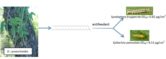
2.1. Choice Assay against S. frugiperda and E. paenulata

Compound 1 showed antifeedant activity against the generalist insect, S. frugiperda, and the specialist insect, E. paenulata, with the ED50 (0.82 µg/cm2) for the polyphagous S. frugiperda being ten times higher than that for the specialist E. paenulata (ED50 = 8.53 µg/cm2) (Table 1). We compared the ED50 of 1 with that of azadirachtin (2), and found an eight-fold difference for S. frugiperda (Table 1), but a 14-fold difference in E. paenulata. As far as we know, this is the first report about antifeedant activity exerted by 1. There are many reports that alkanes in particular have harmful effects on insects [2], but little is known about the effect of wax esters. Other structurally related compounds are bioactive against different species of insects. For example, octanoic acid (C8) was toxic against Drosophila melanogaster, but inactive against D. sechellia [9]. Fatty acids, such as oleic, linoleic, and linolenic acids, deterred the settling of Myzus persicae and Rhopalosiphum padi [10]. Pentacosanol (C25 alcohol) was a deterrent against Plutella xylostella larvae [11] and triacontanol (C30 alcohol) exhibited antifeedant activity against S. litura with an ED50 of 5.42 µg/cm2 [2,12].
Although many iridoid glycoside compounds have been isolated from D. cynanchoides [13,14] and iridoid glycosides have been reported as deterring consumption by generalist insect herbivores [15], we did not find iridoids to be among the most active antifeedant compounds. Compound 1 did show strong antifeedant activity, suggesting that D. cynanchoides mostly relies on 1 for its defense against herbivores.

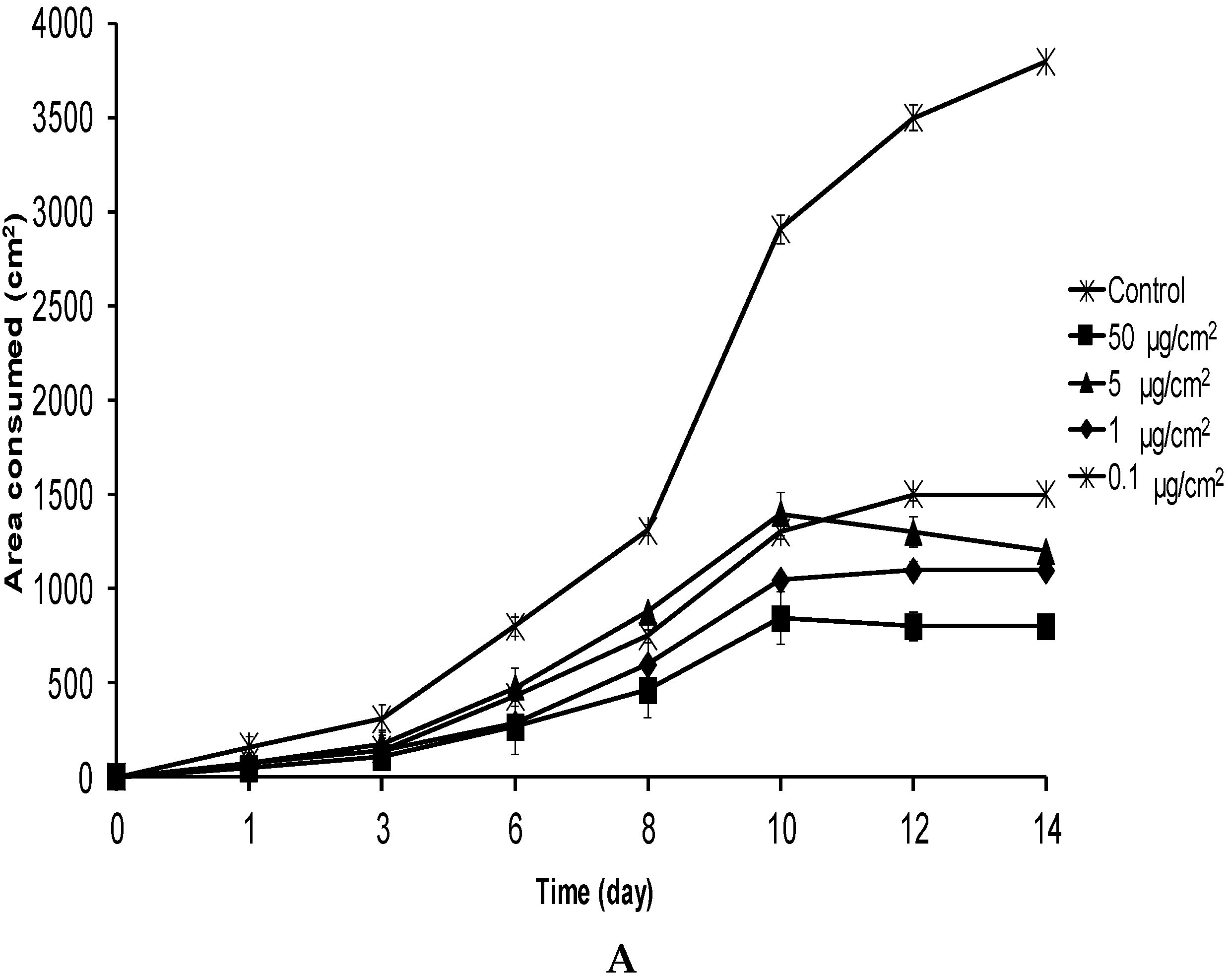
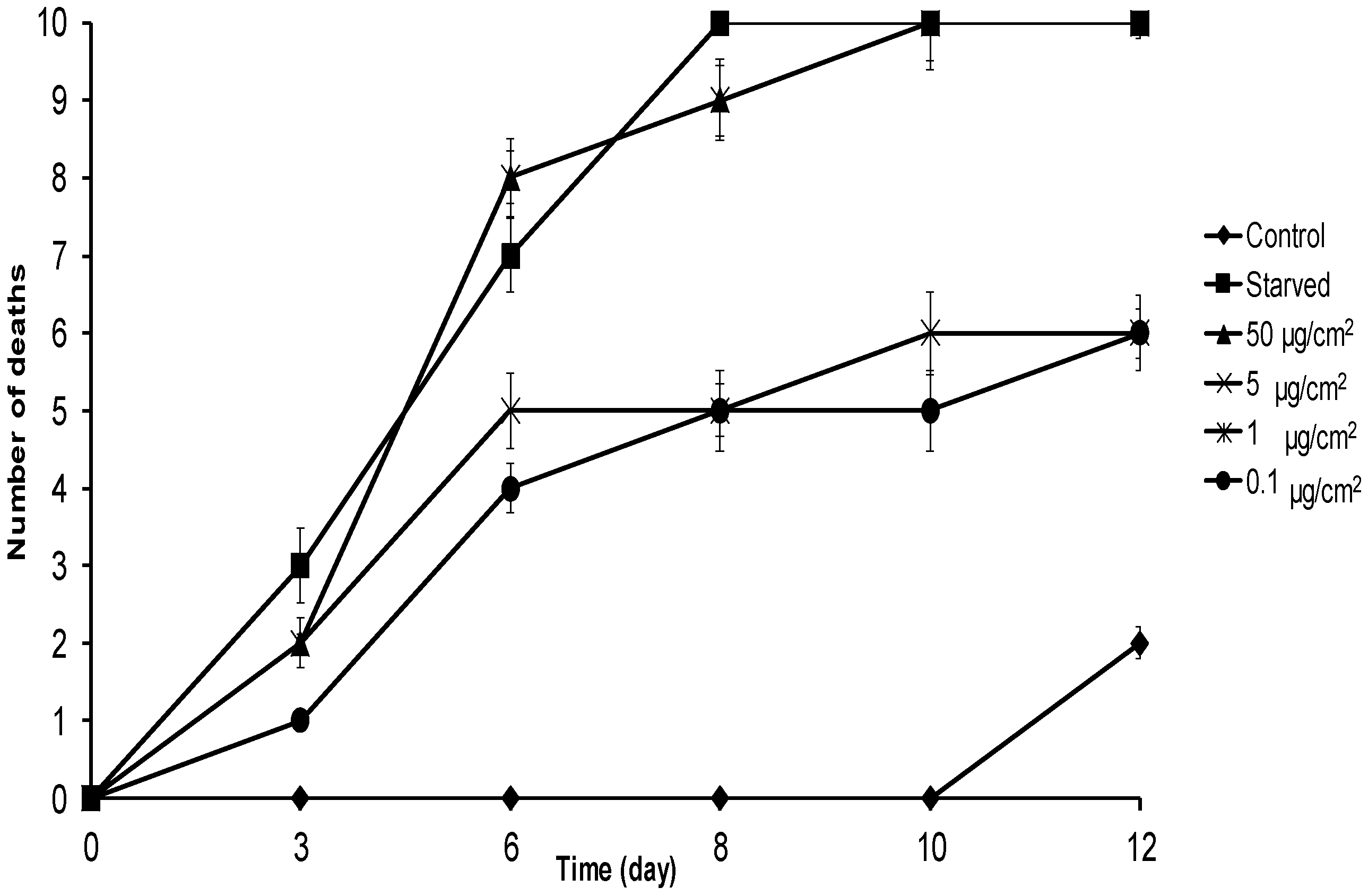
2.2. No-Choice and Mortality Assays against S. frugiperda

S. frugiperda larvae were fed on leaves treated with 1 at 0.1–50 µg/cm2, and at six days, they were strongly deterred, consuming up to three times less food than the controls (Figure 2A) with the observation of some mortality (20%–40%). After 10 days of exposure, larvae receiving 1 at 1, 5, and 50 µg/cm2 were not feeding at all, and larvae treated at 0.1 µg/cm2 consumed significantly less food than controls (H = 5.07; p = 0.019). This behavior was maintained until the end of the experiment (d14) when all the individuals were in pupae in the control, while in the treatments of 50, 5, 1 and 0.1 µg/cm2, all the remaining larvae (two, two, four, and three individuals, respectively) were in instar LII–III. The effect of 1 at 0.1 µg/cm2 was remarkable because few natural compounds are effective at this concentration [16], and even more so considering that the molar concentration of 1 was equivalent to 130 pmol/cm2.
This effect was clearly reflected in insect weight gain. While control larvae body weight steadily increased, the body weight of all larvae fed on leaves treated with 1 at 0.1–50 µg/cm2 was highest at 10 days after treatment and then decreased, probably due to starvation (Figure 2B). The initial average weight of control larvae was 6 ± 0.1 mg, reaching 150 ± 18 mg at day 14. In the case of larvae treated with 50 µg/cm2 of 1, the average weight was 6 ± 0.1 mg at the beginning and only 10 ± 2.2 mg at 14 days after initiation of the experiment. The larval weights for insects treated with the 5, 1, and 0.1 µg/cm2 concentrations at 14 days were 8 ± 2, 46 ± 5, and 45 ± 5 mg, respectively, reflecting the dose-dependent, high impact of 1 in this insect.
In the mortality assay, all larvae treated with 1 at 50 µg/cm2 died 10 days after the beginning of the assay, while all food-deprived larvae died by day 8. Doses of 0.1 and 1 µg/cm2 showed 50% mortality at day 12 (Figure 3). The LD50 of 1 at eight days was 0.39 (0.05–2.5) µg/cm2, while lethal times 50 (LT50) for 1 (Table 2) were 3.14 and 3.31 days for the 50 and 5 µg/cm2 treated groups, respectively. The LT50 for food-deprived larvae was 3.15 days. These results suggest that 1 strongly affects the food intake of S. frugiperda, consequently disrupting insect development and provoking low body weight and high mortality.
Waxes play an important role in plant–insect interactions, adversely affecting insects through direct toxicity or by hindering their physical movement [17]. The latter was extensively studied [18], but few reports were found for the former. Yang et al. found that five wild peanut species were more resistant to S. frugiperda larvae in feeding bioassays than a peanut cultivar. Larvae reared on foliage of the wild peanut species showed 79%–91% mortality compared to 17% for the cultivated peanut [19]. When they removed the epicuticular lipids from the foliage of wild and cultivated peanut species, S. frugiperda exhibited increased larval weights and earlier pupation and adult emergence than insects reared on a diet with untreated foliage, indicating that it was the wax mixture that negatively affected insect survival.
In a similar study, S. frugiperda larvae grew better when they were fed a diet containing corn foliage with the epicuticular lipids extracted than one with unextracted foliage [20], but diets incorporating lipid extracts containing no or trace amounts of wax esters had no effect on the insects [20]. Some resistant alfalfa plants (Medicago sativa L.) were found to have leaf surfaces with as much as 50% more wax esters than plants that are susceptible to spotted alfalfa aphids (Therioaphis maculata) [21], suggesting that wax esters are involved in this preference. In spite of many reports about the role of wax and wax esters and their effects against insects, we found no reports of the antifeedant action of the ester isolated here or similar ones.

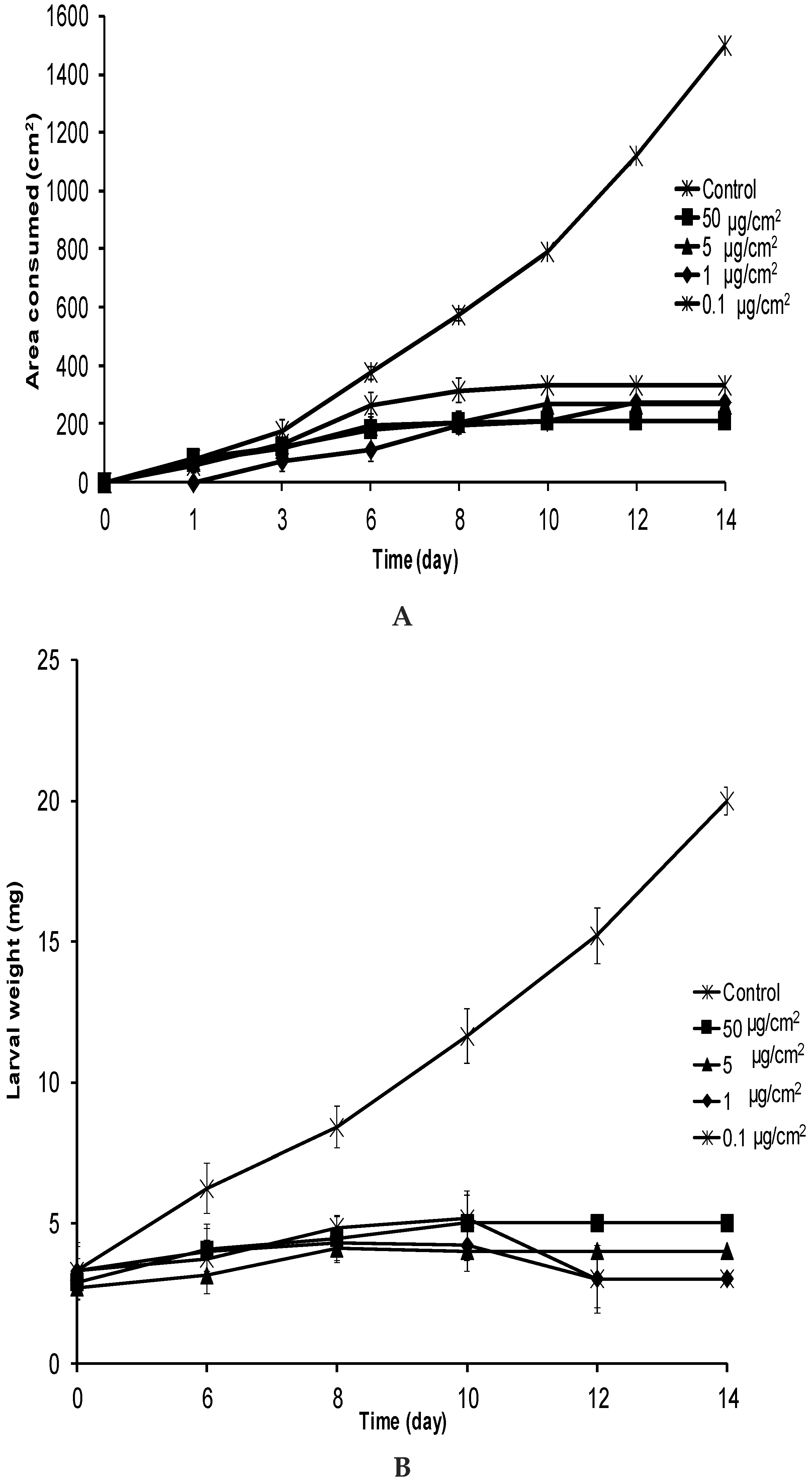
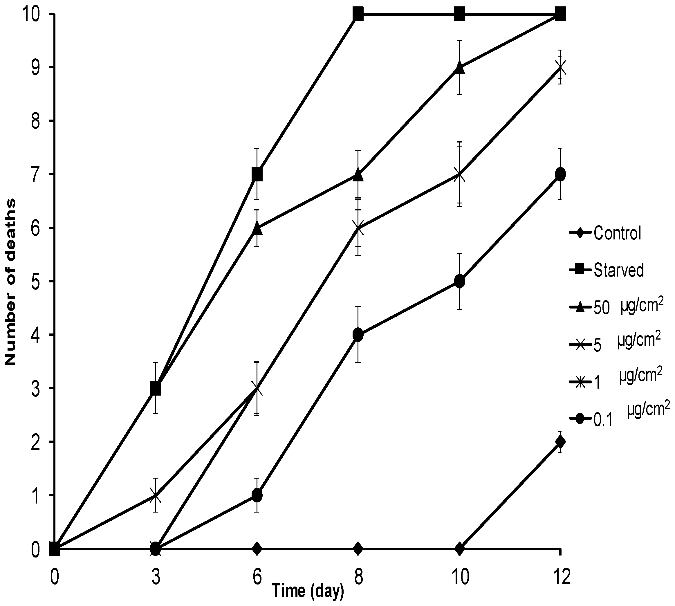
2.3. No-Choice and Mortality Assays against E. paenulata

After 3 days, E. paenulata larvae significantly reduced their intake of food treated with 1 at 0.1, 1, 5, or 50 µg/cm2 (p < 0.05) (Figure 4A). After six days of exposure to 1 at 1, 5, and 50 µg/cm2, E. paenulata larvae had consumed less than half the amount of leaf material as larvae facing untreated leaves (H = 21.87; p < 0.001) (Figure 4A). These differences increased with time and, after 12 days of exposure, larvae receiving 1 at all concentrations were not feeding at all, with four, three, three and two remaining individuals at 0.1, 1, 5, and 50 µg/cm2, respectively. The presence of 1 on insect food clearly led to larval starvation. E. paenulata larvae exposed to 1 at all concentrations did not gain weight (Figure 4B), whereas the body weights of the control larvae continually increased. At day 6, there were significant differences in body weight (H = 18.85; p < 0.001) between control larvae and those receiving food treated with 1 at any concentration (Figure 4B). After 14 days, larval weight had declined to less than the initial body weight.
In the mortality assay, after six days, the mortality rates of E. paenulata larvae receiving treated food were significantly higher than those of controls (H = 14.21; p = 0.004) for all concentrations including 0.1 µg/cm2 (Figure 5). Leaf treatment with 50 µg/cm2 of 1 induced 100% mortality after 12 days (Figure 5). With 5 µg/cm2 and 1 µg/cm2, mortality was 90%, and with 0.1 µg/cm2, it was 60% at the same time point. Food-deprived larvae did not survive longer than eight days. The LD50 of 1 at eight days was 0.68 µg/cm2 (0.01–1.99). Comparing this value with that reported for 2 (LD50 =1.24 µg/cm2) at four days [22] for the same insect suggests that 1 is as toxic as 2, one of the strongest known natural insect toxicants. Likewise, the toxicity of 1 against E. paenulata was similar to that of the limonoid meliartenin (LD50 = 0.76 µg/cm2 at 4 days) [22], another very strong natural insecticide. It was also more potent than other plant-derived compounds such as the flavonoid pinocembrin (LD50 = 18.4 µg/cm2 at six days), quercetin (LD50 > 50 µg/cm2 at 12 days) [23], and the sesquiterpene artemisinin (LD50 ~30 µg/cm2 at seven days) [24]. Lethal times 50 (LT50) for 1 (Table 2) was 4.69 days for 50 µg/cm2 compared to 4.10 days for food-deprived larvae, suggesting that larvae fed on leaves treated with 1 died from starvation. The feeding deterrent effects of 1 against E. paenulata larvae had strong consequences for their performance and survival.
Wax esters are biodegradable, non-toxic, and are obtained from renewable sources [25] including vegetable oil crops such as olive [26], soya [27] and sunflower [28]. Similar esters of different chain lengths may also act as insecticides. Consequently, our results encourage the study of fatty esters from crop residues, such as leaves and seeds, from vegetable oil processing waste, etc., in order to find new sources of potent anti-insect compounds.

3. Materials and Methods

3.1. Plant Material

Aerial parts of D. cynanchoides were collected in the Calamuchita Valley, Córdoba, Argentina (31°68′ S 64°43′ W) in March 2013. A voucher specimen (UCCOR 120) has been deposited at the Herbarium Marcelino Sayago of the School of Agricultural Sciences, Catholic University of Córdoba, and was identified by the agronomist Gustavo Ruiz. Air-dried aerial parts of D. cynanchoides (602 g) were extracted with ethanol for 96 h at room temperature. After removal of the solvent (reduced pressure), an extract was obtained (45.60 g, 7.57% yield).

3.2. General Experimental Procedures and Apparatus

1D- and 2D-NMR spectra were recorded in CDCl3 with a Bruker AVANCE II 400 spectrometer (Bruker, Billerica, MA, USA) operated at 400 MHz for 1H and at 100 MHz for the 13C nucleus. Tetramethylsilane (TMS) was used as the internal reference (δ 0.00). The high resolution mass spectra were determined in a Bruker MicroQTOF-II mass spectrometer (Bruker), equipped with an ESI source operated in positive mode at 180 °C with a capillary voltage of 4500 V. Mass accuracy was verified by calibration before and after sample introduction, using sodium formate (1 mM). Both samples and calibrant were introduced using a syringe pump at 10 µL min−1. Electron impact mass spectra (EI-MS) were obtained at 70 eV by GC–MS on a Hewlett–Packard 5970 Series mass spectrometer interfaced (Hewlwtt-Packard, Palo Alto, CA, USA) with a Hewlett–Packard 5890 gas chromatograph fitted with a column (HP-5MS, 15 m × 0.25 mm i.d., temperature from 200–290 °C, 10 °C/min). HPLC was performed in a Shimadzu chromatograph with a diode array UV-Vis detector (Shimadzu, Kyoto, Japan). Fourier transform infrared spectra were acquired on a FTIR Nicolet 510P spectrometer (Thermo Scientific, Waltham, MA, USA). Spectra over a range of 4000–500 cm−1 with a resolution of 2 cm−1 (50 scans) were recorded using KBr pellets.

3.3. Chemicals and Chromatographic Absorbents

Analytical TLC was performed on silica gel 60 F254 Merck plates (Darmstadt, Germany). Silica gel grade 200–400 mesh, 60 Å, for column chromatography was purchased from Sigma Chemical Co., Inc. (St. Louis, MO, USA). All solvents were purchased from Merck and Fisher Scientific (Buenos Aires, Argentina). Azadirachtin (2) (95% purity) was purchased from Sigma-Aldrich Argentina (Buenos Aires, Argentina).

3.4. Insects

S. frugiperda and E. paenulata larvae were obtained from a laboratory colonies. E. paenulata was reared on a natural diet of Curcubita maxima leaves [23] while S. frugiperda was reared on an artificial diet [29]. Both insects were maintained in a growth chamber at 24 ± 1 °C and 70%–75% relative humidity with a photoperiod of 16/8 h light-dark cycle, and periodically renewed with field specimens.

3.5. Feeding Choice Assay

The antifeedant experiments were carried out by the leaf-disk choice test for compounds 1 and 2 [30]. Two circular sections of the leaves of Lactuca sativa or C. maxima for S. frugiperda and E. paenulata, respectively, were placed in a Petri dish and a glass disk with two 1 cm2 holes was placed on top. One disk was treated with 10 µL of test solution and the other with 10 µL of Et2O (solvent control), and then both pieces were allowed to stand for 15 min until the solvent was completely evaporated. A third-instar larva of the corresponding insect was placed equidistant from the treated and untreated leaf disks and allowed to feed for 3 h or 24 h for S. frugiperda or E. paenulata larvae, respectively. Test solutions were prepared dissolving the necessary amount of 1 or 2 (reference of a potent natural antifeedant compound) in 10 mL of analytical grade Et2O. The dosages applied on the leaf offered for each compound were 0.1, 1, 5 and 50 µg/cm2. Ten replicates were conducted for each treatment. The relative amounts (recorded in percentages from 0 to 100) of the treated and untreated substrate area eaten in each test were estimated visually by dividing the food area into imaginary quarters or by determining the area of the leaf disks consumed by the larvae using the ImageJ screener software program (Fiji project. Available online: http://imagej.net/Downloads). An antifeedant index (AI%) was calculated as [(C − T)/(C + T)] × 100 [31], where T and C represent consumption of treated and untreated foods, respectively. Then, the ED50 (effective dose to obtain 50% feeding inhibition) was determined with the AI% values, from the Probit regression.

3.6. No-Choice Feeding Assay

One S. frugiperda or E. paenulata first instar larva was placed in a Petri dish and fed on L. sativa or C. maxima leaves, respectively, on which aliquots of 0.1, 1, 5 and 50 μg/cm2 (10 µL) of either 1 or Et2O (control, 10 µL) were applied with a Hamilton syringe. The leaves were renewed every 24 h. Ten replicates were conducted for each treatment and each trial was repeated twice. Leaf consumption and body weight were recorded every 24 h and 72 h, respectively [23].

3.7. Mortality Assay

Groups of 10 S. frugiperda or E. paenulata larvae (first instar) were continuously fed on leaves treated with either 0.1, 1, 5 or 50 μg/cm2 of 1 or with solvent (Et2O 10 µL) as control. A similar set of larvae was not fed at all and acted as food-deprived controls. Three replicates were performed for each treatment. Mortality was recorded every 24 h. Lethal dose LD50 and lethal time LT50 values for 1 were determined from mortality data by Probit analysis.

3.8. Isolation of Pentacosyl Heptacosanoate (1)

The extract of D. cynanchoides (3.50 g) was partitioned between Et2O/H2O. The Et2O soluble extract (0.87 g) showed AI% = 100, while the aqueous soluble fraction (2.53 g) was inactive. The organic fraction was fractionated by silica column chromatography and eluted with a gradient of hexane/Et2O and MeOH, obtaining 33 fractions according the TLC (eluted with hexane/Et2O (80:20)). The active fractions (F1 6–10) (AI% = 90% at 50 µg/cm2) were re-chromatographed on silica gel with hexane/Et2O 80:20, yielding 10 fractions. The most active fraction, F2 4 (AI% = 94%), presented a major component that precipitated as a solid. This compound was isolated (TLC with toluene/Et2O (70:30) Rf = 0.38), recrystallized from Et2O, and assayed against E. paenulata, showing AI% = 100% at 50 µg/cm2. The purity of this compound was determined by HPLC performed on a Kromasil direct-phase column (4.6 mm i.d. × 250 mm) (Akzo Nobel, Pasadena, CA, USA), eluting with hexane–isopropanol 95:5 as the mobile phase and UV detection at 210 nm. This methodology was also used to determine the yield of the active compound in plant material. The compound was identified as pentacosyl heptacosanoate (1) (Figure 1) by 1H-NMR, 13C-NMR, MS, and IR spectroscopies.
Pentacosyl heptacosanoate (1) white solid, IR, 2919, 2653, 1732, 1730, 1454, 1374, 1281, 1069 cm−1. 1H-NMR (CDCl3, 400 MHz) δ 0.88 (6H, t, J = 6.8 Hz, 2 × CH3) 1.25 (90H, 45 × CH2), 1.57 (2H, m; CH2), 1.63 (2H, m; CH2), 2.35 (2H, t, J = 7.4, CH2-CO), 3.64 (2H, t, J = 6.6, CH2-O); 13C-NMR (CDCl3, 100 MHz) δ 14.13 (CH3), 22.71 (CH2), 25.75 (CH2), 29.71 (CH2), 31.94 (CH2), 32.82 (CH2-CO), 63.13 (CH2-O), 178.13 (C=O). ESI-Q-TOF MS: m/z 761.3829 (calcd. for C52H104O2, 761.3823, error 0.78 ppm). EIMS: m/z 393 (C27H53O), 365 (C26H53), 351 (C25H51), 111, 97, 83, 71, 57.

3.9. Statistical Analysis

Results from choice tests were analyzed by the Wilcoxon Signed Rank Test. Data were analyzed for normal distribution and homoscedasticity and, as they did not meet these requirements, non-parametric tests were used. Results on average larval body weight, accumulated consumption values from no-choice tests, and mortality from the mortality assay were compared with respect to concentrations by the Kruskal–Wallis non-parametric analysis of variance, followed by the Dunn test. Statistical analyses were performed using the InfoStat statistical package (version 2008, UNC, Córdoba, Argentina). Differences were considered significant at p ≤ 0.05.

4. Conclusions

The antifeedant effect of 1 on generalist and specialist insects, in both the choice and no-choice tests, showed negative consequences for insect survival, suggesting that the use of this compound is worth evaluating as an insecticide. This wax, with its simple chemical structure that is easy to synthesize or mimic, may even be considered as a model for designing new eco-friendly insecticides. In addition, many edible crops produce wax esters that are discarded during processing for food. Research on these materials could lead to the detection of similar waxes with insecticidal activity.

Acknowledgments

G.D.N. gratefully acknowledges receipt of a fellowship from CONICET (Buenos Aires, Argentina). We thank San Francisco Edit for editing and revising the English language. Financial support for this work was provided by the Agencia Nacional de Promoción Científica y Técnica, FONCYT, PICT 33593 and CONICET PIP 11220100100236.

Author Contributions

S.M.P., M.C.C., and G.D.N. conceived and designed the experiments. G.D.N performed the experiments: G.D.N. analyzed the data. S.M.P., M.C.C., and G.D.N. wrote the paper.

Conflicts of Interest

The authors declare no conflict of interest in the decision to publish the results. The founding sponsors had no role in the design of the study; in the collection, analyses, or interpretation of data; in the writing of the manuscript, and in the decision to publish the results.

References

  1. González-Coloma, A.; Reina, M.; Gutiérrez, C.; Fraga, B.M. Natural insecticides: Structure diversity, effects and structure-activity relationships. A case study. In Studies in Natural Products Chemistry; Atta ur, R., Ed.; Elsevier: Amsterdam, The Netherlands, 2002; Volume 26, Part G; pp. 849–879. [Google Scholar]
  2. Céspedes, C.L.; Molina, S.C.; Muñoz, E.; Lamilla, C.; Alarcon, J.; Palacios, S.M.; Carpinella, M.C.; Avila, J.G. The insecticidal, molting disruption and insect growth inhibitory activity of extracts from Condalia microphylla cav. (rhamnaceae). Ind. Crops Prod. 2013, 42, 78–86. [Google Scholar] [CrossRef]
  3. Palacios, S.M.; Maggi, M.E.; Bazán, C.M.; Carpinella, M.C.; Turco, M.; Muñoz, A.; Alonso, R.A.; Nuñez, C.; Cantero, J.J.; Defago, M.T.; et al. Screening of argentinian plants for pesticide activity. Fitoterapia 2007, 78, 580–584. [Google Scholar] [CrossRef] [PubMed]
  4. Del Corral, S.; Diaz Napal, G.N.; Zaragoza, M.H.; Carpinella, M.C.; Ruiz, G.M.; Palacios, S.M. Screening for extracts with insect antifeedant properties in native plants from central argentina. Bol. Latinoamer. Caribe Plant. Med. Aromat. 2014, 13, 498–505. [Google Scholar]
  5. Barboza, G.E. Flora Medicinal de la provincia de Córdoba (Argentina): Pteridófitas Y Antófitas Silvestres O Naturalizadas; Museo Botánico: Córdoba, Spain, 2006. [Google Scholar]
  6. Castillo, L.; González-Coloma, A.; González, A.; Díaz, M.; Santos, E.; Alonso-Paz, E.; Bassagoda, M.J.; Rossini, C. Screening of uruguayan plants for deterrent activity against insects. Ind. Crops Prod. 2009, 29, 235–240. [Google Scholar] [CrossRef] [PubMed]
  7. Yeats, T.H.; Rose, J.K.C. The formation and function of plant cuticles. Plant Physiol. 2013, 163, 5–20. [Google Scholar] [CrossRef] [PubMed]
  8. Eigenbrode, S.D.; Espelie, K.E. Effects of plant epicuticular lipids on insect herbivores. Annu. Rev. Entomol. 1995, 40, 171–194. [Google Scholar] [CrossRef]
  9. Legal, L.; Moulin, B.; Jallon, J.M. The relation between structures and toxicity of oxygenated aliphatic compounds homologous to the insecticide octanoic acid and the chemotaxis of two species of Drosophila. Pestic. Biochem. Physiol. 1999, 65, 90–101. [Google Scholar] [CrossRef]
  10. Castillo, L.; Díaz, M.; González-Coloma, A.; González, A.; Alonso-Paz, E.; Bassagoda, M.J.; Rossini, C. Clytostoma callistegioides (bignoniaceae) wax extract with activity on aphid settling. Phytochemistry 2010, 71, 2052–2057. [Google Scholar] [CrossRef] [PubMed]
  11. Eigenbrode, S.D.; Pillai, S.K. Neonate Plutella xylostella responses to surface wax components of a resistant cabbage (Brassica oleracea). J. Chem. Ecol. 1998, 24, 1611–1627. [Google Scholar] [CrossRef]
  12. Pande, A.; Shukla, Y.N.; Tripathi, A.K. Lipid constituents from Stellaria media. Phytochemistry 1995, 39, 709–711. [Google Scholar] [CrossRef]
  13. Bonini, C.; Davini, E.; Iavarone, C.; Trogolo, C. Cynanchoside, a highly oxygenated iridoid glucoside from Macfadyena cynanchoides. Phytochemistry 1981, 20, 1587–1590. [Google Scholar] [CrossRef]
  14. Adriani, C.; Iavarone, C.; Trogolo, C. 5,7-bisdeoxycynanchoside, an iridoid glucoside from Macfadyena cynanchoides. Phytochemistry 1982, 21, 231–233. [Google Scholar] [CrossRef]
  15. Dobler, S.; Petschenka, G.; Pankoke, H. Coping with toxic plant compounds—The insect’s perspective on iridoid glycosides and cardenolides. Phytochemistry 2011, 72, 1593–1604. [Google Scholar] [CrossRef] [PubMed]
  16. Koul, O. Insect Antifeedants; CRC Press LLC: Boca Raton, FL, USA, 2005. [Google Scholar]
  17. Eigenbrode, S.D.; Jetter, R. Attachment to plant surface waxes by an insect predator. Integr. Comp. Biol. 2002, 42, 1091–1099. [Google Scholar] [CrossRef] [PubMed]
  18. Müller, C.; Riederer, M. Plant surface properties in chemical ecology. J. Chem. Ecol. 2005, 31, 2621–2651. [Google Scholar] [CrossRef] [PubMed]
  19. Yang, G.; Espelie, K.E.; Todd, J.W.; Culbreath, A.K.; Pittman, R.N.; Demski, J.W. Cuticular lipids from wild and cultivated peanuts and the relative resistance of these peanut species to fall armyworm and thrips. J. Agric. Food Chem. 1993, 41, 814–818. [Google Scholar] [CrossRef]
  20. Yang, G.; Wiseman, B.R.; Isenhour, D.; Espelie, K. Chemical and ultrastructural analysis of corn cuticular lipids and their effect on feeding by fall armyworm larvae. J. Chem. Ecol. 1993, 19, 2055–2074. [Google Scholar] [CrossRef] [PubMed]
  21. Bergman, D.K.; Dillwith, J.W.; Zarrabi, A.A.; Caddel, J.L.; Berberet, R.C. Epicuticular lipids of alfalfa relative to its susceptibility to spotted alfalfa aphids (homoptera: Aphididae). Environ. Entomol. 1991, 20, 781–785. [Google Scholar] [CrossRef]
  22. Carpinella, M.C.; Defago, M.T.; Valladares, G.; Palacios, S.M. Antifeedant and insecticide properties of a limonoid from Melia azedarach (meliaceae) with potential use for pest management. J. Agric. Food Chem. 2003, 51, 369–374. [Google Scholar] [CrossRef] [PubMed]
  23. Diaz Napal, G.N.; Defagó, M.; Valladares, G.; Palacios, S.M. Response of Epilachna paenulata to two flavonoids, pinocembrin and quercetin, in a comparative study. J. Chem. Ecol. 2010, 36, 898–904. [Google Scholar] [CrossRef] [PubMed]
  24. Maggi, M.E.; Mangeaud, A.; Carpinella, M.C.; Ferrayoli, C.G.; Valladares, G.R.; Palacios, S.M. Laboratory evaluation of Artemisia annua L. Extract and artemisinin activity against Epilachna paenulata and Spodoptera eridania. J. Chem. Ecol. 2005, 31, 1527–1536. [Google Scholar] [CrossRef] [PubMed]
  25. Rani, K.N.P.; Neeharika, T.S.V.R.; Kumar, T.P.; Satyavathi, B.; Sailu, C.; Prasad, R.B.N. Kinetics of enzymatic esterification of oleic acid and decanol for wax ester and evaluation of its physico-chemical properties. J. Taiwan Inst. Chem. Eng. 2015, 55, 12–16. [Google Scholar] [CrossRef]
  26. Bianchi, G.; Murelli, C.; Vlahov, G. Surface waxes from olive fruits. Phytochemistry 1992, 31, 3503–3506. [Google Scholar] [CrossRef]
  27. Palacios, C.E.J.; Negri, G.; Salatino, A. Esters and other constituents of the foliar cuticular wax of a soybean variety. Biochem. Syst. Ecol. 2015, 63, 198–200. [Google Scholar] [CrossRef]
  28. Kanya, T.C.S.; Rao, L.J.; Sastry, M.C.S. Characterization of wax esters, free fatty alcohols and free fatty acids of crude wax from sunflower seed oil refineries. Food Chem. 2007, 101, 1552–1557. [Google Scholar]
  29. Diaz Napal, G.; Palacios, S. Bioinsecticidal effect of the flavonoids pinocembrin and quercetin against Spodoptera frugiperda. J. Pest Sci. 2015, 88, 629–635. [Google Scholar] [CrossRef]
  30. Defagó, M.; Valladares, G.; Banchio, E.; Carpinella, M.C.; Palacios, S.M. Insecticide and antifeedant activity of different plant parts of Melia azedarach on Xanthogaleruca luteola. Fitoterapia 2006, 77, 500–505. [Google Scholar] [CrossRef] [PubMed]
  31. Mazoir, N.; Benharref, A.; Bailén, M.; Reina, M.; González-Coloma, A. Bioactive triterpene derivatives from latex of two Euphorbia species. Phytochemistry 2008, 69, 1328–1338. [Google Scholar] [CrossRef] [PubMed]
  • Sample Availability: Samples of the compounds are available from the authors.
Figure 1. Chemical structure of pentacosyl heptacosanoate (1).
Molecules 21 01039 g001
Figure 2. Average leaf area consumed (A) and average body weight (B) of S. frugiperda larvae fed on leaves treated with 1 in a no-choice feeding assay. Error bars <60 and <10 for area consumed and body weight, respectively, are embedded in symbols.
Molecules 21 01039 g002aMolecules 21 01039 g002b
Figure 3. Cumulative mortality of S. frugiperda larvae fed with leaves treated with 1 at different concentrations. All treatments were in triplicate (n = 10). Starved represents food-deprived treatment. Error bars show standard deviation; where error is not shown, it is smaller than the symbol.
Molecules 21 01039 g003
Figure 4. Average leaf area consumed (A) and average body weight (B) of E. paenulata larvae fed on leaves treated with 1 in a no-choice feeding assay. Error bars <30 and <2 for area consumed and body weight, respectively, are embedded in symbols.
Molecules 21 01039 g004
Figure 5. Cumulative mortality of E. paenulata larvae fed with leaves treated with 1 at different concentrations. All treatments were in triplicate (n = 10). Starved represents food-deprived treatment. Error bars show standard deviation; where error is not shown, it is smaller than the symbol.
Molecules 21 01039 g005
Table 1. Effective antifeedant dose 50 (ED50) of 1 and 2 against Spodoptera frugiperda and Epilachna paenulata determined by choice test.
CompoundInsectED50 (µg/cm2) (Values and 95% Confidence Interval)
Pentacosyl heptacosanoate (1)S. frugiperda0.82 (0.15–4.50)
Pentacosyl heptacosanoate (1)E. paenulata8.53 (2.73–26.59)
Azadirachtin (2)S. frugiperda0.10 (0.05–0.19)
Azadirachtin (2)E. paenulata0.59 (0.07–4.37)
Table 2. Lethal time (LT50) of 1 against Spodoptera frugiperda and Epilachna paenulata in mortality test.
Dosage µg/cm2LT50 in Days (Values and 95% Confidence Interval) a
1Food-Deprived Larvae
Spodoptera frugiperda
503.14 (2.88–6.40)
53.31 (3.10–6.70)
16.96 (3.62–16.00)
0.17.71 (4.81–16.53)
3.16 (2.63–6.03)
Epilachna paenulata
504.69 (2.78–7.80)
57.56 (5.62–9.96)
17.80 (4.89–10.06)
0.19.62 (7.06–13.13)
4.10 (2.59–6.19)
a LT50 is the time required to obtain 50% mortality. LT50 was calculated by Probit log regression.

