
Synthesis, Structural Studies and Molecular Modelling of a Novel Imidazoline Derivative with Antifungal Activity

 , ,
, ,
Abstract
:1. Introduction
2. Results and Discussion
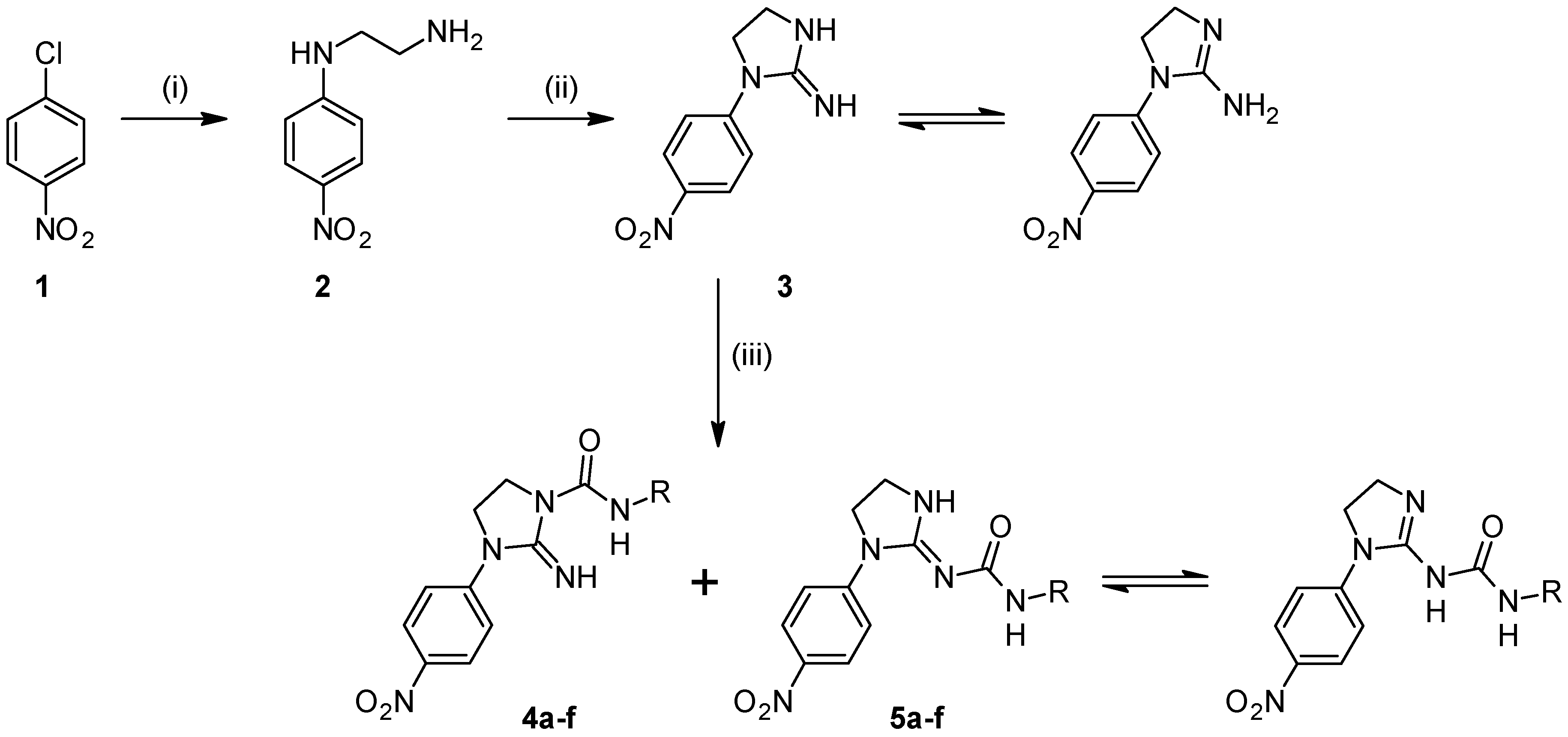
2.1. Chemistry

| Compound | R |
|---|---|
| a |  |
| b |  |
| c |  |
| d |  |
| e |  |
| f |  |


2.2. Microbiology
| MIC Value | 4f | Clotrimazole | ||||||
|---|---|---|---|---|---|---|---|---|
| No. of Strains (n = 41) | % of Strains | MIC50 | MIC90 | No. of Strains (n = 31) | % of Strains | MIC50 | MIC90 | |
| 0.015 | 0 (0.0) | 0 | 250 | 1000 | 3 | 9.7 | 0.24 | 1.95 |
| 0.03 | 0 | 0 | 5 | 16.1 | ||||
| 0.06 | 0 | 0 | 2 | 6.5 | ||||
| 0.12 | 0 | 0 | 3 | 9.7 | ||||
| 0.24 | 0 | 0 | 5 | 16.1 | ||||
| 0.49 | 0 | 0 | 5 | 16.1 | ||||
| 0.98 | 1 | 2.4 | 3 | 9.7 | ||||
| 1.95 | 1 | 2.4 | 2 | 6.5 | ||||
| 3.9 | 1 | 2.4 | 1 | 3.2 | ||||
| 7.82 | 2 | 4.9 | 2 | 6.5 | ||||
| 15.63 | 1 | 2.4 | 0 | 0 | ||||
| 31.25 | 2 | 4.9 | 0 | 0 | ||||
| 62.5 | 3 | 7.3 | 0 | 0 | ||||
| 125 | 7 | 17.1 | 0 | 0 | ||||
| 250 | 5 | 12.2 | 0 | 0 | ||||
| 500 | 10 | 24.4 | 0 | 0 | ||||
| 1000 | 7 | 17.1 | 0 | 0 | ||||
| >1000 | 1 | 2.4 | 0 | 0 | ||||
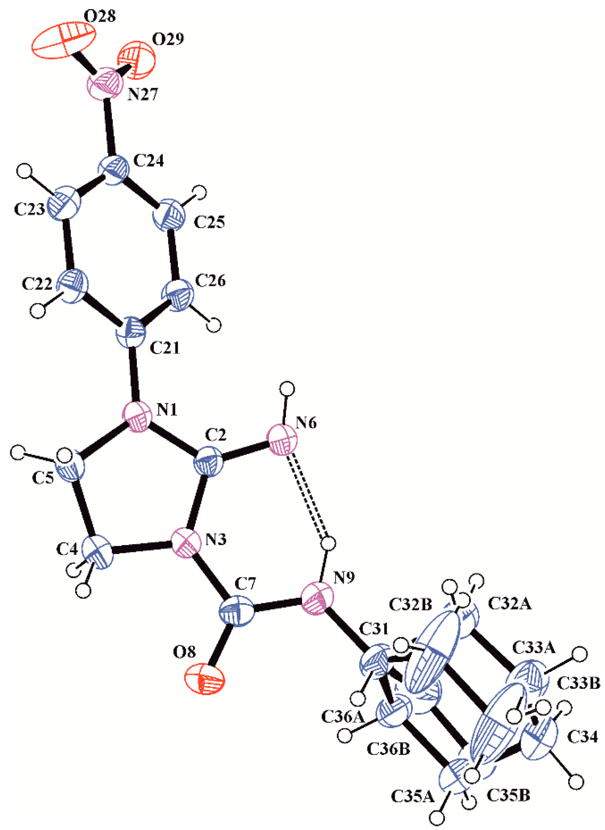
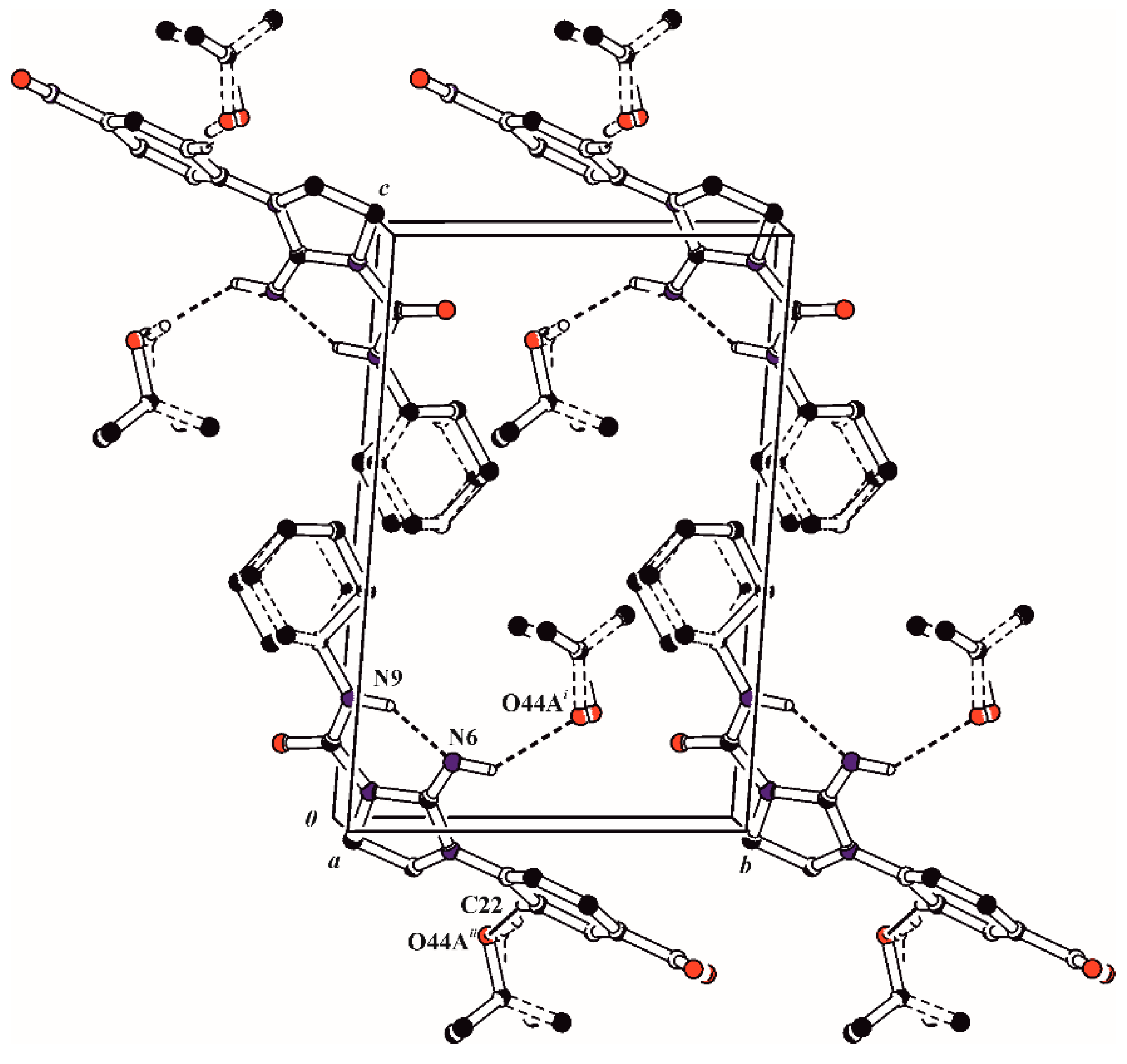
2.3. X-ray Analysis

| D–H···A | D–H | H···A | D···A | D–H···A | Symmetry Codes |
|---|---|---|---|---|---|
| N6–H6···O44A | 0.86(4) | 2.57(4) | 3.263(11) | 139(3) | i = x, y, z |
| C22–H22···O44A | 0.93 | 2.56 | 3.462(15) | 164 | ii = 1 − x, 1 − y, −z |
| N9–H9···N6 | 0.94(4) | 1.88(4) | 2.692(3) | 143(4) |

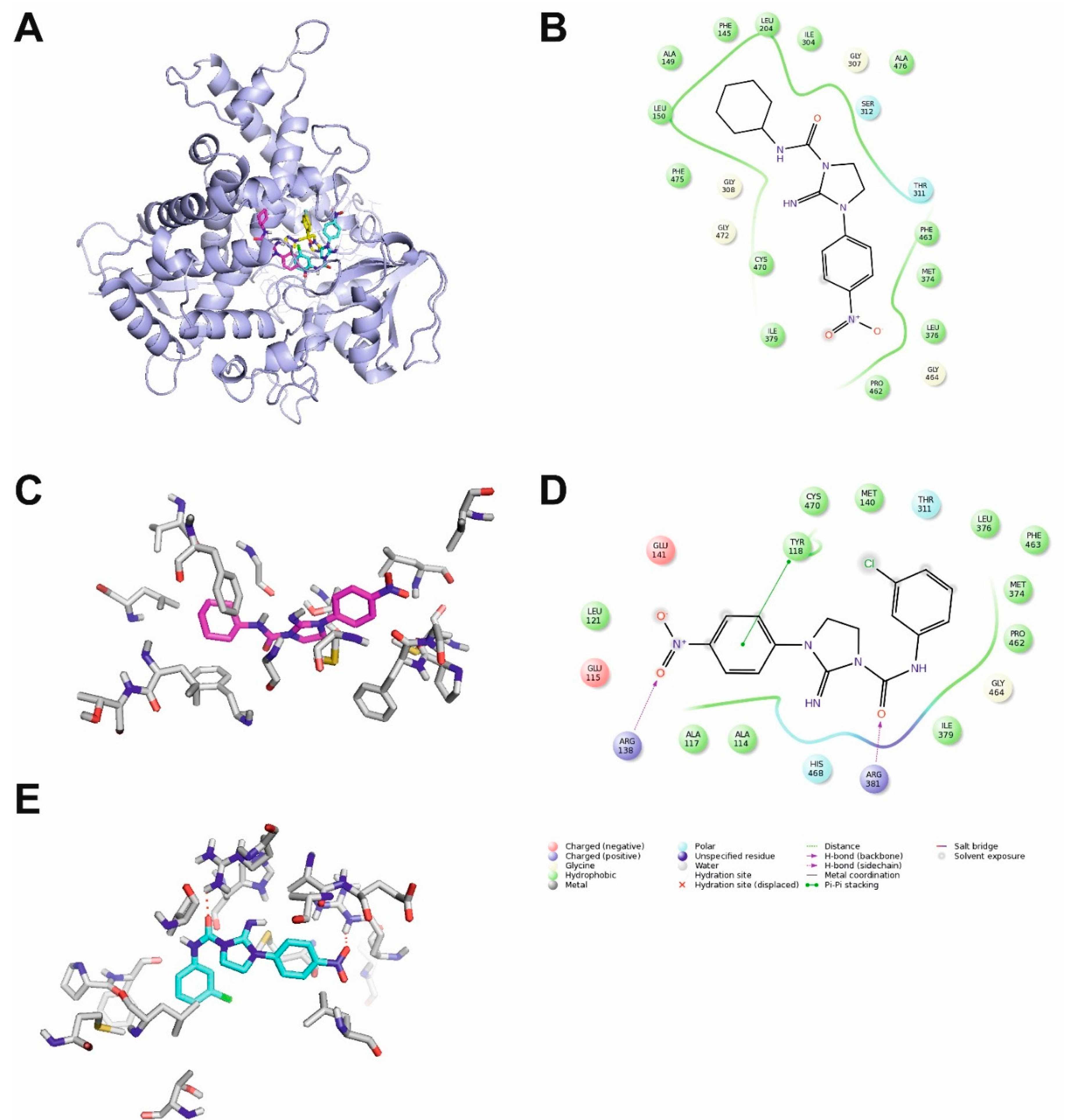
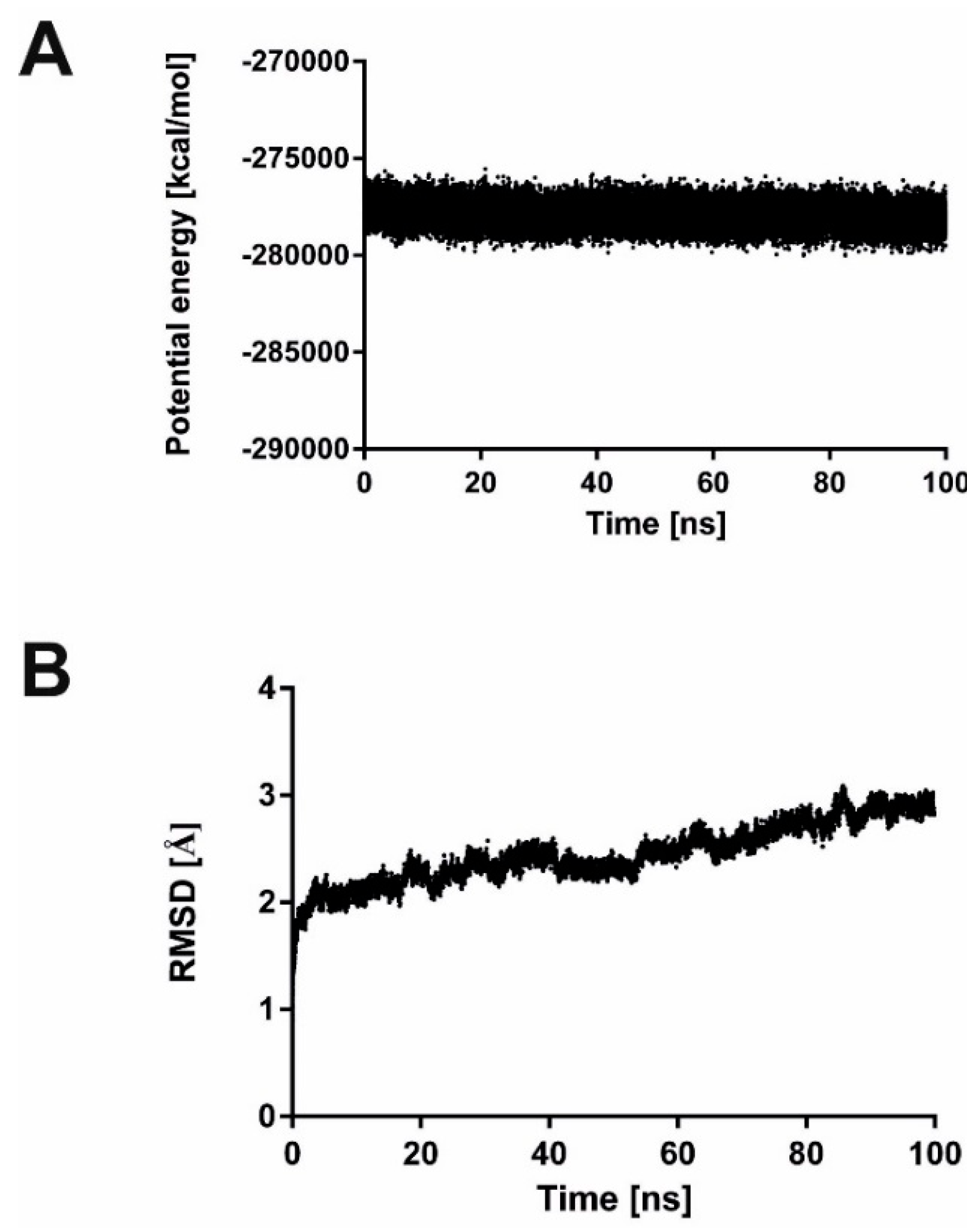
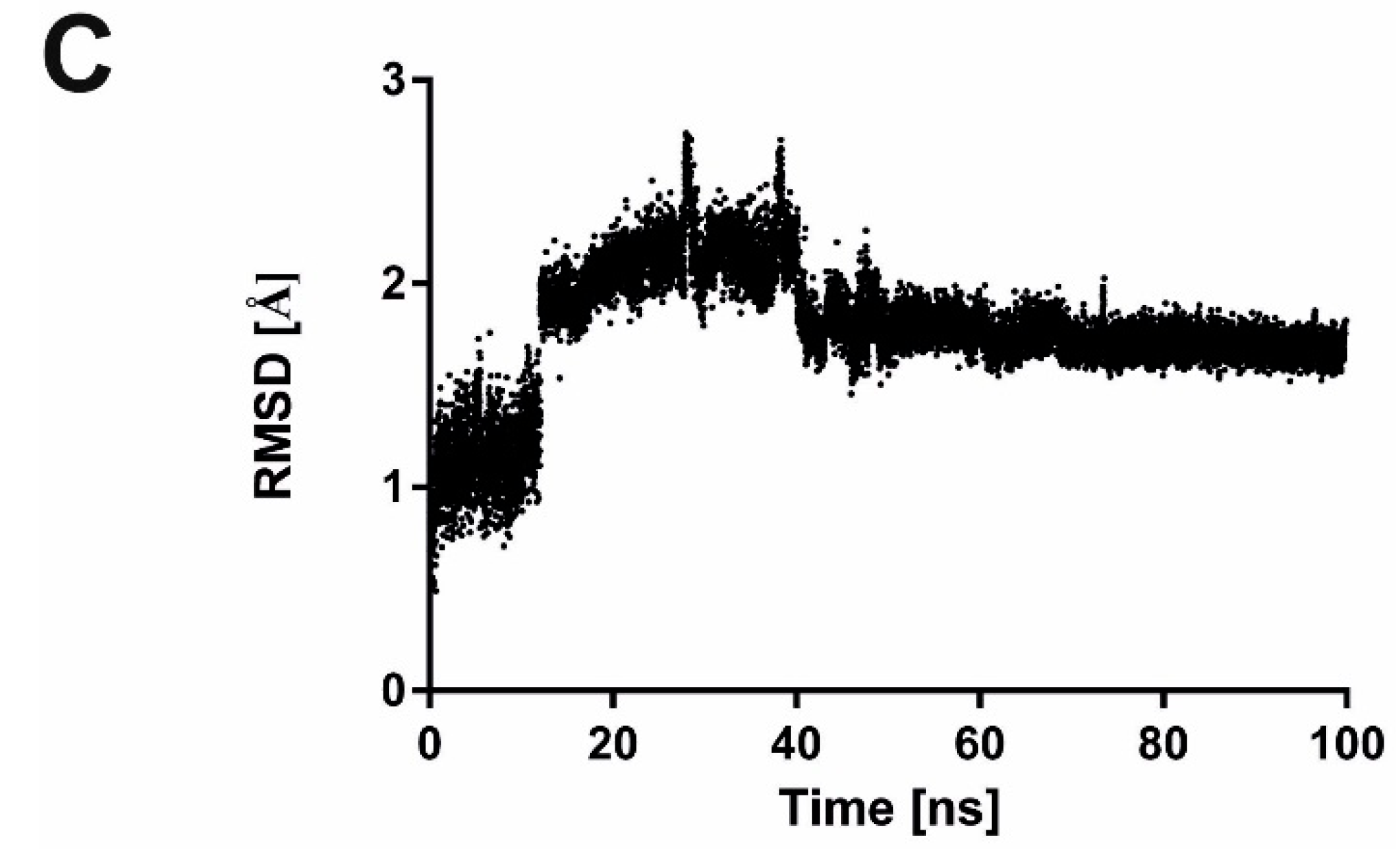
2.4. Molecular Modelling


3. Experimental Section
3.1. Synthesis
General Procedure to Obtain Compounds 4
3.2. Microbiology
3.3. X-ray Analysis
3.4. Molecular Modelling
4. Conclusions
Acknowledgments
Author Contributions
Conflicts of Interest
References
- Cui, L.; Morris, A.; Ghedin, E. The human mycobiome in health and disease. Genome Med. 2013, 5, 63. [Google Scholar] [CrossRef] [PubMed]
- Oever, J.T.; Netea, M.G. The bacteriome-mycobiome interaction and antifungal host defense. Eur. J. Immunol. 2014, 44, 3182–3191. [Google Scholar] [CrossRef] [PubMed]
- Puebla, L.E.J. Fungal Infections in Immunosuppressed Patients. In Immunodeficiency; INTECH Open Access Publisher: Rijeka, Croatia, 2012. [Google Scholar]
- Williams, C.; Ramage, G. Fungal Biofilms in Human Disease. In Biofilm-Based Healthcare-Associated Infections; Donelli, G., Ed.; Springer International Publishing: Cham, Switzerland, 2015; Volume 831, pp. 11–27. [Google Scholar]
- Hube, B.; Hay, R.; Brasch, J.; Veraldi, S.; Schaller, M. Dermatomycoses and inflammation: The adaptive balance between growth, damage, and survival. J. Mycol. Med. 2015, 25, e44–e58. [Google Scholar] [CrossRef] [PubMed]
- Eldridge, M.L.; Chambers, C.J.; Sharon, V.R.; Thompson, G.R. Fungal infections of the skin and nail: New treatment options. Expert Rev. Anti Infect. Ther. 2014, 12, 1389–1405. [Google Scholar] [CrossRef] [PubMed]
- Pappas, P.G. Invasive candidiasis. Infect. Dis. Clin. N. Am. 2006, 20, 485–506. [Google Scholar] [CrossRef] [PubMed]
- Lai, C.C.; Tan, C.K.; Huang, Y.T.; Shao, P.L.; Hsueh, P.R. Current challenges in the management of invasive fungal infections. J. Infect. Chemother. 2008, 14, 77–85. [Google Scholar] [CrossRef] [PubMed]
- Castelli, M.V.; Butassi, E.; Monteiro, M.C.; Svetaz, L.A.; Vicente, F.; Zacchino, S.A. Novel antifungal agents: A patent review (2011–present). Expert Opin. Ther. Pat. 2014, 24, 323–338. [Google Scholar] [CrossRef] [PubMed]
- Calderone, R.; Sun, N.; Gay-Andrieu, F.; Groutas, W.; Weerawarna, P.; Prasad, S.; Alex, D.; Li, D. Antifungal drug discovery: The process and outcomes. Future Microbiol. 2014, 9, 791–805. [Google Scholar] [CrossRef] [PubMed]
- Ganal, S.C.; Sanos, S.L.; Kallfass, C.; Oberle, K.; Johner, C.; Kirschning, C.; Lienenklaus, S.; Weiss, S.; Staeheli, P.; Aichele, P.; et al. Priming of natural killer cells by nonmucosal mononuclear phagocytes requires instructive signals from commensal microbiota. Immunity 2012, 37, 171–186. [Google Scholar] [CrossRef] [PubMed]
- Viaud, S.; Saccheri, F.; Mignot, G.; Yamazaki, T.; Daillère, R.; Hannani, D.; Enot, D.P.; Pfirschke, C.; Engblom, C.; Pittet, M.J.; et al. The intestinal microbiota modulates the anticancer immune effects of cyclophosphamide. Science 2013, 342, 971–976. [Google Scholar] [CrossRef] [PubMed]
- Wróbel, T.M.; Kiełbus, M.; Kaczor, A.A.; Kryštof, V.; Karczmarzyk, Z.; Wysocki, W.; Fruziński, A.; Król, S.K.; Grabarska, A.; Stepulak, A.; et al. Discovery of nitroaryl urea derivatives with antiproliferative properties. J. Enzyme Inhib. Med. Chem. 2015. [Google Scholar] [CrossRef] [PubMed]
- Dobrowolski, M.A.; Cyrański, M.K.; Pisklak, M.; Wawer, I.; Matosiuk, D. Structural studies of 1-aryl-2-aminoimidazolinium bromides: Focus on tautomer preference of the 2-aminoimidazoline moiety in the solid state. Pol. J. Chem. 2007, 81, 1037–1048. [Google Scholar]
- Allen, F.H.; Kennard, O.; Watson, D.G.; Brammer, L.; Orpen, A.G.; Taylor, R. Tables of bond lengths determined by X-ray and neutron diffraction. Part 1. Bond lengths in organic compounds. J. Chem. Soc. Perkin Trans. 1987, 2, S1–S19. [Google Scholar] [CrossRef]
- Wysocki, W.; Matosiuk, D.; Karczmarzyk, Z.; Fidecka, S.; Fruzinski, A. 1-(3-chlorophenyl)-3-(1-p-tolylimidazolidin-2-ylidene)urea. Acta Crystallogr. Sect. E 2009, 65, o40. [Google Scholar] [CrossRef] [PubMed]
- Saczewski, J.; Korcz, M.; Karczewska, P.; Gdaniec, M. Reactivity of cyclic alkoxyguanidines—Experimental and theoretical studies. ARKIVOC 2013, 346–362. [Google Scholar] [CrossRef]
- Cremer, D.; Pople, J.A. General definition of ring puckering coordinates. J. Am. Chem. Soc. 1975, 97, 1354–1358. [Google Scholar] [CrossRef]
- Rossello, A.; Bertini, S.; Lapucci, A.; Macchia, M.; Martinelli, A.; Rapposelli, S.; Herreros, E.; Macchia, B. Synthesis, antifungal activity, and molecular modeling studies of new inverted oxime ethers of oxiconazole. J. Med. Chem. 2002, 45, 4903–4912. [Google Scholar] [CrossRef] [PubMed]
- Podust, L.M.; Poulos, T.L.; Waterman, M.R. Crystal structure of cytochrome p450 14α-sterol demethylase (cyp51) from mycobacterium tuberculosis in complex with azole inhibitors. Proc. Natl. Acad. Sci. USA 2001, 98, 3068–3073. [Google Scholar] [CrossRef] [PubMed]
- Xiao, L.; Madison, V.; Chau, A.S.; Loebenberg, D.; Palermo, R.E.; McNicholas, P.M. Three-dimensional models of wild-type and mutated forms of cytochrome p450 14α-sterol demethylases from aspergillus fumigatus and candida albicans provide insights into posaconazole binding. Antimicrob. Agents Chemother. 2004, 48, 568–574. [Google Scholar] [CrossRef] [PubMed]
- Fukuoka, T.; Johnston, D.A.; Winslow, C.A.; de Groot, M.J.; Burt, C.; Hitchcock, C.A.; Filler, S.G. Genetic basis for differential activities of fluconazole and voriconazole against candida krusei. Antimicrob. Agents Chemother. 2003, 47, 1213–1219. [Google Scholar] [CrossRef] [PubMed]
- Macchiarulo, A.; Costantino, G.; Fringuelli, D.; Vecchiarelli, A.; Schiaffella, F.; Fringuelli, R. 1,4-benzothiazine and 1,4-benzoxazine imidazole derivatives with antifungal activity: A docking study. Biorg. Med. Chem. 2002, 10, 3415–3423. [Google Scholar] [CrossRef]
- Allali, M.; Benoist, E.; Habbadi, N.; Gressier, M.; Souizi, A.; Dartiguenave, M. Design and synthesis of new ethylenediamine or propylenediamine diacetic acid derivatives for re(i) organometallic chemistry. Tetrahedron 2004, 60, 1167–1174. [Google Scholar] [CrossRef]
- Popiołek, Ł.; Kosikowska, U.; Wujec, M.; Malm, A. Synthesis and antimicrobial evaluation of new schiff base hydrazones bearing 1,2,4-triazole moiety. Phosphorus Sulfur Silicon Relat. Elem. 2013, 189, 1611–1623. [Google Scholar]
- Sadabs, 2004/1; Bruker AXS Inc.: Madison, WI, USA, 2005.
- Sheldrick, G. A short history of SHELX. Acta Crystallogr. Sect. A 2008, 64, 112–122. [Google Scholar] [CrossRef] [PubMed]
- Farrugia, L. Wingx and ortep for windows: An update. J. Appl. Crystallogr. 2012, 45, 849–854. [Google Scholar] [CrossRef]
- Edgar, R.C. Muscle: Multiple sequence alignment with high accuracy and high throughput. Nucleic Acids Res. 2004, 32, 1792–1797. [Google Scholar] [CrossRef] [PubMed]
- Martí-Renom, M.A.; Stuart, A.C.; Fiser, A.; Sánchez, R.; Melo, F.; Šali, A. Comparative protein structure modeling of genes and genomes. Annu. Rev. Biophys. Biomol. Struct. 2000, 29, 291–325. [Google Scholar] [CrossRef] [PubMed]
- Shen, M.Y.; Sali, A. Statistical potential for assessment and prediction of protein structures. Protein Sci. 2006, 15, 2507–2524. [Google Scholar] [CrossRef] [PubMed]
- Maestro, Version 10.2; Schrödinger LLC: New York, NY, USA, 2015.
- Pettersen, E.F.; Goddard, T.D.; Huang, C.C.; Couch, G.S.; Greenblatt, D.M.; Meng, E.C.; Ferrin, T.E. Ucsf chimera—A visualization system for exploratory research and analysis. J. Comput. Chem. 2004, 25, 1605–1612. [Google Scholar] [CrossRef] [PubMed]
- The PYMOL Molecular Graphics System, Version 0.99; Schrödinger LLC: New York, NY, USA, 2010.
- Bowers, K.J.; Chow, E.; Xu, H.; Dror, R.O.; Eastwood, M.P.; Gregersen, B.A.; Klepeis, J.L.; Kolossvary, I.; Moraes, M.A.; Sacerdoti, F.D.; et al. Scalable algorithms for molecular dynamics simulations on commodity clusters. In Proceedings of the 2006 ACM/IEEE Conference on Supercomputing, Tampa, FL, USA, 11–17 November 2006; p. 84.
- Sample Availability: Samples of the compound 4f are available from the authors.
© 2015 by the authors. Licensee MDPI, Basel, Switzerland. This article is an open access article distributed under the terms and conditions of the Creative Commons Attribution license ( http://creativecommons.org/licenses/by/4.0/).
Share and Cite
Wróbel, T.M.; Kosikowska, U.; Kaczor, A.A.; Andrzejczuk, S.; Karczmarzyk, Z.; Wysocki, W.; Urbańczyk-Lipkowska, Z.; Morawiak, M.; Matosiuk, D. Synthesis, Structural Studies and Molecular Modelling of a Novel Imidazoline Derivative with Antifungal Activity. Molecules 2015, 20, 14761-14776. https://doi.org/10.3390/molecules200814761
Wróbel TM, Kosikowska U, Kaczor AA, Andrzejczuk S, Karczmarzyk Z, Wysocki W, Urbańczyk-Lipkowska Z, Morawiak M, Matosiuk D. Synthesis, Structural Studies and Molecular Modelling of a Novel Imidazoline Derivative with Antifungal Activity. Molecules. 2015; 20(8):14761-14776. https://doi.org/10.3390/molecules200814761
Chicago/Turabian StyleWróbel, Tomasz M., Urszula Kosikowska, Agnieszka A. Kaczor, Sylwia Andrzejczuk, Zbigniew Karczmarzyk, Waldemar Wysocki, Zofia Urbańczyk-Lipkowska, Maja Morawiak, and Dariusz Matosiuk. 2015. "Synthesis, Structural Studies and Molecular Modelling of a Novel Imidazoline Derivative with Antifungal Activity" Molecules 20, no. 8: 14761-14776. https://doi.org/10.3390/molecules200814761