Comparative Studies on Polyphenolic Composition, Antioxidant and Antimicrobial Activities of Schisandra chinensis Leaves and Fruits

 ,
,  ,
,
Abstract
:
1. Introduction
2. Results and Discussion
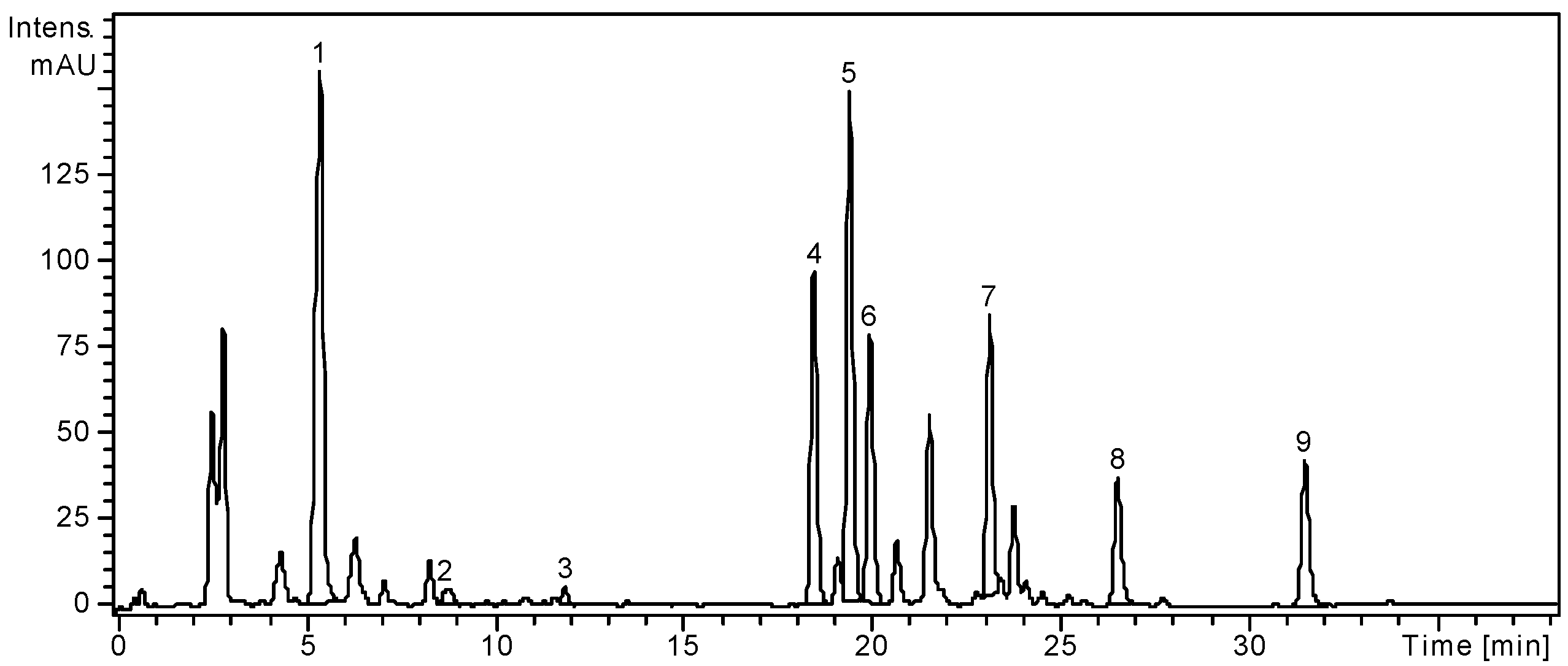
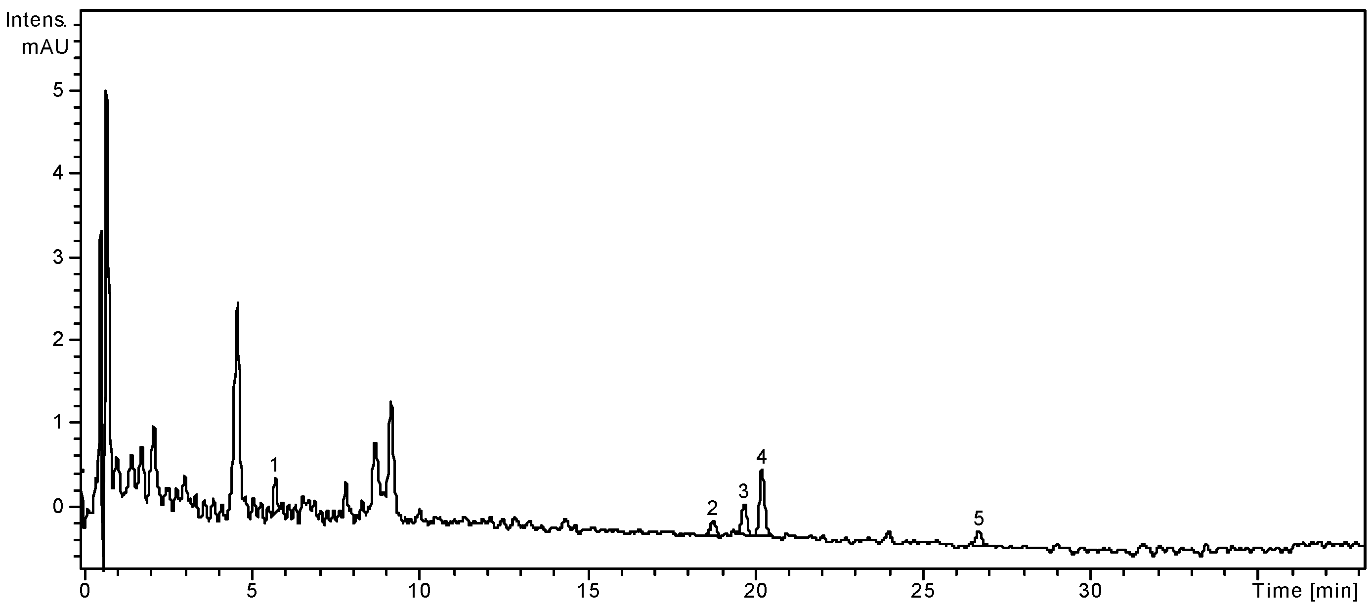
2.1. HPLC Analysis of Polyphenols

| Polyphenolic Compounds | m/z Value | RT ± SD (min) | S. chinensis Leaves | S. chinensis Fruits |
|---|---|---|---|---|
| Gentisic acid | 179 | 3.52 ± 0.04 | <0.02 | <0.02 |
| Caffeic acid | 179 | 5.60 ± 0.04 | <0.02 | NF |
| Chlorogenic acid | 353 | 5.62 ± 0.05 | 1636.19 ± 4.46 | 3.26 ± 0.25 |
| p-Coumaric acid | 163 | 9.48 ± 0.08 | 47.74 ± 0.73 | <0.02 |
| Ferulic acid | 193 | 12.8 ± 0.10 | 22.76 ± 0.67 | NF |
| Hyperoside | 463 | 19.32 ± 0.12 | 1096.61 ± 5.02 | 1.96 ± 0.02 |
| Isoquercitrin | 463 | 19.60 ± 0.10 | 2486.18 ± 5.72 | 6.59 ± 0.07 |
| Rutin | 609 | 20.20 ± 0.15 | 1365.39 ± 3.32 | 13.02 ± 0.21 |
| Myricetin | 317 | 21.13 ± 0.12 | <0.02 | NF |
| Quercitrin | 447 | 23.64 ± 0.13 | 1645.14 ± 2.12 | NF |
| Quercetin | 301 | 26.80 ± 0.15 | 263.25 ± 1.06 | 1.74 ± 0.04 |
| Kaempferol | 285 | 32.48 ± 0.17 | 378.27 ± 1.73 | NF |


2.2. Determination of Phenolic Compounds Content
| Samples | TPC (mg GAE/g Plant Material) | Flavonoids (mg RE/g Plant Material) |
|---|---|---|
| S. chinensis leaves | 62.36 ± 1.38 | 35.10 ± 1.23 |
| S. chinensis fruits | 9.20 ± 0.43 | 7.65 ± 0.95 |
2.3. Antioxidant Activity Assays
| Samples | DPPH (µg QE/mg Plant Material) | TEAC (µg TE/mg Plant Material) | HAPX (%) |
|---|---|---|---|
| S. chinensis leaves | 26.87 ± 0.84 | 45.97 ± 0.31 | 31.47 ± 1.36 |
| S. chinensis fruits | 7.80 ± 0.55 | 15.95 ± 0.68 | 28.86 ± 0.21 |

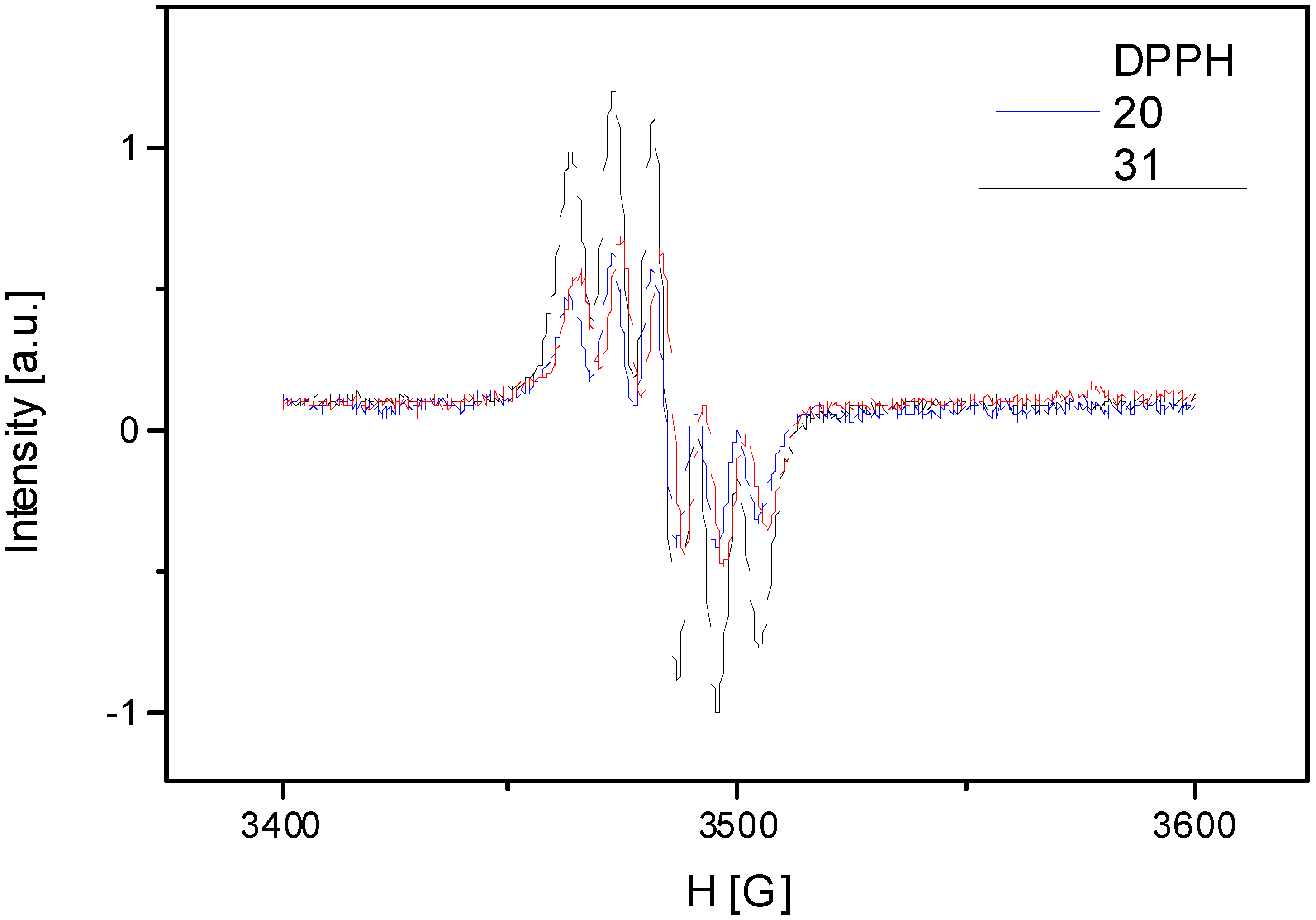
| DPPH | S. chinensis Leaves | S. chinensis Fruits |
|---|---|---|
| 578.85 ± 10.32 | 167.32 ± 3.24 | 277.05 ± 7.05 |

2.4. Antimicrobial Activity Assays
| Bacterial Strains | Standard Antibiotic | Inhibition Zone (mm) | |
|---|---|---|---|
| Gentamicin | S. chinensis Fruits | S. chinensis Leaves | |
| Staphylococcus aureus | 2 ± 0.1 | 4 ± 0.1 | 9 ± 0.1 |
| Bacillus subtilis | 6 ± 0.2 | 7 ± 0.2 | 11 ± 0.2 |
| Listeria monocytogenes | 5 ± 0.5 | 6 ± 0.4 | 12 ± 0.2 |
| Escherichia coli | 3 ± 0.3 | 3 ± 0.1 | 8 ± 0.2 |
| Salmonella typhimurium | 6 ± 0.1 | 6 ± 0.5 | 9 ± 0.2 |
| Bacterial Strains | MIC (µg/mL) | |
|---|---|---|
| S. chinensis Fruits | S. chinensis Leaves | |
| Staphylococcus aureus | 25 | 10 |
| Bacillus subtilis | 50 | 50 |
| Listeria monocytogenes | >100 | 75 |
| Escherichia coli | >100 | 75 |
| Salmonella typhimurium | >100 | 75 |
3. Experimental Section
3.1. Plant Materials and Extraction Procedure
3.2. Chemical and Instrumentation
3.3. HPLC/MS Analysis
3.3.1. Apparatus and Chromatographic Conditions for the Analysis of Polyphenols
3.3.2. Identification and Quantification of Polyphenols
| Peak No. | Phenolic Compound | m/z | RT ± SD | Peak No. | Phenolic Compound | m/z | RT ± SD |
|---|---|---|---|---|---|---|---|
| 1. | Caftaric acid | 311 | 3.54 ± 0.05 | 11. | Rutin | 609 | 20.76 ± 0.15 |
| 2. | Gentisic acid | 153 | 3.69 ± 0.04 | 12. | Myricetin | 317 | 21.13 ± 0.12 |
| 3. | Caffeic acid | 179 | 6.52 ± 0.04 | 13. | Fisetin | 285 | 22.91 ± 0.15 |
| 4. | Chlorogenic acid | 353 | 6.43 ± 0.05 | 14. | Quercitrin | 447 | 23.64 ± 0.13 |
| 5. | p-Coumaric acid | 163 | 9.48 ± 0.08 | 15. | Quercetin | 301 | 27.55 ± 0.15 |
| 6. | Ferulic acid | 193 | 12.8 ± 0.10 | 16. | Patuletin | 331 | 29.41 ± 0.12 |
| 7. | Sinapic acid | 223 | 15.00 ± 0.10 | 17. | Luteolin | 285 | 29.64 ± 0.19 |
| 8. | Cichoric acid | 473 | 15.96 ± 0.13 | 18. | Kaempferol | 285 | 32.48 ± 0.17 |
| 9. | Hyperoside | 463 | 19.32 ± 0.12 | 19. | Apigenin | 279 | 39.45 ± 0.15 |
| 10. | Isoquercitrin | 463 | 20.29 ± 0.10 |
3.4. Determination of Total Polyphenols and Flavonoids Content
3.5. In Vitro Antioxidant Activity Assays
3.5.1. DPPH Bleaching Assay
3.5.2. TEAC Assay (Trolox Equivalent Antioxidant Capacity)
3.5.3. Hemoglobin/Ascorbate Peroxidase Activity Inhibition (HAPX) Assay
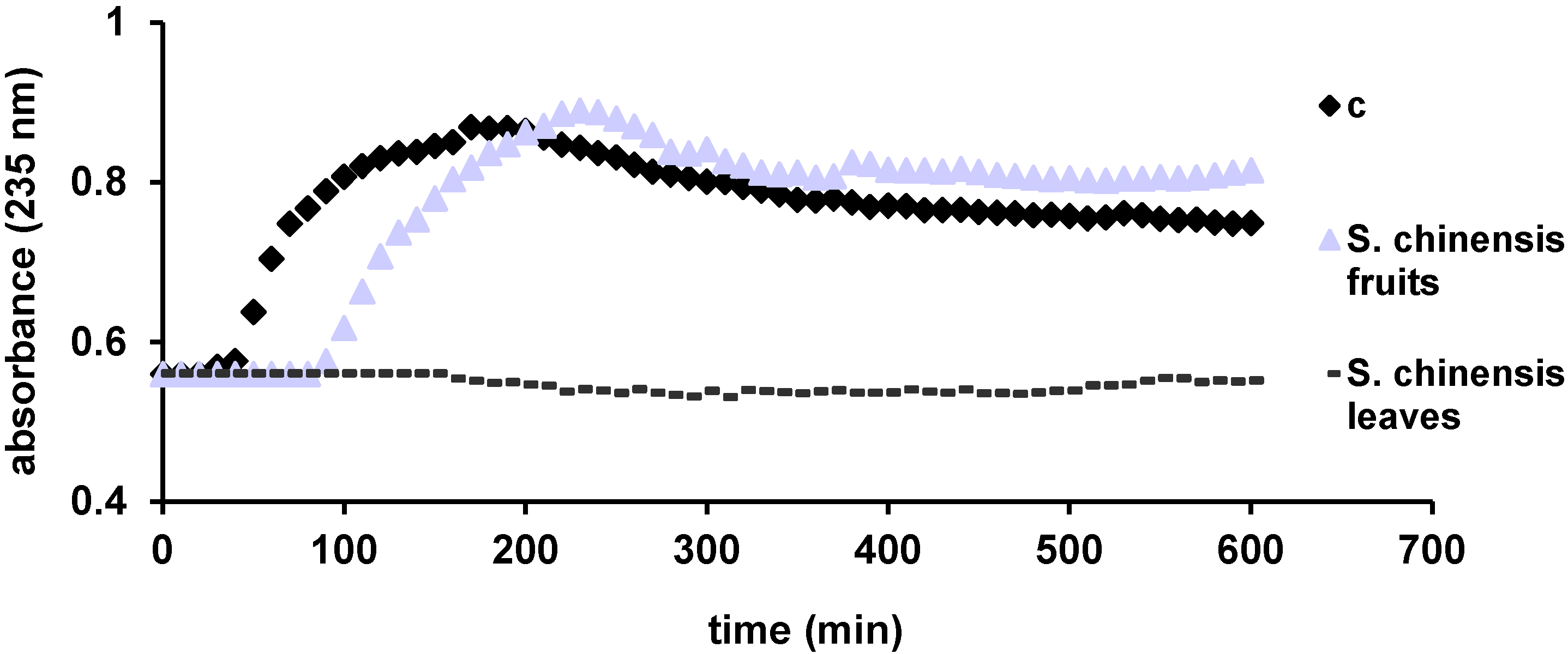
3.5.4. Inhibition of Lipid Peroxidation Catalyzed by Cytochrome c
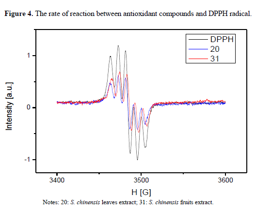
3.5.5. EPR Measurements
3.6. Determination of Antimicrobial Activity
3.6.1. Microorganisms and Culture Growth
3.6.2. Antimicrobial Activity Assay
3.6.3. Minimum Inhibitory Concentration
3.7. Statistical Analysis
4. Conclusions
Acknowledgments
Authors Contributions
Abbreviations
| HPLC | high performance liquid chromatography |
| MS | mass spectrometry |
| RT | retention time |
| SD | standard deviation |
| TPC | total phenolic content |
| GAE | galic acid equivalents |
| RE | rutin equivalents |
| TEAC | Trolox equivalent antioxidant capacity |
| HAPX | hemoglobin ascorbate peroxidase activity |
| EPR | electron paramagnetic resonance |
| QE | quercetin equivalents |
| TE | trolox equivalents |
| MIC | minimal inhibitory concentration |
| CFU | colony-forming unit |
Conflicts of Interest
References
- Vlase, L.; Pârvu, M.; Pârvu, E.A.; Toiu, A. Chemical constituents of three Allium Species from Romania. Molecules 2013, 18, 114–127. [Google Scholar]
- Cheng, N.; Ren, N.; Gao, H.; Lei, X.; Zheng, J.; Cao, W. Antioxidant and hepatoprotective effects of Schisandra. chinensis pollen extract on CCl4-induced acute liver damage in mice. Food Chem. Toxicol. 2013, 55, 234–240. [Google Scholar]
- Wu, X.; Yu, X.; Jing, H. Optimization of phenolic antioxidant extraction from Wuweizi (Schisandra chinensis) pulp using random-centroid optimization methodology. Int. J. Mol. Sci. 2011, 12, 6255–6266. [Google Scholar]
- Chen, X.; Zhang, Y.; Zu, Y.; Fu, Y.; Fu, Y.; Wang, W. Composition and biological activities of essential oil from Schisandra. chinensis obtained by solvent-free microwave extraction. LWT-Food Sci. Technol. 2011, 44, 2047–2052. [Google Scholar]
- Pop, A.; Berce, C.; Bolfă, P.; Nagy, A.; Cătoi, C.; Dumitrescu, I.B.; Silaghi-Dumitrescu, L.; Loghin, F. Evaluation of the possible endocrine disruptive effect of butylated hydroxitoluene and propyl gallate in immature female rats. Farmacia 2013, 61, 202–211. [Google Scholar]
- Popa, D.S.; Bolfă, P.; Kiss, B.; Vlase, L.; Păltinean, R.; Pop, A.; Cătoi, C.; Crișan, G.; Loghin, F. Influence of Genista tinctoria L. or methylparaben on subcronic toxicity of bisphenol A in rats. Biomed. Environ. Sci. 2014, 27, 85–96. [Google Scholar]
- Vlase, L.; Benedec, D.; Hanganu, D.; Damian, D.; Csillag, I.; Sevastre, B.; Mot, A.C.; Silaghi-Dumitrescu, R.; Tilea, I. Evaluation of antioxidant and antimicrobial activities and phenolic profile for Hyssopus officinalis, Ocimum basilicum and Teucrium chamaedrys. Molecules 2014, 19, 5490–5507. [Google Scholar]
- Dai, J.; Mumper, R.J. Plant phenolics: Extraction, analysis and their antioxidant and anticancer properties. Molecules 2010, 15, 7313–7352. [Google Scholar]
- Pratt, D.E. Phenolic Compounds in Food and Their Effects on Health II; American Chemical Society: Washington, DC, USA, 1992; pp. 352–391. [Google Scholar]
- Serafini, M.; Bellocco, R.; Wolk, A.; Ekstrom, A.M. Total antioxidant potential of fruit and vegetables and risk of gastric cancer. Gastroenterology 2002, 123, 985–991. [Google Scholar]
- Sun, Y.; Wen, X.; Huang, H. Population genetic differentiation of Schisandra chinensis and Schisandra sphenanthera as revealed by ISSR analysis. Biochem. Syst. Ecol. 2010, 38, 257–263. [Google Scholar]
- Szopa, A.; Ekiert, H. Production of deoxyschizandrin and γ-schizandrin in shoot-differentiating and undifferentiating callus cultures of Schisandra chinensis (Turcz.) Baill. (Chinese magnolia vine). J. Biotechnol. 2013, 165, 209–213. [Google Scholar]
- Lu, Y.; Chen, D.-F. Analysis of Schisandra chinensis and Schisandra sphenanthera. J. Chromatogr. A 2009, 1216, 1980–1990. [Google Scholar]
- Yang, F.-J.; Ma, C.-H.; Yang, L.; Zhao, C.-J.; Zhang, Y.; Zu, Y.-G. Enrichment and purification of deoxyschizandrin and γ-schizandrin from the extract of Schisandra chinensis fruit by macroporous resins. Molecules 2012, 17, 3510–3523. [Google Scholar]
- Jeong, E.J.; Lee, H.K.; Lee, K.Y.; Jeon, B.J.; Kim, D.H.; Park, J.-H.; Song, J.-H.; Huh, J.; Lee, J.-H.; Sung, S.H. The effects of lignan-riched extract of Schisandra chinensis on amyloid-β-induced cognitive impairment and neurotoxicity in the cortex and hippocampus of mouse. J. Ethnopharmacol. 2013, 146, 347–354. [Google Scholar]
- Zhao, T.; Feng, Y.; Li, J.; Mao, R.; Zou, Y.; Feng, W.; Zheng, D.; Wang, W.; Chen, Y.; Yang, L.; et al. Schisandra polysaccharide evokes immunomodulatory activity through TLR 4-mediated activation of macrophages. Int. J. Biol. Macromol. 2014, 65, 33–40. [Google Scholar]
- Lin, R.-D.; Mao, Y.-W.; Leu, S.-J.; Huang, C.-Y.; Lee, M.-H. The immune-regulatory effects of Schisandra chinensis and its constituents on human monocytic leukemia cells. Molecules 2011, 16, 4836–4849. [Google Scholar]
- Zhao, T.; Mao, G.; Zhang, M.; Zou, Y.; Feng, W.; Gu, X.; Zhu, Y.; Mao, R.; Yang, L.; Wu, X. Enhanced antitumor and reduced toxicity effect of Schisandrae polysaccharide in 5-Fu treated Heps-bearing mice. Int. J. Biol. Macromol. 2014, 63, 114–118. [Google Scholar]
- Chen, H.; Sohn, J.; Zhang, L.; Tian, J.; Chen, S.; Bjeldanes, L.F. Anti-inflammatory effects of chicanine on murine macrophage by down-regulating LPS-induced inflammatory cytokines in IκBα/MAPK/EKR signaling pathways. Eur. J. Pharmacol. 2014, 724, 168–174. [Google Scholar]
- Park, S.Y.; Park, S.J.; Park, T.G.; Rajasekar, S.; Lee, S.-J.; Choi, Y.-W. Schisandrin C exerts anti-neuroinflammatory effects by upregulating phase II detoxifying/antioxidant enzymes in microglia. Int. Immunopharmacol. 2013, 17, 415–426. [Google Scholar]
- Wei, B.; Li, Q.; Fan, R.; Su, D.; Chen, X.; Jia, Y.; Bi, K. Determination of monoamine and amino acid neurotransmitters and their metabolites in rat brain samples by UFLC-MS/MS for the study of sedative-hypnotic effects observed during treatment with S. chinesis. J. Pharm. Biomed. Anal. 2014, 88, 416–422. [Google Scholar]
- Ekiert, R.; Szopa, A.; Ekiert, H.; Krzek, J.; Dzik, E. Analysis of lignans in Schisandra chinensis fruits, leaves, biomasses from in vitro cultures and food supplements. J. Funct. Foods 2013, 5, 1576–1581. [Google Scholar]
- Ciorchină, N.; Onica, E.; Roșca, I.; Dumitraș, A.; Clapa, D.; Fira, A. The biology of the propagation of species Schisandra chinensis (Turcz.) Baill. J. Plant Dev. 2011, 18, 17–26. [Google Scholar]
- Zhao, L.-C.; He, Y.; Deng, X.; Yang, G.-L.; Li, W.; Liang, J.; Tang, Q.-L. Response surface modeling and optimization of accelerated solvent extraction of four lignans from Fructus Schisandrae. Molecules 2012, 17, 3618–3629. [Google Scholar]
- Ma, C.; Yang, L.; Yang, F.; Wang, W.; Zhao, C.; Zu, Y. Content and color stability of anthocyanins isolated from Schisandra chinensis fruit. Int. J. Mol. Sci. 2012, 13, 14294–14310. [Google Scholar]
- Benedec, D.; Vlase, L.; Oniga, I.; Mot, A.C.; Damian, G.; Hanganu, D.; Duma, M.; Silaghi-Dumitrescu, R. Polyphenolic Composition, Antioxidant and Antimicrobial Activities for Two Romanian Subspecies of Achillea distans Waldst. et Kit. ex Willd. Molecules 2013, 18, 8725–8739. [Google Scholar]
- Szopa, A; Ekiert, H. In vitro cultures of Schisandra chinensis (Turcz.) Baill. (Chinese Magnolia Vine)—A potential biotechnological rich source of therapeutically important phenolic acids. Appl. Biochem. Biotechnol. 2012, 166, 1941–1948. [Google Scholar]
- Wang, Z.; Chen, H.; Zhang, W.; Lan, G.; Zhang, L. Comparative studies on the chemical composition and antioxidant activities of Schisandra chinensis and Schisandra sphenanthera fruits. J. Med. Plants Res. 2011, 5, 1207–1216. [Google Scholar]
- Mot, A.C.; Bischin, C.; Damian, G.; Silaghi-Dumitrescu, R. Antioxidant activity evaluation involving hemoglobin-related free radical reactivity. In Advanced Protocols in Oxidative Stress III. Methods in Molecular Biology; Springer: New York, NY, USA, 2013; in press. [Google Scholar]
- Bischin, C.; Tusan, C.; Bartok, A.; Septelean, R.; Damian, G.; Silaghi-Dumitrescu, R. Evalution of the biochemical effects of silyl-phosphaalkenes on oxidative and nitrosative stress pathways involving metallocenters. Phosphorus Sulfur Silicon Relat. Elem. 2014. [Google Scholar] [CrossRef]
- Yang, J.; Guo, J.; Yuan, J. In vitro antioxidant properties of rutin. LWT-Food Sci. Technol. 2008, 41, 1060–1066. [Google Scholar]
- Bischin, C.; Deac, F.; Silaghi-Dumitrescu, R.; Worrall, J.A.; Rajagopal, B.S.; Damian, G.; Cooper, C.E. Ascorbate peroxidase activity of cytochrome c. Free Radic. Res. 2011, 45, 439–444. [Google Scholar]
- Marinova, E.M.; Toneva, A.; Yanishlieva, N. Comparison of the antioxidative properties of caffeic and chlorogenic acids. Food Chem. 2009, 114, 1498–1502. [Google Scholar]
- Cox, S.D.; Mann, C.M.; Markham, J.L.; Gustafson, J.E.; Warmington, J.R.; Wyllie, S.G. Determination the antimicrobial action of tea tree oil. Molecules 2001, 6, 87–91. [Google Scholar]
- Hussain, A.I.; Anwar, F.; Nigam, P.S.; Sarker, S.D.; Moore, J.E.; Rao, J.R.; Mazumdar, A. Antibacterial activity of some Lamiaceae essential oils using resazurin as an indicator of cell growth. LWT-Food Sci. Technol. 2011, 44, 1199–1206. [Google Scholar]
- Antonini, E.; Brunori, M. Hemoglobin and Myoglobin in Their Reaction with Ligands; North-Holland Publishing Company: Amsterdam, The Netherlands, 1971; pp. 98–134. [Google Scholar]
- Mot, A.C.; Damian, G.; Sarbu, C.; Silaghi-Dumitrescu, R. Redox reactivity in propolis: Direct detection of free radicals in basic medium and interaction with hemoglobin. Redox Rep. 2009, 14, 267–274. [Google Scholar]
- Romanian Pharmacopoeia Commission National Medicines Agency. Romanian Pharmacopoeia, Xth ed.; Medical Publishing House: Bucharest, Romania, 1993; p. 335. [Google Scholar]
- Espinoza, M.; Olea-Azar, C.; Speisky, H.; Rodríguez, J. Determination of reactions between free radicals and selected Chilean wines and transition metals by ESR and UV-vis technique. Spectrochim. Acta A Mol. Biomol. Spectrosc. 2009, 71, 1638–1643. [Google Scholar]
- Bauer, A.W.; Kirby, W.M.; Sherris, J.C.; Turck, M. Antibiotic susceptibility testing by a standardized single disk method. Am. J. Clin. Pathol. 1966, 45, 493–496. [Google Scholar]
- Mocan, A.; Vlase, L.; Vodnar, D.C.; Bischin, C.; Hanganu, D.; Gheldiu, A.-M.; Oprean, R.; Silaghi-Dumitrescu, R.; Crișan, G. Polyphenolic Content, Antioxidant and Antimicrobial Activities of Lycium barbarum L. and Lycium chinense Mill. Leaves. Molecules 2014, 19, 10056–10073. [Google Scholar]
- Salvat, A.; Antonacci, L.; Fortunato, R.H.; Suarez, E.Y.; Godo, H.M. Antimicrobial activity in methanolic extracts of several plant species from Northern Argentina. Phytomedicine 2004, 11, 230–234. [Google Scholar]
- Sample Availability: Samples of the compounds are not available from the authors.
© 2014 by the authors. Licensee MDPI, Basel, Switzerland. This article is an open access article distributed under the terms and conditions of the Creative Commons Attribution license ( http://creativecommons.org/licenses/by/3.0/).
Share and Cite
Mocan, A.; Crișan, G.; Vlase, L.; Crișan, O.; Vodnar, D.C.; Raita, O.; Gheldiu, A.-M.; Toiu, A.; Oprean, R.; Tilea, I. Comparative Studies on Polyphenolic Composition, Antioxidant and Antimicrobial Activities of Schisandra chinensis Leaves and Fruits. Molecules 2014, 19, 15162-15179. https://doi.org/10.3390/molecules190915162
Mocan A, Crișan G, Vlase L, Crișan O, Vodnar DC, Raita O, Gheldiu A-M, Toiu A, Oprean R, Tilea I. Comparative Studies on Polyphenolic Composition, Antioxidant and Antimicrobial Activities of Schisandra chinensis Leaves and Fruits. Molecules. 2014; 19(9):15162-15179. https://doi.org/10.3390/molecules190915162
Chicago/Turabian StyleMocan, Andrei, Gianina Crișan, Laurian Vlase, Ovidiu Crișan, Dan Cristian Vodnar, Oana Raita, Ana-Maria Gheldiu, Anca Toiu, Radu Oprean, and Ioan Tilea. 2014. "Comparative Studies on Polyphenolic Composition, Antioxidant and Antimicrobial Activities of Schisandra chinensis Leaves and Fruits" Molecules 19, no. 9: 15162-15179. https://doi.org/10.3390/molecules190915162