Role of Oxidant Scavengers in the Prevention of Ca2+ Homeostasis Disorders


Cell Physiology Research Group, Department of Physiology, University of Extremadura, Caceres, 10071, Spain
*
Author to whom correspondence should be addressed.
†
These authors contributed equally to this work.
Molecules 2010, 15(10), 7167-7187; https://doi.org/10.3390/molecules15107167
Submission received: 1 July 2010
/
Revised: 9 September 2010
/
Accepted: 14 October 2010
/
Published: 15 October 2010
(This article belongs to the Special Issue Antioxidants)
Abstract
:A number of disorders, such as Alzheimer disease and diabetes mellitus, have in common the alteration of the redox balance, resulting in an increase in reactive oxygen species (ROS) generation that might lead to the development of apoptosis and cell death. It has long been known that ROS can significantly alter Ca2+ mobilization, an intracellular signal that is involved in the regulation of a wide variety of cellular functions. Cells have a limited capability to counteract the effects of oxidative stress, but evidence has been provided supporting the beneficial effects of exogenous ROS scavengers. Here, we review the effects of oxidative stress on intracellular Ca2+ homeostasis and the role of antioxidants in the prevention and treatment of disorders associated to abnormal Ca2+ mobilization induced by ROS.

1. Calcium Homeostasis
In eukaryotic cells, Ca2+ is the most versatile signal involved in the control of cellular processes and functions [1,2,3]. This versatility derives from the fact that Ca2+ signalling works in a variety of ways, and the processes involved in Ca2+ mobilization are widely dynamic in range and amplitude. For example, in the cardiac myocyte, Ca2+ entering through L-type Ca2+ channels leads to a signal known as ‘spark’ that triggers contraction within microseconds; on the other hand, the duration of processes like gene transcription or cell proliferation ranges from minutes to hours.
Cytosolic Ca2+ concentration ([Ca2+]c) is determined by a balance between the mechanisms that introduce Ca2+ into the cytoplasm, termed “on”, and those that remove it, termed “off”. These processes combine the action of a variety of channels, both in the plasma membrane and in the membrane of the intracellular stores, such as the endoplasmic or sarcoplasmic reticulum, including Ca2+ pumps, exchangers, and buffers. Many components of this large set of molecules have different isoforms with different characteristics and properties, which gives them the ability to make the system extremely versatile [4]. In the extracellular medium, the free Ca2+ concentration is about 1 mM, while in resting cells [Ca2+]c is approximately 100 nM and in certain intracellular organelles, such as the endoplasmic reticulum (ER) or sarcoplasmic reticulum (SR), the free luminal Ca2+ concentration ([Ca2+]L) is around 0.2–1 mM; therefore, there is a clear concentration gradient between compartments that is essential for the regulation of the cellular processes in which Ca2+ participates [5]. In order to maintain these Ca2+ gradients, a strong homeostatic mechanism acts in the cell.
Agonist-induced changes in [Ca2+]c is determined by the balance between the Ca2+ “on” mechanisms, including Ca2+ influx from the extracellular medium and the output from intracellular stores, and Ca2+ “off” mechanisms, involving Ca2+ extrusion across the plasma membrane and sequestration into the stores or mitochondria [4]. The Ca2+ “off” mechanisms involve four different transporters: the plasma membrane Ca2+ ATPase (PMCA), that mediates Ca2+ extrusion across the plasma membrane, the sarco/endoplasmic reticulum Ca2+ ATPase (SERCA), which reintroduce Ca2+ into the ER/SR, the Na+/Ca2+ exchanger (NCX) that participates in cytosolic Ca2+ clearance through its exchange by Na+ and the mitochondrial Ca2+ uniporter (MCU), which that transports Ca2+ across the inner mitochondrial membrane. The two ATPases (PMCA and SERCA) carry out such transport using the energy provided by ATP hydrolysis. Four PMCA isoforms have been described in humans, all with similar molecular structure, consisting of ten membrane-spanning segments and five extracellular domains while their amino and carboxyl termini are located within the cell [6]. There are three different types of SERCA genes identified, giving rise to three SERCA isoforms [7]. These isoforms can be co-expressed in the same cell type, which could be related to the co-existence of different types of Ca2+ pools in the same cell [8]. Phosphorylation of an aspartic acid residue results in SERCA conformational changes that leads the enzyme to capture in a first step two Ca2+ per ATP molecule hydrolyzed, and then release them inside the ER/SR lumen [9]. In addition, when [Ca2+]c reaches the micromolar range, both NCX and MCU can also transport Ca2+. NCX is a bidirectional transporter that combines the movement of three Na+ ions towards the cytosol with the transport of one Ca2+ in the opposite direction. NCX participates in the regulation of [Ca2+]c in a number of cell types acting either in forward mode, as explained above, or in reverse mode, introducing Ca2+ in the cell when the Na+ concentration at the inner face of the plasma membrane substantially increases. Mitochondrial membrane potential confers the driving force necessary for the activity of the MCU. The threshold [Ca2+]c for the activation of these four transporters is different as well as the transport rate. The lowest threshold is for the PMCA, which, by the way, has the lowest transport rate. This characteristic confers to PMCA an important role in maintaining resting [Ca2+]c [10]. SERCA has lower affinity for Ca2+ but higher transport rates than PMCA [11]. On the other hand, the threshold [Ca2+]c for NCX and MCU is the highest but they also display much higher transport rates than the ATPases.
Agonist-activated Ca2+ “on” mechanisms includes Ca2+ release from the intracellular pools and entry through plasma membrane channels. There are three major types of intracellular Ca2+ channels responsible for Ca2+ release from the ER and the SR: the inositol 1,4,5-trisphosphate receptor (IP3R), the ryanodine receptor (RyR) and the nicotinic acid-adenine dinucleotide phosphate (NAADP) receptor. Mammalian cells possess three different isoforms for IP3R and RyR. Occupation of phospholipase C (PLC)-coupled membrane receptors by agonists results in the activation of phosphoinositide-specific PLC. As a result, inositol 1,4,5-trisphosphate (IP3) is generated and activates IP3R leading to Ca2+ efflux from the ER [12]. Ca2+ stored in the ER can also be released through RyR. The RyR is structurally and functionally similar to IP3R and is activated by cyclic ADP-ribose. These two channels are sensitive to Ca2+ itself, a phenomenon that underlies Ca2+-induced Ca2+ release and contributes to the rapid rise in [Ca2+]c upon agonist stimulation and the development of regenerative Ca2+ waves. Finally, NAADP-mediated Ca2+ efflux from intracellular stores involves endolysosomal two-pore channels (TPC), especially TPC1 [13].
Agonist-induced Ca2+ entry from the extracellular medium occurs through a variety of Ca2+ channels in the plasma membrane depending on the cell type, which are gated by voltage (only in electrically excitable cells), agonists or second messengers [14,15]. In addition to these signals, other physical stimuli, such as mechanical stretch, are able to induce Ca2+ entry [16]. Special attention deserves a Ca2+ entry mechanism regulated by the filling state of the Ca2+ stores known as store-operated Ca2+ entry (SOCE) or capacitative Ca2+ entry. In 1986, Putney proposed that depletion of the intracellular Ca2+ stores lead to a sustained influx of Ca2+ through the plasma membrane independently of the elevation of [Ca2+]c [17]. Subsequently, using biophysical techniques such as patch clamp, the existence of store-operated Ca2+ channels has been demonstrated, which can be opened in response to store depletion by various agents [18]. These channels, known as CRAC (Ca2+ release-activated Ca2+) channels, those mediating the highly Ca2+ selective current ICRAC, and SOC (store-operated channels), those conducting the non-selective cation current ISOC, have been characterized electrophysiologically [19,20,21,22]; however, their molecular identities have remained elusive for almost two decades. The Orai1 protein is the most relevant candidate to form the pore of the CRAC channels [23]. This protein has been demonstrated to form multimeric ion channel complexes in the plasma membrane [24]. The multimeric structure of the channel has recently been demonstrated as a tetramer. The pore structure consists of four separate units of Orai1, where charged residues are essential for Ca2+ selectivity [25]. Transient receptor potential proteins, alone or in combination with Orai1, have been reported to form the SOC channels, with lower Ca2+ selectivity than CRAC channels [26,27,28,29]. Especial attention has been given to the members of the canonical transient receptor potential (TRPC) subfamily, some of them can be activated by Ca2+ store depletion [30,31]. Orai1 and TRPC proteins can independently regulate ion current through CRAC channels and SOCs (ICRAC and ISOCs) or might interact to form SOCs with different biophysical properties, thus providing the cell of some valuable tools to regulate specific Ca2+ signals (this has been extensively reviewed in [5]). SOC and CRAC channels have been reported to be sensitive to Ca2+ store depletion through the cooperative intraluminal Ca2+ sensor, STIM1 [32,33,34,35]. STIM1 is a transmembrane protein located in the Ca2+ stores that has been identified as the intraluminal Ca2+ sensor that communicates the amount of stored Ca2+ to plasma membrane channels [32,36]. This protein has a single transmembrane domain with an EF hand motif near the N-terminus, which is located in the lumen of the ER (or extracellularly when STIM1 is located in the plasma membrane [37]). A number of studies have demonstrated that the EF hand domain of STIM1, senses [Ca2+]L and inhibits STIM1 activity when stores are filled. When a decrease in [Ca2+]L occurs, Ca2+ dissociates from the EF hand motif and STIM1 activates Ca2+ channels [32,36,38,39].
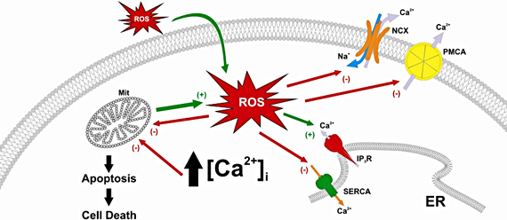
2. Calcium Homeostasis Abnormalities Induced by Reactive Oxygen Species
ROS are small molecules that can be formed as a result of the normal aerobic metabolism [40], the activity of the immune system [41,42], the xenobiotic metabolism [43], or environmental pollution [44]. The sources of physiological ROS production are, among others, mitochondrial activity and the activity of enzymes such as xanthine oxidase, NADPH oxidase, cyclooxygenase and lipoxygenase. ROS were early classified as toxic; however, they have more recently been reported to act also as signalling molecules, in processes like transcription, gene expression or cell death [45,46,47]. At low concentrations ROS have been reported to act as secondary messengers in intracellular pathways involving Ca2+ mobilization, but at high concentrations they produce oxidative stress and cell damage [48,49,50,51].
The interrelationship between ROS production and Ca2+ homeostasis was first reported in the 70s. It is known that the involvement of the redox state in Ca2+ homeostasis is mediated by the modification of disulfide bonds between cysteine residues of some Ca2+ “off”-handling proteins, including PMCA, NCX and SERCA. Most studies reveal that ROS inactivates these transporters, leading to a rise in [Ca2+]c and subsequent cell dysfunction. PMCA can be reversibly inactivated by ROS by an unclear mechanism that is suggested to be a reversible cysteine modification [52,53,54]. Current hypotheses propose that ROS alter the PMCA Tyr589, Met622 and Met831 residues [55]. Furthermore, there is evidence indicating that PMCA inactivation by ROS can be a protective system to avoid high consumption of ATP under stress conditions [56]. As a result of PMCA inactivation [Ca2+]c rises over the resting value.
ROS can also alter NCX function although the effect of ROS on NCX activity remains controversial. Hydrogen peroxide formed by the xanthine/xanthine oxidase system and superoxide anion increase the activity of NCX in myocytes [57]. However, in the same cells, oxidative stress induced by xanthine oxidase plus hypoxanthine inhibits the exchanger [58], and this exchanger has also been shown to be inhibited by the oxidant HOCl [59].
The effects of ROS on the activity of SERCA is also controversial. SERCA contains between 22 to 28 cysteine residues; therefore, the redox state is very important for its activity. ROS are capable of attenuating the activity of this pump in vitro by modifying sulphydryl groups [60,61,62]. Distinct SERCA isoforms show different susceptibility to ROS [51,60,61], which might be attributed to the different location of cysteine residues [63]. It has been hypothesized that small concentrations of ROS can stimulate this pump, whether high concentrations can inhibit SERCA [64], which has been reported to be more sensitive to ROS than PMCA [65].
Ca2+ “on” mechanisms are also susceptible to be altered by oxidative stress. Intracellular Ca2+ channels responsible for Ca2+ release from the intracellular stores are sensitive to ROS. IP3R function has been reported to be affected by ROS through the modification of cysteine residues. ROS increase the sensitivity of IP3R to cytosolic IP3 levels, thus IP3R might be sensitive to resting IP3 levels [66,67]. RyR can also been altered by changes in the redox state. The relationship between RyR channels and ROS production is probably the most widely investigated. In skeletal muscle, it has been suggested that the residue that confers ROS sensitivity to type 1 RyR (RyR1) is Cys3635. ROS play a dual role in RyR1 activity, being activated by concentrations of H2O2 between 100 μmol/L and 1 mmol/L [68] and inhibited by high concentrations of hydrogen peroxide (10 mmol/L) [69]. In cardiomyocytes, ROS produced by the activity of NOX enzymes increases type 2 RyR (RyR2) activity, interfering with its association with calmodulin (necessary to inhibit the channel) or FKBP12.6 (which stabilizes the channel) thus suggesting that binding of these two proteins to RyR2 channel is sensitive to the redox state [70]. Furthermore, studies in neurons have reported that ROS increase Ca2+ release mediated by type 3 RyR (RyR3) channels [70,71]. Modulation of RyR by ROS may be a mechanism of interaction between Ca2+ and the redox signalling pathways, and also a mechanism to increase or decrease Ca2+ signals as needed (for example, in neurons, ROS generation alters the activity of RyR channels, that causes long-term potentiation or depression, processes that depend on Ca2+ release through RyR) [54].
Finally, oxidants can also modulate the function of a number of Ca2+ permeable plasma membrane channels. ROS alter the activity of voltage-gated Ca2+ channels, specially the activity of L-type Ca2+ channels [72], which has been associated to the oxidation of SH groups resulting in altered Ca2+ entry in guinea pig ventricular myocytes [73]. A recent study has reported that exposure of cardiac myocites to hydrogen peroxide produces an increase of intracellular ROS and basal L-type channel activity [74]. Moreover, studies in human embryonic kidney 293 cells revealed that hydrogen peroxide increases basal L-type channel gating [75]. The effect of ROS on other voltage-gated Ca2+ channels has been less investigated. It has been shown that external application of hydrogen peroxide is able to activate voltage-dependent P/Q-type channels in neurons [76]. Evidence has also been provided in favour of an inhibitory role of ROS on voltage-dependent Ca2+ channel gating [73]. Although speculative, these discrepancies may be attributed to the different oxidants or the concentrations used.
In addition to voltage-gated Ca2+ channels, ROS can also affect the activity of other Ca2+ permeable channels, such as the channels conducting SOCE, receptor- or second messenger-operated Ca2+ entry. It has been reported that hydrogen peroxide decreases SOCE in thyroid cells through the activation of protein kinase C and not by a direct effect on SOCs and CRAC channels [77,78]. In human platelets, where ROS have been reported to play a physiological role in Ca2+ signalling, including SOCE [79], hydrogen peroxide plays a dual role in the activation of SOCE, with a stimulating effect at low concentrations (10-100 nM) and inhibitory effects at high concentrations (1 mM) [50]. Transient receptor potential channels have been shown to be sensitive to ROS [80]. Transient receptor potential canonical-3 (TRPC3)-forming channels are activated by ROS through the modulation of tyrosine phosphorylation [80] and transient receptor potential melastatin-2 (TRPM2), melastatin-7 (TRPM7) and ankyrin1 (TRPA1) channels are also sensitive to ROS. In neurons, TRPM7 and TRPM2, are activated by oxidative stress and participate in the pathophysiology of neurodegeneration [81,82]. In contrast, transient receptor potential polycystin-2 (TRPP2) channels are inhibited by oxidative stress in human syncytiotrophoblast [83]. The different effect of oxidative stress on TRP function might depend on the type of channel investigated and has been involved in the pathogenesis of a number of disorders. Other channels, such as Orai1, but not Orai3, have been shown to be inhibited by hydrogen peroxide-mediated oxidation. The differential redox sensitivity of these proteins has been attributed to the presence of an extracellularly located reactive cysteine, which is absent in Orai3 [84]. Oxidative stress also alters the ER Ca2+ sensor STIM1 and Ca2+ effectors such as Ca2+/calmodulin-dependent protein kinase II (for a review see [64]).
In summary, Ca2+ signalling is very sensitive to oxidants or reducing agents and changes in the redox state results in relevant changes in Ca2+ homeostasis, either altering Ca2+ mobilization from the internal stores and Ca2+ entry from the extracellular medium or modulating the activity of Ca2+ “off” mechanisms, including Ca2+ pumps and exchangers. The variable effects of ROS on Ca2+-handling mechanisms can be inhibitory or stimulatory, depending on the type of oxidant, its concentration and the time of exposure [85,86,87].
3. Disorders Caused by Reactive Oxygen Species and Therapeutic Strategies Based on the Use of Antioxidants
ROS have divergent effects on cellular function. At low concentrations, ROS have been reported to contribute to vascular tone regulation, mediate vasodilation, and regulate cell growth and differentiation, activation of platelet aggregation and stimulation of many kinases and proinflammatory genes [88,89,90,91,92]. On the other hand, oxidative stress promotes the development of a number of diseases, such as neurodegenerative, cardiovascular and metabolic diseases and certain types of cancer [90,93,94,95,96,97,98,99,100,101].
The controlled generation of ROS is necessary for many vital cellular functions. For instance, the response of macrophages to external agents leads to production of ROS and bioactive lipids derived from the metabolism of arachidonic acid [102,103,104]. A number of studies have shown that the imbalance between ROS generating and scavenging systems leads to oxidative stress which can cause oxidative damage to biomolecules, followed by various apoptotic pathways that lead to cell death [105,106,107]. In recent years, there has been an increasing use of antioxidants with the aim to regulate the redox balance [108,109] despite of only a few antioxidant are in use now in patients [110]. There are two disorders that illustrate the cellular dysfunction induced by abnormal Ca2+ homeostasis due to oxidative stress: diabetes mellitus and Alzheimer disease.
Diabetes mellitus (DM) is a very common disease that affects over 180 million people, whose hallmarks are pancreatic β-cell dysfunction and insulin resistance [101]. Type 2 DM, which affects 90% of diabetics, leads to a number of cardiovascular alterations, including angiopathy, which is the main cause of morbidity and mortality in type 2 DM [111]. In the study of this disease, platelets have become a very important role because platelet hyperactivation and hiperaggregation play a key role in the development of angiopathy [112,113]. Platelets from diabetic patients have altered Ca2+ mobilization [114,115,116], increased ROS production [117,118] and enhanced protein tyrosine phosphorylation [119,120,121]. The reason of the enhanced ROS production during diabetes mellitus is not clear but it has been reported that diabetes mellitus is associated to hyperhomocysteinemia [122,123]. Studies in animal models have revealed that hyperhomocysteinaemia result in increased oxidative stress, impaired endothelial function and increased thrombogenicity. That rise in homocysteine concentration leads to increased production of ROS which can eventually trigger platelet hyperactivity [88,127] but lowering homocysteine levels by daily supplementation with antioxidants did not reduce the risk of developing type 2 DM [128].
Either at rest or after platelet stimulation with thrombin, [Ca2+]c is higher in cells in patients with type 2 DM than in healthy donors [51,124], although platelets from healthy and type 2 DM subjects accumulate the same amount of Ca2+ into intracellular stores [119]. Abnormal Ca2+ homeostasis in platelets from type 2 diabetic patients has been attributed to altered Ca2+ extrusion mechanisms, increased IP3 generation or enhanced Ca2+ entry mechanisms [50]. Altered Ca2+ extrusion mechanisms have been reported for PMCA and SERCA, which are very sensitive to oxidative damage as mentioned above [51,115,125]. PMCA activity is regulated by tyrosine phosphorylation, and activation of platelets by thrombin stimulates Src-dependent PMCA tyrosine phosphorylation, and thus, inhibition of Ca2+ extrusion [119,126]. The increase in [Ca2+]c can trigger the synthesis of thromboxane A2 and hyperaggregability causing platelet hyperactivation.There is evidence linking the decrease in vascular NO production with increased production of ROS that alter platelet function [127]. The generation of ROS in type 2 DM may result in platelet activation due to removal of the inhibitory effect of NO on platelet function. For example, superoxide anion and hydrogen peroxide are constantly produced in the cell, and diabetes is associated with a reduction in the production of antioxidants. High concentrations of ROS can alter platelet function by different pathways, including the activation of protein tyrosine phosphorylation by Bruton’s tyrosine kinases, and the Src family tyrosine kinases [79,128].
Different antioxidants has been shown to reverse DM-associated platelet hyperactivity and hyperaggregability by reducing [Ca2+]c [117,129,130]. The use of antioxidants combined with diet, such as the Mediterranean diet or low-calorie diet and a healthy lifestyle provide big support in therapies for diabetes [131,132,133]. However, current studies are focused on the prevention of ROS production acting directly on ROS sources and possible treatments to reduce the deleterious effects of these oxidants through the development of inhibitors against the main sources of ROS. Mitochondria have been the focus of several studies aimed to treat or prevent cellular dysfunctions associated with oxidative stress [134,135]. For instance, the use of iron-chelators that attenuated hydrogen peroxide-induced mitochondrial membrane potential loss, decreased the release of cytochrome c into the cytoplasm and inhibited the activation of caspase-3, suggesting that these drugs may induce cytoprotective effects via the preservation of mitochondrial function [136]. The enzyme NADPH oxidase has been suggested as a possible target to decreasing ROS generation. A number of drugs used for the treatment of hypertension, hypercholesterolaemia and coronary artery disease such as the statins, AT1 (angiotensin II receptor type 1) antagonists and ACE inhibitors have been shown to decrease NADPH oxidase-derived superoxide and ROS production [137,138]. The use of NO donors suppressed vascular NADPH oxidase-dependent superoxide production have been probed successfully for oxidative stress attenuation [139,140].
Neurodegenerative diseases
In healthy neurons Ca2+ is essential for neuronal development, synaptic transmission and plasticity, and the regulation of various metabolic pathways [141]. Glutamate, the major excitatory neurotransmitter in the central nervous system induced an increase in [Ca2+]c directly by activating α-amino-3-hydroxy-5-methylisoxazole-4-propionate acid (AMPA) and N-methyl-D-aspartate (NMDA) receptor channels and indirectly by activating voltage-dependent Ca2+ channel. Besides, the activation of glutamate receptors coupled to the GTP-binding protein Gq11 stimulates the release of IP3 leading to the opening of channels in the ER, following by a sustained entry of Ca2+ across the plasma membrane by store-operated or/and receptor-operated channels [142], and recent evidences suggest that transient receptor potential channels play an important role in neuronal Ca2+ homeostasis [143,144].
Under pathological conditions, the ability of neurons to control the increases in [Ca2+]c is reduced, and this dysfunction can lead to neuronal death in three ways. First, direct or indirect cysteine proteases activation, such as calpain and caspases, which degrade a variety of substrates including cytoskeletal proteins, membrane receptors and metabolic enzymes. Calpain also has a role in the apoptotic cascade through its ability to activate caspases [145,146,147]. Second, Ca2+ induces oxidative stress through the activation of oxygenases in arachidonic acid metabolism, disturbance of mitochondrial Ca2+ and energy metabolism. ROS generated in response to Ca2+ influx induced by glutamate includes superoxide, hydrogen peroxide, hydroxyl radical and peroxynitrite [148]. Finally, Ca2+ induces apoptosis through activation of pro-apoptotic proteins such Bax, Par-4 and p53, enhancing mitochondrial membrane permeability and release of cytochrome c [149,150].
Oxidative stress plays a key role in the development of many neurodegenerative disorders such as Alzheimer’s and Parkinson’s disease. ROS induce blood-brain barrier disruption, mediate the transendothelial migration of monocytes and myelin phagocytosis, and finally induce cellular damage [151,152,153]. ROS also bind to lipids, proteins and nucleic acids leading to oxidative damage that ultimately leads to necrosis and cell death [154,155,156].
Alzheimer Disease (AD) is a disease that affects more than 35 million people worldwide. It is characterized by progressive loss of memory and other cognitive functions. Neurons progressively die and some areas of the brain show atrophy. The average life of AD patients is 8-10 years after its detection [157].
Alterations in Ca2+ homeostasis in neurons contribute to the neurodegenerative process. It have been shown that in AD patients the proteolytic processing of β-amyloid precursor protein and presenilin 1 and 2 are altered, leading to increased production of neurotoxic Aβ [158,159,160,161]. Amyloid β-peptide breaks the neuronal Ca2+ homeostasis by generating ROS, a process catalyzed by Cu+ and Fe2+. The neurotoxic Aβ induces oxidative stress, which leads to lipid peroxidation and breaks the Ca2+ homeostasis through the production of 4-hydroxy-2,3-nonenal, resulting in damage of the Na+/K+ and Ca2+-ATPases, and glucose and glutamate transporters [162]. Finally, the Aβ generated an increase in basal levels of intracellular Ca2+ that sensitizes neurons to apoptosis [163,164]. Besides breaking Ca2+ homeostasis by oxidative stress, Aβ oligomers can form pores for Ca2+ in cell membranes [165]. These findings suggest that treatments that regulate neuronal Ca2+ homeostasis might be able to prevent or at least delay the onset of AD. However, despite the success of drugs such as L-type Ca2+ channel blockers [166], therapies that disrupt Ca2+ fluxes may affect normal function of neurons, so the therapies should aim at stimulating the production of neurotrophic factors that protect neurons from cell death. Recent advances in AD treatment are focused on the use of the immune system. Current studies are mainly focused on the production of specific Aβ antibodies, on inhibitors of the enzymatic machinery involved in the production of Aβ from APP and also uncovered Aβ aggregation inhibitors [167,168,169].
Finally, because ROS can affect Ca2+ homeostasis, there are treatments that attempt to reduce oxidative stress in the cells, for instance inhibition of NADPH oxidase, responsible for most of the ROS production in microglia [170], which significantly reduces the development of the disease. The use of antioxidants, such vitamins A, C and E, or coenzyme Q, can form a protective barrier in the brain, preventing access of ROS and avoiding neuronal degeneration [171,172,173]. However, the use of other well known antioxidants, such as folic acid and vitamins B6 and B12 has been ineffective [174]. Currently, much attention has been focused on ROS generation by mitochondria, the main cellular source of ROS, with the aim to design different strategies that prevent the development of AD associated to stressful situations or aging [175,176].
Among other antioxidants, the use of flavonoids in therapeutic strategies has been extensively investigated. Flavonoids group a large and complex number of polyphenolic compounds that contain a three-ring structure with two aromatic centers and a central oxygenated heterocycle. On the basis of structural differences flavonoids are classified into flavonols, flavones, flavanones, catechins, anthocyanidins, isoflavones, dihydroflavonols, chalcones and proanthocyanidins [177]. The antioxidant actions of flavonoids has been reported to induce beneficial effects on a number of disorders. Flavonoids have been reported to reduce the risk of platelet-derived thrombotic disorders [178,179,180], an effect that has been attributed to the effect of flavonoids on a large number of signaling events, including prevention of lipid peroxidation [181], inhibition of actin filament polymerization [182], impairment of the thromboxane A2 signalling pathway [183,184] or reduction of Ca2+ mobilization [130,177,185,186]. Furthermore, flavonoids have been reported to exert vasodilatory effects, thus reducing coronary heart disease, through the inhibition of protein kinase C, cyclic nucleotide phosphodiesterases or Ca2+ uptake [187]. Plant flavonoids also exert modulatory effects on the immune response [188] and suppress pathways of lipid biosynthesis and of very low-density lipoprotein production, thus modulating lipid homeostasis [189]. Future studies of the biochemical mechanisms underlying the biological effects of flavonoids may reveal new strategies for the treatment of cardiovascular disease, as well as associated conditions such as obesity, hepatic steatosis, and Type 2 DM [189].
4. Conclusions
In summary, ROS have been reported to play an important functional role at physiological concentrations; however, when the concentration of oxidants exceeds the cellular scavenging mechanisms, ROS might be involved in the development of a number of cellular disorders, including abnormal Ca2+ homeostasis. The use of exogenous oxidant scavengers has been demonstrated to exert beneficial effects on a number of disorders, although further studies are necessary to design therapeutic strategies specific for the different diseases and scavengers.
Acknowledgments
Supported by MEC grant BFU2007-60104 and BFU2010-21043-C02-01 and PCI A/023417/09.
References
- Berridge, M.J.; Lipp, P.; Bootman, M.D. The versatility and universality of calcium signalling. Nat. Rev. Mol. Cell Biol. 2000, 1, 11–21. [Google Scholar] [CrossRef] [PubMed]
- Carafoli, E.; Santella, L.; Branca, D.; Brini, M. Generation, control, and processing of cellular calcium signals. Crit. Rev. Biochem. Molec. Biol. 2001, 36, 107–260. [Google Scholar] [CrossRef] [PubMed]
- Clapham, D.E. Calcium signaling. Cell 2007, 131, 1047–1058. [Google Scholar] [CrossRef] [PubMed]
- Berridge, M.J.; Bootman, M.D.; Roderick, H.L. Calcium signalling: dynamics, homeostasis and remodelling. Nat. Rev. Mol. Cell Biol. 2003, 4, 517–529. [Google Scholar] [CrossRef] [PubMed]
- Salido, G.M.; Sage, S.O.; Rosado, J.A. Biochemical and functional properties of the store-operated Ca2+ channels. Cell. Signal. 2009, 21, 457–461. [Google Scholar] [CrossRef] [PubMed]
- Strehler, E.E.; Zacharias, D.A. Role of alternative splicing in generating isoform diversity among plasma membrane calcium pumps. Physiol. Rev. 2001, 81, 21–50. [Google Scholar] [CrossRef] [PubMed]
- Exton, J.H. New developments in phospholipase D. J. Biol. Chem. 1997, 272, 15579–15582. [Google Scholar] [CrossRef] [PubMed]
- Cavallini, L.; Coassin, M.; Alexandre, A. Two classes of agonist-sensitive Ca2+ stores in platelets, as identified by their differential sensitivity to 2,5-di-(tert-butyl)-1,4-benzohydroquinone and thapsigargin. Biochem. J. 1995, 310, 449–452. [Google Scholar] [CrossRef] [PubMed]
- Carafoli, E. Calcium signaling: a tale for all seasons. Proc. Natl. Acad. Sci. USA 2002, 99, 1115–1122. [Google Scholar] [CrossRef] [PubMed]
- Guerini, D.; Coletto, L.; Carafoli, E. Exporting calcium from cells. Cell Calcium 2005, 38, 281–289. [Google Scholar] [CrossRef] [PubMed]
- Periasamy, M.; Kalyanasundaram, A. SERCA pump isoforms: their role in calcium transport and disease. Muscle Nerve 2007, 35, 430–442. [Google Scholar] [CrossRef] [PubMed]
- Choe, C.U.; Ehrlich, B.E. The inositol 1,4,5-trisphosphate receptor (IP3R) and its regulators: sometimes good and sometimes bad teamwork. Science’s STKE 2006, 2006, re15. [Google Scholar] [CrossRef] [PubMed]
- Brailoiu, E.; Churamani, D.; Cai, X.; Schrlau, M.G.; Brailoiu, G.C.; Gao, X.; Hooper, R.; Boulware, M.J.; Dun, N.J.; Marchant, J.S.; Patel, S. Essential requirement for two-pore channel 1 in NAADP-mediated calcium signaling. J. Cell Biol. 2009, 186, 201–209. [Google Scholar] [CrossRef] [PubMed]
- Catterall, W.A. Structure and regulation of voltage-gated Ca2+ channels. Annu. Rev. Cell Dev. Biol. 2000, 16, 521–555. [Google Scholar] [CrossRef] [PubMed]
- Redondo, P.C.; Harper, M.T.; Rosado, J.A.; Sage, S.O. A role for cofilin in the activation of store-operated calcium entry by de novo conformational coupling in human platelets. Blood 2006, 107, 973–979. [Google Scholar] [CrossRef] [PubMed]
- Montell, C.; Birnbaumer, L.; Flockerzi, V. The TRP channels, a remarkably functional family. Cell 2002, 108, 595–598. [Google Scholar] [CrossRef]
- Putney, J.W., Jr. A model for receptor-regulated calcium entry. Cell Calcium 1986, 7, 1–12. [Google Scholar] [CrossRef]
- Hogan, P.G.; Rao, A. Dissecting ICRAC, a store-operated calcium current. Trends Biochem. Sci. 2007, 32, 235–245. [Google Scholar] [CrossRef] [PubMed]
- Hoth, M.; Penner, R. Depletion of intracellular calcium stores activates a calcium current in mast cells. Nature 1992, 355, 353–356. [Google Scholar] [CrossRef] [PubMed]
- Zweifach, A.; Lewis, R.S. Mitogen-regulated Ca2+ current of T lymphocytes is activated by depletion of intracellular Ca2+ stores. Proc. Natl. Acad. Sci. USA 1993, 90, 6295–6299. [Google Scholar] [CrossRef] [PubMed]
- Zhang, L.; McCloskey, M.A. Immunoglobulin E receptor-activated calcium conductance in rat mast cells. J. Physiol. 1995, 483, 59–66. [Google Scholar] [CrossRef] [PubMed]
- Parekh, A.B.; Putney, J.W., Jr. Store-operated calcium channels. Physiol. Rev. 2005, 85, 757–810. [Google Scholar] [CrossRef] [PubMed]
- Feske, S.; Gwack, Y.; Prakriya, M.; Srikanth, S.; Puppel, S.H.; Tanasa, B.; Hogan, P.G.; Lewis, R.S.; Daly, M.; Rao, A. A mutation in Orai1 causes immune deficiency by abrogating CRAC channel function. Nature 2006, 441, 179–185. [Google Scholar] [CrossRef] [PubMed]
- Vig, M.; Beck, A.; Billingsley, J.M.; Lis, A.; Parvez, S.; Peinelt, C.; Koomoa, D.L.; Soboloff, J.; Gill, D.L.; Fleig, A.; Kinet, J.P.; Penner, R. CRACM1 multimers form the ion-selective pore of the CRAC channel. Curr. Biol. 2006, 16, 2073–2079. [Google Scholar] [CrossRef] [PubMed]
- Mignen, O.; Thompson, J.L.; Shuttleworth, T.J. Orai1 subunit stoichiometry of the mammalian CRAC channel pore. J. Physiol. 2008, 586, 419–425. [Google Scholar] [CrossRef] [PubMed]
- Cheng, K.T.; Liu, X.; Ong, H.L.; Ambudkar, I.S. Functional requirement for Orai1 in store-operated TRPC1-STIM1 channels. J. Biol. Chem. 2008, 283, 12935–12940. [Google Scholar] [CrossRef] [PubMed]
- Jardin, I.; Gomez, L.J.; Salido, G.M.; Rosado, J.A. Dynamic interaction of hTRPC6 with the Orai1-STIM1 complex or hTRPC3 mediates its role in capacitative or non-capacitative Ca2+ entry pathways. Biochem. J. 2009, 420, 267–276. [Google Scholar] [CrossRef] [PubMed]
- Jardin, I.; Lopez, J.J.; Salido, G.M.; Rosado, J.A. Orai1 mediates the interaction between STIM1 and hTRPC1 and regulates the mode of activation of hTRPC1-forming Ca2+ channels. J. Biol. Chem. 2008, 283, 25296–25304. [Google Scholar] [CrossRef] [PubMed]
- Ong, H.L.; Cheng, K.T.; Liu, X.; Bandyopadhyay, B.C.; Paria, B.C.; Soboloff, J.; Pani, B.; Gwack, Y.; Srikanth, S.; Singh, B.B.; Gill, D.L.; Ambudkar, I.S. Dynamic assembly of TRPC1-STIM1-Orai1 ternary complex is involved in store-operated calcium influx. Evidence for similarities in store-operated and calcium release-activated calcium channel components. J. Biol. Chem. 2007, 282, 9105–9116. [Google Scholar]
- Ambudkar, I.S.; Ong, H.L.; Liu, X.; Bandyopadhyay, B.; Cheng, K.T. TRPC1: the link between functionally distinct store-operated calcium channels. Cell Calcium 2007, 42, 213–223. [Google Scholar] [CrossRef] [PubMed]
- Jardin, I.; Lopez, J.J.; Salido, G.M.; Rosado, J.A. Functional relevance of the de novo coupling between hTRPC1 and type II IP3 receptor in store-operated Ca2+ entry in human platelets. Cell. Signal. 2008, 20, 737–747. [Google Scholar] [CrossRef] [PubMed]
- Roos, J.; DiGregorio, P.J.; Yeromin, A.V.; Ohlsen, K.; Lioudyno, M.; Zhang, S.; Safrina, O.; Kozak, J.A.; Wagner, S.L.; Cahalan, M.D.; Velicelebi, G.; Stauderman, K.A. STIM1, an essential and conserved component of store-operated Ca2+ channel function. J. Cell Biol. 2005, 169, 435–445. [Google Scholar] [CrossRef] [PubMed]
- Lopez, J.J.; Salido, G.M.; Pariente, J.A.; Rosado, J.A. Interaction of STIM1 with endogenously expressed human canonical TRP1 upon depletion of intracellular Ca2+ stores. J. Biol. Chem. 2006, 281, 28254–28264. [Google Scholar] [CrossRef] [PubMed]
- Jardin, I.; Salido, G.M.; Rosado, J.A. Role of lipid rafts in the interaction between hTRPC1, Orai1 and STIM1. Channels (Austin) 2008, 2, 401–403. [Google Scholar] [CrossRef] [PubMed]
- Lopez, J.J.; Jardin, I.; Bobe, R.; Pariente, J.A.; Enouf, J.; Salido, G.M.; Rosado, J.A. STIM1 regulates acidic Ca2+ store refilling by interaction with SERCA3 in human platelets. Biochem. Pharmacol. 2008, 75, 2157–2164. [Google Scholar] [CrossRef] [PubMed]
- Liou, J.; Kim, M.L.; Heo, W.D.; Jones, J.T.; Myers, J.W.; Ferrell, J.E., Jr.; Meyer, T. STIM is a Ca2+ sensor essential for Ca2+-store-depletion-triggered Ca2+ influx. Curr. Biol. 2005, 15, 1235–1241. [Google Scholar] [CrossRef] [PubMed]
- Jardin, I.; Lopez, J.J.; Redondo, P.C.; Salido, G.M.; Rosado, J.A. Store-operated Ca2+ entry is sensitive to the extracellular Ca2+ concentration through plasma membrane STIM1. Biochim. Biophys. Acta 2009, 1793, 1614–1622. [Google Scholar] [CrossRef] [PubMed]
- Zhang, S.L.; Yu, Y.; Roos, J.; Kozak, J.A.; Deerinck, T.J.; Ellisman, M.H.; Stauderman, K.A.; Cahalan, M.D. STIM1 is a Ca2+ sensor that activates CRAC channels and migrates from the Ca2+ store to the plasma membrane. Nature 2005, 437, 902–905. [Google Scholar] [CrossRef] [PubMed]
- Luik, R.M.; Wu, M.M.; Buchanan, J.; Lewis, R.S. The elementary unit of store-operated Ca2+ entry: local activation of CRAC channels by STIM1 at ER-plasma membrane junctions. J. Cell Biol. 2006, 174, 815–825. [Google Scholar] [CrossRef] [PubMed]
- Turrens, J.F. Mitochondrial formation of reactive oxygen species. J. Physiol. 2003, 552, 335–344. [Google Scholar] [CrossRef] [PubMed]
- Swain, J.H.; Alekel, D.L.; Dent, S.B.; Peterson, C.T.; Reddy, M.B. Iron indexes and total antioxidant status in response to soy protein intake in perimenopausal women. Amer. J. Clin. Nutr. 2002, 76, 165–171. [Google Scholar] [CrossRef] [PubMed]
- Winterbourn, C.C. Biological reactivity and biomarkers of the neutrophil oxidant, hypochlorous acid. Toxicology 2002, 181–182, 223–227. [Google Scholar]
- Yasui, H.; Hayashi, S.; Sakurai, H. Possible involvement of singlet oxygen species as multiple oxidants in p450 catalytic reactions. Drug Metab. Pharmacokinet. 2005, 20, 1–13. [Google Scholar] [CrossRef] [PubMed]
- Finkel, T.; Holbrook, N.J. Oxidants, oxidative stress and the biology of ageing. Nature 2000, 408, 239–247. [Google Scholar] [CrossRef] [PubMed]
- Sen, C.K. Cellular thiols and redox-regulated signal transduction. Curr. Topics Cell Regul. 2000, 36, 1–30. [Google Scholar]
- Lopez, J.J.; Salido, G.M.; Gomez-Arteta, E.; Rosado, J.A.; Pariente, J.A. Thrombin induces apoptotic events through the generation of reactive oxygen species in human platelets. J. Thromb Haemost. 2007, 5, 1283–1291. [Google Scholar] [CrossRef] [PubMed]
- Rosado, J.A.; Lopez, J.J.; Gomez-Arteta, E.; Redondo, P.C.; Salido, G.M.; Pariente, J.A. Early caspase-3 activation independent of apoptosis is required for cellular function. J. Cell Physiol. 2006, 209, 142–152. [Google Scholar]
- Rosado, J.A.; Gonzalez, A.; Salido, G.M.; Pariente, J.A. Effects of reactive oxygen species on actin filament polymerisation and amylase secretion in mouse pancreatic acinar cells. Cell. Signal. 2002, 14, 547–556. [Google Scholar] [CrossRef]
- Pariente, J.A.; Camello, C.; Camello, P.J.; Salido, G.M. Release of calcium from mitochondrial and nonmitochondrial intracellular stores in mouse pancreatic acinar cells by hydrogen peroxide. J. Membr. Biol. 2001, 179, 27–35. [Google Scholar] [CrossRef] [PubMed]
- Redondo, P.C.; Salido, G.M.; Pariente, J.A.; Rosado, J.A. Dual effect of hydrogen peroxide on store-mediated calcium entry in human platelets. Biochem. Pharmacol. 2004, 67, 1065–1076. [Google Scholar] [CrossRef] [PubMed]
- Redondo, P.C.; Salido, G.M.; Rosado, J.A.; Pariente, J.A. Effect of hydrogen peroxide on Ca2+ mobilisation in human platelets through sulphydryl oxidation dependent and independent mechanisms. Biochem. Pharmacol. 2004, 67, 491–502. [Google Scholar] [CrossRef] [PubMed]
- Zaidi, A.; Barron, L.; Sharov, V.S.; Schoneich, C.; Michaelis, E.K.; Michaelis, M.L. Oxidative inactivation of purified plasma membrane Ca2+-ATPase by hydrogen peroxide and protection by calmodulin. Biochemistry 2003, 42, 12001–12010. [Google Scholar] [CrossRef] [PubMed]
- Zaidi, A.; Michaelis, M.L. Effects of reactive oxygen species on brain synaptic plasma membrane Ca2+-ATPase. Free Radical Biol. Med. 1999, 27, 810–821. [Google Scholar] [CrossRef]
- Hidalgo, C.; Donoso, P. Crosstalk between calcium and redox signaling: from molecular mechanisms to health implications. Antioxid. Redox Signal. 2008, 10, 1275–1312. [Google Scholar] [CrossRef] [PubMed]
- Lushington, G.H.; Zaidi, A.; Michaelis, M.L. Theoretically predicted structures of plasma membrane Ca2+-ATPase and their susceptibilities to oxidation. J. Mol. Graph. Model. 2005, 24, 175–185. [Google Scholar] [CrossRef] [PubMed]
- Osborn, K.D.; Zaidi, A.; Urbauer, R.J.; Michaelis, M.L.; Johnson, C.K. Single-molecule characterization of the dynamics of calmodulin bound to oxidatively modified plasma-membrane Ca2+-ATPase. Biochemistry 2005, 44, 11074–11081. [Google Scholar] [CrossRef] [PubMed]
- Goldhaber, J.I. Free radicals enhance Na+/Ca2+ exchange in ventricular myocytes. Amer. J. Physiol. 1996, 271, H823–833. [Google Scholar] [CrossRef] [PubMed]
- Coetzee, W.A.; Ichikawa, H.; Hearse, D.J. Oxidant stress inhibits Na-Ca-exchange current in cardiac myocytes: mediation by sulfhydryl groups? Amer. J. Physiol. 1994, 266, H909–919. [Google Scholar] [CrossRef] [PubMed]
- Kato, M.; Kako, K.J. Na+/Ca2+ exchange of isolated sarcolemmal membrane: effects of insulin, oxidants and insulin deficiency. Mol. Cell. Biochem. 1988, 83, 15–25. [Google Scholar] [CrossRef] [PubMed]
- Barnes, K.A.; Samson, S.E.; Grover, A.K. Sarco/endoplasmic reticulum Ca2+-pump isoform SERCA3a is more resistant to superoxide damage than SERCA2b. Mol. Cell. Biochem. 2000, 203, 17–21. [Google Scholar] [CrossRef] [PubMed]
- Grover, A.K.; Samson, S.E. Peroxide resistance of ER Ca2+ pump in endothelium: implications to coronary artery function. Amer. J. Physiol. 1997, 273, C1250–C1258. [Google Scholar] [CrossRef] [PubMed]
- Xu, K.Y.; Zweier, J.L.; Becker, L.C. Hydroxyl radical inhibits sarcoplasmic reticulum Ca2+-ATPase function by direct attack on the ATP binding site. Circ. Res. 1997, 80, 76–81. [Google Scholar] [CrossRef] [PubMed]
- Burk, S.E.; Lytton, J.; MacLennan, D.H.; Shull, G.E. cDNA cloning, functional expression, and mRNA tissue distribution of a third organellar Ca2+ pump. J. Biol. Chem. 1989, 264, 18561–18568. [Google Scholar] [PubMed]
- Trebak, M.; Ginnan, R.; Singer, H.A.; Jourd’heuil, D. Interplay between calcium and reactive oxygen/nitrogen species: an essential paradigm for vascular smooth muscle signaling. Antioxid. Redox Signal. 2010, 12, 657–674. [Google Scholar] [CrossRef] [PubMed]
- Kourie, J.I. Interaction of reactive oxygen species with ion transport mechanisms. Amer. J. Physiol. 1998, 275, C1–24. [Google Scholar] [CrossRef] [PubMed]
- Bootman, M.D.; Taylor, C.W.; Berridge, M.J. The thiol reagent, thimerosal, evokes Ca2+ spikes in HeLa cells by sensitizing the inositol 1,4,5-trisphosphate receptor. J. Biol. Chem. 1992, 267, 25113–25119. [Google Scholar] [PubMed]
- Missiaen, L.; Taylor, C.W.; Berridge, M.J. Spontaneous calcium release from inositol trisphosphate-sensitive calcium stores. Nature 1991, 352, 241–244. [Google Scholar] [CrossRef] [PubMed]
- Oba, T.; Kurono, C.; Nakajima, R.; Takaishi, T.; Ishida, K.; Fuller, G.A.; Klomkleaw, W.; Yamaguchi, M. H2O2 activates ryanodine receptor but has little effect on recovery of releasable Ca2+ content after fatigue. J. Appl. Physiol. 2002, 93, 1999–2008. [Google Scholar] [CrossRef] [PubMed]
- Favero, T.G.; Zable, A.C.; Abramson, J.J. Hydrogen peroxide stimulates the Ca2+ release channel from skeletal muscle sarcoplasmic reticulum. J. Biol. Chem. 1995, 270, 25557–25563. [Google Scholar] [CrossRef] [PubMed]
- Zissimopoulos, S.; Docrat, N.; Lai, F.A. Redox sensitivity of the ryanodine receptor interaction with FK506-binding protein. J. Biol. Chem. 2007, 282, 6976–6983. [Google Scholar] [CrossRef] [PubMed]
- Balshaw, D.M.; Xu, L.; Yamaguchi, N.; Pasek, D.A.; Meissner, G. Calmodulin binding and inhibition of cardiac muscle calcium release channel (ryanodine receptor). J. Biol. Chem. 2001, 276, 20144–20153. [Google Scholar] [CrossRef] [PubMed]
- Hool, L.C.; Corry, B. Redox control of calcium channels: from mechanisms to therapeutic opportunities. Antioxid. Redox Signal. 2007, 9, 409–435. [Google Scholar] [CrossRef] [PubMed]
- Lacampagne, A.; Duittoz, A.; Bolanos, P.; Peineau, N.; Argibay, J.A. Effect of sulfhydryl oxidation on ionic and gating currents associated with L-type calcium channels in isolated guinea-pig ventricular myocytes. Cardiovasc. Res. 1995, 30, 799–806. [Google Scholar] [CrossRef]
- Viola, H.M.; Arthur, P.G.; Hool, L.C. Transient exposure to hydrogen peroxide causes an increase in mitochondria-derived superoxide as a result of sustained alteration in L-type Ca2+ channel function in the absence of apoptosis in ventricular myocytes. Circ. Res. 2007, 100, 1036–1044. [Google Scholar] [CrossRef] [PubMed]
- Hudasek, K.; Brown, S.T.; Fearon, I.M. H2O2 regulates recombinant Ca2+ channel alpha1C subunits but does not mediate their sensitivity to acute hypoxia. Biochem. Biophys. Res. Commun. 2004, 318, 135–141. [Google Scholar] [CrossRef] [PubMed]
- Li, A.; Segui, J.; Heinemann, S.H.; Hoshi, T. Oxidation regulates cloned neuronal voltage-dependent Ca2+ channels expressed in Xenopus oocytes. J. Neurosci. 1998, 18, 6740–6747. [Google Scholar] [CrossRef] [PubMed]
- Rizzuto, R.; Bernardi, P.; Pozzan, T. Mitochondria as all-round players of the calcium game. J. Physiol. 2000, 529 Pt 1, 37–47. [Google Scholar] [CrossRef]
- Tornquist, K.; Vainio, P.J.; Bjorklund, S.; Titievsky, A.; Dugue, B.; Tuominen, R.K. Hydrogen peroxide attenuates store-operated calcium entry and enhances calcium extrusion in thyroid FRTL-5 cells. Biochem. J. 2000, 351, 47–56. [Google Scholar] [CrossRef] [PubMed]
- Rosado, J.A.; Redondo, P.C.; Salido, G.M.; Gomez-Arteta, E.; Sage, S.O.; Pariente, J.A. Hydrogen peroxide generation induces pp60src activation in human platelets: evidence for the involvement of this pathway in store-mediated calcium entry. J. Biol. Chem. 2004, 279, 1665–1675. [Google Scholar] [CrossRef] [PubMed]
- Groschner, K.; Rosker, C.; Lukas, M. Role of TRP channels in oxidative stress. Novart. Fdn. Symp. 2004, 258, 222–230. [Google Scholar]
- Aarts, M.M.; Tymianski, M. TRPMs and neuronal cell death. Pflugers Arch. 2005, 451, 243–249. [Google Scholar] [CrossRef] [PubMed]
- Aarts, M.M.; Tymianski, M. TRPM7 and ischemic CNS injury. Neuroscientist 2005, 11, 116–123. [Google Scholar] [CrossRef] [PubMed]
- Montalbetti, N.; Cantero, M.R.; Dalghi, M.G.; Cantiello, H.F. Reactive oxygen species inhibit polycystin-2 (TRPP2) cation channel activity in term human syncytiotrophoblast. Placenta 2008, 29, 510–518. [Google Scholar] [CrossRef] [PubMed]
- Bogeski, I.; Kummerow, C.; Al-Ansary, D.; Schwarz, E.C.; Koehler, R.; Kozai, D.; Takahashi, N.; Peinelt, C.; Griesemer, D.; Bozem, M.; Mori, Y.; Hoth, M.; Niemeyer, B.A. Differential redox regulation of ORAI ion channels: a mechanism to tune cellular calcium signaling. Sci. Signal. 2010, 3, ra24. [Google Scholar] [CrossRef] [PubMed]
- Trebak, M.; Ginnan, R.; Singer, H.A.; Jourd’heuil, D. Interplay between calcium and reactive oxygen/nitrogen species: an essential paradigm for vascular smooth muscle signaling. Antioxid. Redox Signal. 2010, 12, 657–674. [Google Scholar] [CrossRef] [PubMed]
- Yan, Y.; Wei, C.L.; Zhang, W.R.; Cheng, H.P.; Liu, J. Cross-talk between calcium and reactive oxygen species signaling. Acta Pharmacol. Sin. 2006, 27, 821–826. [Google Scholar] [CrossRef] [PubMed]
- Waring, P. Redox active calcium ion channels and cell death. Arch. Biochem. Biophys. 2005, 434, 33–42. [Google Scholar] [CrossRef] [PubMed]
- Alexandru, N.; Jardin, I.; Popov, D.; Simionescu, M.; Garcia-Estan, J.; Salido, G.M.; Rosado, J.A. Effect of homocysteine on calcium mobilization and platelet function in type 2 diabetes mellitus. J. Cell. Mol. Med. 2008, 12, 2586–2597. [Google Scholar] [CrossRef] [PubMed]
- Feng, J.; Damrauer, S.M.; Lee, M.; Sellke, F.W.; Ferran, C.; Abid, M.R. Endothelium-dependent coronary vasodilatation requires NADPH oxidase-derived reactive oxygen species. Arterioscler. Thromb. Vasc. Biol. 2010, 30, 1703–1710. [Google Scholar] [CrossRef] [PubMed]
- Touyz, R.M.; Schiffrin, E.L. Reactive oxygen species in vascular biology: implications in hypertension. Histochemistry Cell Biol. 2004, 122, 339–352. [Google Scholar] [CrossRef] [PubMed]
- Ushio-Fukai, M.; Alexander, R.W. Reactive oxygen species as mediators of angiogenesis signaling: role of NAD(P)H oxidase. Mol. Cell. Biochem. 2004, 264, 85–97. [Google Scholar] [CrossRef] [PubMed]
- Droge, W. Free radicals in the physiological control of cell function. Physiol. Rev. 2002, 82, 47–95. [Google Scholar] [CrossRef] [PubMed]
- Uttara, B.; Singh, A.V.; Zamboni, P.; Mahajan, R.T. Oxidative stress and neurodegenerative diseases: a review of upstream and downstream antioxidant therapeutic options. Curr. Neuropharmacol. 2009, 7, 65–74. [Google Scholar] [CrossRef] [PubMed]
- Wang, J.; Yi, J. Cancer cell killing via ROS: to increase or decrease, that is the question. Cancer Biol. Ther. 2008, 7, 1875–1884. [Google Scholar] [CrossRef] [PubMed]
- Jardin, I.; Redondo, P.C.; Salido, G.M.; Pariente, J.A.; Rosado, J.A. Endogenously generated reactive oxygen species reduce PMCA activity in platelets from patients with non-insulin-dependent diabetes mellitus. Platelets 2006, 17, 283–288. [Google Scholar] [CrossRef] [PubMed]
- Griendling, K.K.; FitzGerald, G.A. Oxidative stress and cardiovascular injury: Part II: animal and human studies. Circulation 2003, 108, 2034–2040. [Google Scholar] [CrossRef] [PubMed]
- Adibhatla, R.M.; Hatcher, J.F. Altered Lipid Metabolism in Brain Injury and Disorders. Subcell. Biochem. 2008, 48, nihpa41041. [Google Scholar]
- Madamanchi, N.R.; Runge, M.S. Mitochondrial dysfunction in atherosclerosis. Circ. Res. 2007, 100, 460–473. [Google Scholar] [CrossRef] [PubMed]
- Heitzer, T.; Schlinzig, T.; Krohn, K.; Meinertz, T.; Munzel, T. Endothelial dysfunction, oxidative stress, and risk of cardiovascular events in patients with coronary artery disease. Circulation 2001, 104, 2673–2678. [Google Scholar] [CrossRef] [PubMed]
- Wang, M.; Monticone, R.E.; Lakatta, E.G. Arterial aging: a journey into subclinical arterial disease. Curr. Opin. Nephrol. Hypertens. 2010, 19, 201–207. [Google Scholar] [CrossRef] [PubMed]
- Stumvoll, M.; Goldstein, B.J.; van Haeften, T.W. Type 2 diabetes: principles of pathogenesis and therapy. Lancet 2005, 365, 1333–1346. [Google Scholar] [CrossRef]
- Bedard, K.; Krause, K.H. The NOX family of ROS-generating NADPH oxidases: physiology and pathophysiology. Physiol. Rev. 2007, 87, 245–313. [Google Scholar] [CrossRef] [PubMed]
- Bosca, L.; Zeini, M.; Traves, P.G.; Hortelano, S. Nitric oxide and cell viability in inflammatory cells: a role for NO in macrophage function and fate. Toxicology 2005, 208, 249–258. [Google Scholar] [CrossRef] [PubMed]
- Flamand, L.; Tremblay, M.J.; Borgeat, P. Leukotriene B4 triggers the in vitro and in vivo release of potent antimicrobial agents. J. Immunol. 2007, 178, 8036–8045. [Google Scholar] [CrossRef] [PubMed]
- Janssen-Heininger, Y.M.; Aesif, S.W.; van der Velden, J.; Guala, A.S.; Reiss, J.N.; Roberson, E.C.; Budd, R.C.; Reynaert, N.L.; Anathy, V. Regulation of apoptosis through cysteine oxidation: implications for fibrotic lung disease. Ann. N. Y. Acad. Sci. 2010, 1203, 23–28. [Google Scholar] [CrossRef] [PubMed]
- Cardaci, S.; Filomeni, G.; Rotilio, G.; Ciriolo, M.R. Reactive oxygen species mediate p53 activation and apoptosis induced by sodium nitroprusside in SH-SY5Y cells. Mol. Pharmacol. 2008, 74, 1234–1245. [Google Scholar] [CrossRef] [PubMed]
- Matsuzawa, A.; Ichijo, H. Redox control of cell fate by MAP kinase: physiological roles of ASK1-MAP kinase pathway in stress signaling. Biochim. Biophys. Acta 2008, 1780, 1325–1336. [Google Scholar] [CrossRef] [PubMed]
- D’Archivio, M.; Santangelo, C.; Scazzocchio, B.; Vari, R.; Filesi, C.; Masella, R.; Giovannini, C. Modulatory effects of polyphenols on apoptosis induction: relevance for cancer prevention. Int. J. Mol. Sci. 2008, 9, 213–228. [Google Scholar] [CrossRef] [PubMed]
- Ristow, M.; Zarse, K.; Oberbach, A.; Kloting, N.; Birringer, M.; Kiehntopf, M.; Stumvoll, M.; Kahn, C.R.; Bluher, M. Antioxidants prevent health-promoting effects of physical exercise in humans. Proc. Natl. Acad. Sci. USA 2009, 106, 8665–8670. [Google Scholar] [CrossRef] [PubMed]
- Suzuki, K. Anti-oxidants for therapeutic use: why are only a few drugs in clinical use? Advan. Drug Delivery Rev. 2009, 61, 287–289. [Google Scholar] [CrossRef] [PubMed]
- Cimminiello, C.; Milani, M. Diabetes mellitus and peripheral vascular disease: is aspirin effective in preventing vascular events? Diabetologia 1996, 39, 1402–1404. [Google Scholar] [CrossRef] [PubMed]
- Sobol, A.B.; Watala, C. The role of platelets in diabetes-related vascular complications. Diabetes Res. Clin. Pract. 2000, 50, 1–16. [Google Scholar] [CrossRef]
- Vericel, E.; Januel, C.; Carreras, M.; Moulin, P.; Lagarde, M. Diabetic patients without vascular complications display enhanced basal platelet activation and decreased antioxidant status. Diabetes 2004, 53, 1046–1051. [Google Scholar] [CrossRef] [PubMed]
- Alexandru, N.; Constantin, A.; Popov, D. Carbonylation of platelet proteins occurs as consequence of oxidative stress and thrombin activation, and is stimulated by ageing and type 2 diabetes. Clin. Chem. Lab. Med. 2008, 46, 528–536. [Google Scholar] [CrossRef] [PubMed]
- Rosado, J.A.; Saavedra, F.R.; Redondo, P.C.; Hernandez-Cruz, J.M.; Salido, G.M.; Pariente, J.A. Reduced plasma membrane Ca2+-ATPase function in platelets from patients with non-insulin-dependent diabetes mellitus. Haematologica 2004, 89, 1142–1144. [Google Scholar] [PubMed]
- Belia, S.; Santilli, F.; Beccafico, S.; De Feudis, L.; Morabito, C.; Davi, G.; Fano, G.; Mariggio, M.A. Oxidative-induced membrane damage in diabetes lymphocytes: effects on intracellular Ca(2 +) homeostasis. Free Radical Res. 2009, 43, 138–148. [Google Scholar] [CrossRef] [PubMed]
- Redondo, P.C.; Jardin, I.; Hernandez-Cruz, J.M.; Pariente, J.A.; Salido, G.M.; Rosado, J.A. Hydrogen peroxide and peroxynitrite enhance Ca2+ mobilization and aggregation in platelets from type 2 diabetic patients. Biochem. Biophys. Res. Commun. 2005, 333, 794–802. [Google Scholar] [CrossRef] [PubMed]
- Kaneto, H.; Katakami, N.; Matsuhisa, M.; Matsuoka, T.A. Role of reactive oxygen species in the progression of type 2 diabetes and atherosclerosis. Mediat. Inflamm. 2010, 2010, 453892. [Google Scholar] [CrossRef] [PubMed]
- Saavedra, F.R.; Redondo, P.C.; Hernandez-Cruz, J.M.; Salido, G.M.; Pariente, J.A.; Rosado, J.A. Store-operated Ca2+ entry and tyrosine kinase pp60src hyperactivity are modulated by hyperglycemia in platelets from patients with non insulin-dependent diabetes mellitus. Arch. Biochem. Biophys. 2004, 432, 261–268. [Google Scholar] [CrossRef] [PubMed]
- Hers, I. Insulin-like growth factor-1 potentiates platelet activation via the IRS/PI3Kalpha pathway. Blood 2007, 110, 4243–4252. [Google Scholar] [CrossRef] [PubMed]
- Matrougui, K. Diabetes and microvascular pathophysiology: role of epidermal growth factor receptor tyrosine kinase. Diabetes Metab. Res. Rev. 2010, 26, 13–16. [Google Scholar] [CrossRef] [PubMed]
- van Guldener, C.; Stehouwer, C.D. Diabetes mellitus and hyperhomocysteinemia. Semin. Vasc. Med. 2002, 2, 87–95. [Google Scholar] [CrossRef] [PubMed]
- Signorello, M.G.; Viviani, G.L.; Armani, U.; Cerone, R.; Minniti, G.; Piana, A.; Leoncini, G. Homocysteine, reactive oxygen species and nitric oxide in type 2 diabetes mellitus. Thromb Res. 2007, 120, 607–613. [Google Scholar] [CrossRef] [PubMed]
- Yamaguchi, T.; Kadono, K.; Tetsutani, T.; Yasunaga, K. Platelet free Ca2+ concentration in non-insulin-dependent diabetes mellitus. Diabetes Res. 1991, 18, 89–94. [Google Scholar] [PubMed]
- Vangheluwe, P.; Raeymaekers, L.; Dode, L.; Wuytack, F. Modulating sarco(endo)plasmic reticulum Ca2+ ATPase 2 (SERCA2) activity: cell biological implications. Cell Calcium 2005, 38, 291–302. [Google Scholar] [CrossRef] [PubMed]
- Dean, W.L.; Chen, D.; Brandt, P.C.; Vanaman, T.C. Regulation of platelet plasma membrane Ca2+-ATPase by cAMP-dependent and tyrosine phosphorylation. J. Biol. Chem. 1997, 272, 15113–15119. [Google Scholar] [CrossRef] [PubMed]
- Begonja, A.J.; Teichmann, L.; Geiger, J.; Gambaryan, S.; Walter, U. Platelet regulation by NO/cGMP signaling and NAD(P)H oxidase-generated ROS. Blood Cells Molecules Dis. 2006, 36, 166–170. [Google Scholar] [CrossRef] [PubMed]
- Redondo, P.C.; Ben-Amor, N.; Salido, G.M.; Bartegi, A.; Pariente, J.A.; Rosado, J.A. Ca2+-independent activation of Bruton’s tyrosine kinase is required for store-mediated Ca2+ entry in human platelets. Cell. Signal. 2005, 17, 1011–1021. [Google Scholar] [CrossRef] [PubMed]
- Chapado, L.; Linares-Palomino, P.J.; Salido, S.; Altarejos, J.; Rosado, J.A.; Salido, G.M. Synthesis and evaluation of the platelet antiaggregant properties of phenolic antioxidants structurally related to rosmarinic acid. Bioorg. Chem. 2010, 38, 108–114. [Google Scholar] [CrossRef] [PubMed]
- Bouaziz, A.; Salido, S.; Linares-Palomino, P.J.; Sanchez, A.; Altarejos, J.; Bartegi, A.; Salido, G.M.; Rosado, J.A. Cinnamtannin B-1 from bay wood reduces abnormal intracellular Ca2+ homeostasis and platelet hyperaggregability in type 2 diabetes mellitus patients. Arch. Biochem. Biophys. 2007, 457, 235–242. [Google Scholar] [CrossRef] [PubMed]
- Esposito, K.; Maiorino, M.I.; Ceriello, A.; Giugliano, D. Prevention and control of type 2 diabetes by Mediterranean diet: a systematic review. Diabetes Res. Clin. Pract. 2010, 89, 97–102. [Google Scholar] [CrossRef] [PubMed]
- Walker, K.Z.; O’Dea, K.; Gomez, M.; Girgis, S.; Colagiuri, R. Diet and exercise in the prevention of diabetes. J. Hum. Nutr. Diet. 2010, 23, 344–352. [Google Scholar] [CrossRef] [PubMed]
- Fenercioglu, A.K.; Saler, T.; Genc, E.; Sabuncu, H.; Altuntas, Y. The effects of polyphenol-containing antioxidants on oxidative stress and lipid peroxidation in Type 2 diabetes mellitus without complications. J. Endocrinol. Invest. 2010, 33, 118–124. [Google Scholar] [CrossRef] [PubMed]
- Martinez, J.A. Mitochondrial oxidative stress and inflammation: an slalom to obesity and insulin resistance. J. Physiol. Biochem. 2006, 62, 303–306. [Google Scholar] [CrossRef] [PubMed]
- Wenzel, P.; Schulz, E.; Oelze, M.; Muller, J.; Schuhmacher, S.; Alhamdani, M.S.; Debrezion, J.; Hortmann, M.; Reifenberg, K.; Fleming, I.; Munzel, T.; Daiber, A. AT1-receptor blockade by telmisartan upregulates GTP-cyclohydrolase I and protects eNOS in diabetic rats. Free Radical Biol. Med. 2008, 45, 619–626. [Google Scholar] [CrossRef] [PubMed]
- Mechlovich, D.; Amit, T.; Mandel, S.A.; Bar-Am, O.; Bloch, K.; Vardi, P.; Youdim, M.B. The novel multifunctional, iron-chelating drugs M30 and HLA20 protect pancreatic beta-cell lines from oxidative stress damage. J. Pharmacol. Exp. Ther. 2010, 333, 874–882. [Google Scholar] [CrossRef] [PubMed]
- San Jose, G.; Fortuno, A.; Beloqui, O.; Diez, J.; Zalba, G. NADPH oxidase CYBA polymorphisms, oxidative stress and cardiovascular diseases. Clin. Sci. (Lond). 2008, 114, 173–182. [Google Scholar] [CrossRef] [PubMed]
- Judge, E.P.; Phelan, D.; O’Shea, D. Beyond statin therapy: a review of the management of residual risk in diabetes mellitus. J. Roy. Soc. Med. 2010, 103, 357–362. [Google Scholar] [CrossRef] [PubMed]
- Selemidis, S. Suppressing NADPH oxidase-dependent oxidative stress in the vasculature with nitric oxide donors. Clin. Exp. Pharmacol. Physiol. 2008, 35, 1395–1401. [Google Scholar] [CrossRef] [PubMed]
- Gresele, P.; Marzotti, S.; Guglielmini, G.; Momi, S.; Giannini, S.; Minuz, P.; Lucidi, P.; Bolli, G.B. Hyperglycemia-induced platelet activation in type 2 diabetes is resistant to aspirin but not to a nitric oxide-donating agent. Diabetes Care 2010, 33, 1262–1268. [Google Scholar] [CrossRef] [PubMed]
- Missiaen, L.; Robberecht, W.; van den Bosch, L.; Callewaert, G.; Parys, J.B.; Wuytack, F.; Raeymaekers, L.; Nilius, B.; Eggermont, J.; De Smedt, H. Abnormal intracellular Ca2+ homeostasis and disease. Cell Calcium 2000, 28, 1–21. [Google Scholar] [CrossRef] [PubMed]
- Putney, J.W., Jr. Capacitative calcium entry in the nervous system. Cell Calcium 2003, 34, 339–344. [Google Scholar] [CrossRef]
- Yamamoto, S.; Wajima, T.; Hara, Y.; Nishida, M.; Mori, Y. Transient receptor potential channels in Alzheimer’s disease. Biochim. Biophys. Acta 2007, 1772, 958–967. [Google Scholar] [CrossRef] [PubMed]
- Selvaraj, S.; Sun, Y.; Singh, B.B. TRPC channels and their implication in neurological diseases. CNS Neurol. Disord. Drug Target. 2010, 9, 94–104. [Google Scholar] [CrossRef]
- Rao, M.V.; Mohan, P.S.; Peterhoff, C.M.; Yang, D.S.; Schmidt, S.D.; Stavrides, P.H.; Campbell, J.; Chen, Y.; Jiang, Y.; Paskevich, P.A.; Cataldo, A.M.; Haroutunian, V.; Nixon, R.A. Marked calpastatin (CAST) depletion in Alzheimer’s disease accelerates cytoskeleton disruption and neurodegeneration: neuroprotection by CAST overexpression. J. Neurosci. 2008, 28, 12241–12254. [Google Scholar] [CrossRef] [PubMed]
- Raynaud, F.; Marcilhac, A. Implication of calpain in neuronal apoptosis. A possible regulation of Alzheimer’s disease. FEBS J. 2006, 273, 3437–3443. [Google Scholar]
- Fifre, A.; Sponne, I.; Koziel, V.; Kriem, B.; Yen Potin, F.T.; Bihain, B.E.; Olivier, J.L.; Oster, T.; Pillot, T. Microtubule-associated protein MAP1A, MAP1B, and MAP2 proteolysis during soluble amyloid beta-peptide-induced neuronal apoptosis. Synergistic involvement of calpain and caspase-3. J. Biol. Chem. 2006, 281, 229–240. [Google Scholar]
- Lipton, S.A.; Choi, Y.B.; Pan, Z.H.; Lei, S.Z.; Chen, H.S.; Sucher, N.J.; Loscalzo, J.; Singel, D.J.; Stamler, J.S. A redox-based mechanism for the neuroprotective and neurodestructive effects of nitric oxide and related nitroso-compounds. Nature 1993, 364, 626–632. [Google Scholar] [CrossRef] [PubMed]
- Dargusch, R.; Piasecki, D.; Tan, S.; Liu, Y.; Schubert, D. The role of Bax in glutamate-induced nerve cell death. J. Neurochem. 2001, 76, 295–301. [Google Scholar] [CrossRef] [PubMed]
- Culmsee, C.; Zhu, C.; Landshamer, S.; Becattini, B.; Wagner, E.; Pellecchia, M.; Blomgren, K.; Plesnila, N. Apoptosis-inducing factor triggered by poly(ADP-ribose) polymerase and Bid mediates neuronal cell death after oxygen-glucose deprivation and focal cerebral ischemia. J. Neurosci. 2005, 25, 10262–10272. [Google Scholar] [CrossRef] [PubMed]
- Huppert, J.; Closhen, D.; Croxford, A.; White, R.; Kulig, P.; Pietrowski, E.; Bechmann, I.; Becher, B.; Luhmann, H.J.; Waisman, A.; Kuhlmann, C.R. Cellular mechanisms of IL-17-induced blood-brain barrier disruption. FASEB J. 2010, 24, 1023–1034. [Google Scholar] [CrossRef] [PubMed]
- Rattan, V.; Sultana, C.; Shen, Y.; Kalra, V.K. Oxidant stress-induced transendothelial migration of monocytes is linked to phosphorylation of PECAM-1. Amer. J. Physiol. 1997, 273, E453–461. [Google Scholar] [CrossRef] [PubMed]
- Liu, Y.; Hao, W.; Letiembre, M.; Walter, S.; Kulanga, M.; Neumann, H.; Fassbender, K. Suppression of microglial inflammatory activity by myelin phagocytosis: role of p47-PHOX-mediated generation of reactive oxygen species. J. Neurosci. 2006, 26, 12904–12913. [Google Scholar] [CrossRef] [PubMed]
- Adibhatla, R.M.; Hatcher, J.F. Phospholipase A(2), reactive oxygen species, and lipid peroxidation in CNS pathologies. BMB Rep. 2008, 41, 560–567. [Google Scholar] [CrossRef] [PubMed]
- Butterfield, D.A.; Koppal, T.; Howard, B.; Subramaniam, R.; Hall, N.; Hensley, K.; Yatin, S.; Allen, K.; Aksenov, M.; Aksenova, M.; Carney, J. Structural and functional changes in proteins induced by free radical-mediated oxidative stress and protective action of the antioxidants N-tert-butyl-alpha-phenylnitrone and vitamin E. Ann. N. Y. Acad. Sci. 1998, 854, 448–462. [Google Scholar] [CrossRef] [PubMed]
- Hayashi, Y.; Yoshida, M.; Yamato, M.; Ide, T.; Wu, Z.; Ochi-Shindou, M.; Kanki, T.; Kang, D.; Sunagawa, K.; Tsutsui, H.; Nakanishi, H. Reverse of age-dependent memory impairment and mitochondrial DNA damage in microglia by an overexpression of human mitochondrial transcription factor a in mice. J. Neurosci. 2008, 28, 8624–8634. [Google Scholar] [CrossRef] [PubMed]
- Bettens, K.; Sleegers, K.; Van Broeckhoven, C. Current status on Alzheimer disease molecular genetics: from past, to present, to future. Hum. Mol Genet. 2010, 19, R4–R11. [Google Scholar] [CrossRef] [PubMed]
- Vetrivel, K.S.; Thinakaran, G. Amyloidogenic processing of beta-amyloid precursor protein in intracellular compartments. Neurology 2006, 66, S69–S73. [Google Scholar] [CrossRef] [PubMed]
- Vetrivel, K.S.; Zhang, Y.W.; Xu, H.; Thinakaran, G. Pathological and physiological functions of presenilins. Mol. Neurodegeneration 2006, 1, 4. [Google Scholar] [CrossRef] [PubMed]
- Guo, Q.; Robinson, N.; Mattson, M.P. Secreted beta-amyloid precursor protein counteracts the proapoptotic action of mutant presenilin-1 by activation of NF-kappaB and stabilization of calcium homeostasis. J. Biol. Chem. 1998, 273, 12341–12351. [Google Scholar] [CrossRef] [PubMed]
- Mattson, M.P. Calcium and neurodegeneration. Aging Cell 2007, 6, 337–350. [Google Scholar] [CrossRef] [PubMed]
- Cenini, G.; Cecchi, C.; Pensalfini, A.; Bonini, S.A.; Ferrari-Toninelli, G.; Liguri, G.; Memo, M.; Uberti, D. Generation of reactive oxygen species by beta amyloid fibrils and oligomers involves different intra/extracellular pathways. Amino Acids 2010, 38, 1101–1106. [Google Scholar] [CrossRef] [PubMed]
- Jang, J.H.; Surh, Y.J. Beta-amyloid-induced apoptosis is associated with cyclooxygenase-2 up-regulation via the mitogen-activated protein kinase-NF-kappaB signaling pathway. Free Radical Biol. Med. 2005, 38, 1604–1613. [Google Scholar] [CrossRef] [PubMed]
- Mattson, M.P.; Cheng, B.; Davis, D.; Bryant, K.; Lieberburg, I.; Rydel, R.E. beta-Amyloid peptides destabilize calcium homeostasis and render human cortical neurons vulnerable to excitotoxicity. J. Neurosci. 1992, 12, 376–389. [Google Scholar] [CrossRef] [PubMed]
- Kawahara, M.; Kuroda, Y. Molecular mechanism of neurodegeneration induced by Alzheimer’s beta-amyloid protein: channel formation and disruption of calcium homeostasis. Brain Res. Bull. 2000, 53, 389–397. [Google Scholar] [CrossRef]
- Yagami, T.; Ueda, K.; Sakaeda, T.; Itoh, N.; Sakaguchi, G.; Okamura, N.; Hori, Y.; Fujimoto, M. Protective effects of a selective L-type voltage-sensitive calcium channel blocker, S-312-d, on neuronal cell death. Biochem. Pharmacol. 2004, 67, 1153–1165. [Google Scholar] [CrossRef] [PubMed]
- Kasturirangan, S.; Boddapati, S.; Sierks, M.R. Engineered proteolytic nanobodies reduce Abeta burden and ameliorate Abeta-induced cytotoxicity. Biochemistry 2010, 49, 4501–4508. [Google Scholar] [CrossRef] [PubMed]
- Biscaro, B.; Lindvall, O.; Hock, C.; Ekdahl, C.T.; Nitsch, R.M. Abeta immunotherapy protects morphology and survival of adult-born neurons in doubly transgenic APP/PS1 mice. J. Neurosci. 2009, 29, 14108–14119. [Google Scholar] [CrossRef] [PubMed]
- Bacher, M.; Depboylu, C.; Du, Y.; Noelker, C.; Oertel, W.H.; Behr, T.; Henriksen, G.; Behe, M.; Dodel, R. Peripheral and central biodistribution of (111)In-labeled anti-beta-amyloid autoantibodies in a transgenic mouse model of Alzheimer’s disease. Neurosci. Lett. 2009, 449, 240–245. [Google Scholar] [CrossRef] [PubMed]
- Block, M.L. NADPH oxidase as a therapeutic target in Alzheimer’s disease. BMC Neurosci. 2008, 9, S8. [Google Scholar] [CrossRef] [PubMed]
- Zimmermann, M.; Gardoni, F.; Di Luca, M. Molecular rationale for the pharmacological treatment of Alzheimer’s disease. Drugs Aging 2005, 22, 27–37. [Google Scholar] [CrossRef] [PubMed]
- Butterfield, D.A.; Castegna, A.; Lauderback, C.M.; Drake, J. Evidence that amyloid beta-peptide-induced lipid peroxidation and its sequelae in Alzheimer’s disease brain contribute to neuronal death. Neurobiol. Aging 2002, 23, 655–664. [Google Scholar] [CrossRef]
- Butterfield, D.A. Amyloid beta-peptide (1-42)-induced oxidative stress and neurotoxicity: implications for neurodegeneration in Alzheimer’s disease brain. A review. Free Radical Res. 2002, 36, 1307–1313. [Google Scholar] [CrossRef]
- Nelson, C.; Wengreen, H.J.; Munger, R.G.; Corcoran, C.D. Dietary folate, vitamin B-12, vitamin B-6 and incident Alzheimer’s disease: the cache county memory, health and aging study. J. Nutr. Health Aging 2009, 13, 899–905. [Google Scholar] [CrossRef] [PubMed]
- Reddy, P.H. Role of mitochondria in neurodegenerative diseases: mitochondria as a therapeutic target in Alzheimer’s disease. CNS Spectr. 2009, 14, 8–13. [Google Scholar] [CrossRef] [PubMed]
- Liu, Y.; Schubert, D.R. The specificity of neuroprotection by antioxidants. J. Biomed. Sci. 2009, 16, 98. [Google Scholar] [CrossRef] [PubMed]
- Lopez, J.J.; Jardin, I.; Salido, G.M.; Rosado, J.A. Cinnamtannin B-1 as an antioxidant and platelet aggregation inhibitor. Life Sci. 2008, 82, 977–982. [Google Scholar] [CrossRef] [PubMed]
- Tzeng, S.H.; Ko, W.C.; Ko, F.N.; Teng, C.M. Inhibition of platelet aggregation by some flavonoids. Thromb Res. 1991, 64, 91–100. [Google Scholar] [CrossRef]
- Bucki, R.; Pastore, J.J.; Giraud, F.; Sulpice, J.C.; Janmey, P.A. Flavonoid inhibition of platelet procoagulant activity and phosphoinositide synthesis. J. Thromb Haemost. 2003, 1, 1820–1828. [Google Scholar] [CrossRef] [PubMed]
- Beretz, A.; Cazenave, J.P.; Anton, R. Inhibition of aggregation and secretion of human platelets by quercetin and other flavonoids: structure-activity relationships. Agent. Action 1982, 12, 382–387. [Google Scholar]
- Polette, A.; Lemaitre, D.; Lagarde, M.; Vericel, E. N-3 fatty acid-induced lipid peroxidation in human platelets is prevented by catechins. Thromb Haemost. 1996, 75, 945–949. [Google Scholar] [CrossRef] [PubMed]
- Pastore, J.J.; Funaki, M.; Janmey, P.A.; Bucki, R. Flavonoid-mediated inhibition of actin polymerization in cold-activated platelets. Platelets 2005, 16, 362–367. [Google Scholar] [CrossRef] [PubMed]
- Guerrero, J.A.; Navarro-Nunez, L.; Lozano, M.L.; Martinez, C.; Vicente, V.; Gibbins, J.M.; Rivera, J. Flavonoids inhibit the platelet TxA(2) signalling pathway and antagonize TxA(2) receptors (TP) in platelets and smooth muscle cells. Br. J. Clin. Pharmacol. 2007, 64, 133–144. [Google Scholar] [CrossRef] [PubMed]
- Guerrero, J.A.; Lozano, M.L.; Castillo, J.; Benavente-Garcia, O.; Vicente, V.; Rivera, J. Flavonoids inhibit platelet function through binding to the thromboxane A2 receptor. J. Thromb Haemost. 2005, 3, 369–376. [Google Scholar] [CrossRef] [PubMed]
- Bouaziz, A.; Romera-Castillo, C.; Salido, S.; Linares-Palomino, P.J.; Altarejos, J.; Bartegi, A.; Rosado, J.A.; Salido, G.M. Cinnamtannin B-1 from bay wood exhibits antiapoptotic effects in human platelets. Apoptosis 2007, 12, 489–498. [Google Scholar] [CrossRef] [PubMed]
- Ben Amor, N.; Bouaziz, A.; Romera-Castillo, C.; Salido, S.; Linares-Palomino, P.J.; Bartegi, A.; Salido, G.M.; Rosado, J.A. Characterization of the intracellular mechanisms involved in the antiaggregant properties of cinnamtannin B-1 from bay wood in human platelets. J. Med. Chem. 2007, 50, 3937–3944. [Google Scholar] [CrossRef] [PubMed]
- Duarte, J.; Perez Vizcaino, F.; Utrilla, P.; Jimenez, J.; Tamargo, J.; Zarzuelo, A. Vasodilatory effects of flavonoids in rat aortic smooth muscle. Structure-activity relationships. Gen. Pharmacol. 1993, 24, 857–862. [Google Scholar]
- Middleton, E., Jr. Effect of plant flavonoids on immune and inflammatory cell function. Adv. Exp. Med. Biol. 1998, 439, 175–182. [Google Scholar] [PubMed]
- Peluso, M.R. Flavonoids attenuate cardiovascular disease, inhibit phosphodiesterase, and modulate lipid homeostasis in adipose tissue and liver. Exp. Biol. Med. (Maywood). 2006, 231, 1287–1299. [Google Scholar] [CrossRef] [PubMed]
© 2010 by the authors; licensee MDPI, Basel, Switzerland. This article is an open access article distributed under the terms and conditions of the Creative Commons Attribution license (http://creativecommons.org/licenses/by/3.0/).
Share and Cite
MDPI and ACS Style
Galan, C.; Jardín, I.; Dionisio, N.; Salido, G.; Rosado, J.A. Role of Oxidant Scavengers in the Prevention of Ca2+ Homeostasis Disorders. Molecules 2010, 15, 7167-7187. https://doi.org/10.3390/molecules15107167
AMA Style
Galan C, Jardín I, Dionisio N, Salido G, Rosado JA. Role of Oxidant Scavengers in the Prevention of Ca2+ Homeostasis Disorders. Molecules. 2010; 15(10):7167-7187. https://doi.org/10.3390/molecules15107167
Chicago/Turabian StyleGalan, Carmen, Isaac Jardín, Natalia Dionisio, Ginés Salido, and Juan A. Rosado. 2010. "Role of Oxidant Scavengers in the Prevention of Ca2+ Homeostasis Disorders" Molecules 15, no. 10: 7167-7187. https://doi.org/10.3390/molecules15107167