Article

The RNA Chaperone Hfq Participates in Persistence to Multiple Antibiotics in the Fish Pathogen Yersinia ruckeri

by
Iván L. Calderón
1,*,†,
María José Barros
1,†,
Fernanda Montt
1,
Fernando Gil
2,3,
Juan A. Fuentes
4 and
Lillian G. Acuña
1,5,*
1
Laboratorio de RNAs Bacterianos, Departamento de Ciencias Biológicas, Facultad de Ciencias de la Vida, Universidad Andres Bello, Santiago 8370186, Chile
2
Microbiota-Host Interactions and Clostridia Research Group, Departamento de Ciencias Biológicas, Facultad de Ciencias de la Vida, Universidad Andres Bello, Santiago 8370186, Chile
3
ANID-Millennium Science Initiative Program-Millennium Nucleus in the Biology of the Intestinal Microbiota, Santiago 8370186, Chile
4
Laboratorio de Genética y Patogénesis Bacteriana, Departamento de Ciencias Biológicas, Facultad de Ciencias de la Vida, Universidad Andres Bello, Santiago 8370186, Chile
5
Interdisciplinary Center for Aquaculture Research (INCAR), Universidad Andres Bello, Viña del Mar 2531015, Chile
*
Authors to whom correspondence should be addressed.
Authors contributed equally to this study.
Microorganisms 2021, 9(7), 1404; https://doi.org/10.3390/microorganisms9071404
Submission received: 29 May 2021 / Revised: 11 June 2021 / Accepted: 25 June 2021 / Published: 29 June 2021
(This article belongs to the Section Antimicrobial Agents and Resistance)

Abstract

:
Yersinia ruckeri causes outbreaks of enteric redmouth disease in salmon aquaculture all over the world. The transient antibiotic tolerance exhibited by bacterial persisters is commonly thought to be responsible for outbreaks; however, the molecular factors underlying this behavior have not been explored in Y. ruckeri. In this study, we investigated the participation of the RNA chaperone Hfq from Y. ruckeri in antibiotic persistence. Cultures of the hfq-knockout mutant (Δhfq) exhibited faster replication, increased ATP levels and a more reductive environment than the wild type. The growth curves of bacteria exposed to sublethal concentrations of ampicillin, oxolinic acid, ciprofloxacin and polymyxin B revealed a greater susceptibility for the Δhfq strain. The time-kill curves of bacteria treated with the antibiotics mentioned above and florfenicol, using inoculums from exponential, stationary and biofilm cultures, demonstrated that the Δhfq strain has significant defects in persister cells production. To shed more light on the role of Hfq in antibiotic persistence, we analyzed its dependence on the (p)ppGpp synthetase RelA by determining the persister cells production in the absence of the relA gene. The ΔrelA and ΔrelAΔhfq strains displayed similar defects in persister cells formation, but higher than Δhfq strain. Similarly, stationary cultures of the ΔrelA and ΔrelAΔhfq strains exhibited comparable levels of ATP but higher than that of the Δhfq strain, indicating that relA is epistatic over hfq. Taken together, our findings provide valuable information on antibiotic persistence in Y. ruckeri, shedding light on the participation of Hfq in the persistence phenomenon.

1. Introduction

Bacterial persistence is increasingly recognized as one of the major causes of antibiotic treatment failure and recurrence of infections [1,2]. Persisters are a subpopulation of slow-growing or growth-arrested bacterial cells with a decreased susceptibility to bactericidal antibiotics within a susceptible clonal population [3]. Antibiotic persistence is an ability widely described in bacterial species irrespective of whether they possess genes enabling antibiotic resistance [3,4,5]. However, the underlying mechanism of persister production is a complex and still unclear phenomenon that includes multiple systems and pathways such as the (p)ppGpp-mediated stringent response, toxin-antitoxins (TA), SOS response to DNA damage, RpoS-mediated general stress response, modulation of energy metabolism, and drug efflux pumps [1,6,7]. In recent years, some studies have explored the participation of the small RNA (sRNA) chaperone Hfq in antibiotic persistence, showing differences among bacteria and conditions [5,7,8]. Hfq is conserved in a wide range of bacteria and regulates several physiological processes to maintain intracellular homeostasis [9]. The importance of Hfq under stressful environments is evident from the pleiotropic effects observed when the hfq gene is inactivated in different bacteria. The hfq gene disruption produces a decreased growth rate, cell filamentation, and increased sensitivity to stresses and alters motility and biofilm formation [9]. The regulatory role of Hfq on antibiotic persistence is still a poorly explored field and differs from one species to another [5,7,8]. For instance, Escherichia coli Δhfq persister cells formation capacity increases when challenged by ampicillin [8], and defective persistence increases following exposure to gentamicin, levofloxacin or cefotaxime [7]. In the case of the fish pathogen Aeromonas veronii, hfq deletion plays a negative role in persister cells formation under the treatments of tetracycline, cefotaxime, ciprofloxacin and chloramphenicol [5].
Yersinia ruckeri is a facultative, Gram-negative pathogen responsible for the enteric redmouth disease (ERM), a severe septicemic disease that mainly affects salmonids and, consequently, the fish farming industry. In recent years, the number of outbreaks provoked by this pathogen has substantially increased in the aquaculture sector [10], affecting fish in all development stages and exhibiting higher mortality in adult specimens [11]. The ability of Y. ruckeri to survive in fish farms is based on its adaptation to disadvantageous environmental conditions such as antibiotic exposure [12]. The most commonly used antibiotics worldwide, such as prophylaxis and to treat ERM outbreaks, include oxytetracycline, florfenicol, oxolinic acid, amoxicillin and sulfadiazine, in combination with trimethoprim [13,14,15,16]. In vitro studies have demonstrated that Y. ruckeri can develop resistance to oxolinic acid, oxytetracycline and the potentiated sulphonamide [17]. However, to our knowledge, studies focused on antibiotic persistence in this pathogen have not been reported to date. To investigate the regulatory role of Y. ruckeri Hfq in antibiotic persistence, we compared Y. ruckeri wild-type and Δhfq regarding their growth and metabolic status, the susceptibility to sublethal concentrations of bactericidal antibiotics, and persister cells production upon exposure to lethal concentrations of such antibiotics. Furthermore, to better understand the participation of Hfq in antibiotic persistence, its dependence on relA ((p)ppGpp synthetase encoding gene) was also analyzed. Our results provide evidence for the participation of Hfq from Y. ruckeri in antibiotic persistence and contribute to understanding the phenomenon of antibiotic persistence in this salmonid pathogen.

2. Materials and Methods

Bacterial strains and culture conditions: Y. ruckeri strains were routinely grown at 26 °C in Trypticase soy broth (TSB) or Trypticase soy agar (TSA). The strains and plasmids used in this work are listed in Table 1. pCM433 was donated by Christopher Marx (Addgene plasmid # 15670; http://n2t.net/addgene:15670, accessed on 29 May 2021, RRID:Addgene_15670), and pDiGc was donated by Sophie Helaine and David Holden (Addgene plasmid # 59322; http://n2t.net/addgene:59322, accessed on 29 May 2021; RRID:Addgene_59322).
Construction of gene deletions mutants: The deletion mutant strains ΔrelA and Δhfq ΔrelA (double mutant) were constructed as we previously described for Δhfq [18], using an unmarked allelic exchange method based on the sacB-dependent suicide plasmid pCM433 [19]. Briefly, the relA gene sequence of Y. ruckeri CD2 was amplified by PCR with forward and reverse primers containing the NdeI and BglII restriction sites, respectively. Both the PCR product and the pCM433 plasmid were digested with NdeI and BglII and then ligated. The plasmid carrying relA gene was then transformed into Y. ruckeri CD2 to obtain the ΔrelA strain. Transformants were first selected on TSA plates supplemented with chloramphenicol (0.075 mg mL−1) and then plated on TSA supplemented with 5% sucrose for the selection of sucrose-resistance clones. To select for recombinants that have excised the vector, we performed a negative selection on TSA plates supplemented with chloramphenicol (0.075 mg mL−1). The ΔrelA strain was confirmed by PCR, using primers flanking the substitution site. To obtain the double mutant strain ΔrelA Δhfq, the pCM433 plasmid carrying the relA gene was transformed into the single Δhfq strain and the selection of the double knock-out mutant was performed as described above. For the complementation assays, hfq was cloned with its promoter into pBR322. The PCR product obtained with primers Hfq_pBR322_EcoRI_Fw and Hfq_pBR322_HindIII_Rv was digested with EcoRI and HindIII and then cloned into pBR322, in the respective restriction sites, to yield pHfq. Specific primers used in this work are listed in Supplementary Table S1.
Minimum inhibitory concentration (MIC) and minimum bactericidal concentration (MBC) determination: Bacteria were grown overnight in TSB medium, washed three times with sterile phosphate-buffered saline (PBS), diluted in fresh TSB medium and aliquoted (100 μL) into the wells of sterile microtitre plates (108 CFU mL−1 in each well). MIC and MBC were determined using serial 10-fold microdilution of the antibiotics in TSB medium. The lowest concentration of antibiotics that inhibited growth (measured as the OD600) by at least 50%, relative to growth in the absence of antibiotics, was defined as the MIC. MBC was defined as 99.9% killing of the starting inoculum.
Persister assay. Persistence was determined by measuring the viable bacteria as colony-forming units (CFU) per mL. The cultures were prepared from different inoculums: exponential phase cells (OD600 of 0.3), stationary phase cells (OD600 of 0.9) and bacteria recovered from biofilm. For biofilm assay 100 μL of an overnight culture was inoculated in a 24-well polystyrene microtiter plate containing 900 μL of TSB. The plates were incubated without agitation at 26 °C for 24 h and then washed three times with PBS. Adherent bacteria were recovered by sonication in PBS and resuspended in fresh TSB. The OD600 was measured to estimate total cell number for each sample: exponential, stationary and biofilm recovered cells. Sample were adjusted to 108 cells/mL in TSB and exposed to antibiotics (1 mg mL−1 ampicillin, 0.05 mg mL−1 ciprofloxacin, 0.01 mg mL−1 florfenicol, 0.05 mg mL−1 oxolinic acid or 1 mg mL−1 polymyxin B), at desired time points and washed in PBS before plating on TSA without antibiotics to determine CFU count.
Growth curves: Bacteria were cultured in TSB media at 26 °C overnight, diluted into 100 mL of fresh TSB medium at a ratio of 1:100, and incubated with shaking at 200 rpm until stationary phase. When required, the cultures were treated with sublethal concentrations of each antibiotic (ampicillin, oxolinic acid, ciprofloxacin, florfenicol and polymyxin B). The OD600 was measured every 30 min until stationary phase of growth (16 h), using a Synergy H1 microplate reader (Biotek). The experiments were repeated three times.
Intracellular ATP levels and NAD+/NADH ratios: Bacteria were grown in TSB until OD600 0.9 and the quantification of ATP and NAD+/NADH was performed with the ATP Fluorometric Assay Kit and NAD+/NADH quantification kit, respectively (BioVision Research Products, Milpitas, CA, USA), as previously described [18]. The ATP levels and NAD+/NADH ratios were normalized to the bacterial cell concentration.
Measurement of bacterial replication by fluorescence dilution: The procedure was performed as previously described [18] with some modifications. Bacterial strains carrying pDiGc plasmid [21] were aerobically grown overnight at 26 °C in TSB medium supplemented with 4% arabinose to allow production of red fluorescent protein. These bacteria were purified by cell sorting using FACSAria III (Becton Dickinson, New Jersey, USA). Bacteria (6 × 10⁶ cells) were resuspended in 2 mL of TSB and incubated at 26 °C for 12 h. Aliquots of 200 µL were taken at different times (0, 3, 6, 9 and 12 h), and bacteria were analyzed using a FACSCalibur flow cytometer (Becton Dickinson). Data were analyzed using FlowJo 8.6.3 software.
Statistics: The data was statistically analyzed with Student’s T-test. Values of p < 0.05 were considered statistically significant.

3. Results

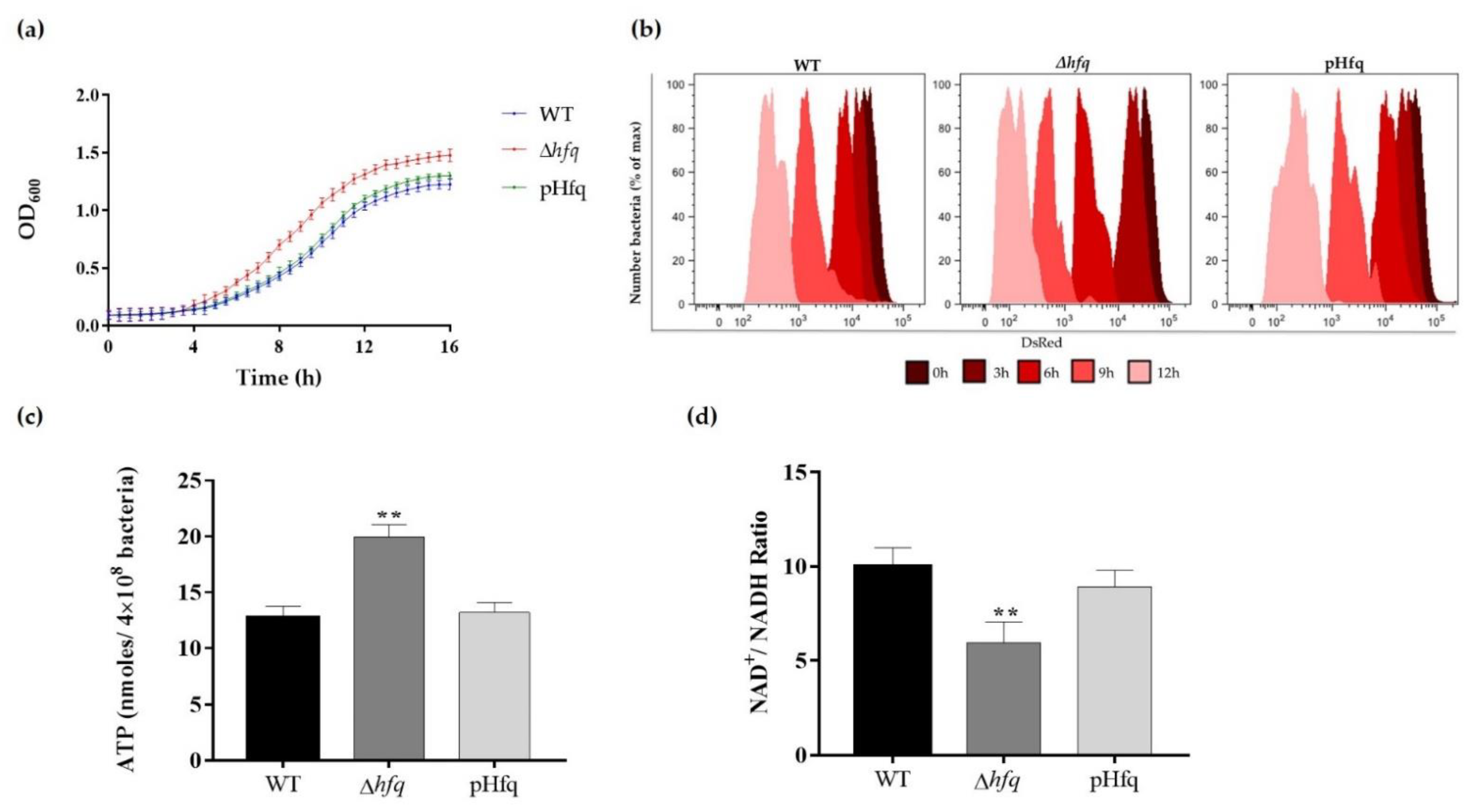
3.1. Hfq Deletion Results in Bacteria with Faster Growth and More Active Metabolism

Since Hfq is a global regulator that impacts growth and metabolism in bacteria [22,23], we first analyzed these processes in Y. ruckeri cultures. For this purpose, a mutant strain deficient in this regulatory protein was constructed (∆hfq strain) prior to analyzing the growth curves, replication profiles and metabolic status. We observed that Y. ruckerihfq exhibited an accelerated bacterial growth in comparison with the wild-type strain (Figure 1a). The bacterial replication in the culture medium was accurately determined by a fluorescence dilution (FD) assay based on a replication reporter system, as previously described [18]. The FD results supported the analysis of growth curves, showing that the Δhfq strain replicates faster than the wild type, displaying a more accelerated profile of red fluorescence dilution as compared to the wild-type strain (Figure 1b). In accordance with the growth and replication phenotypes, the intracellular levels of ATP from stationary cultures of the ∆hfq strain were higher than that of the wild-type (Figure 1c). In agreement with a more active metabolic state, Y. ruckeri Δhfq also showed a lower NAD+/NADH ratio than the wild-type (Figure 1d), evidencing a more reductive intracellular environment. The pHfq complemented strain showed phenotypes comparable to the wild-type strain. All these results suggest that Hfq modulates bacterial proliferation and energy metabolism.

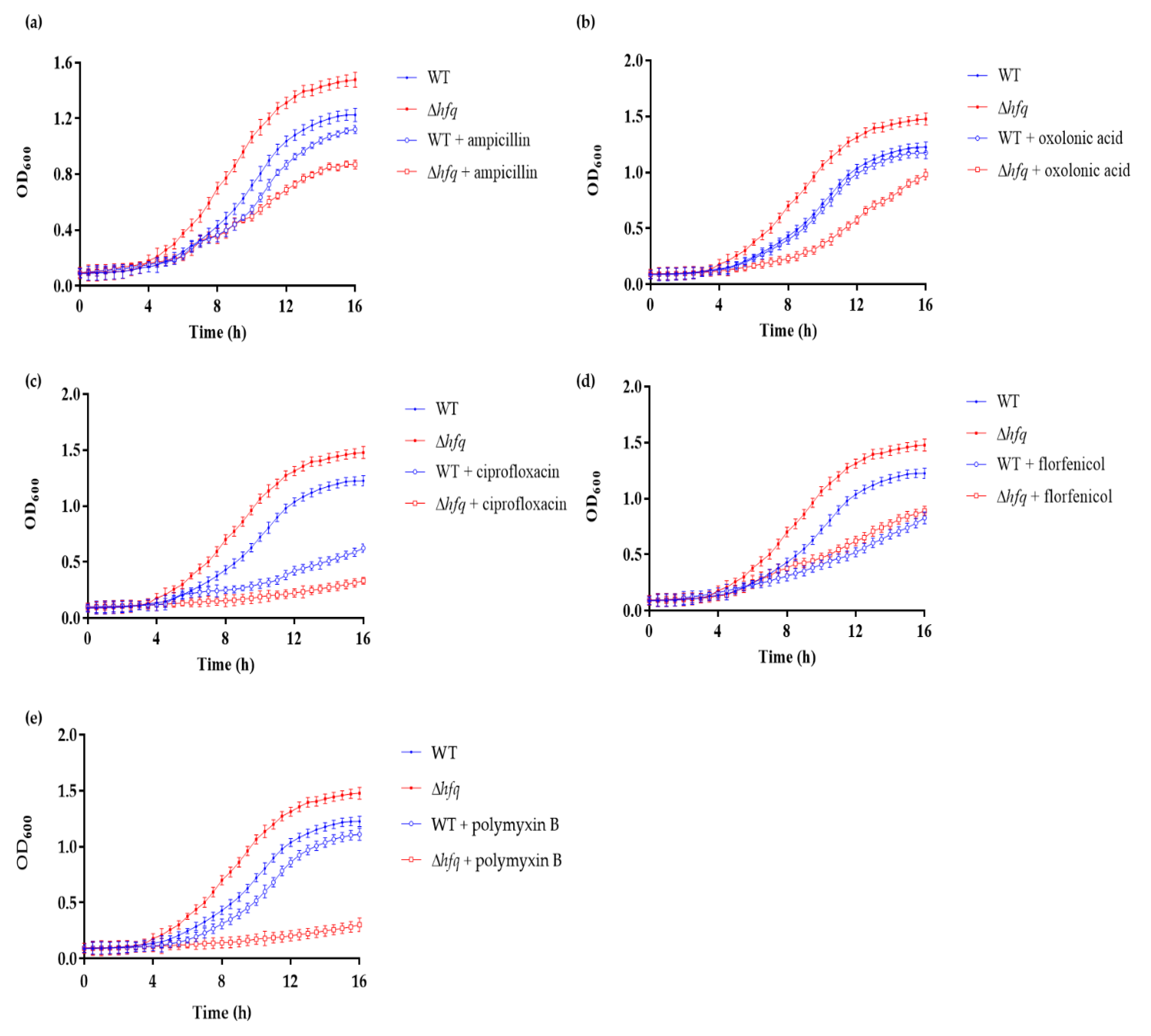
3.2. Y. ruckeri Δhfq Exhibits Increased Susceptibility to Sublethal Concentrations of Different Antibiotics

To gain better insights into the role of Hfq in antibiotic response in Y. ruckeri, we first evaluated the susceptibilities of the Δhfq and wild-type strains by determining the minimal inhibitory concentration (MIC) and minimum bactericidal concentration (MBC) with different bactericidal antibiotics. The deletion of hfq increased the susceptibility 10-fold compared to all the tested antibiotics, except for florfenicol (Table 2). Then, the strains were grown in the presence of sublethal concentrations of the antibiotics, confirming that the Δhfq strain presented enhanced susceptibility toward most of the antibiotics tested, except for florfenicol (Figure 2a–e). The highest susceptibility was observed for polymyxin B, suggesting that Hfq is critical for regulating outer membrane homeostasis. Overall, these results revealed that the efficiency of the antibiotics on the bacterial growth was higher with the rapidly dividing Δhfq cells having high metabolic rates.

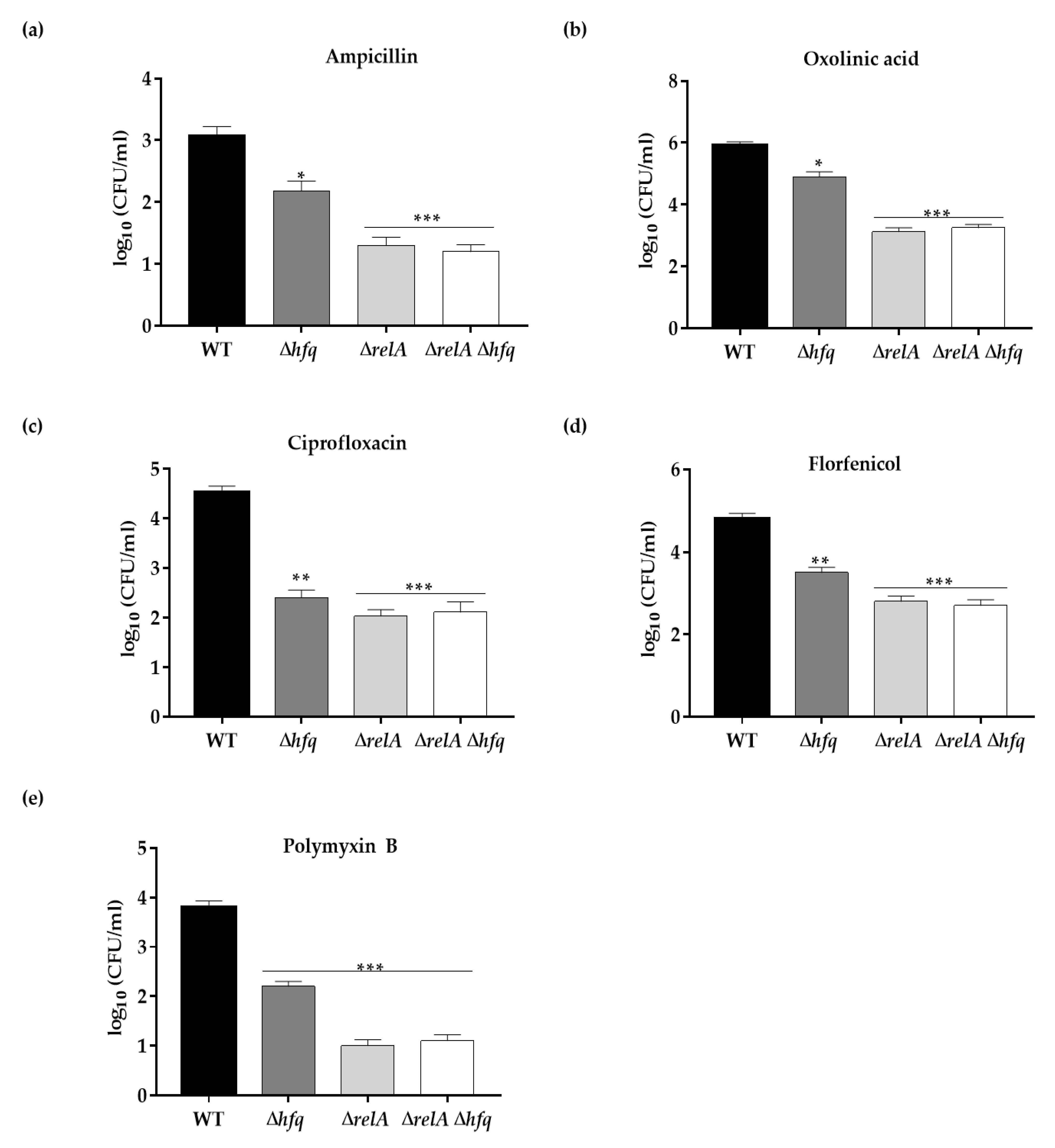
3.3. Hfq Is Required for Antibiotics Persistence in Y. ruckeri

To analyze the role of Hfq in antibiotic persistence of Y. ruckeri, susceptibility of stationary-phase cells of the Δhfq and the wild-type strains was examined during the exposure to lethal concentrations of bactericidal antibiotics (time-kill curves) as an indicator for persister cells production. Survival was significantly diminished in the Δhfq strain upon treatment with ampicillin for 48 h (3-fold), oxolinic acid for 72 h (4-fold), ciprofloxacin for 72 h (~4-fold), polymyxin B for 48 h (~3-fold) and florfenicol for 42 h (~2-fold) (Figure 3a–e), indicating that the persister cells production is affected in the Δhfq strain compared with the wild type. Since the age of inoculum strongly influences persister cells formation [6,24,25], we analyzed whether Hfq also mediates the persister cells production (24 h) from bacteria at exponential phase of growth (OD600 of 0.3). We observed that the survival of the Δhfq strain was also decreased, but to a lesser extent, compared to the wild type (Figure 3f). Since persisters have also been described as dormant subpopulations arising within bacterial biofilms that are characteristically highly tolerant to antibiotics [26], we also analyzed the time-kill curves using resuspended cells that had just been sampled from 24 h-biofilms from both Δhfq and the wild-type strain. As previously reported in other persister assays with cells suspension from biofilm cultures [27], we observed that cells harvested from biofilms produce more persister cells than those from liquid cultures. However, persister cells production in the Δhfq strain was also decreased for all the antibiotics treatments for 24 h, in comparison with the wild-type (Figure 3g). These results indicate that Hfq was involved in antibiotic persistence in bacteria harvested from liquid and biofilm cultures.

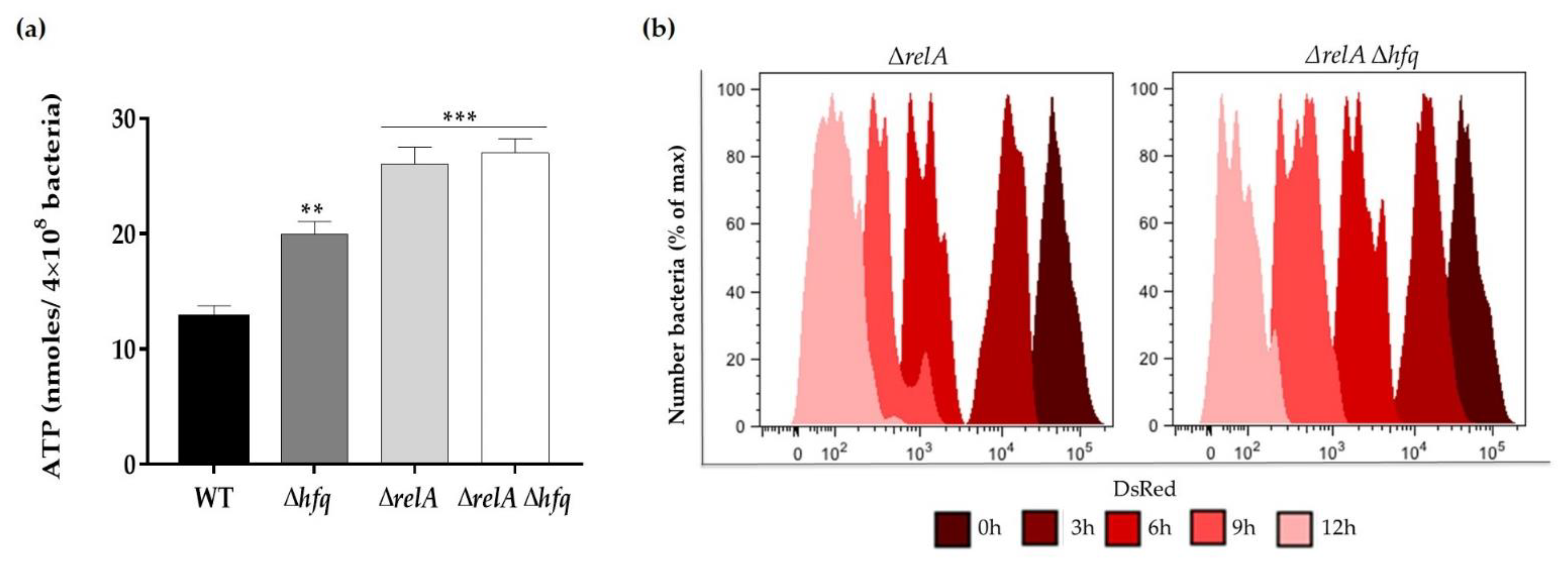
3.4. The Effect of Hfq on Persister Cells Production Is Further Aggravated in the ΔrelA ((p)ppGpp synthetase) Genetic Background

To better understand the role of Hfq in antibiotic persistence and since it is well known that (p)ppGpp is a key regulator of persistence [28], we evaluated whether the role of Hfq in persister cells production is dependent on RelA, the (p)ppGpp synthetase described as a central regulator of persistence [29]. To address this purpose, we constructed the single and double knockout mutants for the relA gene, namely, the ΔrelA and ΔrelA Δhfq strains. We determined the persister levels of the single and double knockout mutants ΔrelA, Δhfq, ΔrelA Δhfq and the wild-type strain from stationary phase cultures treated with lethal concentrations of ampicillin (1 mg mL−1), ciprofloxacin (0.05 mg mL−1), florfenicol (0.01 mg mL−1), oxolinic acid (0.05 mg mL−1) and polymyxin B (1 mg mL−1) for 24 h. We found that the single-gene knockout of relA affected the persister level to the same extent as the double-gene knockout mutant ΔrelA Δhfq but to a greater extent than the single-gene knockout of hfq, for all the antibiotics treatments (Figure 4a–e). These results indicates that the relA gene is epistatic over hfq regarding this phenotype.
To further investigate the interaction between the hfq and relA genes, the replication in bacterial cultures and the ATP levels of stationary phase cells, from the single and double ΔrelA backgrounds strains, were determined as described above. We observed that the faster replication and hypermetabolic status previously detected in the Δhfq strain was increased in the absence of relA, in both the single and double deletion backgrounds (ΔrelA and ΔrelA Δhfq strains), confirming the epistatic effect observed above (Figure 5a,b). Together, these data indicate that the regulatory effect of RelA on persistence, energy metabolism and bacterial proliferation exceeds that of Hfq.

4. Discussion

Vaccination against the major serotypes of Y. ruckeri is used in most fish farms worldwide, although this practice is not effective in all cases [10], and the emergence of outbreaks has substantially increased since 2003 [11]. This situation has awakened interest to investigate the molecular mechanisms of virulence [10,18], and the antimicrobial resistance in Y. ruckeri isolates from aquaculture [13,14,30,31]. However, studies focused on persister cells production, i.e., the formation of cells highly tolerant to killing by antibiotics, have not been reported to date. In the present study, we investigated the contribution of the sRNA chaperone Hfq from Y. ruckeri in persistence to multiple antibiotics, using an isolate from a salmon farm in Chile.
Among the most frequently used antimicrobials to treat ERM and other bacterial diseases affecting aquaculture, we can mention oxytetracycline and florfenicol [13,14,15,16]. Other antibiotics used, but to a lesser extent, are oxolinic acid and amoxicillin [15,16]. ERM outbreaks have been commonly related to the bacterial resistance to these antibiotics [13,17,32]. However, persister cells could also be responsible for outbreaks and the recalcitrance of infections, since they remain viable and repopulate the fish tanks forming biofilms when the level of antibiotics drops [26]. Persisters are thought to be less sensitive to antimicrobials because they are not undergoing cellular processes that antibiotics can affect, i.e., these cells are in an arrest-growth state that enables antibiotic tolerance. Hfq is a global regulator that impacts growth and metabolism in many bacteria, including some human pathogens of the Yersinia genus [22,23,33]. However, contrary to what is commonly observed in those bacteria, the hfq deletion in Y. ruckeri resulted in faster growth than the wild-type (Figure 1a), as well as in a more active metabolic state (Figure 1b–d). These results suggest global modulatory roles for Hfq in the restraint of growth and/or energy metabolism in this bacterium. To our knowledge, similar phenotypes have been previously described in this and other species lacking Hfq-dependent sRNAs [18,34,35], but not in hfq knockout mutants from other species, where the phenotypes are even dissimilar. For example, in Aeromonas veronii, the knockout of the hfq gene caused a slower growth rate at the exponential phase and produced less cell mass at the stationary phase [5]. By contrast, the knockouts of hfq in other species such as Y. pestis [33], E. coli [36] and S. Typhimurium [37] were also affected on growth but in Y. pseudotuberculosis displayed no effect [33]. Although Hfq is highly conserved among bacteria, homologs from different species may exhibit variations in their functions intimately linked to the lifestyle or niches that bacteria inhabit.
As we expected, given that reduced ATP levels have been associated with diminished antibiotic efficacy and vice versa [38,39], the Y. ruckeri Δhfq strain was more susceptible to most of the antibiotics tested under the sub-lethal concentrations evaluated (Figure 2, Table 2). Differential increased susceptibility to different classes of antibiotics has also been described in other hfq deletion strains such as Pseudomonas aeruginosa [40], E. coli [41], Salmonella Typhimurium [42] and Proteus mirabilis [43], indicating that the regulatory functions of Hfq enable bacteria to adapt to multiple antibiotic challenges. Similarly, some studies have demonstrated that some Hfq functions involved in antibiotic susceptibility are indirectly regulated via multiple sRNAs [44,45], thus explaining the pleiotropic phenotypes observed in hfq knockouts of different species.
For its part, the knowledge on the impact of Hfq on antibiotic persistence is still limited. Previous studies have reported discrepant results regarding the persister cells formation in hfq knockouts backgrounds. While in A. veronnii the deletion of hfq reduced the persister cells formation at different extents, under treatment with ciprofloxacin, cefotaxime, chloramphenicol and tetracycline [5], in E. coli, the deletion of hfq increased persister cells production by 11-fold under treatment with ampicillin [8]. Another study with the uropathogenic E. coli strain UIT89 revealed that the sRNA RyhB mediates the persister cells production to levofloxacin, cefotaxime and gentamicin in an Hfq-independent manner; however, the single deletion of hfq also has a detrimental effect on persister cells formation [7]. Together, our results and the data obtained from other studies reporting differential phenotypes for hfq mutants highlight the complexity of the persistence phenomenon governed by many different and heterogeneous molecular mechanisms and factors. Among these factors, the bacterial genetic background that evolved from their different lifestyles also played a pivotal role.
Since greater ATP levels are predictive of bactericidal antibiotic efficacy, and taking into account that the downregulation of transcription of genes involved in energy production and the ATP depletion are conditions that explain bacterial tolerance to antibiotics [38,39,46], we hypothesized that the Y. ruckeri Δhfq strain would be affected in persister cells formation. Our persister assays were conducted with five classes of antibiotics with bactericidal properties, whose choice was also based on their aquaculture usage. As we expected, the hfq deletion mutant showed defects in persister cells formation to different extents for all the antibiotics tested. This result is consistent with previous reports where ΔryhB (sRNA) and ΔphoU (regulator for phosphate metabolism) mutants from E. coli with hyperactive metabolic states have significant defects in persistence [7,24]. A previous study showed that metabolic stimulation can be used to eliminate persisters [47], suggesting that Hfq might be a potential target for anti-persisters strategies.
As the central stringent-response regulator, RelA represents the main (p)ppGpp synthetase, whose deficiency affects the production of persister cells [48]. We observed a significant reduction in persister cells production when the ΔrelA mutant was treated with the five antibiotics (Figure 4). The reduction in persister levels of the ΔrelA strain was comparable to that of the ΔrelA Δhfq strain for each antibiotic treatment, being more drastic than the effect observed in the Δhfq strain, indicating that relA is epistatic over hfq. This epistatic effect was further confirmed when the ATP levels were measured from stationary cultures without antibiotic treatments, as well as the replication analysis (Figure 5). As mentioned above, RelA is the major synthetase of (p)ppGpp, the second messenger that plays a crucial role in persister formation. By controlling diverse physiological processes, (p)ppGpp leads to dormancy or slow growth in bacteria [29,49]. In addition, it is known that, in E. coli, RelA fulfills functions independent of its (p)ppGpp synthetase activity. By interacting with Hfq, RelA stimulates the regulatory activity of the Hfq-dependent sRNA RyhB to arrest growth during iron depletion [50,51]. Based on this information, we hypothesize that RelA has a regulatory effect on Hfq by direct protein–protein interaction, and it also plays a role with the (p)ppGpp synthetase activity, which could explain the above-mentioned epistatic effect. However, the possibility of a mechanism involving RelA, Hfq and sRNAs in antibiotic persistence in Y. ruckeri needs to be further studied.
In summary, our results provide evidence that the sRNA chaperone Hfq from Y. ruckeri restrains the growth and energy metabolism and mediates persistence to multiple antibiotics. In addition, the RelA-mediated persistence displayed by several bacterial pathogens was also evidenced in Y. ruckeri. Thus, this study contributes to understanding the phenomenon of antibiotic persistence in this salmonid pathogen and will aid in the rational design of novel strategies to prevent or treat ERM outbreaks.

Supplementary Materials

The following are available online at https://www.mdpi.com/article/10.3390/microorganisms9071404/s1, Table S1: Primers (Oligos) used in this study.

Author Contributions

Conceptualization, L.G.A. and I.L.C.; methodology, L.G.A., I.L.C., M.J.B. and F.M.; software, L.G.A.; validation, L.G.A., M.J.B. and F.M.; formal analysis, L.G.A., I.L.C., F.G. and J.A.F.; investigation, L.G.A., I.L.C. and M.J.B.; resources, L.G.A. and I.L.C.; data curation, L.G.A. and I.L.C.; writing—original draft preparation, L.G.A. and I.L.C.; writing—review and editing, I.L.C., J.A.F. and F.G.; visualization, L.G.A. and I.L.C.; supervision, L.G.A. and I.L.C.; project administration, L.G.A., I.L.C.; funding acquisition, L.G.A., and I.L.C. All authors have read and agreed to the published version of the manuscript.

Funding

This research received financial support from Agencia Nacional de Investigación y Desarrollo (ANID, Chile), FONDECYT 11201070 to L.G.A., FONDECYT 1171655 to I.L.C. and CONICYT/FONDAP Grant No. 15110027 to L.G.A.

Conflicts of Interest

The authors declare no conflict of interest.

References

  1. Harms, A.; Maisonneuve, E.; Gerdes, K. Mechanisms of bacterial persistence during stress and antibiotic exposure. Science 2016, 354, aaf4268. [Google Scholar] [CrossRef]
  2. Moldoveanu, A.L.; Rycroft, J.A.; Helaine, S. Impact of bacterial persisters on their host. Curr. Opin. Microbiol. 2021, 59, 55–71. [Google Scholar] [CrossRef]
  3. Fisher, R.A.; Gollan, B.; Helaine, S. Persistent bacterial infections and persister cells. Nat. Rev. Microbiol. 2017, 15, 453–464. [Google Scholar] [CrossRef]
  4. Salcedo-Sora, J.E.; Kell, D.B. A Quantitative Survey of Bacterial Persistence in the Presence of Antibiotics: Towards Antipersister Antimicrobial Discovery. Antibiotics 2020, 9, 508. [Google Scholar] [CrossRef] [PubMed]
  5. Zhang, L.; Yu, W.; Tang, Y.; Li, H.; Ma, X.; Liu, Z. RNA chaperone Hfq mediates persistence to multiple antibiotics in Aeromonas veronii. Microb. Pathog. 2019, 132, 124–128. [Google Scholar] [CrossRef] [PubMed]
  6. Zhang, Y. Persisters, persistent infections and the Yin-Yang model. Emerg. Microbes Infect. 2014, 3, e3. [Google Scholar] [CrossRef]
  7. Zhang, S.; Liu, S.; Wu, N.; Yuan, Y.; Zhang, W.; Zhang, Y. Small Non-coding RNA RyhB Mediates Persistence to Multiple Antibiotics and Stresses in Uropathogenic Escherichia coli by Reducing Cellular Metabolism. Front. Microbiol. 2018, 9, 136. [Google Scholar] [CrossRef] [PubMed]
  8. Kim, Y.; Wood, T.K. Toxins Hha and CspD and small RNA regulator Hfq are involved in persister cell formation through MqsR in Escherichia coli. Biochem. Biophys. Res. Commun. 2010, 391, 209–213. [Google Scholar] [CrossRef] [Green Version]
  9. Chao, Y.; Vogel, J. The role of Hfq in bacterial pathogens. Curr. Opin. Microbiol. 2010, 13, 24–33. [Google Scholar] [CrossRef]
  10. Wrobel, A.; Leo, J.C.; Linke, D. Overcoming Fish Defences: The Virulence Factors of Yersinia ruckeri. Genes 2019, 10, 700. [Google Scholar] [CrossRef] [Green Version]
  11. Guijarro, J.A.; García-Torrico, A.I.; Cascales, D.; Méndez, J. The Infection Process of Yersinia ruckeri: Reviewing the Pieces of the Jigsaw Puzzle. Front. Cell Infect. Microbiol. 2018, 8, 218. [Google Scholar] [CrossRef]
  12. Bystritskaya, E.; Stenkova, A.; Chistuylin, D.; Chernysheva, N.; Khomenko, V.; Anastyuk, S.; Novikova, O.; Rakin, A.; Isaeva, M. Adaptive responses of outer membrane porin balance of Yersinia ruckeri under different incubation temperature, osmolarity, and oxygen availability. Microbiol. Open 2016, 5, 597–603. [Google Scholar] [CrossRef] [Green Version]
  13. Michel, C.; Kerouault, B.; Martin, C. Chloramphenicol and florfenicol susceptibility of fish-pathogenic bacteria isolated in France: Comparison of minimum inhibitory concentration, using recommended provisory standards for fish bacteria. J. Appl. Microbiol. 2003, 95, 1008–1015. [Google Scholar] [CrossRef] [PubMed]
  14. Duman, M.; Altun, S.; Cengiz, M.; Saticioglu, I.B.; Buyukekiz, A.G.; Sahinturk, P. Genotyping and antimicrobial resistance genes of Yersinia ruckeri isolates from rainbow trout farms. Dis. Aquat. Organ. 2017, 125, 31–44. [Google Scholar] [CrossRef] [PubMed]
  15. Kumar, G.; Menanteau-Ledouble, S.; Saleh, M.; El-Matbouli, M. Yersinia ruckeri, the causative agent of enteric redmouth disease in fish. Vet. Res. 2015, 46, 103. [Google Scholar] [CrossRef] [Green Version]
  16. Miranda, C.D.; Godoy, F.A.; Lee, M.R. Current Status of the Use of Antibiotics and the Antimicrobial Resistance in the Chilean Salmon Farms. Front. Microbiol. 2018, 9, 1284. [Google Scholar] [CrossRef] [PubMed]
  17. Rodgers, J.C. Resistance of Yersinia ruckeri to antimicrobial agents in vitro. Aquaculture 2001, 196, 325–345. [Google Scholar] [CrossRef]
  18. Acuña, L.G.; Barros, M.J.; Montt, F.; Peñaloza, D.; Núñez, P.; Valdés, I.; Gil, F.; Fuentes, J.A.; Calderón, I.L. Participation of two sRNA RyhB homologs from the fish pathogen Yersinia ruckeri in bacterial physiology. Microbiol. Res. 2021, 242, 126629. [Google Scholar] [CrossRef]
  19. Marx, C.I. Development of a broad-host-range sacB-based vector for unmarked allelic exchange. BMC Res. Notes 2008, 1, 1. [Google Scholar] [CrossRef] [Green Version]
  20. Bolivar, F.; Rodriguez, R.L.; Greene, P.J.; Betlach, M.C.; Heyneker, H.L.; Boyer, H.W.; Crosa, J.H.; Falkow, S. Construction and characterization of new cloning vehicles. II. A multipurpose cloning system. Gene 1977, 2, 95–113. [Google Scholar] [CrossRef]
  21. Helaine, S.; Thompson, J.A.; Watson, K.G.; Liu, M.; Boyle, C.; Holden, D.W. Dynamics of intracellular bacterial replication at the single cell level. Proc. Natl. Acad. Sci. USA 2010, 107, 3746–3751. [Google Scholar] [CrossRef] [PubMed] [Green Version]
  22. Kakoschke, T.; Kakoschke, S.; Magistro, G.; Schubert, S.; Borath, M.; Heesemann, J.; Rossier, O. The RNA chaperone Hfq impacts growth, metabolism and production of virulence factors in Yersinia enterocolitica. PLoS ONE 2014, 9, e86113. [Google Scholar] [CrossRef] [Green Version]
  23. Vo, P.N.L.; Lee, H.M.; Ren, J.; Na, D. Optimized expression of Hfq protein increases Escherichia coli growth. J. Biol. Eng. 2021, 15, 7. [Google Scholar] [CrossRef] [PubMed]
  24. Li, Y.; Zhang, Y. PhoU is a persistence switch involved in persister formation and tolerance to multiple antibiotics and stresses in Escherichia coli. Antimicrob. Agents Chemother. 2007, 51, 2092–2099. [Google Scholar] [CrossRef] [Green Version]
  25. Luidalepp, H.; Jõers, A.; Kaldalu, N.; Tenson, T. Age of inoculum strongly influences persister frequency and can mask effects of mutations implicated in altered persistence. J. Bacteriol. 2011, 193, 3598–3605. [Google Scholar] [CrossRef] [Green Version]
  26. Lewis, K. Persister cells. Annu. Rev. Microbiol. 2010, 64, 357–372. [Google Scholar] [CrossRef]
  27. Miyaue, S.; Suzuki, E.; Komiyama, Y.; Kondo, Y.; Morikawa, M.; Maeda, S. Bacterial Memory of Persisters: Bacterial Persister Cells Can Retain Their Phenotype for Days or Weeks After Withdrawal from Colony-Biofilm Culture. Front. Microbiol. 2018, 9, 1396. [Google Scholar] [CrossRef] [Green Version]
  28. Hauryliuk, V.; Atkinson, G.C.; Murakami, K.S.; Tenson, T.; Gerdes, K. Recent functional insights into the role of (p)ppGpp in bacterial physiology. Nat. Rev. Microbiol. 2015, 13, 298–309. [Google Scholar] [CrossRef] [Green Version]
  29. Pacios, O.; Blasco, L.; Bleriot, I.; Fernandez-Garcia, L.; Ambroa, A.; López, M.; Bou, G.; Cantón, R.; Garcia-Contreras, R.; Wood, T.K.; et al. (p)ppGpp and Its Role in Bacterial Persistence: New Challenges. Antimicrob. Agents Chemother. 2020, 64, e01283-20. [Google Scholar] [CrossRef]
  30. Calvez, S.; Gantelet, H.; Blanc, G.; Douet, D.G.; Daniel, P. Yersinia ruckeri Biotypes 1 and 2 in France: Presence and antibiotic susceptibility. Dis. Aquat. Organ. 2014, 109, 117–126. [Google Scholar] [CrossRef]
  31. Huang, Y.; Michael, G.B.; Becker, R.; Kaspar, H.; Mankertz, J.; Schwarz, S.; Runge, M.; Steinhagen, D. Pheno- and genotypic analysis of antimicrobial resistance properties of Yersinia ruckeri from fish. Vet. Microbiol. 2014, 171, 406–412. [Google Scholar] [CrossRef]
  32. Subpesca. Instituto de Fomento Pesquero (IFOP). Informe Final: Vigilancia de la Resistencia Bacteriana en la Salmonicultura. FIP N°2008-65. 2011. Available online: https://www.subpesca.cl/fipa/613/articles-89266_informe_final.pdf (accessed on 3 May 2021).
  33. Bai, G.; Golubov, A.; Smith, E.A.; McDonough, K.A. The importance of the small RNA chaperone Hfq for growth of epidemic Yersinia pestis, but not Yersinia pseudotuberculosis, with implications for plague biology. J. Bacteriol. 2010, 192, 4239–4245. [Google Scholar] [CrossRef] [Green Version]
  34. Padalon-Brauch, G.; Hershberg, R.; Elgrably-Weiss, M.; Baruch, K.; Rosenshine, I.; Margalit, H.; Altuvia, S. Small RNAs encoded within genetic islands of Salmonella typhimurium show host-induced expression and role in virulence. Nucleic Acids Res. 2008, 36, 1913–1927. [Google Scholar] [CrossRef] [PubMed]
  35. Peñaloza, D.; Acuña, L.G.; Barros, M.J.; Núñez, P.; Montt, F.; Gil, F.; Fuentes, J.A.; Calderón, I.L. The Small RNA RyhB Homologs from Salmonella Typhimurium Restrain the Intracellular Growth and Modulate the SPI-1 Gene Expression within RAW264.7 Macrophages. Microorganisms 2021, 9, 635. [Google Scholar] [CrossRef] [PubMed]
  36. Tsui, H.C.; Leung, H.C.; Winkler, M.E. Characterization of broadly pleiotropic phenotypes caused by an hfq insertion mutation in Escherichia coli K-12. Mol. Microbiol. 1994, 13, 35–49. [Google Scholar] [CrossRef] [PubMed]
  37. Sittka, A.; Pfeiffer, V.; Tedin, K.; Vogel, J. The RNA chaperone Hfq is essential for the virulence of Salmonella typhimurium. Mol. Microbiol. 2007, 63, 193–217. [Google Scholar] [CrossRef] [Green Version]
  38. Conlon, B.P.; Rowe, S.E.; Gandt, A.B.; Nuxoll, A.S.; Donegan, N.P.; Zalis, E.A.; Clair, G.; Adkins, J.N.; Cheung, A.L.; Lewis, K. Persister formation in Staphylococcus aureus is associated with ATP depletion. Nat. Microbiol. 2016, 1, 16051. [Google Scholar] [CrossRef] [Green Version]
  39. Shan, Y.; Brown Gandt, A.; Rowe, S.E.; Deisinger, J.P.; Conlon, B.P.; Lewis, K. ATP-Dependent Persister Formation in Escherichia coli. mBio 2017, 8, e02267-16. [Google Scholar] [CrossRef] [Green Version]
  40. Pusic, P.; Sonnleitner, E.; Krennmayr, B.; Heitzinger, D.A.; Wolfinger, M.T.; Resch, A.; Bläsi, U. Harnessing Metabolic Regulation to Increase Hfq-Dependent Antibiotic Susceptibility in Pseudomonas aeruginosa. Front. Microbiol. 2018, 9, 2709. [Google Scholar] [CrossRef] [Green Version]
  41. Yamada, J.; Yamasaki, S.; Hirakawa, H.; Hayashi-Nishino, M.; Yamaguchi, A.; Nishino, K. Impact of the RNA chaperone Hfq on multidrug resistance in Escherichia coli. J. Antimicrob. Chemother. 2010, 65, 853–858. [Google Scholar] [CrossRef]
  42. Hayashi-Nishino, M.; Fukushima, A.; Nishino, K. Impact of Hfq on the intrinsic drug resistance of Salmonella enterica serovar typhimurium. Front. Microbiol. 2012, 3, 205. [Google Scholar] [CrossRef] [Green Version]
  43. Wang, M.C.; Chien, H.F.; Tsai, Y.L.; Liu, M.C.; Liaw, S.J. The RNA chaperone Hfq is involved in stress tolerance and virulence in uropathogenic Proteus mirabilis. PLoS ONE 2014, 9, e85626. [Google Scholar] [CrossRef] [PubMed]
  44. Dersch, P.; Khan, M.A.; Mühlen, S.; Görke, B. Roles of Regulatory RNAs for Antibiotic Resistance in Bacteria and Their Potential Value as Novel Drug Targets. Front. Microbiol. 2017, 8, 803. [Google Scholar] [CrossRef] [Green Version]
  45. Zhang, Y.F.; Han, K.; Chandler, C.E.; Tjaden, B.; Ernst, R.K.; Lory, S. Probing the sRNA regulatory landscape of P. aeruginosa: Post-transcriptional control of determinants of pathogenicity and antibiotic susceptibility. Mol. Microbiol. 2017, 106, 919–937. [Google Scholar] [CrossRef] [PubMed] [Green Version]
  46. Kwan, B.W.; Valenta, J.A.; Benedik, M.J.; Wood, T.K. Arrested protein synthesis increases persister-like cell formation. Antimicrob. Agents Chemother. 2013, 57, 1468–1473. [Google Scholar] [CrossRef] [Green Version]
  47. Allison, K.R.; Brynildsen, M.P.; Collins, J.J. Metabolite-enabled eradication of bacterial persisters by aminoglycosides. Nature 2011, 473, 216–220. [Google Scholar] [CrossRef] [Green Version]
  48. Korch, S.B.; Henderson, T.A.; Hill, T.M. Characterization of the hipA7 allele of Escherichia coli and evidence that high persistence is governed by (p)ppGpp synthesis. Mol. Microbiol. 2003, 50, 1199–1213. [Google Scholar] [CrossRef]
  49. Svenningsen, M.S.; Veress, A.; Harms, A.; Mitarai, N.; Semsey, S. Birth and Resuscitation of (p)ppGpp Induced Antibiotic Tolerant Persister Cells. Sci. Rep. 2019, 9, 6056. [Google Scholar] [CrossRef]
  50. Argaman, L.; Elgrably-Weiss, M.; Hershko, T.; Vogel, J.; Altuvia, S. RelA protein stimulates the activity of RyhB small RNA by acting on RNA-binding protein Hfq. Proc. Natl. Acad. Sci. USA 2012, 109, 4621–4626. [Google Scholar] [CrossRef] [Green Version]
  51. Basu, P.; Elgrably-Weiss, M.; Hassouna, F.; Kumar, M.; Wiener, R.; Altuvia, S. RNA binding of Hfq monomers promotes RelA-mediated hexamerization in a limiting Hfq environment. Nat. Commun. 2021, 12, 2249. [Google Scholar] [CrossRef]
Figure 1. Growth curves, bacterial replication and metabolic status of Y. ruckeri strains. (a) Wild-type (WT), ∆hfq and pHfq (complemented) strains were grown in TSB medium and OD600 was measured at different time points. Data represent the means ± standard deviations (n = 3). (b) Flow cytometric detection of DsRed and EGFP fluorescence at different time points from the WT, ∆hfq and pHfq strains (carrying pDiGc plasmid) cultured in TSB medium for 12 h (n = 30,000 events analyzed at each time point). WT, ∆hfq and pHfq strains were grown in TSB to OD600 of 0.9 and both the intracellular ATP levels (c) and NAD+/NADH ratio were determined (d). Asterisks represent statistical differences with respect to the WT strain (** p < 0.001). Data represent the means ± standard deviations (n = 3).
Microorganisms 09 01404 g001
Figure 2. Growth curves of the WT and ∆hfq strains exposed to sublethal concentrations of antibiotics. WT and ∆hfq strains were grown in TSB medium supplemented with 0.01 mg/mL ampicillin (a), 0.0001 mg/mL oxolinic acid (b), 0.001 mg/mL ciprofloxacin (c), 0.001 mg/mL florfenicol (d) and 0.002 mg/mL polymyxin B (e). OD600 was measured every 30 min until the bacteria reached the stationary phase (16 h). Data represent the means ± standard deviations (n = 3).
Microorganisms 09 01404 g002
Figure 3. Time-kill curves and persister cells production of the WT and ∆hfq strains exposed to antibiotics. Stationary cells of the WT and Δhfq strains were exposed to 1 mg mL−1 ampicillin (a), 0.05 mg mL−1 oxolinic acid (b), 0.05 mg mL−1 ciprofloxacin (c), 0.01 mg mL−1 florfenicol (d) or 1 mg mL−1 polymyxin B (e), and the survival of persister cells was measured as CFUs per 1 mL at different time points. Exponential (f) and biofilm resuspended (g) cells of the WT and Δhfq strains were exposed to 1 mg mL−1 ampicillin, 0.05 mg mL−1 oxolinic acid, 0.05 mg mL−1 ciprofloxacin, 0.01 mg mL−1 florfenicol or 1 mg mL−1 polymyxin B for 24 h, and the survival of persister cells was measured as CFUs per 1 mL at 24 h. Asterisks represent statistical differences with respect to the wild-type strain (* p < 0.01; ** p < 0.001; *** p = 0.0001). Data represent the means ± standard deviations (n = 3).
Microorganisms 09 01404 g003
Figure 4. Persister cells production of the ΔrelA and ΔrelA Δhfq strains exposed to antibiotics. Stationary cells of the WT, Δhfq, ΔrelA and ΔrelA Δhfq strains were exposed to 1 mg mL−1 ampicillin (a), 0.05 mg mL−1 oxolinic acid (b), 0.05 mg mL−1 ciprofloxacin (c), 0.01 mg mL−1 florfenicol (d) or 1 mg mL−1 polymyxin B (e) for 24 h, and the survival of persister cells was measured as CFUs per 1 mL at 24 h. Asterisks represent statistical differences with respect to the wild-type strain (* p < 0.01; ** p < 0.001; *** p = 0.0001). Data represent the means ± standard deviations (n = 3).
Microorganisms 09 01404 g004
Figure 5. Bacterial replication and metabolic status of the ΔrelA and ΔrelA Δhfq strains. (a) WT, Δhfq, ΔrelA and ΔrelA Δhfq strains were grown in TSB to OD600 of 0.9 and the intracellular ATP levels were determined. Asterisks represent statistical differences with respect to the WT strain (** p < 0.001; *** p = 0.0001). Data represent the means ± standard deviations (n = 3). (b) Flow cytometric detection of DsRed and EGFP fluorescence at different time points from the ΔrelA and ΔrelA Δhfq strains (carrying pDiGc plasmid) cultured in TSB medium for 12 h (n = 30,000 events analyzed at each time point).
Microorganisms 09 01404 g005
Table 1. Bacterial strains and plasmids used in this study.
StrainCharacteristicsReference
Y. ruckeri CD2 Wild-type strain of Y. ruckeri [18]
ΔhfqY. ruckeri CD2 lacking hfq gene[18]
ΔrelAY. ruckeri CD2 lacking relA geneThis study
ΔrelA ΔhfqY. ruckeri CD2 lacking realA and hfq genesThis study
PlasmidGenotypeReference
pCM433ApR, CmR, TcR, oriT, tet, cat, sacB and ColEI ori; broad-host-range sacB-based allelic exchange vector[19]
pCM433-hfqhfq region of Y. ruckeri CD2 cloned into pCM433This study
pCM433-relArelA region of Y. ruckeri CD2 cloned into pCM433This study
pBR322ApR, TcR, ColEl Ori.[20]
pHfqhfq region of Y. ruckeri CD2 cloned into pBR322This study
pDiGcbla GFP pBAD DsRed ori f1 AmpR[21]
Table 2. MIC and MBC measurements for wild-type (WT), Δhfq and pHfq (complemented) strains in TSB medium supplemented with antibiotics.
AntibioticMIC (mg mL−1)MBC (mg mL−1)
WT ΔhfqpHfqWT ΔhfqpHfq
Ampicillin10.1110.11
Oxolinic acid0.0050.00050.0050.050.50.05
Ciprofloxacin0.0030.0030.0030.0030.30.003
Florfenicol0.010.010.010.010.10.01
Polymyxin B20.2220.22
Publisher’s Note: MDPI stays neutral with regard to jurisdictional claims in published maps and institutional affiliations.

