Article

Leishmanicidal Activity of Betulin Derivatives in Leishmania amazonensis; Effect on Plasma and Mitochondrial Membrane Potential, and Macrophage Nitric Oxide and Superoxide Production

by
Wilmer Alcazar
1,
Sami Alakurtti
2,3,
Maritza Padrón-Nieves
1,
Maija Liisa Tuononen
2,3,
Noris Rodríguez
4,
Jari Yli-Kauhaluoma
2,* and
Alicia Ponte-Sucre
1,*
1
Laboratory of Molecular Physiology, Institute of Experimental Medicine, School of Medicine Luis Razetti, Faculty of Medicine, Universidad Central de Venezuela, P.O. Box 50587, Caracas 1050, Venezuela
2
Drug Research Program, Division of Pharmaceutical Chemistry and Technology, Faculty of Pharmacy, University of Helsinki, Viikinkaari 5 E, P.O. Box 56, FI-00014 Helsinki, Finland
3
VTT Technical Research Centre of Finland, Biologinkuja 7, P.O. Box 1000, FI-02044 Espoo, Finland
4
Laboratory of Genetic Engineering, Institute of Biomedicine, Universidad Central de Venezuela, P.O. Box 4043, Caracas 1010A, Venezuela
*
Authors to whom correspondence should be addressed.
Microorganisms 2021, 9(2), 320; https://doi.org/10.3390/microorganisms9020320
Submission received: 19 October 2020 / Revised: 24 January 2021 / Accepted: 29 January 2021 / Published: 4 February 2021

Abstract

:
Herein, we evaluated in vitro the anti-leishmanial activity of betulin derivatives in Venezuelan isolates of Leishmania amazonensis, isolated from patients with therapeutic failure. Methods: We analyzed promastigote in vitro susceptibility as well as the cytotoxicity and selectivity of the evaluated compounds. Additionally, the activity of selected compounds was determined in intracellular amastigotes. Finally, to gain hints on their potential mechanism of action, the effect of the most promising compounds on plasma and mitochondrial membrane potential, and nitric oxide and superoxide production by infected macrophages was determined. Results: From the tested 28 compounds, those numbered 18 and 22 were chosen for additional studies. Both 18 and 22 were active (GI50 ≤ 2 µM, cytotoxic CC50 > 45 µM, SI > 20) for the reference strain LTB0016 and for patient isolates. The results suggest that 18 significantly depolarized the plasma membrane potential (p < 0.05) and the mitochondrial membrane potential (p < 0.05) when compared to untreated cells. Although neither 18 nor 22 induced nitric oxide production in infected macrophages, 18 induced superoxide production in infected macrophages. Conclusion: Our results suggest that due to their efficacy and selectivity against intracellular parasites and the potential mechanisms underlying their leishmanicidal effect, the compounds 18 and 22 could be used as tools for designing new chemotherapies against leishmaniasis.

1. Introduction

Leishmaniasis is endemic in 98 countries, in which 14 million people remain infected and more than 350 million are at risk [1,2]. Approximately 1.3 million people contract the disease annually; 300,000 cases correspond to visceral leishmaniasis (VL) and 1,000,000 to tegumentary leishmaniasis (TL). The estimated number of deaths from VL ranges between 20,000 and 50,000 each year [3]. However, these numbers reflect the tip of the iceberg, since only 40 countries where the disease is endemic regularly report their health statistics to the World Health Organization (WHO) [4].
In Venezuela, VL has a low incidence (0.03 to 0.24 per 100,000 inhabitants) [5], although a notable under-registration of cases remains [6]. The main focus of VL is located at the Nueva Esparta State [5], a geographical area in which the whole life cycle of Leishmania (L.) infantum has been described, including the dog as the main reservoir, as well as Lutzomyia longipalpis as the insect vector [7]. TL is officially recognized as a public health problem [5,8]. A total of 66,550 cases in its various clinical forms, with an annual incidence rate that varied between 5.3 per 100,000 inhabitants (2014) and 12 per 100,000 inhabitants (1992), have been reported during the period 1990–2018. TL represents 99% of the cases of leishmaniasis reported each year, of which 98% correspond to localized cutaneous leishmaniasis (LCL), 1% mucocutaneous leishmaniasis (MCL), and 0.2% diffuse cutaneous leishmaniasis (DCL) [9].
After malaria, leishmaniasis is the protozoan parasitic disease with the highest prevalence worldwide (14 million people infected). It is the second in number of disability-adjusted life years or healthy years of life lost (1.7 million), the second with the highest mortality rate (up to 50,000 deaths per year), and the third cause of morbidity due to communicable diseases (1,300,000 new cases per year), after malaria and tuberculosis [1,2,3,4].
There are no pharmacological treatments sufficiently effective, safe, and affordable to combat diseases caused by kinetoplastid parasites, including Leishmania [2,10]. Progress in the biology, epidemiology, and immunology of leishmaniasis and the characterization of the parasite genome and proteome form the framework to design leishmaniasis chemotherapy of the future. Approaches to the development of chemotherapeutic strategies against the disease include optimization of the available therapy, use of active compounds against other diseases, pharmacological target-oriented design, and evaluation of natural products and their derivatives.
Prospects for developing an effective leishmaniasis vaccine are not immediate and disease control is based primarily on chemotherapeutic treatment, although the arsenal of available drugs is reduced and with significant associated toxicity; vector control is problematic generating environmental risk; comorbidity with HIV is increasing; and finally, almost all endemic regions exhibit a growing incidence of therapeutic failure [11,12,13,14]. Consequently, the design of more effective and safer therapeutic alternatives has become an extraordinary and urgent need [15,16,17]. Meanwhile, there is a delay in the development of new drugs, mainly due to the lack of economic stimuli that this disease represents for the pharmaceutical companies. For example, in the 2000s, the global investment in new antiparasitic drugs was only about 0.1% of the global asset in research [18]. Currently, with the exception of miltefosine (MLF), the same anti-leishmanial drugs of the early last century are still used.
Experimental development of new anti-leishmanial agents either starts from a validated target or they are searched by phenotypic screening [19]. The first approach has proved to be unsuccessful, because a powerful inhibitory agent found against an enzyme or another particular target usually fails to have activity in the entire microorganism [20]. The second alternative involves all possible therapeutic targets and simultaneously allows evaluation of compound activity in the cellular environment, in a much more real condition. Thus, phenotypic screening that includes in vitro susceptibility tests and the macrophage-amastigote model stands out as the cornerstone of studies prior to the development of new chemotherapeutics against this disease [21]. For example, trifluoromethyl-substituted quinolones and their analogues, as well as metronidazole derivatives have emerged as an interesting platform to design agents against L. (L.) mexicana and L. (V.) braziliensis [22,23].
According to the WHO, plants are the largest source of medicines for humans [24]. In fact, natural products and their derivatives are the source of 30% of the world pharmaceutical market [25]. In this scenario, extracts or compounds of plant origin, including terpenes, phenolics, and alkaloids, can be used as a valuable starting point for the search for new anti-leishmanial agents [18]. Among terpenoids, betulin, a pentacyclic triterpene, isolated from the bark of trees of the genus Betula (birches), stands out [26]. The leishmanicidal potential of some betulin derivatives has already been documented in the literature [27,28,29,30]. However, for this type of compounds, their effectiveness in New World species, or in parasites isolated from patients with therapeutic failure, is not clear, nor is their mechanism of action well known.
Herein, we evaluated in vitro the anti-leishmanial activity of betulin derivatives in Venezuelan isolates of Leishmania amazonensis [L. (L.) amazonensis], isolated from patients with therapeutic failure. Our results suggest that, due to their efficacy and selectivity against intracellular parasites and the mechanisms underlying their leishmanicidal effect, some of these compounds could be used as tools for designing new chemotherapies against leishmaniasis.

2. Materials and Methods

2.1. Parasites

Three L. (L.) amazonensis isolates were used: a reference species certified by the WHO (MHOM/BR/77/LTB0016, LTB0016) donated by Dr. Lionel Schnur, Hebrew University-Hadassah Medical School, Jerusalem, Israel, and two isolates (MHOM/VE/1998/MR, VE98MR; and MHOM/VE/2000/MM, VE2000MM) obtained from patient lesions identified as such at the Dermatology Department, Institute of Biomedicine (SAIB-MPPS-UCV), Caracas, Venezuela. The patients were diagnosed DCL and suffered therapeutic failure against meglumine antimoniate (Glucantime®, Sanofi-Aventis (Suzano, SP, Brazil). Species identification was performed by PCR and molecular hybridization [31]. This project (CDCH UCV PI-09-8717-2013) was approved by the Ethics Committee from the Institute of Biomedicine (SAIB-MPPS-UCV), Caracas, Venezuela. Isolates were kept in liquid nitrogen until use, when parasites were thawed and cultured in blood agar-glucose enriched with human urine [32,33].
Leishmania promastigotes were grown at 26 °C in semisolid blood agar supplemented with glucose-NaCl medium (glucose 1.5%, NaCl 0.85%, w/v). Promastigotes at late log growth phase were collected by centrifugation at 125× g for 10 min at room temperature (RT). The medium was decanted and the cells were suspended in the appropriate medium up to the desired cell density.

2.2. Murine Macrophages

J774.1 macrophages [34], donated by Dr. Concepción Hernández, Institute of Experimental Biology-Universidad Central de Venezuela, were used. Culture monolayers were established in complete Roswell Park Memorial Institute medium (RPMIc; 25 mM HEPES, 2 mM L-glutamine, 5 mg L−1 phenol red, supplemented with 10% fetal calf serum, 2 g L−1 NaHCO3, pH 7.2, 0.02 mM 2-mercaptoethanol, and 100 U mL−1 penicillin plus 100 µg mL−1 streptomycin), at 37 °C and 5% CO2. Culture medium was replaced weekly. For the experimental procedures, cells were detached from the flasks by mechanical procedures, washed twice with phosphate-buffered saline (NaCl 136 mM, KCl 2.6 mM, Na2HPO4⋅2H2O 10 mM, KH2PO4 1.7 mM; PBS, pH 7.4), and suspended at the desired density in RPMIc.

2.3. Compounds

Twenty-eight semisynthetic betulin derivatives (20 structurally simple and 8 heterocyclic derivatives, compounds 120 and 2128, respectively) were tested. The compounds, stable at RT, were dissolved in DMSO at a concentration of 10 mM and stored at −20 °C until use. Their synthesis and properties have been described by Alcazar et al. and references therein [28]. A summary of their chemical formulae, structures, and molecular weights is included in Supplementary Materials I.

2.4. Promastigote In Vitro Susceptibility

The fluorometric method of Alamar Blue® (resazurin) was used [35]. Promastigotes grown in biphasic agar-blood medium, at the exponential phase of growth, were seeded in flat-bottom 96-well culture plates, at a cell density of 1 × 10⁶ parasites mL−1 (200,000 parasites/well), in 180 µL of RPMIc without phenol red. Subsequently, either a sole concentration (50 µM), or increasing concentrations of compounds or drugs were applied, according to each treatment group, in volumes of 2 µL. Plates were kept at 26 °C, in a humid chamber, for 72 h. Resazurin was then added (20 µL of 10% v/v), to reach a final concentration of 250 µM and plates were incubated for an additional 4 h. Live parasites are metabolically active and capable of reducing non-fluorescent resazurin (blue) to fluorescent resorufin (red). Fluorescence intensity is directly proportional to the number of viable cells. The fluorometric reading was carried out using the appropriate wavelengths (ex: 540 nm/em: 580 nm) in a spectrofluorometer Victor2 Wallac (Perkin Elmer, Turku, Finland). At least two independent experiments were performed in duplicate. The effect of trivalent antimonial [SbIII, potassium antimonyl tartrate trihydrate, Sigma-Aldrich, (St. Louis, MO, USA)] was included as positive control of the assay. Based on growth inhibition caused by the tested compounds, the percentage of growth inhibition compared to untreated cells was used to calculate the GI50 as the concentration that causes 50% inhibition of cell proliferation [36].

2.5. Cytotoxicity and Selectivity

Cytotoxicity of “active compounds” was evaluated by the fluorometric method of Alamar Blue® [37]. In flat-bottom 96-well culture plates, J774.1 macrophages were seeded at a cell density of 5 × 10⁴ mL−1 (10,000 macrophages/well) in 200 µL of RPMIc. After 24 h at 37 °C, 5% CO2, medium was replaced by 180 µL of RPMIc, without phenol red, and increasing concentrations of test compounds in 2-µL volumes. Plates were kept at 37 °C, 5% CO2 for 72 h; then resazurin was added (20 µL, 10% v/v) to a final concentration of 250 µM and plates were incubated for additional 4 h. Fluorometric readings were carried out using the appropriate wavelengths (ex: 540 nm/em: 580 nm) in a spectrofluorometer Victor2 Wallac (Perkin Elmer, Turku, Finland). At least two independent experiments were performed in duplicate. Based on the inhibition of cell growth caused by the tested compounds, the percentage of growth inhibition compared to untreated cells was calculated. With these percentages, the concentration that causes cytotoxicity in 50% of macrophages (CC50) of macrophages was calculated. Selectivity indices (SI) were calculated, dividing the macrophage CC50 by the parasites GI50 (SI = CC50/GI50) [38].

2.6. Macrophage-Amastigote Model

We used a previously described methodology [39], with some modifications. Macrophage monolayers, grown in RMPIc medium, for 3 days, were detached from the bottom of the culture chamber, by physical treatment (low temperature and light strokes). Obtained macrophages were seeded in flat-bottom 96-well culture plates, at a density of 2 × 10⁵ cells mL−1 (40,000 macrophages/well), in a final volume of 200 µL. Plates were incubated for 2 h, and the supernatant was discarded to remove non-adhered macrophages. Medium was replaced with stationary-phase promastigotes suspended in 200 µL of RPMIc (2 × 10⁶ parasites mL−1, 400,000 parasites/well), at a macrophage: parasite ratio 1:10 [40]. The plates were incubated for 4 h. Afterwards the medium was replaced with fresh RPMIc and increasing concentrations of compounds or drugs tested were applied, in volumes of 2 µL, according to the different treatment groups. Plates were kept for 72 h, at 37 °C and 5% CO2. The medium was discarded and the wells were washed twice with PBS. Next, a Giemsa staining was performed in situ. It consisted of adding 200 µL of methanol/well, incubating for 5 min, discarding the fixative, and letting it dry at room temperature. Later, the Giemsa solution (10% in PBS w/v, 200 µL/well) was added, incubated for 10 min, and washed with PBS, allowing it to dry. Finally, the bottom of the plates was detached by percussion and the circular bottoms (wells) were fixed to slides with Permount synthetic resin, for later evaluation (Alcazar W., manuscript in preparation). Cell counting was performed with a 100× immersion objective. A minimum of 200 random macrophages were examined. The percentage of infected macrophages (% infection) and the average number of amastigotes present in infected macrophages (amastigotes) were determined. From these data, the infection index (inf-I) was calculated: inf-I = % infection × amastigotes. The infection rate represents the number of intracellular amastigotes per 100 macrophages evaluated [40,41]. Additionally, the GI50 for the decrease in the infection rate was calculated. At least three independent experiments were performed. The effect of amphotericin-B (AmB) [Sigma-Aldrich, (St. Louis, MO, USA)] was included as positive control of the assay.

2.7. Plasma Membrane Potential (ΔΨp)

To evaluate the plasma membrane potential (ΔΨp), the fluorescent compound, bisoxonol, bis[1,3-diethylthiobarbituric acid] trimethine oxonol, was used, as well as a method [41] optimized by Padrón-Nieves et al. [42,43,44]. Initially, calibration curves for each strain, using increasing concentrations of KCl (0.5 to 75 mM), were performed in the late log growth phase Leishmania promastigotes in the presence of the potassium ionophore valinomycin. By this procedure, fluorescence values could be transformed into millivolts (mV), applying the Nernst equation, ΔΨp = −59.4 log(120/[KCl]). In 96-well plates, 200-µL aliquots of parasitic suspension were seeded at a cell density of 1 × 10⁶ parasites mL−1, dissolved in a solution free of monovalent cations, N-methyl-D-glucamine hydrochloride (140 mM NMGHCl; 0.8 mM MgCl2; 1 mM CaCl2; 10 mM HEPES; 11 mM glucose, pH 7.2). After incubation at RT and in darkness, for 10 min, in the presence of 1 μM of valinomycin, 100 nM bisoxonol was added, and then, increasing concentrations of KCl. After 10 min of incubation in the dark, fluorescence was measured in a Victor2 Wallac spectrofluorometer (Perkin Elmer, Turku, Finland), using the appropriate wavelengths (ex: 540 nm/em: 580 nm). This method allowed us also to measure the resting ΔΨp. Once the corresponding calibration curve for each strain was obtained, the effect of compounds on the ΔΨp was evaluated. A similar procedure was followed. Briefly, in 96-well plates, 200-µL aliquots of parasitic suspension were seeded at a cell density of 1 × 10⁶ parasites mL−1, dissolved in HEPES solution (HEPESs: 20 mM HEPES, 132 mM NaCl, 3.5 mM KCl, 1 mM CaCl2, 0.5 mM MgCl2, pH 7.2) with glucose (5 mM, HEPES-G). After incubation at RT and in darkness, for 15 min, in the presence of test compounds and controls (depolarizers: 1 µM valinomycin, 300 µM cytochalasin B; hyperpolarizers: 500 µM sodium 4,4′-diisothiocyano-2,2′-stilbenedisulfonate, DIDS), 100 nM bisoxonol was added. After 10 min of incubation in the dark, fluorescence was measured, using the appropriate wavelengths (ex: 540 nm/em: 580 nm). At least two independent experiments were performed in duplicate. The treatments were applied in volumes of 2 µL. Valinomycin, a selective ionophore for potassium, and cytochalasin-B, an inhibitor of glucose transport, were used as depolarizing control compounds. DIDS, a voltage-dependent chloride channel inhibitor, was used as a hyperpolarizing control compound. To transform the fluorescence values in millivolts (mV), calibration curves were prepared using increasing concentrations of KCl (0.5, 1.15, 2.5, 5, 10, 20, 40, and 75 mM). This was done for the reference strain LTB0016, as well as for the patient isolates. Finally, to calculate the plasma membrane potential, the Nernst equation was used [ΔΨp = −59.4 log(120/[KCl])] [42,43,44].

2.8. Mitochondrial Membrane Potential (ΔΨm)

To evaluate the mitochondrial membrane potential (ΔΨm), the fluorescent compound JC-1 (5,5′,6,6′-tetrachloro-1,1′,3,3′-tetraethylbenzimidazolocarbocyanine iodide) was used, according to a modification of a previously described method [45]. Exponential phase promastigotes were suspended at 26 °C, in RPMIc without phenol red, in a volume of 1 mL, at a cell density of 1 × 10⁷ parasites mL−1. The parasites were left to grow for 48 h, in the presence and absence of the test compounds. Then, after centrifuging (500× g for 10 min) and washing the parasites twice with PBS, they were resuspended in 200 µL of HEPES-G, and placed in 96-well plates. In the depolarizing control wells, 1 µM carbonyl cyanide 4-(trifluoromethoxy) phenylhydrazone (FCCP) and 20 µM oligomycin were added. The JC-1 probe was then added, at a final concentration of 15 µM. Then, the plates were incubated for 30 min, at RT in the dark. Finally, fluorescence was measured in a Victor2 Wallac spectrofluorometer (Perkin Elmer, Turku, Finland) at the appropriate wavelengths (ex: 485 nm/em: 535 and 580 nm). As the mitochondrial membrane is polarized, JC-1 produces intra-mitochondrial aggregates “J-aggregates” that emit a red fluorescence (λ 590 nm). Mitochondrial membrane depolarization induces JC-1 monomeric conformation in the cytoplasm, then the fluorescence turns green (λ 530 nm). The red/green ratio of fluorescence then is an indirect measurement of the mitochondrial membrane potential. A more depolarized mitochondria means a lower ratio of red/green fluorescence.
Relative fluorescence (RF) was calculated, where RF = 580 nm fluorescence/535 nm fluorescence (red/green). A decrease in the value of RF would indicate the collapse of the mitochondrial membrane potential. At least two independent experiments were performed in duplicate. Compounds used as depolarizing controls were: FCCP, an oxidative phosphorylation inhibitor and ionophore, which acts by decoupling the respiratory chain, interrupting the synthesis of ATP by transporting hydrogen ions through the mitochondrial membrane before they can be used as a source of energy; and oligomycin, an antibiotic that inhibits mitochondrial ATPase by binding to the F0 subunit and interfering with the transport of H+, thus inhibiting the synthesis of ATP [43].

2.9. Nitric Oxide (NO) Production

Following a previously described procedure [46,47], infected and uninfected macrophage cultures were treated with test compounds to analyze their effect in Leishmania host cell nitric oxide (NO) production. J774.1 macrophages, at the third day of growth, were collected and seeded in flat-bottom 96-well culture plates, at a density of 1 × 10⁶ cells mL−1 (2 × 10⁵ macrophages/well), in 200 µL of RPMIc. The plates were incubated for 2 h; once the supernatant was discarded, the medium was replaced with 200 µL of RPMIc mixed with infectious promastigotes in the stationary phase of growth (1 × 10⁷ parasites/mL, 2 × 10⁶ parasites/well), so that the ratio macrophage: parasites was 1:10 [41]. The plates were then incubated for 4 h, the medium was replaced with fresh RPMIc and the test compounds were applied, according to the different treatment groups. The plates were kept in incubation for 48 h, at 37 °C and 5% CO2.
From the culture supernatants, 50-µL aliquots were recovered and transferred to a new 96-well plate to perform the Griess reaction [48], 50 µL of Solution 1 [1% sulfanilamide dissolved in 2.5% phosphoric acid] was added, incubated for 5 min at RT and in the dark. Subsequently, 50 µL of Solution 2 [0.1% N-(1-naphthyl) ethylenediamine dihydrochloride dissolved in distilled water] was added, stirred for an additional 5 min, at RT and in the dark. Finally, the colorimetric reading was performed at 550 nm. Three independent experiments were performed in duplicate. The Griess reaction quantifies NO, using nitrite (NO2) as the most stable species. NO2 concentrations in culture medium reflect indirectly the levels of NO produced by macrophages. Levels of NO2 (expressed as µM of NO2) were calculated from a calibration curve constructed with increasing concentrations of sodium nitrite (NaNO2). MLF (5 µM) and AmB (0.1 µM), known to induce NO production in macrophages, were included as controls.

2.10. Superoxide (O2) Production

The intracellular oxidative burst was evaluated by the production of the superoxide anion O2, using the method of reduction of ferricytochrome c, previously described [46]. Macrophages from preceding experiments were used after removing the supernatant; the cells pretreated with compounds or AmB as positive control of the experiment were resuspended in 100 µL of Hanks saline (HBSS: 137 mM NaCl, 5.4 mM KCl, 1 mM MgSO4⋅7H2O, 1.3 mM CaCl2⋅H2O, 0.3 mM Na2HPO4, 0.44 mM KH2PO4, 4.2 mM NaHCO3, 5 mM glucose, pH 7.2) and incubated for 1 h in the presence of 2 mg mL−1 (160 µM) of cytochrome c type III, at 37 °C and 5% CO2. The supernatant was saved to measure the change in optical density at 550 nm. Three independent experiments were performed in duplicate. The superoxide anion concentration was calculated using the reduced molar extinction coefficient of cytochrome c, 21 mM−1 cm−1. Therefore, [O2] = (ΔOD/6.3) × 100, where ΔOD is the variation of the optical density (reading-blank), and the result obtained corresponds to the concentration of superoxide anion expressed in nanomoles of O2 released/2 × 105 cells. MLF (5 µM) and amphotericin-B (AmB, 0.1 µM), known to induce NO production in macrophages, were included as controls.

2.11. Statistics

GraphPad Prism 8 software (2018, San Diego, CA, USA) was used. The Kolmogorov–Smirnov normality test and the Barlett test for homogeneity of variances, with a 95% confidence, were used to test the null hypothesis that the data were taken from a Gaussian distribution. Since the value of p > 0.05 showed that the data did not deviate from the normal distribution, the variance analysis and the Bonferroni multiple comparisons test were used to compare the results between different test conditions [49]. Results were expressed as mean ± standard error of the mean. In all tests, a value of p < 0.05 was considered statistically significant. GI50 and CC50 values were calculated by non-linear regression of the sigmoid response dose curve with variable slope [50]. Supplementary Materials III includes tables of the statistical analysis performed for each of the methods used to analyze the potential mechanism of action of compounds.

3. Results

3.1. Selection of the Tool Compounds

To select compounds that may be interesting for their potential anti-leishmanial activity, 20 structurally simple (compounds 120) and 8 heterocyclic (compounds 2128) betulin derivatives [28,29] were evaluated by their capacity to inhibit reference strain (LTB0016) growth at one sole concentration of 50 µM (upper limit to consider their clinical value) [38]. For details on their chemical formula, structure, and molecular weight, please consult Supplementary Materials I.
A total of eight compounds that reduced at least 75% of parasite growth were forwarded to the next step of analysis as “active compounds”. These compounds were tested at increasing concentrations to find out their GI50 against the Leishmania reference strain.
Three simple betulin derivatives (1, 17, and 18) reduced parasite growth to less than 25%, with 18 being the most active compound, reducing promastigote growth to 3.32% compared to untreated cells. Five heterocyclic betulin derivatives (22, 24, 25, 26, 27) reduced parasite growth to less than 25%, with 26 being the most active compound, reducing promastigotes growth to 4.28%.
To quantify the leishmanicidal effect of the best compounds of both groups of betulin derivatives, GI50 was determined in promastigotes of LTB0016, at increasing concentrations from 1 to 100 µM and 72 h of incubation (see Table 1).
One-way ANOVA analysis, followed by Bonferroni test, allowed identification of significant differences (p < 0.05) of the tested compounds vs. SbIII [GI50 = 5.51 (IL = 4.92–SL = 6.08 µM] against promastigotes. Compounds 17, 18, 22, 26, and 27 were as effective as the reference compound to inhibit LTB0016 growth. However, a significant difference against SbIII (p = 0.0413) was only detected for 18, being even stronger than it. Potential cytotoxicity was evaluated in J774.1 macrophages. Due to their low cytotoxicity, 18 and 22 presented the best SI (SI > 15) and were forwarded to the next step of research.

3.2. Effect of Compounds on Intracellular Amastigotes

To confirm the anti-leishmanial effect of the compounds 18 and 22, GI50 was determined for intracellular amastigotes of reference strain LTB0016 and patient isolates, using the amastigote-macrophage model. Thus, infected macrophages were exposed to increasing concentrations of 18 and 22 (0.1, 0.5, 1, 5, and 10 µM). The infectious index data is included as Supplementary Materials (Supplementary Materials II). Final results are summarized in Table 2. Compounds 18 and 22 showed GI50 < 1 µM and can be considered safe in their toxicity against J774.1 macrophages (SI > 20), both for reference strain LTB0016 and patient isolates.

3.3. In the Search of the Mechanism of Action of Betulin Derivatives

In the search of the mechanism of action of the selected compounds, their effect on the plasma membrane potential and mitochondrial membrane potential was evaluated in promastigotes. In non-infected and infected macrophages, the effect of selected compounds was evaluated on nitric oxide production and on superoxide production.

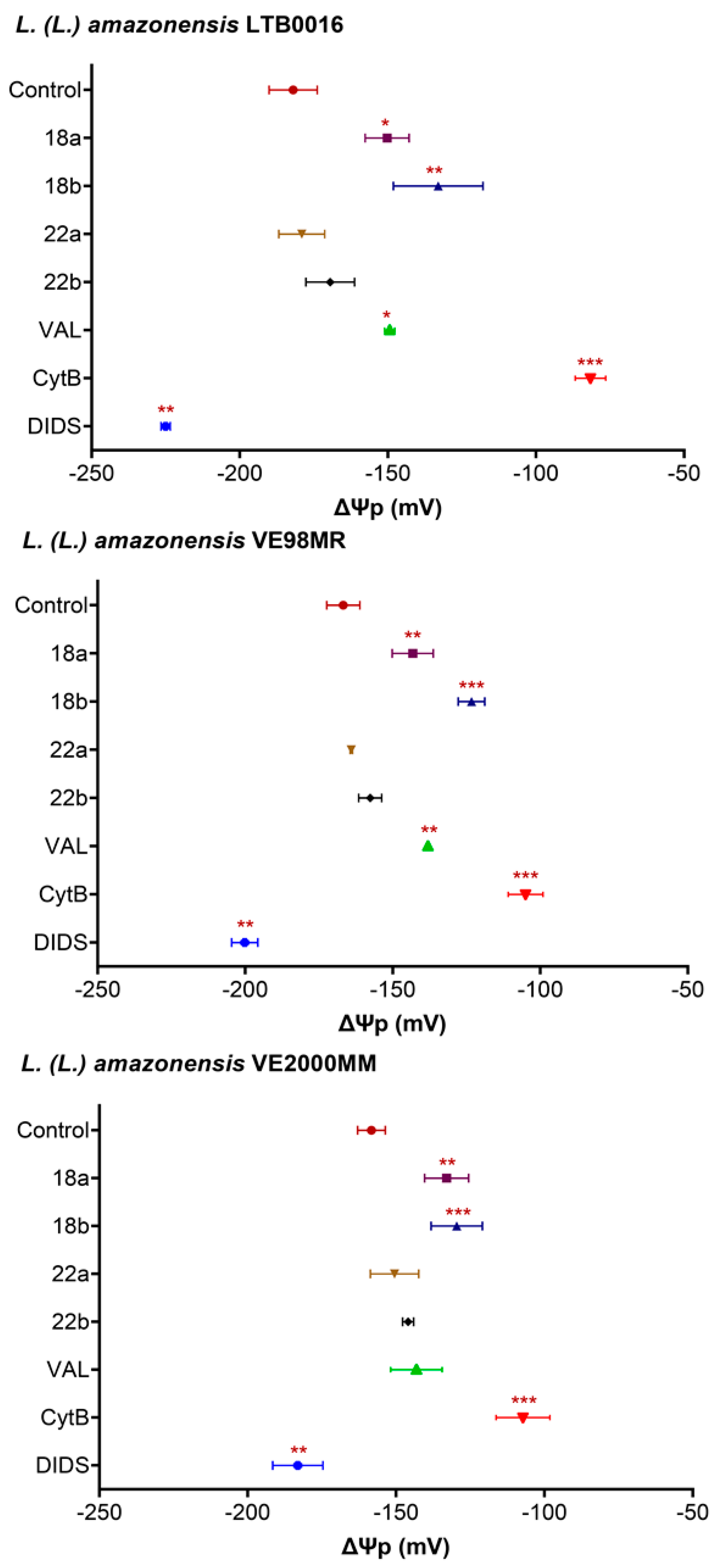
3.3.1. Plasma Membrane Potential (ΔΨp) in Promastigotes

To evaluate the effect of the most promising compounds on the plasma membrane potential (ΔΨp), promastigotes were exposed during 15 min to 18 and 22. Selected concentrations were their GI50 and twice GI50 (18, 1 and 2 µM; 22, 2 and 4 µM). Untreated cells, as well as cells treated with depolarizing molecules (valinomycin, 1 µM; cytochalasin B, 300 µM) and one hyperpolarizing compound (DIDS, 500 µM) were included. As the depolarizing probe, the lipophilic anion bisoxonol [100 nM] was used.
The resting ΔΨp of parasites was then established (LTB0016: −181.97 ± 19 mV; VE98MR = −166.75 ± 11 mV and VE2000MM = −158.30 ± 8 mV). The isolates presented more depolarized resting membrane potentials than the reference strain (see Supplementary Materials III for statistics).
Figure 1 demonstrates that only 18 was able to significantly depolarize the ΔΨp in all evaluated strains when compared to untreated controls (p < 0.05). The effect was similar to that depicted by valinomycin and concentration dependent, except in the case of VE2000MM. This strain was not sensitive to valinomycin.

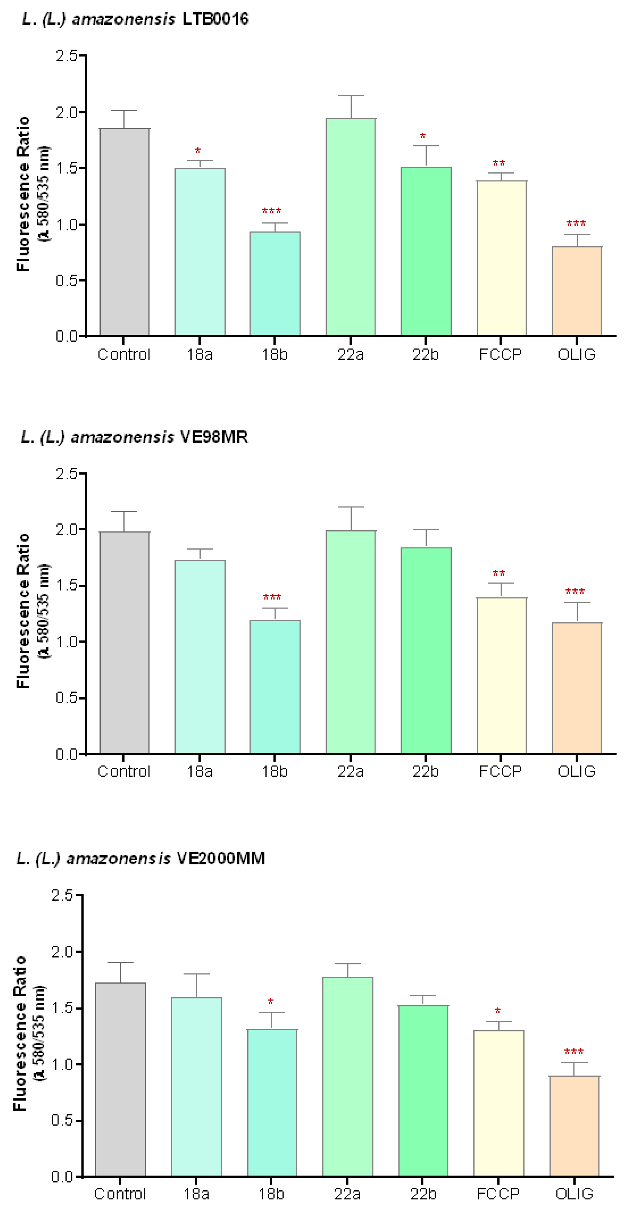
3.3.2. Mitochondrial Membrane Potential (ΔΨm) in Promastigotes

To evaluate if the effect of the betulin derivatives on parasites was related to their action on mitochondria, or related to programmed cell death, their effect on the mitochondrial membrane potential (ΔΨm) was evaluated. Promastigotes were exposed to two concentrations of the compounds, equivalent to their GI50 and twice GI50 (as before, 18: 1 and 2 µM; 22: 2 and 4 µM), during 48 h. As controls, untreated cells, as well as cells treated with depolarizing compounds (FCCP 1 µM and oligomycin 20 µM) were included. As the label for ΔΨm, the lipophilic cationic probe JC-1 was used.
Figure 2 demonstrates how 18 depolarizes ΔΨm in a concentration-dependent way when compared to untreated cells (p < 0.05) in all studied cells. The effect of 18 at its higher concentration (2 µM) in LTB0016 and VE98MR was comparable to that of oligomycin 20 µM, a compound that quickly inhibits the mitochondrial ATPase and ATP synthesis. 22 also inhibited ΔΨm, and a significant difference (p = 0.025) was only present at the highest concentration used (4 µM) and for LTB0016 (see Supplementary Materials III for statistical details).

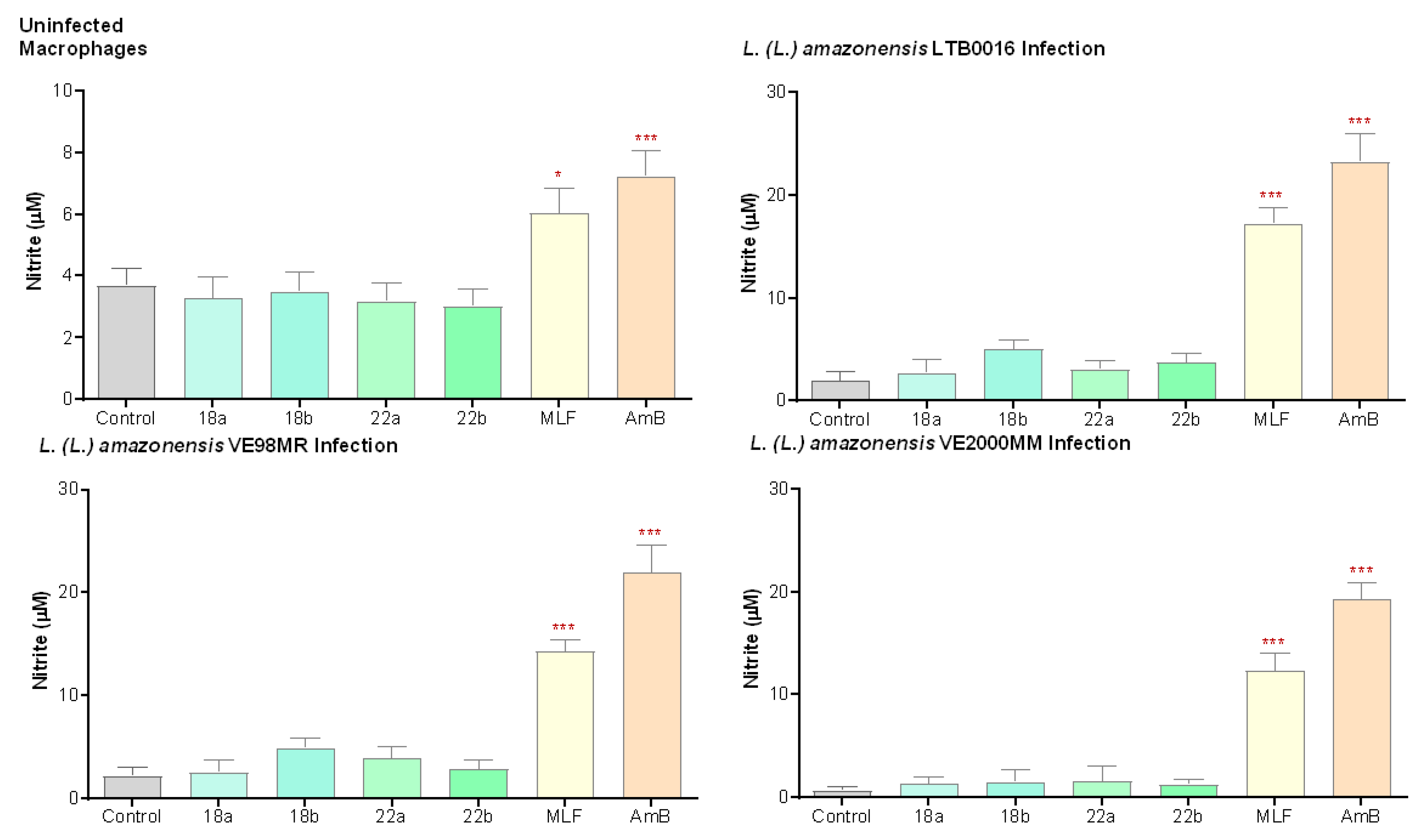
3.3.3. Nitric Oxide Production (NO) in Macrophages
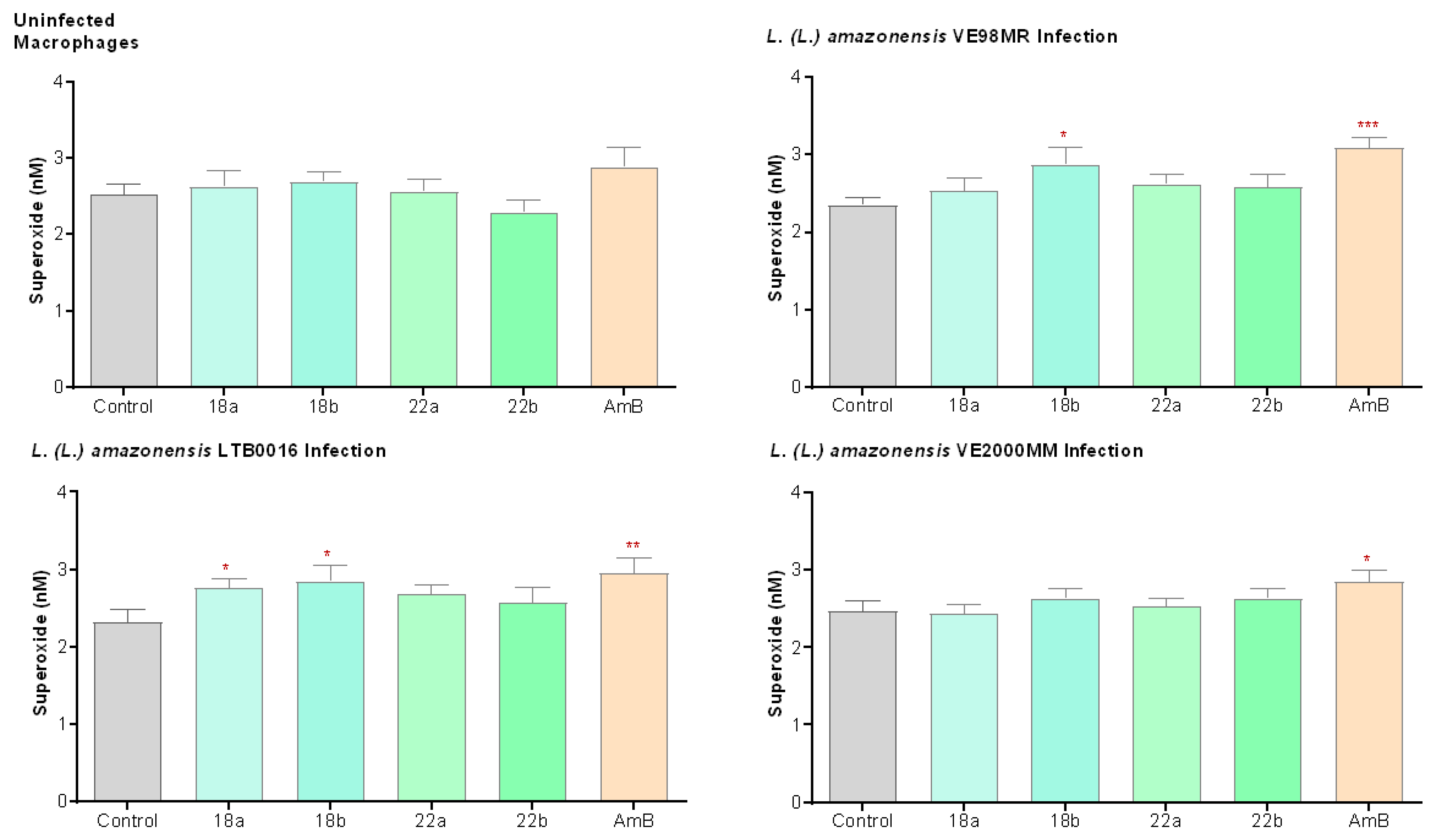
To evaluate if the anti-leishmanial activity of compounds 18 and 22 is related to modulating effects on the microbicidal activity of macrophages, non-infected and infected cultured macrophages were exposed to two concentrations of the compounds, promastigote GI50 and twice GI50 (18: 1 and 2 µM; 22: 2 and 4 µM). The NO production was indirectly determined through the evaluation of nitrite (NO2) levels by the Griess reaction. Compounds 18 and 22 were not capable of inducing NO production in any of the strains tested (Figure 3). Supplementary Materials III details the associated statistics.

3.3.4. Superoxide Production (O2) in Macrophages

Additionally, the O2 was evaluated in healthy macrophages, as well as in macrophages infected either with the reference strain LTB0016 or with the patient isolates VE98MR or VE2000MM and treated for 48 h with 18 and 22 (18: 1 and 2 µM; 22: 2 and 4 µM; respectively). Cells were further incubated for 1 h in the presence of cytochrome C-III (CYT-CIII 160 µM). By using the method of cytochrome reduction and the molar extinction coefficient of CYT-CIII, superoxide production, expressed in nanomoles of O2 released by 2 × 10⁵ cells, was determined. In healthy macrophages, betulin derivatives did not induce superoxide production. However, in LTB0016- and VE98MR-infected macrophages, the betulin derivative 18 induced superoxide production (Figure 4). Supplementary Materials III details the associated statistics.

4. Discussion

Pentacyclic triterpenes like betulin and lup-20(29)-ene-3ß,28-diol, are isolated from birch outer bark (Betula spp. Betulaceae) with a yield around 30% of dry matter [51]. Derivatives from the original active betulin compound can be chemically modified at the hydroxyl group C-3, the double bond C-20, and the primary hydroxyl C-28. Betulin derivatives with anti-leishmanial properties have been described previously [27,28,29,30].
Although plant extracts and compounds against L. (L.) amazonensis [52,53,54,55] have been thoroughly evaluated, the betulin derivatives herein described have been tested against New World Leishmania spp. but not against patient isolates with therapeutic failure. In the present work, two L. (L.) amazonensis Venezuelan isolates, obtained from patients with therapeutic failure against SbV, were used to characterize the activity of betulin derivatives.
Here, 28 semisynthetic betulin derivatives selected according to Alakurtti et al. [29,55] were tested at 50 µM. Eight compounds showed an anti-leishmanial effect (1, 17, 18, 22, 24, 25, 26, and 27), inhibiting L. (L.) amazonensis growth by at least 75%. Previously, we demonstrated that derivatives 14, 17, 18, 25, 26, and 27 were active at less than 10 µM in L. (L.) braziliensis, reference strain MHOM/BR/LTB300, and that 18, 26, and 27 inhibited parasite migration at nano and picomolar concentrations, suggesting a chemotactic effect of betulin derivatives that might be vital for host–parasite interaction [28].
The corresponding promastigote GI50 against the reference compounds (AmB, PNT, SbIII, MLF) were defined. As pentavalent antimonials (SbV) constitute pro-drugs that need biological reduction to their trivalent form (SbIII) to be leishmanicidal [56], we used the latter as potassium antimonyl tartrate trihydrate instead of the pentavalent form, meglumine antimoniate.
Promastigotes of Venezuelan isolates herein analyzed were less susceptible to SbIII (VE98MR, GI50 = 9.08 µM; VE2000MM, GI50 = 13.60 µM) than the reference strain (LTB0016 GI50 = 5.51 µM). Additionally, VE2000MM was slightly less susceptible to MLF (GI50 = 5.13 µM) than the reference strain (LTB0016 GI50 = 4.17 µM). These results suggest that VE98MR, and specifically VE2000MM, might be parasites refractive to conventional treatments, thus being suitable isolates for in vitro evaluation of new anti-leishmanial compounds.
Data not detailed in this manuscript suggest that the isolates’ susceptibility against NaNO2 was such that GI50 was four-fold higher (VE98MR, GI50 = 3.28 µM; VE2000MM, GI50 = 4.79 µM) than the reference strain (LTB0016, GI50 = 0.91 µM), thus suggesting that the Leishmania isolates, obtained from patients suffering therapeutic failure against meglumine antimoniate, can be classified as chemoresistant as has been described for antimonial-resistant L. (L.) infantum isolated from Brazilian patients with therapeutic failure [57]. Additionally, the GI50 against K2Cr2O7 (>2.5 mM) for both isolates was significantly higher than that obtained for the reference strain (LTB0016 GI50 = 1.06 mM); similar results have been published for SbV-resistant L. (L.) donovani tolerant to K2Cr2O7 [58].
As compounds 18 and 22 [25,59] were classified as effective and relatively secure (GI50 ≤ 2 µM, cytotoxic CC50 > 45 µM, SI > 20), they were furthered to explore their mechanism of action.
In the amastigote-macrophage model, the isolates (GI50: VE98MR = 1.76 µM, VE2000MM = 0.72 μM) were less susceptible to 18 than the reference strain LTB0016 (GI50: = 0.20 µM). Similar results were obtained for 22; the less susceptible isolate, VE2000MM, demonstrated a GI50 of 1.69 µM, and the reference strain of 0.21 µM. VE98MR and VE2000MM might be less susceptible due to potential cross-resistance mechanisms between SbV and betulin derivatives as more than 3-fold doses were needed to reach similar effects in isolates vs. the reference strain.
These concentrations did not affect macrophages’ viability (SI > 20), suggesting that 18 and 22 are worthy of being analyzed as potential candidates against parasites refractory to conventional treatment, as has been tested against L. (L.) donovani susceptible to SbV or selected as resistant to the same compound [60], as well as against Plasmodium falciparum (clone W2), resistant to chloroquine [61] for which betulin derivatives like O-disuccinoyl betulin, betulonic acid, and other betulinic acid derivatives have proved to be effective.
Our results suggest that GI50 values for 18 in L. (L.) amazonensis LTB0016 (GI50 promastigote = 0.90 µM; GI50 amastigote = 0.20 µM) were lower than those described for the same compound in L. (L.) donovani axenic amastigotes (GI50 = 22.80 µM) [29]. Differences may respond to species-specific internal factors, with L. (L.) donovani being intrinsically less susceptible to 18 than L. (L.) amazonensis. Alternatively, the used parasitic stage, axenic amastigotes vs. intracellular amastigotes, may be influencing the obtained results.
Our data suggest that the carboxyl group at position C28 is not indispensable for betulin anti-leishmanial activity, as suggested for L. (L.) major [62]. For example, 11 (betulinic acid), at 50 µM, inhibited L. (L.) amazonensis LTB0016 growth by 44%; other carboxyl compounds, (13 and 14) did not show enough anti-leishmanial activity.
In L. (V.) braziliensis promastigotes, the most potent compound was 26 (3,28-diformylbetulin triazolodione) with a GI50 < 1 µM [29]. Likewise, a betulin aldehyde derivative reduced infection up to 88% at 136 µM and 58% at 68 µM of intracellular L. (L.) amazonensis amastigotes [63]. Herein, 26 was also within the initially selected compounds. However, as previously mentioned, only 18 and 22 showed significant differences that made them worthy of further analysis. These results support our data regarding the moderate activity identified for betulinic aldehyde (12) that at 50 µM inhibited L. (L.) amazonensis growth by 67% and suggest that there might be species variances that could explain the differences herein found when compared to previous data in L. (V.) braziliensis.
Compound 22 is a heterocyclic adduct between 3,28-di-O-acetyllupa-12,18-diene and 4-ethylurazine, while the structurally related 25 is another heterocyclic adduct between 3,28-di-O-acetyllupa-12,18-diene and 4-methylurazine [55]. Compound 25, although cytotoxic at all evaluated concentrations, is an effective derivative with a GI50 of 8.9 µM, in L. (L.) donovani axenic amastigotes. On the other hand, 22, was slightly toxic for J774.1 macrophages (CC50 = 61.13 µM) with minimal GI50 against L. (L.) amazonensis intracellular amastigotes = 0.21 µM), and very high selectivity index (SI = 291). Heterocyclic derivatives bulkier in C3, C28, or N4 were less active.
The anti-leishmanial activity of betulin derivatives associates in Old World Leishmania with inhibitory effects over enzymes fundamental for parasite survival, such as topoisomerase IB [64] and trypanothione synthetase, TryS [65] as well as on the promoted parasite oxidative stress that ends up in programmed cell death [66]. However, specific targets and molecular mechanisms involved in their action still are not completely understood [67], and for New World species are not known at all. Herein, we explored their mechanism of action.
In organisms like protozoa, bacteria, pathogenic fungi, and tumor cells, betulin derivatives’ target seems to be located at the plasma membrane [68]. Examples also exist in Leishmania that illustrate the effect of compounds on the membrane, specifically affecting the ΔΨp, i.e., AmB in L. (L.) infantum [69], clomipramine and imipramine in L. (L.) donovani and L. (L.) major [70] or peptides like cecropin-A and melittin in L. (L.) donovani [71], or lupeol, an anti-leishmanial triterpene from Sterculia villosa for L. (L.) infantum [72], and L. (L.) donovani [73]. In this line of evidence, we report that betulin derivative 18, induced, in a very short time (15 min), the collapse of the plasma membrane potential (ΔΨp) in a concentration-dependent way. The effect was comparable to that imposed by valinomycin (1 µM) and cytochalasin B (300 µM).
On the other hand, betulin 1 and betulin derivatives induce programmed cell death in L. (L.) donovani promastigotes [66] and the betulin derivative O-disuccinoyl betulin induces non-dependent caspase apoptosis [64]. The effect of 1 in L. (L.) donovani correlates with the inhibition of TryS [65], resulting in intracellular redox homeostasis loss [66].
Besides that, medicinal plants and their secondary metabolites may have immunomodulatory properties [74] [superoxide (O2) and nitric oxide (NO) production], potentially useful against leishmaniasis [75]. L. major amastigote elimination depends preferentially on NO production [75] and L. (V.) braziliensis abolition seems to strictly depend on both O2 and ROS generation [76]. Of note, in macrophages infected with L. (L.) donovani, lupeol induced NO production and increased the production of proinflammatory cytokines, decreasing the production of anti-inflammatory cytokines [73].
Nitric oxide is essential for macrophage activation and is produced in Leishmania-infected macrophages [77,78]; additionally, metacyclic L. amazonensis promastigotes produce NO and axenic amastigotes produce NO levels 10-fold higher than those produced by metacyclic promastigotes as has been described by Acuña and collaborators (2017) [79] and references within. However, similar to what we herein described in both the reference strain and patient isolates and in L. amazonensis-infected macrophages, there is a lower production of NO than in un-infected macrophages. L. amazonensis seems to have an adaptive strategy to escape from host defense. This strategy seems to be produced by the activation of the NF-κB repressor complex p50/p50, a specific host transcriptional response that negatively regulates the expression of inducible nitric oxide synthase. This feature seems to favor the establishment and success of L. amazonensis infection [80].
In our hands, 18 and 22 were not capable of inducing NO production for any of the strains tested. However, compound 18 seems to influence the oxidative response of J774.1 macrophages infected with L. (L.) amazonensis LTB0016 and VE98MR, inducing an oxidative burst and thus increasing O2 production.
Considering the structural similarity between lupeol and betulin derivatives, both terpenoids may be addressing the same cellular target. Molecular docking analysis [73] suggests that these compounds may be capable of attachment to the parasite surface protein GP63, thus affecting the cells’ signaling processes that lead to macrophage activation [81]. On the other hand, an in silico analysis of a 58-member library of betulin derivatives (including 18) identified the membrane transport protein D1 as a potential target for this compound in L. (L.) donovani [67].
Finally, whether molecular functional events associated with the production of non-caspase-dependent apoptosis in L. (L.) donovani [64] and produced by O-disuccinoyl betulin [66] or inhibition of TryS [65]) are affected by betulin derivatives like compound 18 in L. (L.) amazonensis has to be further analyzed.
Altogether, our results suggest that diverse targets might be addressed by 18 in L. (L.) amazonensis. Therefore, further experiments are needed to fully elucidate the final mechanism of action of this compound.

Supplementary Materials

The following are available online at https://www.mdpi.com/2076-2607/9/2/320/s1.

Author Contributions

Conceptualization, A.P.-S., S.A. and J.Y.-K.; methodology, A.P.-S., W.A., M.P.-N., N.R., S.A., M.L.T. and J.Y.-K.; software, A.P.-S. and W.A.; validation, W.A.; formal analysis, W.A.; investigation, W.A., N.R., S.A. and M.L.T.; resources, M.P.-N., N.R. and J.Y-K.; data curation, W.A. and A.P.-S.; writing—original draft preparation, A.P.-S.; writing—review and editing, A.P.-S., W.A., M.P.-N., N.R., S.A., M.L.T. and J.Y.-K.; visualization, A.P.-S., W.A. and M.P.-N.; supervision, A.P.-S., S.A. and J.Y.-K.; project administration, M.P.-N., A.P.-S. and J.Y.-K.; funding acquisition, A.P.-S., M.P.-N. and J.Y.-K. All authors have read and agreed to the published version of the manuscript.

Funding

This research was funded by the Universidad Central de Venezuela Council for Research, grants CDCH-UCV PI-09-8717-2013/1 (M.P.-N.) and PG-09-8646-2013/1 (A.P.-S.). This study also was supported by the Foundation for Research of Natural Resources in Finland, Marjatta ja Eino Kollin Säätiö, the Academy of Finland (Projects 252308, 264020, 265481) and the COST Action CM-0801 (New drugs for neglected diseases). Finally, A.P.-S. is grateful for the support conferred by the Alexander von Humboldt Foundation and the Siebold-Collegium Institute for Advanced Studies, University of Würzburg, Germany.

Institutional Review Board Statement

The study (CDCH UCV PI-09-8717-2013) was conducted according to the guidelines of the Declaration of Helsinki and approved by the Institutional review Board from the Institute of Biomedicine (SAIB-MPPS-UCV), Caracas, Venezuela.

Informed Consent Statement

Informed consent was obtained from all subjects involved in the study.

Data Availability Statement

Not applicable.

Acknowledgments

The authors are grateful to Anja Salakari and Irene Kylänlahti for their excellent technical assistance as well as late Salme Koskimies for valuable discussions. Emilia Díaz is thanked for fruitful discussions related to the development of the project and Pilar Rodríguez for her technical assistance.

Conflicts of Interest

The authors declare no conflict of interest. The funders had no role in the design of the study; in the collection, analyses, or interpretation of data; in the writing of the manuscript, or in the decision to publish the results.

References

  1. Savoia, D. Recent updates and perspectives on leishmaniasis. J. Infect. Dev. Ctries 2015, 9, 588–596. [Google Scholar] [CrossRef] [PubMed] [Green Version]
  2. Makala, L.H.C.; Baban, B. Novel therapeutic approaches to Leishmania infection. In Leishmaniasis—Trends in Epidemiology, Diagnosis and Treatment; Claborn, D.M., Ed.; InTechOpen: London, UK, 2014; pp. 495–523. Available online: https://www.intechopen.com/books/-novel-therapeutic-approaches-to-leishmania-infection (accessed on 11 October 2020).
  3. Sundar, S.; Chakravarty, J. Visceral leishmaniasis. In Drug Resistance in Leishmania Parasites: Consequences, Molecular Mechanisms and Possible Treatments, 2nd ed.; Ponte-Sucre, A., Padrón-Nieves, M., Eds.; Springer Nature: Cham, Switzerland, 2018; pp. 159–176. [Google Scholar]
  4. World Health Organization. Investing to Overcome the Global Impact of Neglected Tropical Diseases; Third Who Report on Neglected Tropical Diseases; WHO: Geneva, Switzerland, 2015; pp. 118–126. [Google Scholar]
  5. Guevara, J.R. MPPS. Estadísticas de Leishmaniasis Visceral en Venezuela Histórico/Año 1990–2018; Servicio Autónomo Instituto de Biomedicina “Dr. Jacinto Convit”: Caracas, Venezuela, 2018. [Google Scholar]
  6. Zerpa, O.; Ulrich, M.; Borges, R.; Rodrìguez, V.; Centeno, M.; Negrón, E.; Belizario, D.; Convit, J. Epidemiological aspects of human and canine visceral leishmaniasis in Venezuela. Rev. Panam Salud Publica 2003, 13, 239–245. [Google Scholar] [CrossRef]
  7. Rodríguez, N.M.; De Guglielmo, Z.; Barrios, M.A.; Barrios, R.M.; Zerpa, O.; Feliciangeli, M.D. Nueva Esparta Genetic homogeneity within Leishmania (L.) infantum isolated from human and dogs: The relationship with the sandfly fauna distribution in endemic areas of Nueva Esparta State, Venezuela. Parasitology 2005, 130, 611–619. [Google Scholar] [CrossRef]
  8. Hernández, D.; Rojas, E.; Scorza, J.V.; Jorguera, A. Infectividad del perro (Canis familiaris) para Lutzomyia youngi en Trujillo, Venezuela. Biomédica 2006, 26, 242–248. [Google Scholar] [CrossRef]
  9. Guevara, J.R. MPPS. Estadísticas de Leishmaniasis Cutánea en Venezuela Histórico/Año 1990–2018; Servicio Autónomo Instituto de Biomedicina “Dr. Jacinto Convit”: Caracas, Venezuela, 2018. [Google Scholar]
  10. Peña, I.; Manzano, P.; Cantizani, J.; Kessler, A.; Alonso-Padilla, J.; Bardera, A.I.; Alvarez, E.; Colmenarejo, G.; Cotillo, I.; Roquero, I. New compound sets identified from high throughput phenotypic screening against three kinetoplastid parasites: An open resource. Sci. Rep. 2015, 5, 1–47. [Google Scholar] [CrossRef] [Green Version]
  11. De Vries, H.J.C.; Reedijk, S.H.; Schallig, H.D.F.H. Cutaneous leishmaniasis: Recent developments in diagnosis and management. Am. J. Clin. Dermatol. 2015, 16, 99–109. [Google Scholar] [CrossRef] [Green Version]
  12. Reveiz, L.; Maia-Elkhoury, A.N.S.; Nicholls, R.S.; Sierra Romero, G.A.; Yadon, Z.E. Interventions for American cutaneous and mucocutaneous leishmaniasis: A systematic review update. PLoS ONE 2013, 8, e61843. [Google Scholar] [CrossRef] [Green Version]
  13. De Menezes, J.P.B.; Guedes, C.E.S.; Petersen, A.L.D.O.A.; Fraga, D.B.M.; Veras, P.S.T. Advances in development of new treatment for leishmaniasis. BioMed Res. Int. 2015, 2015, 815023. [Google Scholar] [CrossRef]
  14. Joshi, S.; Rawat, K.; Yadav, N.K.; Kumar, V.; Siddiqi, M.I.; Dube, A. Visceral leishmaniasis: Advancements in vaccine development via classical and molecular approaches. Front. Immunol. 2014, 5, 1–18. [Google Scholar] [CrossRef] [Green Version]
  15. Singh, N.; Kumar, M.; Singh, R.K. Leishmaniasis: Current status of available drugs and new potential drug targets. Asian Pac. J. Trop. Med. 2012, 5, 485–497. [Google Scholar] [CrossRef] [Green Version]
  16. Ponte-Sucre, A.; Diaz, E. Leishmaniasis: The Biology of a Parasite. In Drug Resistance in Leishmania Parasites: Consequences, Molecular Mechanisms and Possible Treatments, 2nd ed.; Ponte-Sucre, A., Padrón-Nieves, M., Eds.; Springer Nature: Cham, Switzerland, 2018; pp. 1–16. [Google Scholar]
  17. Gupta, S.K.; Dandekar, T. Bioinformatics in Leishmania Drug Design. In Drug Resistance in Leishmania Parasites: Consequences, Molecular Mechanisms and Possible Treatments, 2nd ed.; Ponte-Sucre, A., Padrón-Nieves, M., Eds.; Springer Nature: Cham, Switzerland, 2018; pp. 297–317. [Google Scholar]
  18. Vermelho, A.B.; Supuran, C.T.; Cardoso, V.; Menezes, D.; De Andrade Silva, J.R.; Pinto Ferreira, J.L.; Fernandez Amaral, A.C.; Rodrigues, I.A. Leishmaniasis: Possible new strategies for treatment. In Leishmaniasis—Trends in Epidemiology, Diagnosis and Treatment; Claborn, D.M., Ed.; InTechOpen: London, UK, 2014; pp. 351–376. Available online: https://www.intechopen.com/books/leishmaniasis-trends-in-epidemiology-diagnosis-and-treatment/leishmaniasis-possible-new-strategies-for-treatment (accessed on 11 October 2020).
  19. Gilbert, I.H. Drug discovery for neglected diseases: Molecular target-based and phenotypic approaches. J. Med. Chem. 2013, 56, 7719–7726. [Google Scholar] [CrossRef]
  20. Spinks, D.; Ong, H.B.; Mpamhanga, C.P.; Shanks, E.J.; Robinson, D.A.; Collie, I.T.; Read, K.D.; Fearson, J.A.; Wyatt, P.G.; Brenk, R.; et al. Design, synthesis and biological evaluation of novel inhibitors of Trypanosoma brucei pteridine reductase 1. Chem. Med. Chem. 2011, 6, 302–308. [Google Scholar] [CrossRef] [Green Version]
  21. Prada, C.F.; Alvarez-Velilla, R.; Balaña-Fouce, R.; Prieto-Sánchez, R.; Calvo-Álvarez, C.; Escudero-Martínez, E.; Requena, J.M.; Ordoñez, J.M.; Desideri, C.; Pérez-Pertejo, A.; et al. Gimatecan and other camptothecin derivatives poison Leishmania DNA-topoisomerase IB leading to a strong leishmanicidal effect. Biochem. Pharmacol. 2013, 85, 1433–1440. [Google Scholar] [CrossRef] [Green Version]
  22. Romero, A.H.; López, S.E.; Rodríguez, N.M.; Oviedo, H. Anti-leishmanial activity, structure–activity relationship of series of 2-(trifluoromethyl)benzo[b][1,8]naphthyridin-4(1H)-ones. Arch. Pharm. 2018, 351, 1–9. [Google Scholar] [CrossRef]
  23. Rodríguez, M.; Gutiérrez, J.; Domínguez, J.; Pixoto, P.A.; Fernández, A.; Rodríguez, N.M.; Deffieux, D.; Rojas, L.B.; Quideau, S.; Pouysegu, L.; et al. Synthesis and leishmanicidal evaluation of sulfanyl and sulfonyl-tethered functionalized benzoate derivatives featuring a nitroimidazole moiety. Arch. Pharm. 2020, 353, e2000002. [Google Scholar] [CrossRef]
  24. Brito, A.M.; Santos, D.; Rodrigues, S.; Brito, R.G.; Xavier-Filho, L. Plants with anti-Leishmania activity: Integrative review from 2000 to 2011. Pharmacogn. Rev. 2013, 7, 34. [Google Scholar] [CrossRef] [PubMed] [Green Version]
  25. Basso, L.A.; Pereira Da Silva, L.H.; Fett-Neto, A.G.; De Azevedo, W.F., Jr.; De Souza Moreira, I.; Palma, M.S.; Calixto, J.B.; Filho, S.A.; Ribero Dos Santos, R.; Pereira Soares, M.B.; et al. The use of biodiversity as source of new chemical entities against defined molecular targets for treatment of malaria, tuberculosis, and T-cell mediated diseases—A review. Mem. Inst. Oswaldo Cruz 2005, 100, 475–506. [Google Scholar] [CrossRef] [Green Version]
  26. Krasutsky, P. Birch bark research and development. Nat. Prod. Rep. 2006, 23, 919–942. [Google Scholar] [CrossRef]
  27. Sousa, M.C.; Varandas, R.; Santos, R.C.; Santos-Rosa, M.; Alves, V.; Salvador, J.A.R. Anti-leishmanial activity of semisynthetic lupane triterpenoids betulin and betulinic acid derivatives: Synergistic effects with miltefosine. PLoS ONE 2014, 9, e89939. [Google Scholar] [CrossRef] [Green Version]
  28. Alcazar, W.; López, A.S.; Alakurtti, S.; Tuononen, M.L.; Yli-Kauhaluoma, J.; Ponte-Sucre, A. Betulin derivatives impair Leishmania braziliensis viability and host–parasite interaction. Bioorg. Med. Chem. 2014, 22, 6220–6226. [Google Scholar] [CrossRef]
  29. Alakurtti, S.; Bergström, P.; Sacerdoti-Sierra, N.; Jaffe, C.L.; Yli-Kauhaluoma, J. Anti-leishmanial activity of betulin derivatives. J. Antibiot. 2010, 63, 123–126. [Google Scholar] [CrossRef] [Green Version]
  30. Wert, L.; Alakurtti, S.; Corral, M.J.; Sánchez-Fortún, S.; Yli-Kauhaluoma, J.; Alunda, J.M. Toxicity of betulin derivatives and in vitro effect on promastigotes and amastigotes of Leishmania infantum and L. donovani. J. Antibiot. 2011, 64, 475–481. [Google Scholar] [CrossRef] [PubMed] [Green Version]
  31. Rodríguez, N.M.; Guzmán, B.; Rodas, A.; Takiff, H.; Bloom, B.R.; Convit, J. Diagnoses of cutaneous leishmaniasis and species discrimination of parasites by PCR and hybridization. J. Clin. Microbiol. 1994, 32, 2246–2250. [Google Scholar] [CrossRef] [PubMed] [Green Version]
  32. Howard, M.; Pharoah, M.; Ashall, F.; Miles, M.A. Human urine stimulates growth of Leishmania in vitro. Trans. R. Soc. Trop. Med. Hyg. 1991, 85, 477–479. [Google Scholar] [CrossRef]
  33. Allahverdiyev, A.M.; Bagirova, M.; Elcicek, S.; Cakir Koc, R.; Oztel, O.N. Effect of human urine on cell cycle and infectivity of Leismania species promastigotes in vitro. Am. J. Trop. Med. Hyg. 2011, 85, 639–643. [Google Scholar] [CrossRef]
  34. Ralph, P.; Nakoinz, I. Phagocytosis and cytolysis by a macrophage tumour and its cloned cell line. Nature 1975, 257, 393–394. [Google Scholar] [CrossRef]
  35. Le Pape, P.; Pagniez, F.; Abdala, H. A new automatized fluorometric assay for anti-Leishmania drug screening. Acta Parasitol. 2002, 47, 79–81. [Google Scholar]
  36. Tong, F.P. Statistical Methods for Dose-Response Assays; University of California: Berkeley, CA, USA, 2010; Available online: http://escholarship.org/uc/item/9295t422# (accessed on 11 October 2020).
  37. Borborema, S.E.T.; Schwendener, R.A.; Osso, J.A.; De Andrade, H.F., Jr.; Do Nascimento, N. Uptake and anti-leishmanial activity of meglumine antimoniate-containing liposomes in Leishmania major infected macrophages. Int. J. Antimicrob. Agents 2011, 38, 341–347. [Google Scholar] [CrossRef]
  38. Nwaka, S.; Hudson, A. Innovative lead discovery strategies for tropical diseases. Nat. Rev. Drug Discov. 2006, 5, 941–955. [Google Scholar] [CrossRef]
  39. Neal, R.A.; Croft, S.L. An in-vitro system for determining the activity of compounds against the intracellular amastigote form of Leishmania donovani. J. Antimicrob. Chemother. 1984, 14, 463–475. [Google Scholar] [CrossRef]
  40. Da Luz, R.I.; Vermeersch, M.; Dujardin, J.C.; Cos, P.; Maes, L. In vitro sensitivity testing of Leishmania clinical field isolates: Preconditioning of promastigotes enhances infectivity for macrophage host cells. Antimicrob. Agents Chemother. 2009, 53, 5197–5203. [Google Scholar] [CrossRef] [Green Version]
  41. Lira, R.; Sundar, S.; Makharia, A.; Kenney, R.; Gam, A.; Saraiva, E.; Sacks, D. Evidence that the high incidence of treatment failures in Indian kala-azar is due to the emergence of antimony-resistant strains of Leishmania donovani. J. Infect. Dis. 1999, 180, 564–567. [Google Scholar] [CrossRef] [Green Version]
  42. Vieira, L.; Slotki, I.; Cabantchik, Z.I. Chloride conductive pathways which support electrogenic H+ pumping by Leishmania major promastigotes. J. Biol. Chem. 1995, 270, 5299–5304. [Google Scholar] [CrossRef] [Green Version]
  43. Padrón-Nieves, M.; Machuca, C.; Díaz, E.; Cotrim, P.; Rodríguez, N.M.; Ponte-Sucre, A. Correlation between glucose uptake and membrane potential in Leishmania parasites isolated from DCL patients with therapeutic failure: A proof of concept. Parasitol. Res. 2014, 113, 2121–2128. [Google Scholar] [CrossRef] [PubMed]
  44. Padrón-Nieves, M.; Ponte-Sucre, A. Cellular Markers for the Identification of Chemo-Resistant Isolates in Leishmania. In Methods in Molecular Biology. Trypanosomatids Methods and Protocols; Michels, P., Ginger, M., Zilberstein, D., Eds.; Springer Nature: Cham, Switzerland; Humana Press: New York, NY, USA, 2019; Chapter 46; pp. 755–769. [Google Scholar]
  45. De Macedo-Silva, S.T.; De Oliveira Silva, T.L.A.; Urbina, J.A.; Souza, W.D.; Rodrigues, J.C.F. Antiproliferative, ultrastructural, and physiological effects of amiodarone on promastigote and amastigote forms of Leishmania amazonensis. Mol. Biol. Int. 2011, 2011, 876021. [Google Scholar] [CrossRef] [Green Version]
  46. Ponte-Sucre, A.; Mendoza-León, A.; Moll, H. Experimental leishmaniasis: Synergistic effect of ion channel blockers and interferon-gamma on the clearance of Leishmania major by macrophages. Parasitol. Res. 2001, 87, 27–31. [Google Scholar] [CrossRef]
  47. Ponte-Sucre, A.; Vicik, R.; Schultheis, M.; Schirmeister, T.; Moll, H. Aziridine-2,3-dicarboxylates, peptidomimetic cysteine protease inhibitors with anti-leishmanial activity. Antimicrob. Agents Chemother. 2006, 50, 2439–2447. [Google Scholar] [CrossRef] [Green Version]
  48. Granger, D.L. Measurement of nitrate and nitrite in biological samples using nitrate reductase and Griess reaction. Methods Enzymol. 1996, 268, 142–151. [Google Scholar]
  49. Decuypere, S.; Vanaerschot, M.; Brunker, K.; Imamura, H.; Müller, S.; Khanal, B.; Rijal, S.; Dujardin, J.C.; Coombs, G.H. Molecular mechanisms of drug resistance in natural Leishmania populations vary with genetic background. PLoS Negl. Trop. Dis. 2012, 6, e1514. [Google Scholar] [CrossRef] [PubMed]
  50. De Morais-Teixeira, E.; Damasceno, Q.S.; Galuppo, M.K.; Romanha, A.J.; Rabello, A. The in vitro leishmanicidal activity of hexadecylphosphocholine (miltefosine) against four medically relevant Leishmania species of Brazil. Mem. Inst. Oswaldo Cruz 2011, 106, 475–478. [Google Scholar] [CrossRef] [PubMed] [Green Version]
  51. García, M.; Monzote, L.; Montalvo, A.M.; Scull, R. Screening of medicinal plants against Leishmania amazonensis. Pharm. Biol. 2010, 48, 1053–1058. [Google Scholar] [CrossRef]
  52. García, M.C.F.; Soares, D.C.; Santana, R.C.; Saraiva, E.M.; Siani, A.C.; Ramos, M.F.S.; Danielli, M.G.M.; Souto-Padron, T.C.; Pinto da Silva, L.H. The in vitro anti-leishmanial activity of essential oil from Aloysia gratissima and guaiol, its major sesquiterpene against Leishmania amazonensis. Parasitology 2018, 145, 1219–1227. [Google Scholar] [CrossRef]
  53. Al-Sokari, S.S.; Ali, N.A.A.; Monzote, L.; Al-Fatimi, M.A. Evaluation of anti-leishmanial activity of albaha medicinal plants against Leishmania amazonensis. BioMed Res. Int. 2015, 2015, 1–6. [Google Scholar] [CrossRef] [Green Version]
  54. Souza-Fagundes, E.M.; Cota, B.B.; Rosa, L.H.; Romanha, A.J.; Corrêa-Oliveira, R.; Zani, C.L.; Texeira-Carvalho, A.; Martins-Filho, O.A. In vitro activity of hypnophilin from Lentinus strigosus: A potential prototype for Chagas disease and leishmaniasis chemotherapy. Braz. J. Med. Biol. Res. 2010, 43, 1054–1061. [Google Scholar] [CrossRef] [PubMed] [Green Version]
  55. Alakurtti, S.; Heiska, T.; Kiriazis, A.; Sacerdoti-Sierra, N.; Jaffe, C.L.; Yli-Kauhaluoma, J. Synthesis and anti-leishmanial activity of heterocyclic betulin derivatives. Bioorg. Med. Chem. 2010, 18, 1573–1582. [Google Scholar] [CrossRef] [PubMed]
  56. Haldar, A.K.; Sen, P.; Roy, S. Use of antimony in the treatment of leishmaniasis: Current status and future directions. Mol. Biol. Int. 2011, 2011, 1–23. [Google Scholar] [CrossRef] [PubMed] [Green Version]
  57. Holzmuller, P.; Sereno, D.; Lemesre, J.L. Lower nitric oxide susceptibility of trivalent antimony-resistant amastigotes of Leishmania infantum. Antimicrob. Agents Chemother. 2005, 49, 4406–4409. [Google Scholar] [CrossRef] [Green Version]
  58. Vanaerschot, M.; Maes, I.; Ouakad, M.; Adaui, I.; Maes, L.; De Doncker, S. Linking in vitro and in vivo survival of clinical Leishmania donovani strains. PLoS ONE 2010, 5, e12211. [Google Scholar] [CrossRef] [Green Version]
  59. Prayong, P.; Barusrux, S.; Weerapreeyakul, N. Cytotoxic activity screening of some indigenous Thai plants. Fitoterapia 2008, 79, 598–601. [Google Scholar] [CrossRef]
  60. Chowdhury, S.; Mukherjee, T.; Sengupta, S.; Chowdhury, S.R.; Mukhopadhyay, S.; Majumder, H. Novel betulin derivatives as anti-leishmanial agents with mode of action targeting type IB DNA topoisomerase. Mol. Pharmacol. 2011, 80, 694–703. [Google Scholar] [CrossRef] [Green Version]
  61. De Sá, M.S.; Costa, J.F.O.; Krettli, A.U.; Zalis, M.G.; de Azevedo, G.L.M.; Sette, I.V.M.F.; de Amorim, C.C.; Filho, J.M.B.; Giulietti-Harley, A.M.; Dos Santos, R.R.; et al. Antimalarial activity of betulinic acid and derivatives in vitro against Plasmodium falciparum and in vivo in P. berghei-infected mice. Parasitol. Res. 2009, 105, 275–279. [Google Scholar] [CrossRef]
  62. Takahashi, M.; Fuchino, H.; Sekita, S.; Satake, M. In vitro leishmanicidal activity of some scarce natural products. Phytother. Res. 2004, 18, 573–578. [Google Scholar] [CrossRef]
  63. Sauvain, M.; Kunesch, N.; Poisson, J.; Gantier, J.C.; Gayral, P.; Dedet, J.P. Isolation of leishmanicidal triterpenes and lignans from the amazonian liana Doliocarpus dentatus (Dilleniaceae). Phytother. Res. 2002, 10, 1–4. [Google Scholar] [CrossRef]
  64. Chowdhury, S.; Mukherjee, T.; Chowdhury, S.R.; Sengupta, S.; Mukhopadhyay, S.; Parasuraman, J.; Majumder, H.K. Disuccinyl betulin triggers metacaspase-dependent endonuclease G-mediated cell death in unicellular protozoan parasite Leishmania donovani. Antimicrob. Agents Chemother. 2014, 58, 2186–2201. [Google Scholar] [CrossRef] [PubMed] [Green Version]
  65. Saudagar, P.; Dubey, V.K. Cloning, expression, characterization and inhibition studies on trypanothione synthetase, a drug target enzyme, from Leishmania donovani. Biol. Chem. 2011, 392, 1113–1122. [Google Scholar] [CrossRef] [PubMed]
  66. Saudagar, P.; Dubey, V.K. Molecular mechanisms of in vitro betulin-induced apoptosis of Leishmania donovani. Am. J. Trop. Med. Hyg. 2014, 90, 354–360. [Google Scholar] [CrossRef]
  67. Zhang, Y.; Xhaard, H.; Ghemtio, L. Predictive classification models and targets identification for betulin derivatives as Leishmania donovani inhibitors. J. Cheminform. 2018, 10, 1–16. [Google Scholar] [CrossRef]
  68. Camargos, H.S.; Moreira, R.A.; Mendanha, S.A.; Fernandes, K.S.; Dorta, M.L.; Alonso, A. Terpenes increase the lipid dynamics in the Leishmania plasma membrane at concentrations similar to their IC50 values. PLoS ONE 2014, 9, e104429. [Google Scholar] [CrossRef]
  69. Azas, N.; Di Giorgio, C.; Delmas, F.; Gasquet, M.; Timon-David, P. Assessment of amphotericin B susceptibility in Leishmania infantum promastigotes by flow cytometric membrane potential assay. Cytometry 1997, 28, 165–169. [Google Scholar] [CrossRef]
  70. Zilberstein, D.; Dwyer, D.M. Antidepressants cause lethal disruption of membrane function in the human protozoan parasite Leishmania. Science 1984, 226, 977–979. [Google Scholar] [CrossRef]
  71. Díaz-Achirica, P.; Ubach, J.; Guinea, A.; Andreu, D.; Rivas, L. The plasma membrane of Leishmania donovani promastigotes is the main target for CA(1-8)M(1-18), a synthetic cecropin A-melittin hybrid peptide. Biochem. J. 1998, 330, 453–460. [Google Scholar] [CrossRef]
  72. Mazoir, N.; Benharref, A.; Bailén, M.; Reina, M.; González-Coloma, A.; Martínez-Díaz, R.A. Anti-leishmanial and antitrypanosomal activity of triterpene derivatives from latex of two Euphorbia species. Z. Naturforsch. 2011, 66, 360–366. [Google Scholar] [CrossRef] [PubMed] [Green Version]
  73. Das, A.; Jawed, J.J.; Das, M.C.; Sandhu, P.; De Utpal, C.; Dinda, B.; Akhter, Y.; Bhattacharjee, S. Anti-leishmanial and immunomodulatory activity of lupeol a triterpene compound isolated from Sterculia villosa. J. Antimicrob. Agents 2017, 50, 512–522. [Google Scholar] [CrossRef] [PubMed]
  74. Kaur, R.; Kaur, S. Evaluation of in vitro and in vivo anti-leishmanial potential of bergenin rich Bergenia ligulata (Wall.) Engl. root extract against visceral leishmaniasis in inbred BALB/c mice through immunomodulation. J. Tradit. Complement. Med. 2018, 8, 251–260. [Google Scholar] [CrossRef] [PubMed]
  75. Assreuy, J.; Cunha, F.Q.; Epperlein, M.; Noronha-Dutra, A.; O’Donnell, A.; Liew, F.Y.; Moncada, S. Production of nitric oxide and superoxide by activated macrophages and killing of Leishmania major. Eur. J. Immunol. 1994, 24, 672–676. [Google Scholar] [CrossRef]
  76. Carneiro, P.P.; Conceiçao, J.; Macedo, M.; Magalhães, V.; Carvalho, E.M.; Bacellar, O. The role of nitric oxide and reactive oxygen species in the killing of Leishmania braziliensis by monocytes from patients with cutaneous leishmaniasis. PLoS ONE 2016, 11, e0148084. [Google Scholar] [CrossRef] [Green Version]
  77. Gantt, K.R.; Goldman, T.L.; McCormick, M.L.; Miller, M.A.; Jeronimo, S.M.B.; Nascimento, E.T.; Britigan, B.E.; Wilson, M.E. Oxidative responses of human and murine macrophages during phagocytosis of Leishmania chagasi. J. Immunol. 2001, 167, 893–901. [Google Scholar] [CrossRef] [PubMed] [Green Version]
  78. Mukbel, R.M.; Patten, C., Jr.; Gibson, K.; Ghosh, M.; Petersen, C.; Jones, D.E. Macrophage killing of Leishmania amazonensis amastigotes requires both nitric oxide and superoxide. Am. J. Trop. Med. Hyg. 2007, 76, 669–675. [Google Scholar] [CrossRef] [PubMed]
  79. Acuña, S.M.; Aoki, J.I.; Laranjeira-Silva, M.F.; Zampieri, R.A.; Fernandes, J.C.R.; Muxel, S.M.; Floeter-Winter, M.L. Arginase expression modulates nitric oxide production in Leishmania (Leishmania) amazonensis. PLoS ONE 2017, 12, e0187186. [Google Scholar] [CrossRef] [PubMed] [Green Version]
  80. Calegari-Silva, T.C.; Pereira, R.M.S.; Barbosa De-Melo, L.D.; Saraiva, E.M.; Soares, D.C.; Bellio, M.; Ulisses, G.; Lopes, U.G. NF-κB-mediated repression of iNOS expression in Leishmania amazonensis macrophage infection. Immunol. Lett. 2009, 127, 19–26. [Google Scholar] [CrossRef]
  81. Isnard, A.; Shio, M.T.; Olivier, M. Impact of Leishmania metalloprotease GP63 on macrophage signaling. Front. Cell Infect. Microbiol. 2012, 2, 1–9. [Google Scholar] [CrossRef] [PubMed] [Green Version]
Figure 1. Effect of betulin derivatives on plasma membrane potential (ΔΨp) in L. (L.) amazonensis reference strain LTB0016 and isolates VE98MR and VE2000MM promastigotes isolated from patients, at two increasing concentrations (a and b): 18 (1 and 2 µM), 22 (2 and 4 µM), incubated for 15 min. Each bar or point represents the average (n = 3 to 6 independent experiments) with its standard error (SEM). * p < 0.05; ** p < 0.005; *** p < 0.0005 compared to control without treatment. Control: without treatment; VAL: valinomycin, 1 µM; CytB: cytochalasin B, 300 µM; DIDS: sodium 4,4’-diisothiocyano-2,2’-stilbenedisulfonate, 500 µM.
Microorganisms 09 00320 g001
Figure 2. Effect of betulin derivatives on mitochondrial membrane potential (ΔΨm) in L. (L.) amazonensis reference strain LTB0016 and isolates VE98MR and VE2000MM promastigotes isolated from patients, at two increasing concentrations (a and b): 18 (1 and 2 µM), 22 (2 and 4 µM), incubated for 48 h. Each bar represents the average (n = 3 independent experiments in duplicate) with its respective standard error (SEM). * p < 0.05; ** p < 0.005; *** p < 0.0005 with respect to the control without treatment. Control: untreated control; FCCP: carbonyl cyanide 4-(trifluoromethoxy) phenylhydrazone, 1 µM; OLIG: oligomycin, 20 µM.
Microorganisms 09 00320 g002
Figure 3. Effect of betulin derivatives on the production of nitric oxide (as µM nitrite) by healthy macrophages or macrophages infected either with LTB0016, 98MR, or VE2000MM, at two increasing concentrations (a and b): 18 (1 and 2 µM), 22 (2 and 4 µM), incubated for 48 h. Each bar represents the average (n = 3 independent experiments) with its respective standard error (SEM). * p < 0.05; *** p < 0.0005 with respect to the control without treatment. Control: untreated control; MLF: miltefosine, 5 µM; AmB: amphotericin B, 0.1 µM.
Microorganisms 09 00320 g003
Figure 4. Effect of betulin derivatives on the production of superoxide anion (O2) by healthy macrophages, or macrophages infected by LTB0016, VE98MR, or VE2000MM, at two increasing concentrations (a and b): 18 (1 and 2 µM), 22 (2 and 4 µM), incubated for 48 h. Each bar represents the average (n = 3 independent experiments) with its respective standard error (SEM). * p < 0.05; ** p < 0.005; *** p < 0.0005 with respect to the control without treatment. Control: control without treatment; AmB: amphotericin, 0.1 µM.
Microorganisms 09 00320 g004
Table 1. In vitro activity, toxicity, and selectivity indices of the best betulin derivatives against the reference strain LTB0016.
Betulin DerivativesParasite Growth % [50 µM]Promastigote (GI50)Macrophages J774.1 (CC50)SI
X ¯ M)ILSL X ¯ M)ILSLCC50/GI50
1119.7619.3314.1424.5325.088.9541.221.30
1716.491.35−0.032.7419.2610.1028.4114.23
1813.320.900.241.5548.0933.1763.0153.63
2227.981.980.992.9861.1352.9169.3530.82
24210.269.056.9511.1551.0634.7867.345.64
25211.706.462.6310.3065.9351.7680.1010.20
2624.283.85−0.237.9321.8615.7228.005.68
2727.344.230.428.0421.1216.8625.384.99
SbIII ND5.514.926.08NDNDNDND
AmBND0.0680.050.08NDNDNDND
PNTND0.930.511.35NDNDNDND
MLFND4.173.794.54NDNDNDND
LTB0016 promastigotes and J774.1 macrophages were exposed to increasing concentrations (1, 5, 10, 50, and 100 µM) of the compounds for 72 h. Summarized data are expressed as the mean concentration ( X ¯ ) of three independent experiments performed in duplicate, including 95% confidence interval (IL: inferior limit, SL: superior limit). GI50 and CC50 values were determined by lineal regression of dose response curves with variable slope, using the GraphPad Prism 8/2018. GI50: 50% inhibitory growth concentration, CC50: 50% cytotoxic concentration, SI: selectivity index. 1 Simple betulins, 2 heterocyclic betulins. SbIII was used as control to evaluate the susceptibility of extracellular parasites, since it is well described that Leishmania promastigotes are not susceptible to SbV. AmB, PNT, and MLF were used as control commercial drugs to compare the susceptibility of promastigotes.
Table 2. In vitro activity of compounds against intracellular amastigotes of the reference strain LTB0016 and patient isolates VE98MR and VE2000MM.
GI50 (µM)MØ J774.1SI (CC50/GI50)
LTB0016VE98MRVE2000MMCC50 1 (µM)LTB0016VE98MRVE2000MM
180.201.760.7248.09240.4527.3266.79
220.210.631.6961.13291.1097.0336.17
AmB0.0200.0730.080>10>500>136>125
LTB0016, VE98MR, and VE2000MM intracellular amastigotes and J774.1 macrophages were exposed to increasing concentrations of the control drug AmB or experimental compounds 18 and 22 for 72 h. Data are presented as mean values of three independent experiments. GI50 and CC50 values were determined by lineal regression of dose–response curves using GrapPad Prism 8/2018. GI50: 50% inhibitory growth concentration, CC50: 50% cytotoxic concentration, SI: selectivity index. 1 The CC50 values for compounds 18 and 22 come from Table 1.
Publisher’s Note: MDPI stays neutral with regard to jurisdictional claims in published maps and institutional affiliations.

