Article

Disrupting Bordetella Immunosuppression Reveals a Role for Eosinophils in Coordinating the Adaptive Immune Response in the Respiratory Tract

1
Department of Infectious Diseases, College of Veterinary Medicine, University of Georgia, Athens, GA 30602, USA
2
Department of Pathology, Athens Veterinary Diagnostic Laboratory, University of Georgia, Athens, GA 30602, USA
3
Department of Veterinary Medicine, University of Cambridge, Cambridge CB30ES, UK
4
Complex Carbohydrate Research Center, Department of Biochemistry and Molecular Biology, University of Georgia, Athens, GA 30602, USA
*
Author to whom correspondence should be addressed.
Current address: Department of Microbiology and Immunology, LSU Health Science Center, Shreveport, LA 71103, USA.
Microorganisms 2020, 8(11), 1808; https://doi.org/10.3390/microorganisms8111808
Submission received: 30 September 2020 / Revised: 10 November 2020 / Accepted: 12 November 2020 / Published: 17 November 2020
(This article belongs to the Special Issue Infectious Diseases, New Approaches to Old Problems)

Abstract

:
Recent findings revealed pivotal roles for eosinophils in protection against parasitic and viral infections, as well as modulation of adaptive immune responses in the gastric mucosa. However, the known effects of eosinophils within the respiratory tract remain predominantly pathological, associated with allergy and asthma. Simulating natural respiratory infections in mice, we examined how efficient and well-adapted pathogens can block eosinophil functions that contribute to the immune response. Bordetella bronchiseptica, a natural pathogen of the mouse, uses the sigma factor btrS to regulate expression of mechanisms that interfere with eosinophil recruitment and function. When btrS is disrupted, immunomodulators are dysregulated, and eosinophils are recruited to the lungs, suggesting they may contribute to much more efficient generation of adaptive immunity induced by this mutant. Eosinophil-deficient mice failed to produce pro-inflammatory cytokines, to recruit lymphocytes, to organize lymphoid aggregates that resemble Bronchus Associated Lymphoid Tissue (BALT), to generate an effective antibody response, and to clear bacterial infection from the respiratory tract. Importantly, the failure of eosinophil-deficient mice to produce these lymphoid aggregates indicates that eosinophils can mediate the generation of an effective lymphoid response in the lungs. These data demonstrate that efficient respiratory pathogens can block eosinophil recruitment, to inhibit the generation of robust adaptive immune responses. They also suggest that some post-infection sequelae involving eosinophils, such as allergy and asthma, might be a consequence of bacterial mechanisms that manipulate their accumulation and/or function within the respiratory tract.

1. Introduction

Eosinophils are well-known to be involved in asthmatic [1,2,3,4,5,6,7,8,9,10] and allergic reactions [11,12,13,14,15]. However, the eosinophilic dysregulation that contributes to these disorders does not likely reflect the authentic evolutionarily conserved roles of these unique cells in maintaining host health [14,16,17,18,19,20]. Eosinophils are recognized to play crucial roles in immune responses to parasites [16,21,22,23,24,25,26], as well as in the maintenance of homeostasis [27,28,29,30] and regulation of inflammatory responses in the gastrointestinal mucosal epithelium [24,25,26,27,28,29,30,31,32]. In fact, eosinophils have antibacterial [33,34] capabilities themselves, including the ability to engulf bacteria [35,36,37,38,39,40,41,42,43,44], or to form traps [45,46,47] that physically contain bacteria, similar to other immune cells, such as neutrophils. However, how eosinophils modulate adaptive immune responses during bacterial respiratory infections remains not fully understood [48]. Intriguingly, there is a correlation between respiratory infections and increasing risk of asthma and allergies [49,50,51,52,53,54], suggesting that eosinophils might be involved in the immune responses to these infections, and infection-induced dysregulation may contribute to this hyper-responsiveness. However, if the role(s) of eosinophils in the immune response to infection involves complex interactions and communication with multiple other immune cells, it/they might be difficult to detect and/or study in crude models of human pathogens infecting mice. In addition, the more important these cells might be to a vigorous immune response, the stronger the selective pressure on well-adapted pathogens to subvert their functions, obscuring our ability to measure and study them.
The Bordetella bronchiseptica–mouse experimental system is particularly useful as a natural infection model that allows for the detailed study of the interactions between bacteria and host immune system, each of which can be manipulated both independently and in combination, to probe and define their molecular-level interactions. Using this powerful experimental system, we recently discovered a B. bronchiseptica sigma factor (btrS) which is involved in the modulation of host immune responses, inhibiting T- and B-cell-mediated immunity to allow bacteria to persist indefinitely. In its absence, the host immune response is much more robust and can completely clear the infection [55]. Importantly, the resulting robust immune response both clears these otherwise lifelong infections and prevents subsequent infections with B. bronchiseptica or the closely related species B. pertussis [55]. The induced protection is stronger than that induced by vaccination or prior infection with the wild-type parental strain; this is further evidence of a coordinated immunosuppressive strategy that is btrS-dependent. This experimental system now allows for an exploration of the improved protective immunity induced by the mutant, to better understand the immunological mechanisms involved that are able to better control and clear infection, which has important applications to ongoing efforts to improve the currently failing pertussis vaccines.
Transcriptomic data of macrophages challenged with B. bronchiseptica (Bb), or a DbtrS mutant derivative (BbDbtrS), revealed that cytokines associated with eosinophil activation and asthma, such as IL-25, IL-33, and CXCL10, were suppressed via a btrS-dependent mechanism [55]. We further observed that btrS mediates the suppression of eosinophil influx to the lungs during B. bronchiseptica infections. In the absence of btrS, eosinophils are recruited to the respiratory tract in large numbers, and lymphoid cells aggregate in the lungs. Mice lacking eosinophils were severely defective in the recruitment of T and B cells and their organization into recognizable lymphoid aggregates, and they were deficient in the production of IL-17, IL-6, and IL-1b, which are cytokines that are known to be critical for the clearance of Bordetella spp. [56,57,58,59,60,61,62,63,64]. Together, these findings reveal that B. bronchiseptica blocks eosinophil influx via a btrS-dependent mechanism(s), to prevent eosinophil-mediated recruitment and organization of lymphoid aggregates that resemble BALT and stronger protective immunity. Altogether, these results suggest that prior difficulties in detecting eosinophil contributions to the generation of adaptive immunity might be because successful respiratory pathogens have evolved means to suppress eosinophil functions.

2. Materials and Methods

2.1. Bacterial Strains and Culture Conditions

Bordetella bronchiseptica RB50 wild-type (Bb) and RB50 mutant strain, herein BbDbtrS [55], were grown on plates of Difco Bordet-Gengou agar (BD, cat. 248200) supplemented with 10% sheep defibrinated blood and 20 μg/mL concentration of streptomycin, as previously described [55,65].

2.2. Animal Experiments

Wild-type C57BL/6J, Balb/cJ (WT) and Balb/cJ DdblGATA-1 [66] (EO-) mice were obtained from Jackson Laboratories, Bar Harbor, ME, or our breeding colony (established from Jackson laboratories mice). Mice were bred and maintained at Paul D. Coverdell Center for Biomedical and Health Sciences, University of Georgia, GA, (AUP: A2016 02-010-Y2-A3) [55]. All experiments were carried out in accordance with all institutional guidelines (Bordetella Host Interactions AUP: A2016 02-010-Y2-A6). Nasal cavity, trachea, lungs, spleen, and blood were collected, post-mortem, in 1 mL of cold Phosphate-buffered saline (PBS). When tissues were used to enumerate colonies, collection was performed in beaded tubes. When organs were collected for immunological studies, they were collected in 15 mL falcon tubes containing sterile PBS.
For pathological studies, mice were euthanized by using CO2, followed by fixation with Formalin [55,67]. Tissues were subsequently processed and stained with hematoxylin and eosin. The undersigned board-certified pathologist performed, blindly, all microscopic evaluations of these hematoxylin and eosin (HE)-stained sections at the Athens Veterinary Diagnostic Laboratory. Immunohistochemistry was performed as follows. Briefly, the primary antibody used was non-conjugated polyclonal rabbit anti-CD3 antibody (Dako A0452). The antibody was provided as a ready-to-use antiserum with staining time of 60 min. The antigen retrieval performed was heat-induced epitope retrieval, using citrate buffer with a pH of 6.0 (HK086-9K, Biogenex, San Ramon, CA, USA), for 15 min. Endogenous peroxidase was blocked by using 3% hydrogen peroxide (H312-500, Fisher Scientific, Fair Lawn, NJ, USA), for 5 min. All other blocking was completed with Power Block (HK085-5K, Biogenex, San Ramon, CA, USA), for 5 min. Positive tissue controls consisted of formalin-fixed, paraffin-embedded mouse lymph node. As a negative control, the primary antibody was eliminated and substituted with Universal Negative Control provided as the immunoglobulin fraction of serum from non-immunized rabbits. Streptavidin conjugated to horseradish peroxidase antibody conjugate system (4+HRP, HP604 H, L; Bio Care Medical) in PBS (K1016, Dako, Carpinteria, CA, USA) for 10 min was used as a detection for the secondary antibody. The substrate–chromogen system used was 3, 3′ diaminobenzidine (DAB; K3466, Dako, Carpinteria, CA, USA), for 12 min. The tissue sections were counterstained with Gills #2 hematoxylin, followed by bluing, and then they were dehydrated in ethyl alcohol levels of 70%, 95%, and 100%, cleared in xylene, and mounted with xylene-based mounting medium.

2.3. Enzyme-Linked Immunosorbent Assays and Cytokine Assays

Serum samples were utilized to evaluate antibody titers. Then, 96-well microtiter plates (Costar) were coated with heat-killed BbΔbtrS as previously reported [55]. SureBlue (SeraCare, cat. 5120-0076) was added to start the reaction, which was terminated with HCl, after three minutes. The plates were read at an OD of 450 nm. The titer was determined to be the reciprocal of the lowest dilution in which an OD of 0.1 was obtained.
To asses IL-1β, TNF-α, IL-17, and IL-6 expression, the collected trachea was homogenized, using the bead mill. Following homogenization, the tubes were centrifuged for 5 min, at 14,000 rpm and 4 °C, and the supernatant was collected to quantify levels of cytokines via commercially available ELISA kits (DuoSet ELISA system, R&D Systems).

2.4. Flow Cytometry

Spleen and lungs were processed and stained as previously described [55,68,69,70]. Numbers of live cells were enumerated with Countess II (Thermo Fisher) with trypan blue stain. Two million live cells were seeded in each well, for staining (see Table 1, below). The acquisition of the data was performed by using BD-LSR II (Becton Dickinson), and the analysis was completed with FlowJo 10.0, following standard gating strategy. Statistical significance was calculated by using two-way ANOVA in GraphPrism.
We used fluorescence minus one (FMO), unstained control, and compensation beads controls in each one of our experiments. Dilutions were titrated prior to the assay, to guarantee no unspecific stains.
Our gating strategy included first selecting the single cells population, using Forward scatter height versus forward scatter area density plot for doublet exclusion (FSCH/FSC-A); then, with the selected population, we performed a doublet discrimination (SSCW/SSC-H); and, finally, we cleaned the sidescatter by using SSC-A/SSC-H. We then followed the manufacture’s recommendation to specifically select viable cells, using Ghost dye (Tonbo, 13-0871-T500). From there, we continued our grating strategy by following the criteria shown in Table 2.

2.5. Ethics Statement

This study was carried out in strict accordance with the recommendations in the Guide for the Care and Use of Laboratory Animals of the National Institutes of Health. The protocol was approved by the Institutional Animal Care and Use Committee at the University of Georgia, Athens (A2016 02-010-Y2-A3 Bordetella-Host Interactions and A2016 07-006-Y2-A5 Breeding protocol). All animals were anesthetized with 5% isoflurane and euthanized by using carbon dioxide inhalation, followed by cervical dislocation, to minimize animal suffering. Animals were handled by following institutional guidelines, in keeping with full accreditation from the Association for Assessment and Accreditation of Laboratory Animal Care International.

2.6. Statistical Analysis

All results were graphed in GraphPrism (version 8.0.2), and statistical significance was calculated by using two-way ANOVA. A power analysis was used (G-Power 3) to compare the immunological response and colonization, and we utilized at least 6 mice per group (and experiments were performed in triplicate), at alpha 0.05 and power of 80%.
For all of our experiments, we used both genders (male and female), to account for gender variability.

3. Results

3.1. Splenic eosinophils Numbers Increase in Response to B. bronchiseptica

Our prior transcriptomic analysis of macrophages exposed to wild-type Bb or BbDbtrS strains revealed that Bb suppresses the production of IL-33, IL-25, CXCL10, and other factors associated with inflammation [55]. Based on these results, we hypothesized that btrS mediates the modulation of inflammatory cell recruitment and activation in the respiratory tract [55]. To investigate this possibility, we inoculated C57BL/6J mice with the wild-type strain of Bb, and, at different time points, we enumerated bacterial and immune cell numbers in the lungs and spleen. Bb numbers rose rapidly, as expected, and this infection increased eosinophil (CD11b+CD193+SiglecF+ cells) numbers in the spleen, slightly, by day three, and over three-fold by day seven (Figure 1A), coinciding with the peak of bacterial numbers (Figure 1B). However, eosinophil numbers were only slightly increased in the lungs (Figure 1C), despite the large numbers of bacteria there (Figure 1B). The large increase in spleen but not at the site of infection suggested respiratory infection results in a substantial increase in eosinophil numbers produced in the spleen, but Bb somehow interferes with their recruitment to the lungs.

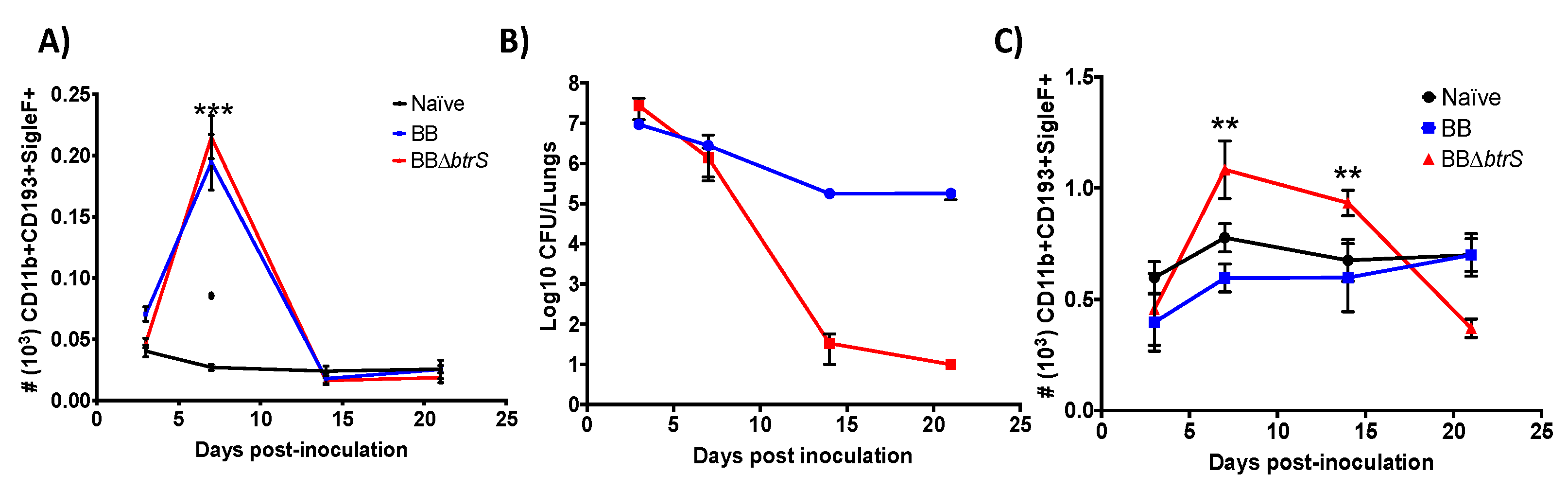
3.2. Bordetella bronchiseptica Blocks Eosinophil Recruitment to the Lungs

Since the BbDbtrS mutant failed to suppress the macrophage inflammatory response, based on our previously published transcriptomic data, and induced much more effective protective immunity in vivo [55], we tested the hypothesis that Bb blocks eosinophil recruitment to the lungs by a btrS-mediated mechanism. Consistent with this hypothesis, mice challenged with BbDbtrS had substantially more eosinophil numbers in both the spleen (Figure 1A) and lungs (Figure 1C), coinciding with the peak of infection (Figure 1B). Moreover, the numbers of eosinophils at the site of infection remained high until BbDbtrS was cleared from the lungs (Figure 1B,C), suggesting that Bb uses a BtrS-dependent mechanism to block eosinophils’ recruitment (Figure 1C). Furthermore, the timing of their recruitment, which peaked just before BbDbtrS numbers began to decline, implicated these eosinophils as potential mediators of the robust immunity induced by the BbDbtrS strain.

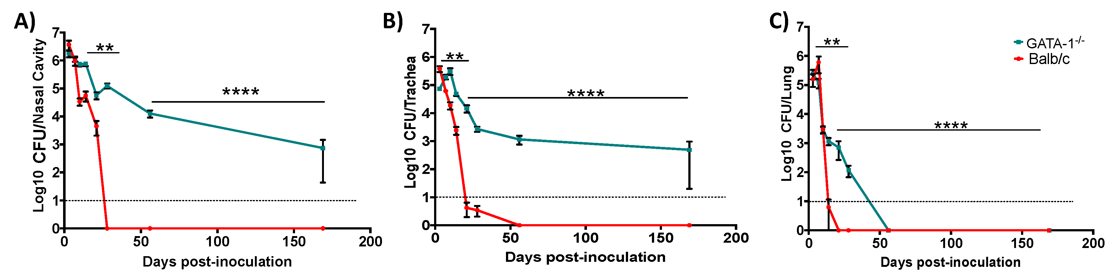
3.3. Eosinophil-Deficient Mice Fail to Clear Respiratory Infection

To investigate the contribution of eosinophils to the robust immune response to BbDbtrS, we compared infections in wild-type (Balb/cJ) and eosinophil-deficient (ΔdblGATA-1) mice, which fail to produce mature eosinophils and are one of the most commonly used models for the study of eosinophilic functions and biology [66]. Wild-type (WT) mice were able to rapidly clear bacteria from the upper and lower respiratory tract (Figure 2). In contrast, eosinophil-deficient mice (EO-) more slowly cleared bacteria from the lower respiratory tract (Figure 2C) and failed to clear bacteria from the upper respiratory tract, even after 169 days (Figure 2A,B). Interestingly, when WT and EO- mice were challenged with wild-type Bb, no differences in the colonization dynamics were detected (Figure S1A), revealing that Bb blocks both eosinophil recruitment and function via btrS-mediated mechanism. When EO- mice were challenged with BbDbtrS, the colonization timescale resembled that caused by wild-type Bb (Figure S1B), indicating that, in the absence of eosinophils, btrS plays a small role in infection. Overall, these results suggest that Bb blocks eosinophil-mediated generation of protective immunity via a btrS-dependent mechanism.

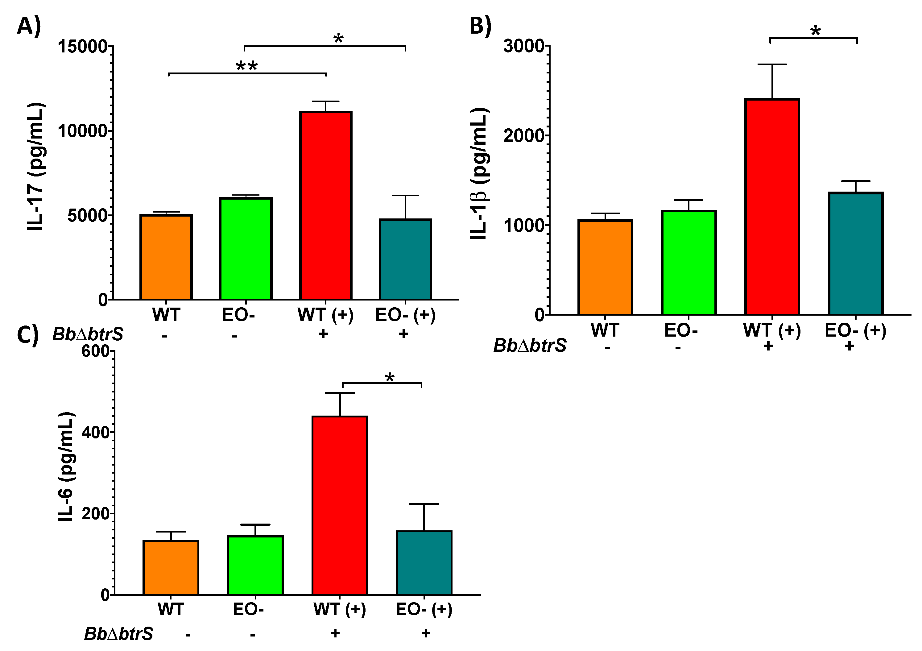
3.4. Eosinophils Contribute to Cytokine Secretion in Respiratory Mucosal Surfaces

The failure of the eosinophil-deficient mice to clear BbDbtrS infection suggests that eosinophils contribute to the generation of protective adaptive immunity within the respiratory tract, analogous to their role in gastric mucosal immunity, also demonstrated in these mice [71]. To explore how eosinophils augment the robust adaptive immunity to BbDbtrS, we inoculated this strain into WT and EO- mice and assessed cytokine levels in the trachea, which is rich in mucosal surfaces [28], and, where such differences can be measured [26], IL-1b and IL-17, key cytokines for the clearance of Bordetella spp. infections [57,64,72,73,74], were increased in WT mice at day seven post-challenge (Figure 3A,B). IL-6 was increased in WT mice at day 14 post-infection, while, in EO- mice, these differences were not detectable (Figure 3C). These results suggest that the milieu of signals instigated by the highly immunogenic BbDbtrS strain are substantially missing in the absence of eosinophils, implicating eosinophils in the cytokine response that shapes the generation of adaptive immunity.

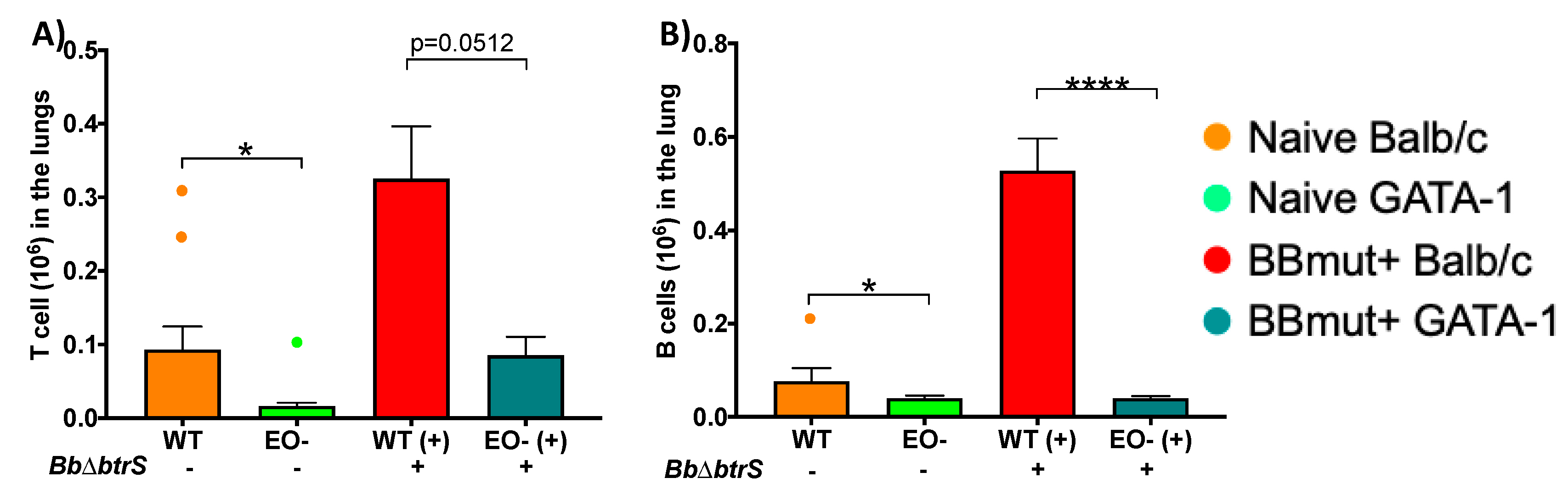
3.5. Eosinophils Mediate Lymphocyte Recruitment to the Lungs

The markedly different cytokine profiles produced by these two different mouse strains suggest that B- and T-cell recruitment might be modulated by eosinophils. It is known that eosinophils can present viral antigen to CD8 T cells [48], and there is evidence that they also modulate B cells [75,76]. Moreover, the modulatory effect of eosinophils in B and T cells can be observed even in naïve uninfected mice [48,75,77]. We have previously reported that the T- and B-cell numbers in the lungs of WT mice were at least twice as high on day 14 post-challenge with BbDbtrS as in WT mice challenged with Bb [55]. To examine the effect of eosinophils on the substantial T- and B-cell recruitment induced by BbDbtrS, we inoculated this strain into WT or EO- mice. T-cell numbers were substantially higher in WT Balb/c mice by day 14 post-infection, as in C57BL/6J mice (Figure 1), while in EO- mice, there were smaller numbers that increased modestly with infection but were still substantially lower than uninfected WT (Figure 4A). B-cell numbers were recruited to the lungs by BbDbtrS in even higher numbers in WT Balb/c mice, but their numbers were unchanged by this severe infection in EO- mice (Figure 4B). While in the wild-type mice the numbers of B cells nearly tripled by 14 days post-challenge, in eosinophil-deficient mice, B-cell numbers remained much lower and did not change measurably in response to 14 days of infection. This substantial defect in the recruitment of B cells to the lungs of EO- mice suggests that eosinophils are critical to the robust local B-cell response to this infection.

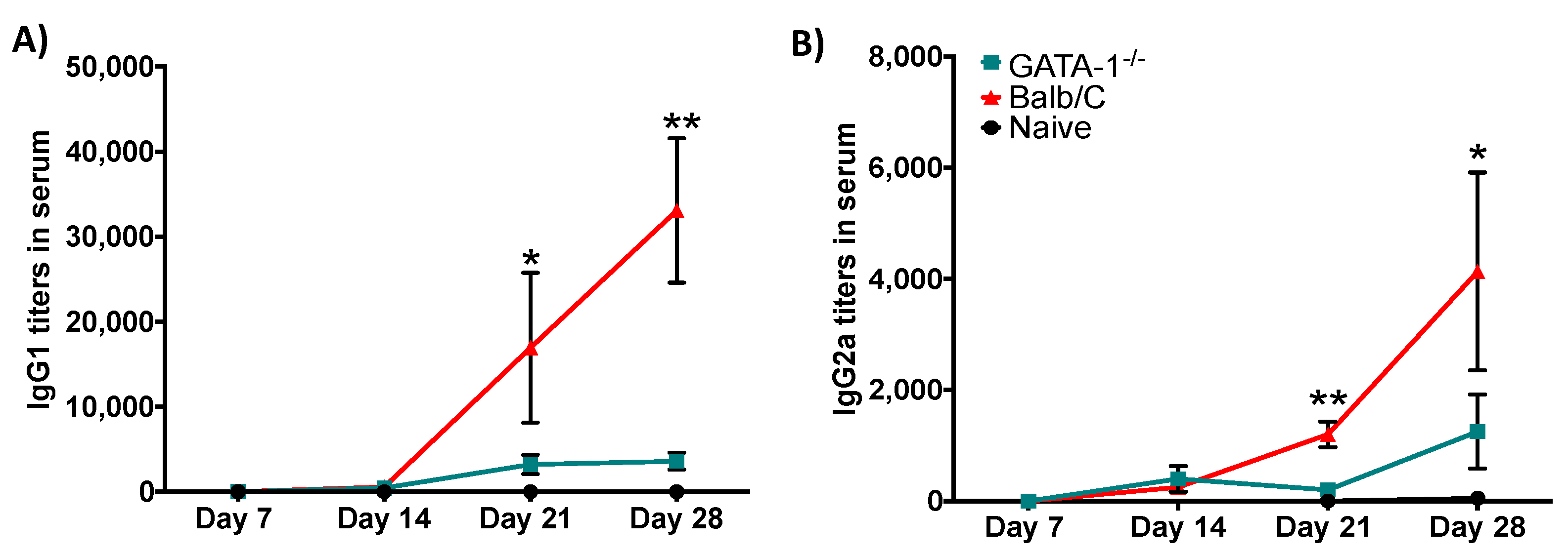
3.6. Eosinophils Are Required for an Efficient Antibody Response

To investigate whether the defect of B cells’ numbers in the lungs of EO- mice is reflective of systemic differences in B-cell response, we measured anti-BbDbtrS antibody titers in the serum of wild-type and EO- mice, at various days after inoculation with BbDbtrS. In WT mice, BbDbtrS-specific antibody titers increased to over 1000 by day 21 and over 4000 by day 28. In stark contrast, EO- mice barely generated detectable antibody titers by day 21, and values had only risen to about 100 by day 28, even though the bacterial numbers, reflecting antigen load, were sustained at much higher numbers than in WT mice (Figure 5A,B). These data indicate that eosinophils play a crucial role in systemic antibody production in response to respiratory infection, which can explain the delay in clearance of the bacterial infection from the respiratory tract.

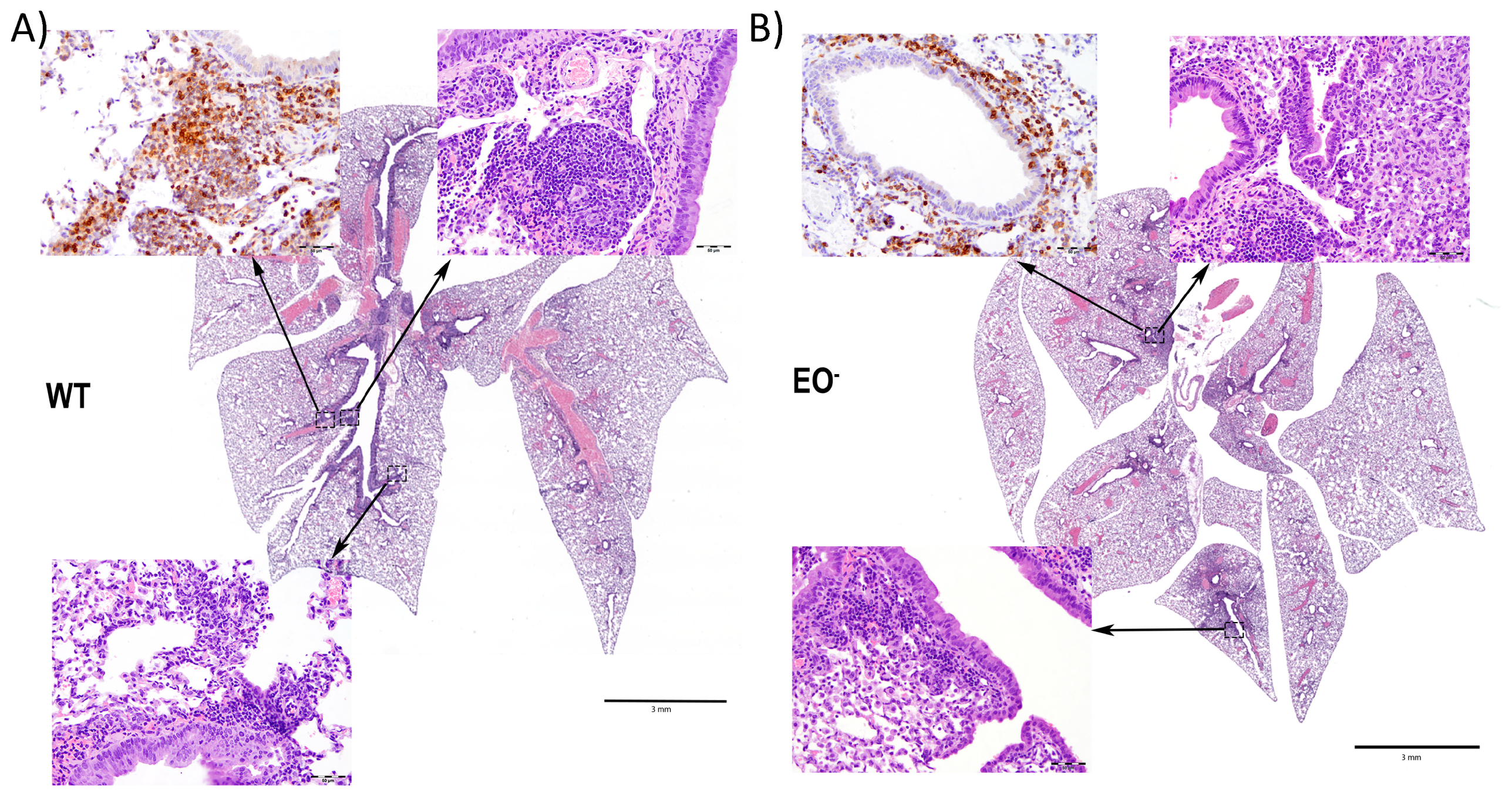
3.7. Eosinophils Contribute to an Organized Local Immune Response

We anticipated that failure to generate a robust adaptive response would lead to differences in the lung inflammatory responses. In order to investigate how the local inflammatory and cellular response might be affected by eosinophils, we challenged WT and EO- mice to examine changes in the induced lung pathology. Wild-type mice developed organized inflammatory cell-mediated responses (Figure 6A), with formation of lymphoid aggregates assembled into recognizable BALT. EO- mice had less lymphoid activity, despite having higher bacterial number, and formed only indistinct structure, characterized by fewer, smaller, and less organized accumulations of macrophages and neutrophils, with few lymphocytes (Figure 6B), suggesting defective generation of localized immune response in these eosinophil-deficient mice. These distinct responses indicate that eosinophils can regulate both the inflammatory response and the generation of protective immunity by mediating the formation of lymphoid aggregates resembling BALT, in response to local respiratory infections, analogous to their role in the gastric mucosa [28].

4. Discussion

We recently demonstrated that btrS-mediated mechanisms contribute to B. bronchiseptica’s ability to suppress immunity to persist in the respiratory tract [55]. Here we demonstrate that btrS-mediated mechanisms suppress the accumulation and function of eosinophils, identifying these cells as targets of immunosuppression. More detailed examination of eosinophil-deficient mice revealed profound defects in aspects of the stimulation and organization of functional protective adaptive immunity. EO- mice were defective in the ability to produce key cytokines, recruit lymphocytes, and organize them in aggregates in which local adaptive immunity can be generated. In addition to revealing important new roles for eosinophils, this work suggests that targeted disruption of eosinophil functions can suppress the local immune response in the respiratory tract. Other persistent commensals and pathogens are likely to have evolved similar strategies, potentially explaining why these important roles of eosinophils were not previously readily observed; successful respiratory pathogens can block them.
The particulars of the role(s) of eosinophils in adaptive immunity in the respiratory tract are intriguing. One hypothesis is that, during bacterial respiratory infections, eosinophils facilitate immune homeostasis and adaptive immunity [78] by influencing the cytokine milieu and immune signaling pathways [79,80,81,82,83,84]. This would generate a strong selective pressure on bacteria to evolve and develop mechanisms to block eosinophilic functions. Excitingly, we report a new role for eosinophils which involves the generation of robust protective immunity against bacterial respiratory infections, apparently mediated by the formation of lymphoid aggregates near the site of infection. A better understanding of the molecular mechanisms by which eosinophils mediate the recruitment, organization and maturation of lymphoid aggregates can provide novel targets for therapeutics and potentially augment vaccines inducing local mucosal immunity.
The defects of EO- mice in cytokine production, lymphocytes recruitment, and lymphocyte organization into aggregates suggest eosinophils may be important in the efficient generation of local lymphoid structures in which lymphocytes are activated and develop. Eosinophils possess Toll-Like Receptors [85,86,87,88,89], indicating that they harbor receptors to respond to infections and trigger the required immune response. This response could be relatively direct; for example, eosinophils might directly present antigens to T cells, as they do during parasitic [24,25,90], viral [48], and allergic reactions [91], indicating they have the required machinery to respond to bacterial infections and activate the subsequent inflammatory response and augment the following adaptive responses.
Bordetella spp. provide an exceptional natural-host experimental system in mice, where aspects of bacterial–host interactions can be probed at the molecular level. However, Bordetella spp. are not the only pathogens well-adapted to manipulate respiratory immunity to persist. The revelation that eosinophils can mediate effective protective immunity leads to the expectation that successful respiratory pathogens have been under strong selection for the ability to disrupt these functions. It appears likely that one or more human pathogens that were previously common can similarly suppress eosinophil functions, and that their partial or complete control (via medicines, vaccines, and “hygiene”) would likely leave eosinophils tending to be excessively activated, potentially contributing to the upsurge in asthma and allergy. The mechanism by which B. bronchiseptica interferes with eosinophil recruitment and function could be an important target for the development of novel therapeutics against eosinophil-mediated pathologies like asthma and allergy. Conversely, understanding how eosinophils contribute to the generation of protective immunity could also facilitate the development of more effective vaccines inducing local mucosal immunity and therapies against a variety of respiratory pathogens.

5. Conclusions

It is well-established that Bordetella spp. sense details of the local host environment [65,92,93,94,95] and carefully and coordinately regulate multiple mechanisms to modulate host immune responses, suppressing the generation of robust immunity [55,92,96]. The type 3 and type 6 secretion systems [97,98], adenylate cyclase toxin [99,100,101], pertussis toxin [102,103], and other immunomodulators can affect immune cells in vitro and in vivo, and disrupting their carefully choreographed expression can lead to much more robust adaptive immunity [55]. Our previous work revealed how classical Bordetella spp. (B. pertussis, B. parapertussis, and B. bronchiseptica) similarly respond to blood and serum [65], and one of the genes that increase expression levels in response to blood and serum is btrS (sigE type sigma factor). When btrS is knocked out, B. bronchiseptica fails to dampen the host response and induces much stronger protective immunity [55]. Here we reveal that btrS is involved in the blockage of eosinophil recruitment to the lungs, reducing adaptive immune responses and promoting persistence. Moreover, our data indicate that eosinophils mediate cytokine production and B-cell responses to respiratory infections, providing new roles for these cells in the generation of protective respiratory immunity. Excitingly, our work associates eosinophils with the generation of lymphoid aggregates, suggesting that a better understanding of the underlying mechanisms can provide targets for therapeutics against a broad spectrum of diseases. There is also a positive correlation between Bordetella spp. infections and the development of asthma and allergy [104,105,106,107,108,109,110,111], highlighting the clinical significance of these findings and suggesting there may be more implications in this complex interaction.

Supplementary Materials

The following are available online at https://www.mdpi.com/2076-2607/8/11/1808/s1. Figure S1: Colonization levels in the lungs of Balb/c and DdblGATA-1 mice following infection with Bb and BbDbtrS.

Author Contributions

M.C.G., conceptualization, developed and performed experiments, analyzed the data, and writing. U.B.-M., performed all the pathological studies included in the manuscript. H.M.J., performed experiments. L.N.R., performed ELISA and analyzed the data. K.K.D., performed part of the pathological studies. C.B., brainstorming and conceptualization. M.T., brainstorming and conceptualization. E.T.H., conceptualization, mentoring, funding, editing of the manuscript, and brainstorming. All authors have read and agreed to the published version of the manuscript.

Funding

This work was supported by the Harvill Lab Start up package and NIH (Grant Number AI122753).

Acknowledgments

The content is solely the responsibility of the authors and does not necessarily represent the official views of the National Institutes of Health. The funders had no role in the study design, data collection, and interpretation, or the decision to submit the work for publication. The authors would like to thank Laura K. Howard and Margaret R. Dedloff for their passion and good work, supporting the generation of data for this manuscript.

Conflicts of Interest

The authors declare no conflict of interest.

References

  1. Hansen, K. Allergic and Psychical Factors in Asthma. Proc. R. Soc. Med. 1929, 22, 789–800. [Google Scholar] [CrossRef] [PubMed] [Green Version]
  2. D’Abrera, V.S.E. Further observations on cases of asthma and bronchitis associated with high eosinophilia and with mites in the sputum. Ind. Med. Gaz. 1946, 81, 414–417. [Google Scholar]
  3. Gundel, R.H.; Letts, L.G.; Gleich, G.J. Human eosinophil major basic protein induces airway constriction and airway hyperresponsiveness in primates. J. Clin. Investig. 1991, 87, 1470–1473. [Google Scholar] [CrossRef] [PubMed]
  4. Michel, F.B.; Bousquet, J.; Godard, P. Bronchial epithelium and asthma. Bull. Acad. Natl. Med. 1992, 176, 683–692. [Google Scholar]
  5. Boushey, H.A.; Fahy, J.V. Basic mechanisms of asthma. Environ. Health Perspect. 1995, 103 (Suppl. 6), 229–233. [Google Scholar] [CrossRef]
  6. Heymann, P.W.; Rakes, G.P.; Hogan, A.D.; Ingram, J.M.; Hoover, G.E.; Platts-Mills, T.A. Assessment of eosinophils, viruses and IgE antibody in wheezing infants and children. Int. Arch. Allergy Immunol. 1995, 107, 380–382. [Google Scholar] [CrossRef]
  7. Koller, D.Y.; Herouy, Y.; Götz, M.; Hagel, E.; Urbanek, R.; Eichler, I. Clinical value of monitoring eosinophil activity in asthma. Arch. Dis. Child. 1995, 73, 413–417. [Google Scholar] [CrossRef] [Green Version]
  8. Thomas, L.H.; Warner, J.A. The eosinophil and its role in asthma. Gen. Pharmacol. 1996, 27, 593–597. [Google Scholar] [CrossRef]
  9. Boushey, H.A. Pathogenesis of asthma. Clin. Cornerstone 1998, 1, 1–8. [Google Scholar] [CrossRef]
  10. Katz, L.E.; Gleich, G.J.; Hartley, B.F.; Yancey, S.W.; Ortega, H.G. Blood eosinophil count is a useful biomarker to identify patients with severe eosinophilic asthma. Ann. Am. Thorac. Soc. 2014, 11, 531–536. [Google Scholar] [CrossRef]
  11. MANSMANN, J.A. The study of the eosinophile in allergic states. Am. J. Med. Technol. 1946, 12, 25–30. [Google Scholar] [PubMed]
  12. Weller, P.F.; Lim, K.; Wan, H.C.; Dvorak, A.M.; Wong, D.T.; Cruikshank, W.W.; Kornfeld, H.; Center, D.M. Role of the eosinophil in allergic reactions. Eur. Respir. J. Suppl. 1996, 22, 109s–115s. [Google Scholar] [PubMed]
  13. Piliponsky, A.M.; Pickholtz, D.; Gleich, G.J.; Levi-Schaffer, F. Human eosinophils induce histamine release from antigen-activated rat peritoneal mast cells: A possible role for mast cells in late-phase allergic reactions. J. Allergy Clin. Immunol. 2001, 107, 993–1000. [Google Scholar] [CrossRef] [PubMed]
  14. Rosenberg, H.F.; Dyer, K.D.; Foster, P.S. Eosinophils: Changing perspectives in health and disease. Nat. Rev. Immunol. 2013, 13, 9–22. [Google Scholar] [CrossRef]
  15. Ueki, S.; Takeda, M. Allergic inflammatory diseases with luminal eosinophilia and extracellular trap cell death (etosis). Arerugi 2019, 68, 1126–1131. [Google Scholar] [CrossRef]
  16. Rothenberg, M.E.; Mishra, A.; Brandt, E.B.; Hogan, S.P. Gastrointestinal eosinophils. Immunol. Rev. 2001, 179, 139–155. [Google Scholar] [CrossRef]
  17. Hogan, S.P.; Rosenberg, H.F.; Moqbel, R.; Phipps, S.; Foster, P.S.; Lacy, P.; Kay, A.B.; Rothenberg, M.E. Eosinophils: Biological properties and role in health and disease. Clin. Exp. Allergy 2008, 38, 709–750. [Google Scholar] [CrossRef]
  18. Shamri, R.; Xenakis, J.J.; Spencer, L.A. Eosinophils in innate immunity: An evolving story. Cell Tissue Res. 2011, 343, 57–83. [Google Scholar] [CrossRef] [Green Version]
  19. Lacy, P.; Rosenberg, H.F.; Walsh, G.M. Eosinophil overview: Structure, biological properties, and key functions. Methods Mol. Biol 2014, 1178, 1–12. [Google Scholar] [CrossRef]
  20. Lee, J.J.; Jacobsen, E.A.; McGarry, M.P.; Schleimer, R.P.; Lee, N.A. Eosinophils in health and disease: The LIAR hypothesis. Clin. Exp. Allergy 2010, 40, 563–575. [Google Scholar] [CrossRef] [Green Version]
  21. Yoshimura, K. Mechanism of parasite killing by eosinophils in parasitic infections. Nihon Rinsho 1993, 51, 657–663. [Google Scholar]
  22. Grezel, D.; Capron, M.; Grzych, J.M.; Fontaine, J.; Lecocq, J.P.; Capron, A. Protective immunity induced in rat schistosomiasis by a single dose of the Sm28GST recombinant antigen: Effector mechanisms involving IgE and IgA antibodies. Eur. J. Immunol. 1993, 23, 454–460. [Google Scholar] [CrossRef] [PubMed]
  23. Yoshimura, K.; Sugaya, H.; Ishida, K. The role of eosinophils in Angiostrongylus cantonensis infection. Parasitol Today 1994, 10, 231–233. [Google Scholar] [CrossRef]
  24. Padigel, U.M.; Lee, J.J.; Nolan, T.J.; Schad, G.A.; Abraham, D. Eosinophils can function as antigen-presenting cells to induce primary and secondary immune responses to Strongyloides stercoralis. Infect. Immun. 2006, 74, 3232–3238. [Google Scholar] [CrossRef] [PubMed] [Green Version]
  25. Padigel, U.M.; Hess, J.A.; Lee, J.J.; Lok, J.B.; Nolan, T.J.; Schad, G.A.; Abraham, D. Eosinophils act as antigen-presenting cells to induce immunity to Strongyloides stercoralis in mice. J. Infect. Dis. 2007, 196, 1844–1851. [Google Scholar] [CrossRef] [Green Version]
  26. Nfon, C.K.; Makepeace, B.L.; Njongmeta, L.M.; Tanya, V.N.; Bain, O.; Trees, A.J. Eosinophils contribute to killing of adult Onchocerca ochengi within onchocercomata following elimination of Wolbachia. Microbes Infect. 2006, 8, 2698–2705. [Google Scholar] [CrossRef] [PubMed]
  27. Chu, D.K.; Jimenez-Saiz, R.; Verschoor, C.P.; Walker, T.D.; Goncharova, S.; Llop-Guevara, A.; Shen, P.; Gordon, M.E.; Barra, N.G.; Bassett, J.D.; et al. Indigenous enteric eosinophils control DCs to initiate a primary Th2 immune response in vivo. J. Exp. Med. 2014, 211, 1657–1672. [Google Scholar] [CrossRef] [PubMed]
  28. Chu, V.T.; Beller, A.; Rausch, S.; Strandmark, J.; Zänker, M.; Arbach, O.; Kruglov, A.; Berek, C. Eosinophils promote generation and maintenance of immunoglobulin-A-expressing plasma cells and contribute to gut immune homeostasis. Immunity 2014, 40, 582–593. [Google Scholar] [CrossRef] [PubMed] [Green Version]
  29. Rosenberg, H.F.; Masterson, J.C.; Furuta, G.T. Eosinophils, probiotics, and the microbiome. J. Leukoc. Biol. 2016, 100, 881–888. [Google Scholar] [CrossRef]
  30. Mishra, A.; Hogan, S.P.; Lee, J.J.; Foster, P.S.; Rothenberg, M.E. Fundamental signals that regulate eosinophil homing to the gastrointestinal tract. J. Clin. Investig. 1999, 103, 1719–1727. [Google Scholar] [CrossRef] [Green Version]
  31. Rothenberg, M.E. Gastrointestinal eosinophils. Allergy 2001, 56 (Suppl. 67), 21–22. [Google Scholar] [CrossRef] [PubMed]
  32. Hogan, S.P.; Waddell, A.; Fulkerson, P.C. Eosinophils in infection and intestinal immunity. Curr. Opin. Gastroenterol. 2013, 29, 7–14. [Google Scholar] [CrossRef] [PubMed] [Green Version]
  33. Femling, J.K.; Cherny, V.V.; Morgan, D.; Rada, B.; Davis, A.P.; Czirják, G.; Enyedi, P.; England, S.K.; Moreland, J.G.; Ligeti, E.; et al. The antibacterial activity of human neutrophils and eosinophils requires proton channels but not BK channels. J. Gen. Physiol. 2006, 127, 659–672. [Google Scholar] [CrossRef] [PubMed]
  34. Linch, S.N.; Kelly, A.M.; Danielson, E.T.; Pero, R.; Lee, J.J.; Gold, J.A. Mouse eosinophils possess potent antibacterial properties in vivo. Infect. Immun. 2009, 77, 4976–4982. [Google Scholar] [CrossRef] [PubMed] [Green Version]
  35. Hatano, Y.; Taniuchi, S.; Masuda, M.; Tsuji, S.; Ito, T.; Hasui, M.; Kobayashi, Y.; Kaneko, K. Phagocytosis of heat-killed Staphylococcus aureus by eosinophils: Comparison with neutrophils. APMIS 2009, 117, 115–123. [Google Scholar] [CrossRef] [PubMed]
  36. Cline, M.J. Microbicidal activity of human eosinophils. J. Reticuloendothel Soc. 1972, 12, 332–339. [Google Scholar] [PubMed]
  37. Cline, M.J.; Hanifin, J.; Lehrer, R.I. Phagocytosis by human eosinophils. Blood 1968, 32, 922–934. [Google Scholar] [CrossRef]
  38. Cohen, S.G.; Sapp, T.M. Phagocytosis of bacteria by eosinophils in infectious-related asthma. J. Allergy 1969, 44, 113–117. [Google Scholar] [CrossRef]
  39. Cohen, S.G.; Sapp, T.M.; Chiampi, P.N. Eosinophil leukocyte responses and hypersensitivity reactions in the Bordetella pertussis-treated mouse. J. Allergy 1970, 46, 205–215. [Google Scholar] [CrossRef]
  40. Baehner, R.L.; Johnston, R.B. Metabolic and bactericidal activities of human eosinophils. Br. J. Haematol. 1971, 20, 277–285. [Google Scholar] [CrossRef]
  41. Bujak, J.S.; Root, R.K. The role of peroxidase in the bactericidal activity of human blood eosinophils. Blood 1974, 43, 727–736. [Google Scholar] [CrossRef]
  42. Leyva Covian, F. Eosinophils. III. Phagocytosis. Rev. Clin. Esp. 1977, 144, 167–171. [Google Scholar] [PubMed]
  43. Baggiolini, M. Phagocytes use oxygen to kill bacteria. Experientia 1984, 40, 906–909. [Google Scholar] [CrossRef] [PubMed]
  44. Kernen, P.; Wymann, M.P.; von Tscharner, V.; Deranleau, D.A.; Tai, P.C.; Spry, C.J.; Dahinden, C.A.; Baggiolini, M. Shape changes, exocytosis, and cytosolic free calcium changes in stimulated human eosinophils. J. Clin. Investig. 1991, 87, 2012–2017. [Google Scholar] [CrossRef] [PubMed] [Green Version]
  45. Ueki, S.; Konno, Y.; Takeda, M.; Moritoki, Y.; Hirokawa, M.; Matsuwaki, Y.; Honda, K.; Ohta, N.; Yamamoto, S.; Takagi, Y.; et al. Eosinophil extracellular trap cell death-derived DNA traps: Their presence in secretions and functional attributes. J. Allergy Clin. Immunol 2016, 137, 258–267. [Google Scholar] [CrossRef] [PubMed] [Green Version]
  46. Gevaert, E.; Zhang, N.; Krysko, O.; Lan, F.; Holtappels, G.; De Ruyck, N.; Nauwynck, H.; Yousefi, S.; Simon, H.U.; Bachert, C. Extracellular eosinophilic traps in association with Staphylococcus aureus at the site of epithelial barrier defects in patients with severe airway inflammation. J. Allergy Clin. Immunol. 2017, 139, 1849–1860.e1846. [Google Scholar] [CrossRef] [PubMed] [Green Version]
  47. Yousefi, S.; Simon, D.; Stojkov, D.; Karsonova, A.; Karaulov, A.; Simon, H.U. In vivo evidence for extracellular DNA trap formation. Cell Death Dis. 2020, 11, 300. [Google Scholar] [CrossRef]
  48. Samarasinghe, A.E.; Melo, R.C.; Duan, S.; LeMessurier, K.S.; Liedmann, S.; Surman, S.L.; Lee, J.J.; Hurwitz, J.L.; Thomas, P.G.; McCullers, J.A. Eosinophils Promote Antiviral Immunity in Mice Infected with Influenza A Virus. J. Immunol. 2017, 198, 3214–3226. [Google Scholar] [CrossRef] [PubMed]
  49. Openshaw, P.J. Immunity and immunopathology to respiratory syncytial virus. The mouse model. Am. J. Respir. Crit. Care Med. 1995, 152, S59–S62. [Google Scholar] [CrossRef]
  50. Ehlenfield, D.R.; Cameron, K.; Welliver, R.C. Eosinophilia at the time of respiratory syncytial virus bronchiolitis predicts childhood reactive airway disease. Pediatrics 2000, 105, 79–83. [Google Scholar] [CrossRef]
  51. Rosenberg, H.F.; Dyer, K.D.; Domachowske, J.B. Respiratory viruses and eosinophils: Exploring the connections. Antivir. Res. 2009, 83, 1–9. [Google Scholar] [CrossRef] [PubMed]
  52. Jesenak, M.; Schwarze, J. Lung Eosinophils—A Novel ‘Virus Sink’ that is Defective in Asthma? Allergy 2019. [Google Scholar] [CrossRef] [PubMed] [Green Version]
  53. Sabogal Piñeros, Y.S.; Bal, S.M.; Dijkhuis, A.; Majoor, C.J.; Dierdorp, B.S.; Dekker, T.; Hoefsmit, E.P.; Bonta, P.I.; Picavet, D.; van der Wel, N.N.; et al. Eosinophils capture viruses, a capacity that is defective in asthma. Allergy 2019. [Google Scholar] [CrossRef] [PubMed]
  54. Zhai, J.; Zou, Y.; Liu, J.; Jin, X.; Ma, C.; Li, J.; Guo, R.; Huang, B. Analysis of the predicting factors of recurrent wheezing in infants. Ital. J. Pediatr. 2019, 45, 19. [Google Scholar] [CrossRef]
  55. Gestal, M.C.; Howard, L.K.; Dewan, K.; Johnson, H.M.; Barbier, M.; Bryant, C.; Soumana, I.H.; Rivera, I.; Linz, B.; Blas-Machado, U.; et al. Enhancement of immune response against Bordetella spp. by disrupting immunomodulation. Sci. Rep. 2019, 9, 20261. [Google Scholar] [CrossRef] [Green Version]
  56. McGuirk, P.; McCann, C.; Mills, K.H. Pathogen-specific T regulatory 1 cells induced in the respiratory tract by a bacterial molecule that stimulates interleukin 10 production by dendritic cells: A novel strategy for evasion of protective T helper type 1 responses by Bordetella pertussis. J. Exp. Med. 2002, 195, 221–231. [Google Scholar] [CrossRef]
  57. Andreasen, C.; Powell, D.A.; Carbonetti, N.H. Pertussis toxin stimulates IL-17 production in response to Bordetella pertussis infection in mice. PLoS ONE 2009, 4, e7079. [Google Scholar] [CrossRef]
  58. Skerry, C.M.; Mahon, B.P. A live, attenuated Bordetella pertussis vaccine provides long-term protection against virulent challenge in a murine model. Clin. Vaccine Immunol. 2011, 18, 187–193. [Google Scholar] [CrossRef]
  59. Zhang, X.; Hester, S.E.; Kennett, M.J.; Karanikas, A.T.; Bendor, L.; Place, D.E.; Harvill, E.T. Interleukin-1 receptor signaling is required to overcome the effects of pertussis toxin and for efficient infection- or vaccination-induced immunity against Bordetella pertussis. Infect. Immun. 2011, 79, 527–541. [Google Scholar] [CrossRef] [Green Version]
  60. Warfel, J.M.; Merkel, T.J. Bordetella pertussis infection induces a mucosal IL-17 response and long-lived Th17 and Th1 immune memory cells in nonhuman primates. Mucosal Immunol. 2013, 6, 787–796. [Google Scholar] [CrossRef]
  61. Place, D.E.; Muse, S.J.; Kirimanjeswara, G.S.; Harvill, E.T. Caspase-1-independent interleukin-1β is required for clearance of Bordetella pertussis infections and whole-cell vaccine-mediated immunity. PLoS ONE 2014, 9, e107188. [Google Scholar] [CrossRef] [PubMed] [Green Version]
  62. da Silva Antunes, R.; Babor, M.; Carpenter, C.; Khalil, N.; Cortese, M.; Mentzer, A.J.; Seumois, G.; Petro, C.D.; Purcell, L.A.; Vijayanand, P.; et al. Th1/Th17 polarization persists following whole-cell pertussis vaccination despite repeated acellular boosters. J. Clin. Investig. 2018, 128, 3853–3865. [Google Scholar] [CrossRef] [Green Version]
  63. Solans, L.; Debrie, A.S.; Borkner, L.; Aguiló, N.; Thiriard, A.; Coutte, L.; Uranga, S.; Trottein, F.; Martín, C.; Mills, K.H.G.; et al. IL-17-dependent SIgA-mediated protection against nasal Bordetella pertussis infection by live attenuated BPZE1 vaccine. Mucosal Immunol. 2018, 11, 1753–1762. [Google Scholar] [CrossRef] [PubMed]
  64. Allen, A.C.; Wilk, M.M.; Misiak, A.; Borkner, L.; Murphy, D.; Mills, K.H.G. Sustained protective immunity against Bordetella pertussis nasal colonization by intranasal immunization with a vaccine-adjuvant combination that induces IL-17-secreting T. Mucosal Immunol. 2018, 11, 1763–1776. [Google Scholar] [CrossRef] [PubMed] [Green Version]
  65. Gestal, M.C.; Rivera, I.; Howard, L.K.; Dewan, K.K.; Soumana, I.H.; Dedloff, M.; Nicholson, T.L.; Linz, B.; Harvill, E.T. Blood or Serum Exposure Induce Global Transcriptional Changes, Altered Antigenic Profile, and Increased Cytotoxicity by Classical Bordetellae. Front. Microbiol. 2018, 9, 1969. [Google Scholar] [CrossRef]
  66. Yu, C.; Cantor, A.B.; Yang, H.; Browne, C.; Wells, R.A.; Fujiwara, Y.; Orkin, S.H. Targeted deletion of a high-affinity GATA-binding site in the GATA-1 promoter leads to selective loss of the eosinophil lineage in vivo. J. Exp. Med. 2002, 195, 1387–1395. [Google Scholar] [CrossRef] [Green Version]
  67. Blas-Machado, U.; Saliki, J.T.; Sánchez, S.; Brown, C.C.; Zhang, J.; Keys, D.; Woolums, A.; Harvey, S.B. Pathogenesis of a bovine enterovirus-1 isolate in experimentally infected calves. Vet. Pathol. 2011, 48, 1075–1084. [Google Scholar] [CrossRef]
  68. Wilk, M.M.; Misiak, A.; McManus, R.M.; Allen, A.C.; Lynch, M.A.; Mills, K.H.G. Lung CD4 Tissue-Resident Memory T Cells Mediate Adaptive Immunity Induced by Previous Infection of Mice with. J. Immunol. 2017, 199, 233–243. [Google Scholar] [CrossRef] [Green Version]
  69. Cossarizza, A.; Chang, H.D.; Radbruch, A.; Akdis, M.; Andrä, I.; Annunziato, F.; Bacher, P.; Barnaba, V.; Battistini, L.; Bauer, W.M.; et al. Guidelines for the use of flow cytometry and cell sorting in immunological studies. Eur. J. Immunol. 2017, 47, 1584–1797. [Google Scholar] [CrossRef] [Green Version]
  70. Halim, T.Y.F.; Takei, F. Current Protocols in Immunology; Wiley Online library: Hoboken, NJ, USA, 2014; Volume 3.25, p. 13. [Google Scholar]
  71. Arnold, I.C.; Artola-Borán, M.; Tallón de Lara, P.; Kyburz, A.; Taube, C.; Ottemann, K.; van den Broek, M.; Yousefi, S.; Simon, H.U.; Müller, A. Eosinophils suppress Th1 responses and restrict bacterially induced gastrointestinal inflammation. J. Exp. Med. 2018, 215, 2055–2072. [Google Scholar] [CrossRef]
  72. Kavanagh, H.; Noone, C.; Cahill, E.; English, K.; Locht, C.; Mahon, B.P. Attenuated Bordetella pertussis vaccine strain BPZE1 modulates allergen-induced immunity and prevents allergic pulmonary pathology in a murine model. Clin. Exp. Allergy 2010, 40, 933–941. [Google Scholar] [CrossRef] [PubMed]
  73. Bancroft, T.; Dillon, M.B.; da Silva Antunes, R.; Paul, S.; Peters, B.; Crotty, S.; Lindestam Arlehamn, C.S.; Sette, A. Th1 versus Th2 T cell polarization by whole-cell and acellular childhood pertussis vaccines persists upon re-immunization in adolescence and adulthood. Cell Immunol. 2016, 304–305, 35–43. [Google Scholar] [CrossRef] [PubMed] [Green Version]
  74. Misiak, A.; Wilk, M.M.; Raverdeau, M.; Mills, K.H. IL-17-Producing Innate and Pathogen-Specific Tissue Resident Memory γδ T Cells Expand in the Lungs of Bordetella pertussis-Infected Mice. J. Immunol. 2017, 198, 363–374. [Google Scholar] [CrossRef] [PubMed] [Green Version]
  75. Wong, T.W.; Doyle, A.D.; Lee, J.J.; Jelinek, D.F. Eosinophils regulate peripheral B cell numbers in both mice and humans. J. Immunol. 2014, 192, 3548–3558. [Google Scholar] [CrossRef] [PubMed] [Green Version]
  76. Viola, J.P.; Kiani, A.; Bozza, P.T.; Rao, A. Regulation of allergic inflammation and eosinophil recruitment in mice lacking the transcription factor NFAT1: Role of interleukin-4 (IL-4) and IL-5. Blood 1998, 91, 2223–2230. [Google Scholar] [CrossRef]
  77. Berek, C. Eosinophils: Important players in humoral immunity. Clin. Exp. Immunol. 2016, 183, 57–64. [Google Scholar] [CrossRef] [Green Version]
  78. Shah, K.; Ignacio, A.; McCoy, K.D.; Harris, N.L. The emerging roles of eosinophils in mucosal homeostasis. Mucosal Immunol. 2020. [Google Scholar] [CrossRef] [Green Version]
  79. Costa, J.J.; Matossian, K.; Resnick, M.B.; Beil, W.J.; Wong, D.T.; Gordon, J.R.; Dvorak, A.M.; Weller, P.F.; Galli, S.J. Human eosinophils can express the cytokines tumor necrosis factor-alpha and macrophage inflammatory protein-1 alpha. J. Clin. Investig. 1993, 91, 2673–2684. [Google Scholar] [CrossRef] [Green Version]
  80. Tong, Y.; Yang, T.; Wang, J.; Zhao, T.; Wang, L.; Kang, Y.; Cheng, C.; Fan, Y. Elevated Plasma Chemokines for Eosinophils in Neuromyelitis Optica Spectrum Disorders during Remission. Front. Neurol. 2018, 9, 44. [Google Scholar] [CrossRef] [Green Version]
  81. Hu, S.; Wong, C.K.; Lam, C.W. Activation of eosinophils by IL-12 family cytokine IL-27: Implications of the pleiotropic roles of IL-27 in allergic responses. Immunobiology 2011, 216, 54–65. [Google Scholar] [CrossRef]
  82. Legrand, F.; Driss, V.; Woerly, G.; Loiseau, S.; Hermann, E.; Fournié, J.J.; Héliot, L.; Mattot, V.; Soncin, F.; Gougeon, M.L.; et al. A functional gammadeltaTCR/CD3 complex distinct from gammadeltaT cells is expressed by human eosinophils. PLoS ONE 2009, 4, e5926. [Google Scholar] [CrossRef]
  83. Spencer, L.A.; Szela, C.T.; Perez, S.A.; Kirchhoffer, C.L.; Neves, J.S.; Radke, A.L.; Weller, P.F. Human eosinophils constitutively express multiple Th1, Th2, and immunoregulatory cytokines that are secreted rapidly and differentially. J. Leukoc. Biol. 2009, 85, 117–123. [Google Scholar] [CrossRef] [PubMed] [Green Version]
  84. Gounni, A.S.; Nutku, E.; Koussih, L.; Aris, F.; Louahed, J.; Levitt, R.C.; Nicolaides, N.C.; Hamid, Q. IL-9 expression by human eosinophils: Regulation by IL-1beta and TNF-alpha. J. Allergy Clin. Immunol. 2000, 106, 460–466. [Google Scholar] [CrossRef]
  85. Yoon, J.; Um, H.N.; Jang, J.; Bae, Y.A.; Park, W.J.; Kim, H.J.; Yoon, M.S.; Chung, I.Y.; Jung, Y. Eosinophil Activation by Toll-Like Receptor 4 Ligands Regulates Macrophage Polarization. Front. Cell Dev. Biol. 2019, 7, 329. [Google Scholar] [CrossRef] [PubMed]
  86. Nagase, H.; Okugawa, S.; Ota, Y.; Yamaguchi, M.; Tomizawa, H.; Matsushima, K.; Ohta, K.; Yamamoto, K.; Hirai, K. Expression and function of Toll-like receptors in eosinophils: Activation by Toll-like receptor 7 ligand. J. Immunol. 2003, 171, 3977–3982. [Google Scholar] [CrossRef] [Green Version]
  87. Mulder, D.J.; Lobo, D.; Mak, N.; Justinich, C.J. Expression of toll-like receptors 2 and 3 on esophageal epithelial cell lines and on eosinophils during esophagitis. Dig. Dis. Sci. 2012, 57, 630–642. [Google Scholar] [CrossRef] [PubMed]
  88. Wong, C.K.; Cheung, P.F.; Ip, W.K.; Lam, C.W. Intracellular signaling mechanisms regulating toll-like receptor-mediated activation of eosinophils. Am. J. Respir. Cell Mol. Biol 2007, 37, 85–96. [Google Scholar] [CrossRef]
  89. Kvarnhammar, A.M.; Cardell, L.O. Pattern-recognition receptors in human eosinophils. Immunology 2012, 136, 11–20. [Google Scholar] [CrossRef] [Green Version]
  90. Xenakis, J.J.; Howard, E.D.; Smith, K.M.; Olbrich, C.L.; Huang, Y.; Anketell, D.; Maldonado, S.; Cornwell, E.W.; Spencer, L.A. Resident intestinal eosinophils constitutively express antigen presentation markers and include two phenotypically distinct subsets of eosinophils. Immunology 2018, 154, 298–308. [Google Scholar] [CrossRef] [Green Version]
  91. Akuthota, P.; Wang, H.; Weller, P.F. Eosinophils as antigen-presenting cells in allergic upper airway disease. Curr. Opin. Allergy Clin. Immunol 2010, 10, 14–19. [Google Scholar] [CrossRef] [Green Version]
  92. Gestal, M.C.; Whitesides, L.T.; Harvill, E.T. Integrated Signaling Pathways Mediate Bordetella Immunomodulation, Persistence, and Transmission. Trends Microbiol. 2019, 27, 118–130. [Google Scholar] [CrossRef] [PubMed]
  93. Hester, S.E.; Lui, M.; Nicholson, T.; Nowacki, D.; Harvill, E.T. Identification of a CO2 responsive regulon in Bordetella. PLoS ONE 2012, 7, e47635. [Google Scholar] [CrossRef] [Green Version]
  94. Brickman, T.J.; Suhadolc, R.J.; Armstrong, S.K. Interspecies variations in Bordetella catecholamine receptor gene regulation and function. Infect. Immun. 2015, 83, 4639–4652. [Google Scholar] [CrossRef] [PubMed] [Green Version]
  95. Armstrong, S.K.; Brickman, T.J.; Suhadolc, R.J. Involvement of multiple distinct Bordetella receptor proteins in the utilization of iron liberated from transferrin by host catecholamine stress hormones. Mol. Microbiol. 2012, 84, 446–462. [Google Scholar] [CrossRef] [PubMed] [Green Version]
  96. Gestal, M.; Jonhson, H.; Harvill, E. Immunomodulation as a Novel Strategy for Prevention and Treatment of Bordetella spp. Infections. Front. Front. Immunol. 2019, in press. [Google Scholar] [CrossRef] [PubMed] [Green Version]
  97. Pilione, M.R.; Harvill, E.T. The Bordetella bronchiseptica type III secretion system inhibits gamma interferon production that is required for efficient antibody-mediated bacterial clearance. Infect. Immun. 2006, 74, 1043–1049. [Google Scholar] [CrossRef] [Green Version]
  98. Siciliano, N.A.; Skinner, J.A.; Yuk, M.H. Bordetella bronchiseptica modulates macrophage phenotype leading to the inhibition of CD4+ T cell proliferation and the initiation of a Th17 immune response. J. Immunol. 2006, 177, 7131–7138. [Google Scholar] [CrossRef] [Green Version]
  99. Gorgojo, J.; Scharrig, E.; Gómez, R.M.; Harvill, E.T.; Rodríguez, M.E. Bordetella parapertussis Circumvents Neutrophil Extracellular Bactericidal Mechanisms. PLoS ONE 2017, 12, e0169936. [Google Scholar] [CrossRef] [Green Version]
  100. Hoffman, C.; Eby, J.; Gray, M.; Heath Damron, F.; Melvin, J.; Cotter, P.; Hewlett, E. Bordetella adenylate cyclase toxin interacts with filamentous haemagglutinin to inhibit biofilm formation in vitro. Mol. Microbiol. 2017, 103, 214–228. [Google Scholar] [CrossRef] [Green Version]
  101. Gonyar, L.A.; Gray, M.C.; Christianson, G.J.; Mehrad, B.; Hewlett, E.L. Albumin, in the Presence of Calcium, Elicits a Massive Increase in Extracellular Bordetella Adenylate Cyclase Toxin. Infect. Immun. 2017, 85. [Google Scholar] [CrossRef] [Green Version]
  102. Freyberg, Z.; Harvill, E.T. Pathogen manipulation of host metabolism: A common strategy for immune evasion. PLoS Pathog. 2017, 13, e1006669. [Google Scholar] [CrossRef] [PubMed] [Green Version]
  103. Kirimanjeswara, G.S.; Agosto, L.M.; Kennett, M.J.; Bjornstad, O.N.; Harvill, E.T. Pertussis toxin inhibits neutrophil recruitment to delay antibody-mediated clearance of Bordetella pertussis. J. Clin. Investig. 2005, 115, 3594–3601. [Google Scholar] [CrossRef] [PubMed] [Green Version]
  104. Cauchi, S.; Locht, C. Non-specific Effects of Live Attenuated Pertussis Vaccine Against Heterologous Infectious and Inflammatory Diseases. Front. Immunol. 2018, 9, 2872. [Google Scholar] [CrossRef] [PubMed] [Green Version]
  105. Zhang, B.Y.; Chi, S.; Sun, Y. Modulation of Toll-like signal path of allergic asthma by CpG-ODNs from Bordetella pertussis. Yao Xue Xue Bao 2011, 46, 285–292. [Google Scholar] [PubMed]
  106. Carbonetti, N.H. Immunomodulation in the pathogenesis of Bordetella pertussis infection and disease. Curr. Opin. Pharmacol. 2007, 7, 272–278. [Google Scholar] [CrossRef] [PubMed]
  107. Ennis, D.P.; Cassidy, J.P.; Mahon, B.P. Whole-cell pertussis vaccine protects against Bordetella pertussis exacerbation of allergic asthma. Immunol. Lett. 2005, 97, 91–100. [Google Scholar] [CrossRef] [PubMed] [Green Version]
  108. Ennis, D.P.; Cassidy, J.P.; Mahon, B.P. Prior Bordetella pertussis infection modulates allergen priming and the severity of airway pathology in a murine model of allergic asthma. Clin. Exp. Allergy 2004, 34, 1488–1497. [Google Scholar] [CrossRef] [Green Version]
  109. Kim, Y.S.; Kwon, K.S.; Kim, D.K.; Choi, I.W.; Lee, H.K. Inhibition of murine allergic airway disease by Bordetella pertussis. Immunology 2004, 112, 624–630. [Google Scholar] [CrossRef]
  110. Dong, W.; Selgrade, M.K.; Gilmour, M.I. Systemic administration of Bordetella pertussis enhances pulmonary sensitization to house dust mite in juvenile rats. Toxicol. Sci. 2003, 72, 113–121. [Google Scholar] [CrossRef] [Green Version]
  111. Donato, L.; Gaugler, C.; Weiss, L.; Krieger, P.; Debry, C. Chronic cough in children: Signs of serious disease and investigations. Arch. Pediatr. 2001, 8 (Suppl. 3), 638–644. [Google Scholar] [CrossRef]
Figure 1. Eosinophils numbers increase in the spleen, but btrS blocks their recruitment to lungs. C57BL/6J mice were intranasally inoculated with PBS (black), or PBS containing 5 × 10⁵ CFU of Bb (blue) or BbΔbtrS (red). Mice were euthanized at different times, to enumerate CFU (B) and eosinophils CD11b+CD193+SiglecF+ in the spleen (A) and the lungs, CD11b+CD193+SiglecF+ (C). The graphs show the average and SEM. ** p < 0.001 and *** p < 0.0001, using two-way ANOVA test N = 10.
Microorganisms 08 01808 g001
Figure 2. Eosinophil-deficient mice failed to clear bacterial infection from the respiratory tract. Balb/cJ—WT (wild-type) (red) or DdblGATA-1—EO- (eosinophil-deficient) (green) mice were intranasally challenged with 50 μL of PBS containing 5 × 10⁵ BbDbtrS. At different days, mice were euthanized, and colonies were enumerated in the nasal cavity (A), trachea (B), or lungs (C). The graphs show the average and SEM. ** p < 0.01, **** p < 0.0001, using two-way ANOVA test. This experiment was conducted multiple independent times, to compensate for possible effects of times of the year and spontaneous allergic reactions on eosinophils counts. n = 22.
Microorganisms 08 01808 g002
Figure 3. Eosinophil-deficient mice failed to secrete IL-17, IL-1β, and IL-6. Groups of mice were intranasally challenged with 50 μL of PBS, or PBS containing 5 × 10⁵ BbDbtrS; they were euthanized on day 7 (A,B) or 14 (C) post-challenge, and trachea was collected and homogenized, to perform ELISA. The graphs show the average and SEM. * p < 0.05 and ** p < 0.01, using two-way ANOVA. n = 6.
Microorganisms 08 01808 g003
Figure 4. Eosinophil-deficient mice failed to recruit lymphocytes to the lung. WT or EO- mice were intranasally challenged with 50 μL of PBS, or PBS containing 5 × 10⁵ BbDbtrS. Mice were euthanized at day 14 post-infection, to enumerate T (A) and B (B) cell populations. WT indicates Balb/c, and EO- stands for DdblGATA-1 mice. BbDbtrS indicates if intranasal challenge has been (+) or not (-) performed. The graphs show the average and SEM. * p < 0.05, **** p < 0.001, using two-way ANOVA. This experiment was conducted multiple independent times, to control for possible effects of times of the year and spontaneous allergic reactions. n = 20.
Microorganisms 08 01808 g004
Figure 5. Eosinophil-deficient mice failed to generate protective antibodies. WT or EO mice were intranasally challenged with 50 μL of PBS or PBS containing 5 × 10⁵ BbDbtrS, in two independent experiments. At different days, mice were euthanized, and serum was collected to perform ELISA, to evaluate anti-Bb antibody titers (results are the average of two independent assays in triplicate). IgG1 (A) and IgG2a (B) were evaluated. The data are shown as average and SEM; * p < 0.05 and ** p < 0.01, using Student t-test. n = 6.
Microorganisms 08 01808 g005
Figure 6. Eosinophil-deficient mice were deficient in generating organized BALT. Groups of 6 Balb/cJ (WT) (A) and GATA-1 (EO-) (B) mice were intranasally challenged with 50 μL of PBS containing 5 × 10⁵ BbDbtrS, and, at day seven post-challenge, lungs were fixed in 10% formalin solution for hematoxylin and eosin staining. Scale bar = 3 mm. n = 6.
Microorganisms 08 01808 g006
Table 1. List of antibodies for flow cytometry.
Flow Cytometry Antibodies
FluorochromeTargetCloneVendorRef
APC-Cy7GR1RB6–8C5Biolegend108–424
PECD11bM1/70Tonbo50–0112–U100
PerCPF4/80BM8Biolegend123,126
APCSiglecFS17007LBiolegend155,508
BV510CD193CCR3BD Biosciences747,820
PerCPCD196D5Biolegend115,532
PECD90.253–2.1Biolegend140,308
AF488CD4GK1.5Biolegend100,423
BV510CD853–6.7BD Biosciences563,068
Table 2. Gating strategy.
+++
EosinophilsCD11bCD193SiglecF
B cellsCD11bCD19
CD4CD11bCD90.2CD4
CD8CD11bCD90.2CD8
Publisher’s Note: MDPI stays neutral with regard to jurisdictional claims in published maps and institutional affiliations.

