Prevalence of Hepatitis B Virus Markers in Patients with Autoimmune Inflammatory Rheumatic Diseases in Italy

 ,
,  , ,
, ,
Abstract
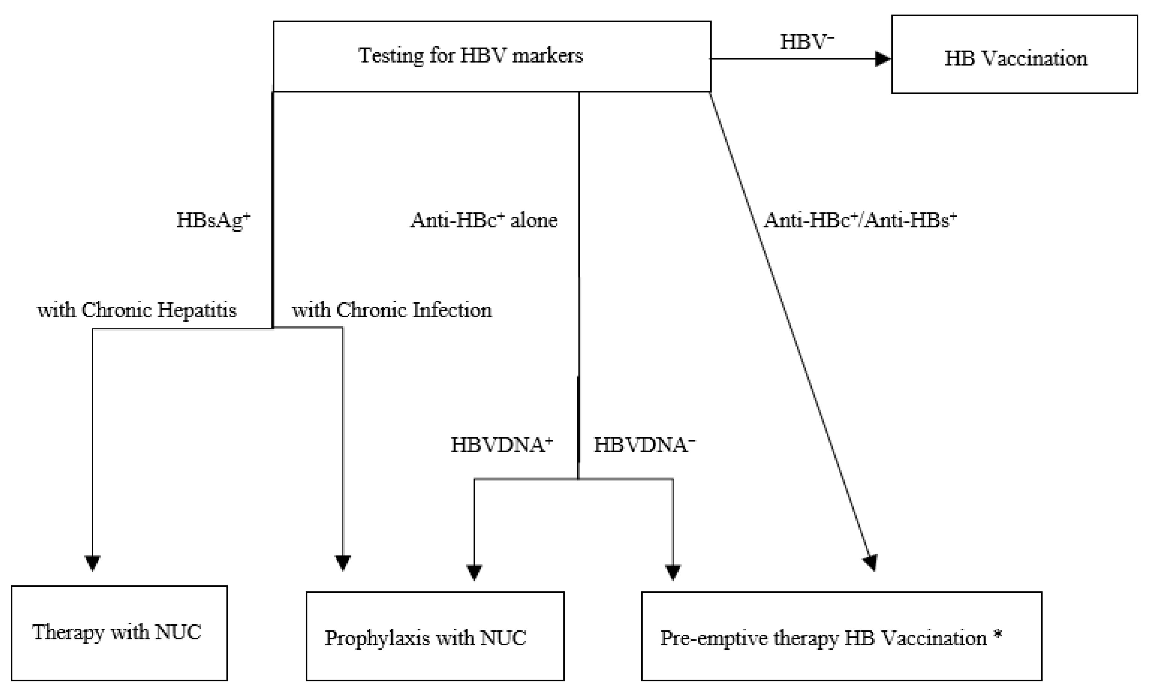
:1. Introduction
2. Materials and Methods
2.1. Laboratory Assay
2.2. Statistical Analysis
3. Results
4. Discussion
5. Conclusions
Author Contributions
Funding
Acknowledgments
Conflicts of Interest
References
- Do, A.; Reau, N.S. Chronic Viral Hepatitis: Current Management and Future Directions. Hepatol. Commun. 2020, 4, 329–341. [Google Scholar] [CrossRef] [Green Version]
- Nelson, N.P.; Easterbrook, P.J.; McMahon, B.J. Epidemiology of Hepatitis B Virus Infection and Impact of Vaccination on Disease. Clin. Liver Dis. 2016, 20, 607–628. [Google Scholar] [CrossRef] [Green Version]
- Trepò, C.; Chan, H.L.Y.; Lok, A. Hepatitis B virus infection. Lancet 2014, 384, 2053–2063. [Google Scholar] [CrossRef]
- Liang, T.J. Hepatitis B: The virus and disease. Hepatology 2009, 49 (Suppl. 5), S13–S21. [Google Scholar] [CrossRef] [PubMed] [Green Version]
- Dienstag, J.L. Hepatitis B virus infection. N. Engl. J. Med. 2008, 359, 1486–1500. [Google Scholar] [CrossRef] [PubMed]
- Lampertico, P.; Agarwal, K.; Berg, T.; Buti, M.; Janssen, H.L.A.; Papatheodoridis, G.; Zoulim, F.; Tacke, F. EASL 2017 Clinical Practice Guidelines on the management of hepatitis B Virus Infection. J. Hepatol. 2017, 67, 370–398. [Google Scholar] [CrossRef] [PubMed] [Green Version]
- Hoofnagle, J.H. Reactivation of hepatitis B. Hepatology 2009, 49 (Suppl. 5), S156–S165. [Google Scholar] [CrossRef] [PubMed]
- Perrillo, R.P.; Gish, R.; Falck-Ytter, Y.T. American Gastroenterological Association Institute technical review on prevention and treatment of hepatitis B virus reactivation during immunosuppressive drug therapy. Gastroenterology 2015, 148, 221–244. [Google Scholar] [CrossRef]
- Sansone, S.; Guarino, M.; Castiglione, F.; Rispo, A.; Auriemma, F.; Loperto, I.; Rea, M.; Caporaso, N.; Morisco, F. Hepatitis B and C virus reactivation in immunosuppressed patients with inflammatory bowel disease. World J. Gastroenterol. 2014, 20, 3516–3524. [Google Scholar] [CrossRef] [Green Version]
- Zanella, A.; Marignani, M.; Begini, P. Hematological malignancies and HBV reactivation risk: Suggestion for clinical management. Viruses 2019, 11, 858. [Google Scholar] [CrossRef] [Green Version]
- Feuchtenberger, M.; Schafer, A.; Nigg, A.P.; Krauss, M.R. Hepatitis B Serology in Patients with Rheumatic Diseases. Open Rheumatol. J. 2016, 10, 39–48. [Google Scholar] [CrossRef] [PubMed] [Green Version]
- Romanò, L.; Velati, C.; Cambiè, G.; Fomiatti, L.; Galli, C.; Zanetti, A.R.; SIMTI study group for HBV infection among first-time blood donors. Hepatitis B virus infection among first-time blood donors in Italy: Prevalence and correlates between serological patterns and occult infection. Blood Transfus. 2013, 11, 281–288. [Google Scholar] [CrossRef]
- Furer, V.; Rondaan, C.; Heijstek, M.; van Assen, S.; Bijl, M.; Agmon-Levin, N.; Breedveld, F.C.; D’Amelio, R.; Dougados, M.; Kapetanovic, M.C.; et al. Incidence and prevalence of vaccine preventable infections in adult patients with autoimmune inflammatory rheumatic diseases (AIIRD): A systemic literature review informing the 2019 update of the EULAR recommendations for vaccination in adult patients with AIIRD. RMD Open 2019, 5, e001041. [Google Scholar] [CrossRef]
- Salemi, S.; D’Amelio, R. Could autoimmunity be induced by vaccination? Int. Rev. Immunol. 2010, 29, 247–269. [Google Scholar] [CrossRef]
- Velati, C.; Romanò, L.; Pati, I.; Marano, G.; Piccinini, V.; Catalano, L.; Pupella, S.; Vaglio, S.; Veropalumbo, E.; Masiello, F.; et al. Prevalence, incidence and residual risk of transfusion-transmitted hepatitis B virus infection in Italy from 2009 to 2018. Blood Transfus. 2019, 17, 409–417. [Google Scholar] [CrossRef]
- Mori, S.; Fujiama, S. Hepatitis B virus reaction associated with anti-rheumatic therapy: Risk and prophylaxis recommendations. World J. Gastroenterol. 2015, 21, 10274–10289. [Google Scholar] [CrossRef] [PubMed]
- Pérez-Alvarez, R.; Díaz-Lagares, C.; García-Hernández, F.; Lopez-Roses, L.; Brito-Zerón, P.; Pérez-de-Lis, M.; Retamozo, S.; Bové, A.; Bosch, X.; Sanchez-Tapias, J.M.; et al. Hepatitis B virus (HBV) reactivation in patients receiving tumor necrosis factor (TNF)-targeted therapy: Analysis of 257 cases. Medicine (Baltimore) 2011, 90, 359–371. [Google Scholar] [CrossRef]
- Chisari, F.V.; Isogawa, M.; Wieland, S.F. Pathogenesis of hepatitis B virus infection. Pathol. Biol. (Paris) 2010, 58, 258–266. [Google Scholar] [CrossRef] [Green Version]
- Puro, R.; Schneider, R.J. Tumor necrosis factor activates a conserved innate antiviral response to hepatitis B virus that destabilizes nucleocapsids and reduces nuclear viral DNA. J. Virol. 2007, 81, 7351–7362. [Google Scholar] [CrossRef] [Green Version]
- Fourati, S.; Cristescu, R.; Loboda, A.; Talla, A.; Filali, A.; Railkar, R.; Schaeffer, A.K.; Favre, D.; Gagnon, D.; Peretz, Y.; et al. Pre-vaccination inflammation and B-cell signalling predict age-related hyporesponse to hepatitis B vaccination. Nat. Commun. 2016, 7, 10369. [Google Scholar] [CrossRef]
- Marzano, A.; Angelucci, E.; Andreone, P.; Brunetto, M.; Bruno, R.; Burra, P.; Caraceni, P.; Daniele, B.; Di Marco, V.; Fabrizi, F.; et al. Prophylaxis and treatment of hepatitis B in immunocompromised patients. Dig. Liver Dis. 2007, 39, 397–408. [Google Scholar] [CrossRef] [PubMed]
- Hwang, J.P.; Lok, A.S. Management of patients with hepatitis B who require immunosuppressive therapy. Nat. Rev. Gastroenterol. Hepatol. 2014, 11, 209–219. [Google Scholar] [CrossRef] [PubMed] [Green Version]
- Hatzakis, A.; Wait, S.; Bruix, J.; Buti, M.; Carballo, M.; Cavaleri, M.; Colombo, M.; Delarocque-Astagneau, E.; Dusheiko, G.; Esmat, G.; et al. The state of hepatitis B and C in Europe: Report from the hepatitis B and C summit conference. J. Viral Hepat. 2011, 18 (Suppl. 1), 1–16. [Google Scholar] [CrossRef] [PubMed] [Green Version]
- Institute of Medicine (US) Committee on the Prevention and Control of Viral Hepatitis Infection. Hepatitis and Liver Cancer: A National Strategy for Prevention and Control. of Hepatitis B and C; Colvin, H.M., Mitchell, A.E., Eds.; National Academies Press: Washington, DC, USA, 2010. [CrossRef]
- Santantonio, T.A.; Fasano, M. Chronic hepatitis B: Advances in treatment. World J. Hepatol. 2014, 6, 284–292. [Google Scholar] [CrossRef] [PubMed]
- Moretto, F.; Catherine, F.X.; Esteve, C.; Blot, M.; Piroth, L. Isolated Anti-HBc: Significance and Management. J. Clin. Med. 2020, 9, 202. [Google Scholar] [CrossRef] [PubMed] [Green Version]
- European Association for the Study of the Liver. EASL clinical practice guidelines: Management of chronic hepatitis B virus infection. J. Hepatol. 2012, 57, 167–185. [Google Scholar] [CrossRef] [Green Version]
- Furer, V.; Rondaan, C.; Heijstek, M.W.; Agmon-Levin, N.; van Assen, S.; Bijl, M.; Breedveld, F.C.; D’Amelio, R.; Dougados, M.; Kapetanovic, M.C.; et al. 2019 update of EULAR recommendations for vaccination in adult patients with autoimmune inflammatory rheumatic diseases. Ann. Rheum. Dis. 2020, 79, 39–52. [Google Scholar] [CrossRef]
- Umemura, T.; Tanaka, E.; Kiyosawa, K.; Kumada, H. Mortality secondary to fulminant hepatic failure in patients with prior resolution of hepatitis B virus infection in Japan. Clin. Infect. Dis. 2008, 47, e52–e56. [Google Scholar] [CrossRef]
- Louthrenoo, W. Treatment considerations in patients with concomitant viral infection and autoimmune rheumatic diseases. Best Pract. Res. Clin. Rheumatol. 2015, 29, 319–342. [Google Scholar] [CrossRef]
- Karadağ, Ö.; Kaşifoğlu, T.; Özer, B.; Kaymakoğlu, S.; Kuş, Y.; İnanç, M.; Keser, G.; Kiraz, S. Viral hepatitis screening guideline before biological drug use in rheumatic patients. Eur. J. Rheumatol. 2016, 3, 25–28. [Google Scholar] [CrossRef]
- Lampertico, P.; Maini, M.; Papatheodoridis, G. Optimal management of hepatitis B virus infection—EASL Special Conference. J. Hepatol. 2015, 63, 1238–1253. [Google Scholar] [CrossRef] [PubMed]
- Haykir Solay, A.; Eser, F. High dose hepatitis B vaccine is not effective in patients using immunomodulatory drugs: A pilot study. Hum. Vaccin. Immunother. 2019, 15, 1177–1182. [Google Scholar] [CrossRef] [PubMed]
- Terrault, N.A.; Lok, A.S.F.; McMahon, B.J.; Chang, K.M.; Hwang, J.P.; Jonas, M.M.; Brown, R.S.; Bzowej, N.H.; Wong, J.B. Update on prevention, diagnosis, and treatment of chronic hepatitis B: AASLD 2018 hepatitis B guidance. Hepatology 2018, 67, 1560–1599. [Google Scholar] [CrossRef]
- Taliani, G.; Lecce, R.; Furlan, C.; Nuti, M.; Bernaschi, P.; De Bac, C. Hepatitis B vaccination for a wider diagnostic purpose? Lancet 1984, 2, 173. [Google Scholar] [CrossRef]
- Draelos, M.; Morgan, T.; Schifman, R.B.; Sampliner, R.E. Significance of isolated antibody to hepatitis B core antigen determined by immune response to hepatitis B vaccination. JAMA 1987, 258, 1193–1195. [Google Scholar] [CrossRef] [PubMed]
- Lok, A.S.F.; Lai, C.L.; Wu, P.C. Prevalence of isolated antibody to hepatitis B core antigen in an area endemic for hepatitis B virus infection: Implications in hepatitis B vaccination programs. Hepatology 1988, 8, 766–770. [Google Scholar] [CrossRef]
- McMahon, B.J.; Parkinson, A.J.; Helminiak, C.; Wainwright, R.B.; Bulkow, L.; Kellerman-Douglas, A.; Schoenberg, S.; Ritter, D. Response to hepatitis B vaccine of persons positive for antibody to hepatitis B core antigen. Gastroenterology 1992, 103, 590–594. [Google Scholar] [CrossRef]
- Silva, A.E.; McMahon, B.J.; Parkinson, A.J.; Sjogren, M.H.; Hoofnagle, J.H.; Di Bisceglie, A.M. Hepatitis B virus DNA in persons with isolated antibody to hepatitis B core antigen who subsequently received hepatitis B vaccine. Clin. Infect. Dis. 1998, 26, 895–897. [Google Scholar] [CrossRef]
- Sünbül, M.; Leblebicioglu, H.; Esen, S.; Eroglu, C.; Barut, S. Response to hepatitis B vaccine in HBsAg/anti-HBs negative and anti-HBc positive subjects. Scand. J. Infect. Dis. 2000, 32, 315–316. [Google Scholar] [CrossRef]
- Ural, O.; Findik, D. The response of isolated anti-HBc positive subjects to recombinant hepatitis B vaccine. J. Infect. 2001, 43, 187–190. [Google Scholar] [CrossRef]
- Koh, H.J.; Kim, S.D.; Choi, J.H.; Kim, S.R.; Lee, J.S. A study of immune response to hepatitis B vaccine & HBV DNA in isolated anti-HBc positive subjects. J. Prev. Med. Public Health 2005, 38, 170–174. [Google Scholar] [CrossRef] [PubMed]
- Gandhi, R.T.; Wurcel, A.; Lee, H.; McGovern, B.; Shopis, J.; Geary, M.; Sivamurthy, R.; Sax, P.E.; Ukomadu, C. Response to hepatitis B vaccine in HIV-1-positive subjects who test positive for isolated antibody to hepatitis B core antigen: Implications for hepatitis B vaccine strategies. J. Infect. Dis. 2005, 191, 1435–1441. [Google Scholar] [CrossRef] [PubMed]
- Gessoni, G.; Beggio, S.; Barin, P.; Favarato, M.; Galli, C.; Valverde, S.; Boscolo Nata, M.; Salvadego, M.M.; Marchiori, G. Significance of anti-HBc only in blood donors: A serological and virological study after hepatitis B vaccination. Blood Transfus. 2014, 12 (Suppl. 1), s63–s68. [Google Scholar] [CrossRef]
- Yao, J.; Ren, W.; Chen, Y.; Jiang, Z.; Shen, L.; Shan, H.; Dai, X.; Li, J.; Liu, Y.; Qiu, Y.; et al. Responses to hepatitis B vaccine in isolated anti-HBc positive adults. Hum. Vaccin. Immunother. 2016, 12, 1847–1851. [Google Scholar] [CrossRef] [PubMed] [Green Version]
- Bahari, A.; Izadi, S.; Bari, Z.; Khosravi, S.; Baghaei, B.; Saneimoghadam, E.; Firouzi, F.; Espiari, A.; Esmaeilzadeh, A.; Mokhtarifar, A.; et al. Significance of Response to Hepatitis B Recombinant Vaccine in Subjects with Isolated Antibody to Hepatitis B Core Antigen. Middle East. J. Dig. Dis. 2015, 7, 233–240. [Google Scholar] [PubMed]
- Starzl, T.E.; Zinkernagel, R.M. Antigen localization and migration in immunity and tolerance. N. Engl. J. Med. 1998, 339, 1905–1913. [Google Scholar] [CrossRef] [PubMed] [Green Version]
- Thakur, V.; Guptan, R.C.; Basir, S.F.; Parvez, M.K.; Sarin, S.K. Enhanced Immunogenicity of Recombinant Hepatitis B Vaccine in Exposed Family Contacts of Chronic Liver Disease Patients. Scand. J. Infect. Dis. 2001, 33, 618–621. [Google Scholar] [CrossRef]
- Stine, J.G.; Khokhar, O.S.; Charalambopoulos, J.; Shanmugam, V.K.; Lewis, J.H. Rheumatologists’ awareness of and screening practices for hepatitis B virus infection prior to initiating immunomodulatory therapy. Arthritis Care Res. (Hoboken) 2010, 62, 704–711. [Google Scholar] [CrossRef]


| Characteristic | N | % | DD ¥ | CRP © | ESR ∞ |
|---|---|---|---|---|---|
| Sex | |||||
| 113 189 0.6 | 63 37 | |||
| Age-groups (years) | |||||
| 39 131 132 57 | 13 43 44 | |||
| Origin | |||||
| Italians | 276 | 91 | |||
| 16 219 41 | 5 72 14 | |||
| Non-Italians | 26 | 9 | |||
| Spondyloarthritis | 146 * | 48 | 4.17 ± 7.47 | 4.07 ± 9.43 | 25.74 ± 20.4 |
| Chronic Arthritis | 75 ‡ | 25 | 3.36 ± 8.21 | 5.96 ± 11.33 † | 33.88 ± 22.19 ↕ |
| Connective Tissue Disease | 83 | 27 | 3.44 ± 9.93 | 3.82 ± 7.88 | 34.41 ± 26 |
| Candidates to immunosuppressive therapy | 167 | 55 | |||
| HBV Markers | N | % |
| HBsAg+ ** | 6 | 2 |
| Anti-HBc+ alone | 12 | 4 |
| Anti-HBc+/Anti-HBs+ | 52 | 18 |
| Any HBV infection marker positivity | 70 | 24 |
| Characteristic | N Positive/N Tested | % | p |
|---|---|---|---|
| Sex | |||
| ● Male | 27/111 | 24 | NS |
| ● Female | 43/181 | 24 | |
| Age-groups | |||
| ● ≤60 | 31/159 | 19 | <0.05 |
| ● >60 | 39/133 | 29 | |
| Spondyloarthritis | 34/143 | 24 | 0.01146 * |
| Chronic Arthritis | 25/75 | 33 | |
| Connective Tissue Disease | 11/76 | 14 | |
| Candidates to immunosuppressive therapy | |||
| ● Yes | 35/160 | 22 | NS |
| ● No | 35/132 | 27 |
| Patients | Non-Immunosuppressive Therapy | Immunosuppressive Therapy | Total |
|---|---|---|---|
| All γ-globulins >0.7 g/dL N (%) | 129 (47) | 147 (53) | 276 (91) |
| All γ-globulins ≤0.7 g/dL N (%) | 6 (23) | 20 (77) * | 26 (9) |
| SpA γ-globulins > 0.7 g/dL N (%) | 52 (40) | 77 (60) | 129 (88) |
| SpA γ-globulins ≤ 0.7 g/dL N (%) | 5 (29) | 12 (71) ^ | 17 (12) |
| ChA γ-globulins > 0.7 g/dL N (%) | 27 (38) | 44 (62) | 71 (95) |
| ChA γ-globulins ≤ 0.7 g/dL N (%) | 1 (25) | 3 (75) ^ | 4 (5) |
| CTD γ-globulins > 0.7 g/dL N (%) | 48 (61.5) | 30 (38.5) | 78 (94) |
| CTD γ-globulins ≤ 0.7 g/dL N (%) | 3 (60) | 2 (40) ^ | 5 (6) |
Publisher’s Note: MDPI stays neutral with regard to jurisdictional claims in published maps and institutional affiliations. |
© 2020 by the authors. Licensee MDPI, Basel, Switzerland. This article is an open access article distributed under the terms and conditions of the Creative Commons Attribution (CC BY) license (http://creativecommons.org/licenses/by/4.0/).
Share and Cite
Canzoni, M.; Marignani, M.; Sorgi, M.L.; Begini, P.; Biondo, M.I.; Caporuscio, S.; Colonna, V.; Casa, F.D.; Conigliaro, P.; Marrese, C.; et al. Prevalence of Hepatitis B Virus Markers in Patients with Autoimmune Inflammatory Rheumatic Diseases in Italy. Microorganisms 2020, 8, 1792. https://doi.org/10.3390/microorganisms8111792
Canzoni M, Marignani M, Sorgi ML, Begini P, Biondo MI, Caporuscio S, Colonna V, Casa FD, Conigliaro P, Marrese C, et al. Prevalence of Hepatitis B Virus Markers in Patients with Autoimmune Inflammatory Rheumatic Diseases in Italy. Microorganisms. 2020; 8(11):1792. https://doi.org/10.3390/microorganisms8111792
Chicago/Turabian StyleCanzoni, Marco, Massimo Marignani, Maria Laura Sorgi, Paola Begini, Michela Ileen Biondo, Sara Caporuscio, Vincenzo Colonna, Francesca Della Casa, Paola Conigliaro, Cinzia Marrese, and et al. 2020. "Prevalence of Hepatitis B Virus Markers in Patients with Autoimmune Inflammatory Rheumatic Diseases in Italy" Microorganisms 8, no. 11: 1792. https://doi.org/10.3390/microorganisms8111792