Article

The Domestic Environment and the Lung Mycobiome

1
Department of Physiology, Genetics and Microbiology, University of Alicante, Sant Vicent del Raspeig, 03690 Alicante, Spain
2
Pneumology Service, General University Hospital, Elda, 03600 Alicante, Spain
3
Respiratory Department, General University Hospital, Institute for Healthcare and Biomedical Research of Alicante (ISABIAL), 03010 Alicante, Spain
4
Medical Mycology Laboratory, Department of Plant Production and Microbiology, Institute for Healthcare and Biomedical Research of Alicante (ISABIAL), Campus of Sant Joan d’Alacant, University Miguel Hernández, 03550 Alicante, Spain
5
Pneumology Department, Vinalopó University Hospital, Elche 03293 Alicante, Spain
6
Respiratory Department, University Hospital of Valencia, 46010 Valencia, Spain
*
Author to whom correspondence should be addressed.
Microorganisms 2020, 8(11), 1717; https://doi.org/10.3390/microorganisms8111717
Submission received: 14 August 2020 / Revised: 16 October 2020 / Accepted: 30 October 2020 / Published: 2 November 2020
(This article belongs to the Special Issue Precision Microbiomics: Environment to Human Health)

Abstract

:
This study analyzes the relationship between the mycobiome of the Lower Respiratory Tract (LRT) and the fungi in the domestic environment. Samples studied consisted of Broncho-Alveolar Lavage (BAL) from 45 patients who underwent bronchoscopy for different diagnostic purposes, and dust and air from the houses (ENV) of 20 of them (44.4%). Additionally, five bronchoscopes (BS) were also analyzed and negative controls were included for every procedure. All samples were processed for DNA extraction and cultures, which were performed in Sabouraud Dextrose and Potato Dextrose Agar. The fungal Internal Transcribed Spacer (ITS2) was sequenced by the Solexa/Illumina system and sequences were analyzed by QIIME 1.8.0 and compared with the UNITE Database for identification. The similarity between the two fungal communities (BAL and ENV) for a specific patient was assessed via the percentage of coincidence in the detection of specific operational taxonomic units (OTUs), and about 75% of co-occurrence was detected between the mycobiome of the LRT and the houses. Cultures confirmed the presence of the core mycobiome species. However, the low rate of isolation from BAL suggests that most of its mycobiome corresponds to non-culturable cells. This likely depends on the patient’s immune system activity and inflammatory status.

1. Introduction

High-throughput sequencing-based methods (HTS) are currently providing a wealth of information about the composition of the human microbiota, now considered an important “organ” playing a key role in maintaining the homeostasis of the body [1,2]. The gut microbiome is the most explored organ by far, but the study of other epithelial tissues such as those of the respiratory tract has also shown that their microbial communities are directly involved in in people’s health status of individuals [2,3,4,5,6]. Therefore, the microbiome of the respiratory tract is a topic of growing interest.
Few studies based on HTS have been performed to describe the lung mycobiome, that is, the study of fungi as members of the microbial community of the lungs. It is presumed that the species comprising that mycobiome reflect exposure to environmental and endogenous fungi [7,8,9], but this fact has not been explored deeply or specifically yet. In fact, until recently, the Lower Respiratory Tract (LRT) was believed to be sterile and this idea only changed after several studies demonstrated the existence of a microbial community in the lower airway of healthy individuals [10]. Nevertheless, in recent years, interesting contributions have shown a relationship between the airway mycobiome and the health status in patients with chronic inflammatory respiratory diseases. Thus, studies of patients suffering from cystic fibrosis (CF), asthma, and chronic obstructive pulmonary disease (COPD) demonstrate that fungal diversity is inversely proportional to the severity of the inflammatory status [3,4,11]. Some approaches to the study of the human mycobiome and its origin show that the colonization of the human body by fungi occurs from very early stages of life, when fungi are mainly acquired from the mother, other caregivers, and likely from the environment [12]. Regarding the fungal components of the LRT, it has been considered that they can be inhaled from the environment [7,8] and/or be aspirated from the upper airway, mouth, and nose [13,14,15]. For some opportunistic infections like pneumocystosis, it has been demonstrated that the fungus is acquired from early stages of life and is carried in the lungs of many children [16,17] and other population groups with different percentages of colonization [18]. The development of the opportunistic infection is attributed to the decrease of immune system efficiency [19]. This highlights the importance of knowing as much as possible about the lung mycobiome and the factors that can influence its stability and composition.
The relationship of the lung mycobiota and the environment has been explored in some works, mainly with the aim of ascertaining the connection between environmental mycobiota and the development of allergies. Several culture-based studies have attempted to demonstrate the link between asthmatic crises in children and the presence of fungal components in the air of their homes [20,21,22]. More recently, some studies have approached the same objective through various molecular methods by studying the mycobiome of environments at work [23,24,25]. Most of these studies have shown that the lung mycobiome mainly proceeds from inhaled air and oral cavity mycobiota [26]. Opportunistic mycoses, especially those known as IFIs (Invasive Fungal Infections), are a problem of growing interest due to the recent increase in the number of patients susceptible to them. Some of the fungi most frequently involved in these processes are commonly found in the environment. Therefore, more precise knowledge of the microbiome of the lower respiratory tract and how it is influenced by the microbiota of the environment can constitute an important tool for the prevention of not only allergies but also opportunistic infections. It would contribute to more personalized medicine. These facts led us to perform a study of the relationship between the mycobiome of the lower respiratory tract of adult patients and the mycobiota detected in their domestic environment. More specifically, we focused on an analysis of patients submitted to bronchoscopy due to causes other than infectious diseases since bronchoscopy samples from healthy patients are scarce and Broncho-Alveolar Lavage (BAL) is considered the best sample to explore the microbiome of the LRT [27]. The study is based on molecular (HTS) methods, but cultures were also performed to assess the presence and viability of the fungi detected.

2. Materials and Methods

2.1. Study Design and Participants

This is an observational study carried out from 2015 to 2019 including 45 patients attending three University Hospitals in the area of the Valencian Autonomous Community (Spain). Selected patients met two important criteria: (1) they permitted a sample of the lower respiratory tract to be taken by bronchoscopy and (2) they were not at risk of suffering lung dysbiosis (at least presumably). The first requirement led us to select patients who had a clinical reason, which justified bronchoscopy. For the second, we had to reject patients under antibiotic treatment, or who had any kind of immunodepression, as these situations can disturb the microbiome and cause a dysbiosis (Supplementary material. Document S1: Patient data sheet). Therefore, the inclusion criteria were: adult patients with an indication for bronchoscopy, not suffering from or showing symptoms of infectious disease and/or immune deficit, and not treated with antibiotics for at least four weeks prior. The study received the approval of the ethics committees of all the hospitals involved (reference codes: PI2017/68, MICROBERESPV4, F-EG-CCI-9) and the University Miguel Hernandez (ref. code: DVM.CVM.01.14). Patients were informed about the objectives of the study and gave their informed consent for the use of their BAL sample. Twenty of them also allowed entrance to their homes for environmental sampling (Supplementary material: Figure S1: Project work flow).

2.2. Obtention of Broncho-Alveolar Lavage Samples by Bronchoscopy

Samples were obtained by bronchoscopy with instillation and subsequent aspiration of about 50 mL of sterile saline solution (NaCl 0.9%) [28], which were collected in sterile plastic tubes. Immediately afterward, they were transported to the laboratory at 4 °C in a polystyrene box with cooling blocks. Once in the lab, the samples were divided into 1–1.5 mL aliquots (4–12 aliquots per sample). Four of these aliquots were separated and kept at 4 °C to be immediately processed for conventional culture-based mycology studies and for DNA extraction. The rest were frozen and kept at −80 °C in the Biobank of the General University Hospital of Alicante. Additionally, five of the bronchoscopes were also tested by washing the device with sterile saline before use. The solution was collected and treated like the BAL samples (BS samples).

2.3. Environmental Sampling

The addresses of patients were located on a map with the aid of Google Maps software [29] and sampling was scheduled for groups of two or three patients per day, according to the proximity of their houses. Environmental sampling was carried out during six different days in nine cities or towns in an area of about 2000 square km in the Province of Alicante (Southeast of Spain). Once in the house, plates with Potato Dextrose Agar (PDA) were distributed in each of the rooms. Open plates were exposed to the air for at least 10 minutes, while swab samples were also taken in the same rooms. Swabs were previously soaked in saline solution with 0.05 g/L of chloramphenicol and subsequently used to rub the exposed surfaces (furniture, walls, air conditioner openings, sinks, shelves, kitchen countertop, and others). When pets or plants were present in the house, samples directly related to them were also collected (swabs of pet beds, birdcages, plant leaves and stems, bird feces, water from aquariums for fish and turtles, etc.). Swabs were kept inside their cases containing 2 mL of the chloramphenicol solution and other samples were kept in sterile tubes [30]. Data about the house and its environment were recorded during the visit (Table 1 and supplemental material: “patients & houses”). All samples were transported to the laboratory in less than three hours. The number of samples per house ranged between seven and 17 (median: 10). In the laboratory, the exposed PDA plates were kept at room temperature for incubation. Swabs with antibiotic solution and liquid samples were vigorously vortexed and allowed to settle for 15 min. Solid samples were weighed, suspended (approximately 1 gr/10 mL) in the same antibiotic solution, vortexed, and allowed to settle for 15 min. From each supernatant, 100 µL were inoculated in plates of Sabouraud Dextrose Agar (SDA) and PDA and another 100 µL were separated in a microtube for the DNA extraction.

2.4. Cultures

For BAL samples, 0.1 mL of each of two aliquots of the sample were inoculated in SDA and PDA plates and subsequently incubated at 37 °C and at room temperature, respectively. The supernatants from the environmental samples were processed in the same way as the BAL samples. All cultures were incubated for four weeks and examined for fungal growth every two or three days. The colonies were isolated in the same media and studied by macro-morphology and micro-morphology and other phenotypic features [31,32] as well as by molecular methods after the extraction of DNA from pure cultures.

2.5. DNA Isolation

One aliquot (1 mL) of BAL sample from every patient, together with one combined sample from the domestic environment (ENV sample), was used for the study of the mycobiome. From the samples obtained in the same house, 100 µL of each supernatant were mixed all together, creating a pool which constituted the unique environmental sample (ENV) corresponding to each patient. Each ENV and BAL sample was submitted to DNA extraction by the same procedure.
For DNA extraction, the E.Z.N.A. Yeast DNA kit (Omega Bio-tek Inc., Norcross, GA, USA) was used. The fungal Internal Transcribed Spacer (ITS1-4) was amplified from the extracted DNA by PCR with primers ITS1 (5′ TCC GTA GGT GAA CCT GCG G 3′) and ITS4 (5′ TCC TCC GCT TAT TGA TAT GC 3′) [33,34]. The product was purified by the GeneJet DNA Purification Kit (ThermoFisher Scientific Lithuania, EU) and the concentration of DNA was measured by NanoDrop Lite (ThermoFisher Scientific Inc.). BAL samples had a mean concentration of 79.2 ng/µL with a median value of 64.7 ng/µL (range 15.9–179.7 ng/µL) while, for ENV samples, the mean value was 27.64 ng/µL and the median value was 11.4 ng/µL (range: 6.4–121.7 ng/µL). The fragment of about 300 bp spanned by ITS86 (5′ GTG AAT CAT CGA ATC TTT GAA C 3′) and ITS4 primers inside the barcode region of the ITS2 of fungi was amplified by semi-nested PCR [34] and sequenced by the Solexa system (Illumina, San Diego, CA, USA). The extraction of DNA from a pure culture of isolated fungi was performed mainly by the InstaGeneTM Matrix (Biorad Laboratories Inc., Hercules, CA, USA).

2.6. Illumina High-Throughput ITS2 Region Sequencing and Bioinformatic Analyses

Forty-five BAL, 20 ENV, and 5 BS samples, together with a negative PCR control, were submitted to massive sequencing after the first amplification of the ITS-5.8S region (SRA accession number: PRJNA474914). The QIIME 1.8.0 pipeline [35] was used for data processing. Sequences were checked for chimeric sequences with identify_chimeric_seqs.py script against the UNITE database (version 2017-12-01) (https://unite.ut.ee/community.php) using UCHIME [36] Afterward, one negative control from PCR and a negative control from each sequencing run were used to filter contaminants. Samples were clustered in different fungal Operational Taxonomic Units (OTUs) at 99% identity and sequences in OTUs present in control samples were removed for the subsequent analysis. After that, 25 samples (35.7%) were discarded to avoid errors in the analysis due to the low number of sequences [37]. Clean sequences were clustered in OTUs at 97% identity and identified using pick_open_reference_otus.py script. Due to the large difference in library size among samples, the OTU table was rarefied to 6000 reads with a single_rarefaction.py script for comparisons across samples [38] (Supplementary material. Figure S2: Rarefaction curves). Alpha diversity metrics (total observed number of OTUs, and Shannon-Wiener diversity) were generated from the rarefied OTU table using alpha_diversity.py script. Differences among microbial assemblages were assessed with UniFrac analysis, which is a β-diversity measure (differentiation at diversity level among habitats) that uses phylogenetic information using jackknifed UPGMA (weighted pair group method with arithmetic mean) clustering based on the weighted UniFrac (Lozupone and Knight, 2005). These results were plotted in three dimensions with the UniFrac-based principal coordinate analysis (PCoA) [39]. Occasionally, NCBI and EMBL nucleotide databases (BLASTn update 2019) were also used for identification of highly prevalent OTUs that could not be assigned to a species or genus by the UNITE database.

3. Results

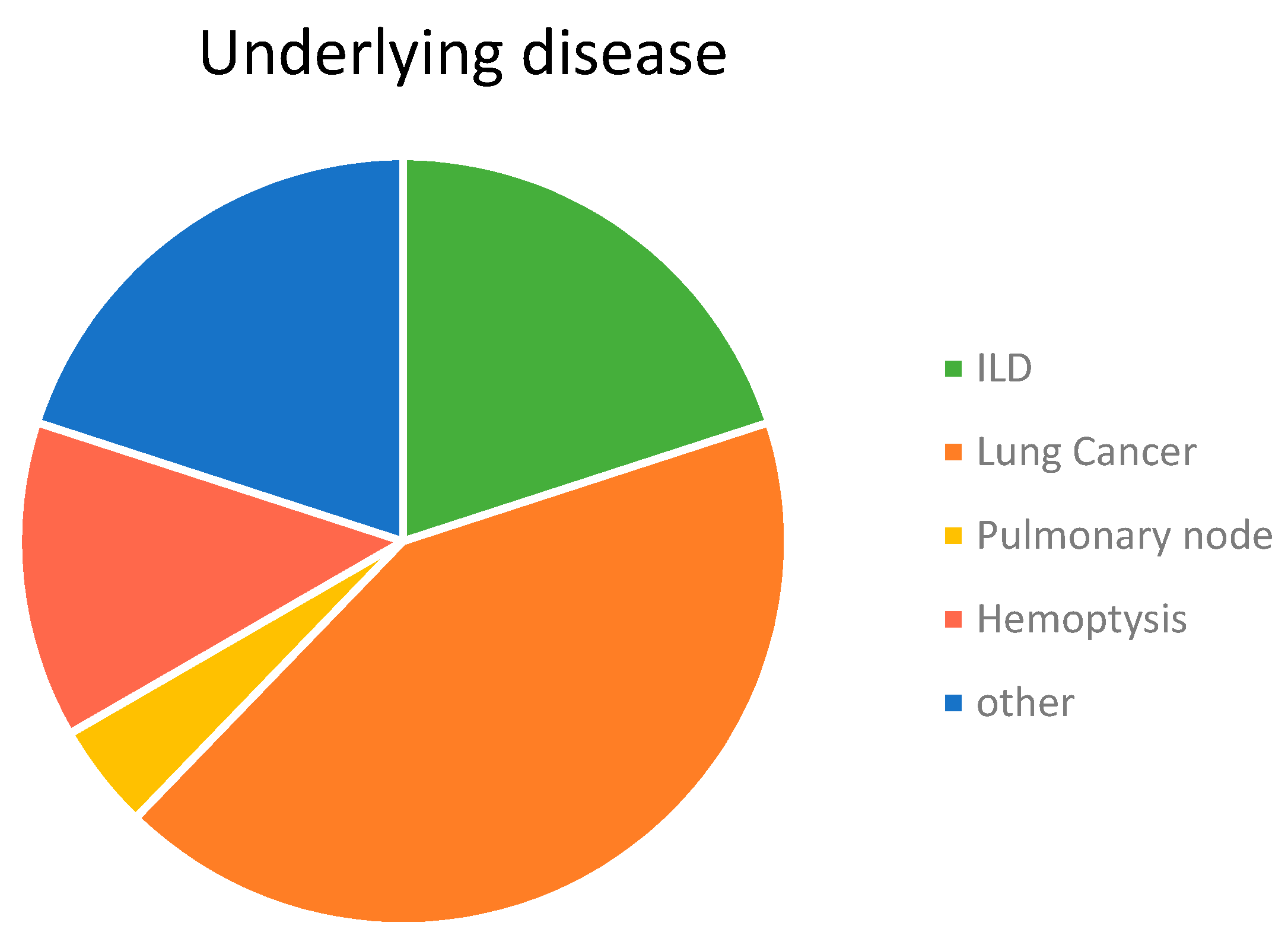
3.1. Characteristics of Participants

Demographic characteristics of the patients and their houses are shown in Table 1. The studied population consisted of 25 male (55.6%) and 20 female (44.4%) patients. Bronchoscopy was indicated on the basis of 15 different non-infectious clinical situations with lung cancer being the most frequent (42.2%), which was followed by diffuse interstitial lung disease (ILD) (20%), hemoptysis (13.3%), and pulmonary nodules (4.4%). Eleven additional pathologies were detected including one in each of eleven patients (Figure 1). The professional activity of the patients was quite varied in which the largest group was related to the footwear industry (20%), known as one of the most important in the area, which was followed by building (15.5%). Nine patients (20%) declared that they had had more than one profession throughout their lives. With regard to foreign travel, almost half of the patients (22) had never left Spain and among the others, 13 (28.9%) had only visited one country/city. Nine patients (22.2%) declared they had travelled to different destinations in the world. Finally, 33.3% of the patients declared having had exposure to animals or animal products. Most of them (6/15) had birds in cages (canaries, a goldfinch, and a siskin) and 40% (6/15) had pigeons, hens, and/or quails. Of the 20 patients who allowed sampling of their homes, 13 (65%) lived in flats in an urban environment and seven (35%) lived in houses located in villages (2), on town limits (3), or in rural areas (2). Pets were present in seven (35%) of the studied dwellings (see Supplementary Materials. Table S1: patients and houses).

3.2. HTS-Based Mycobiome Characterization

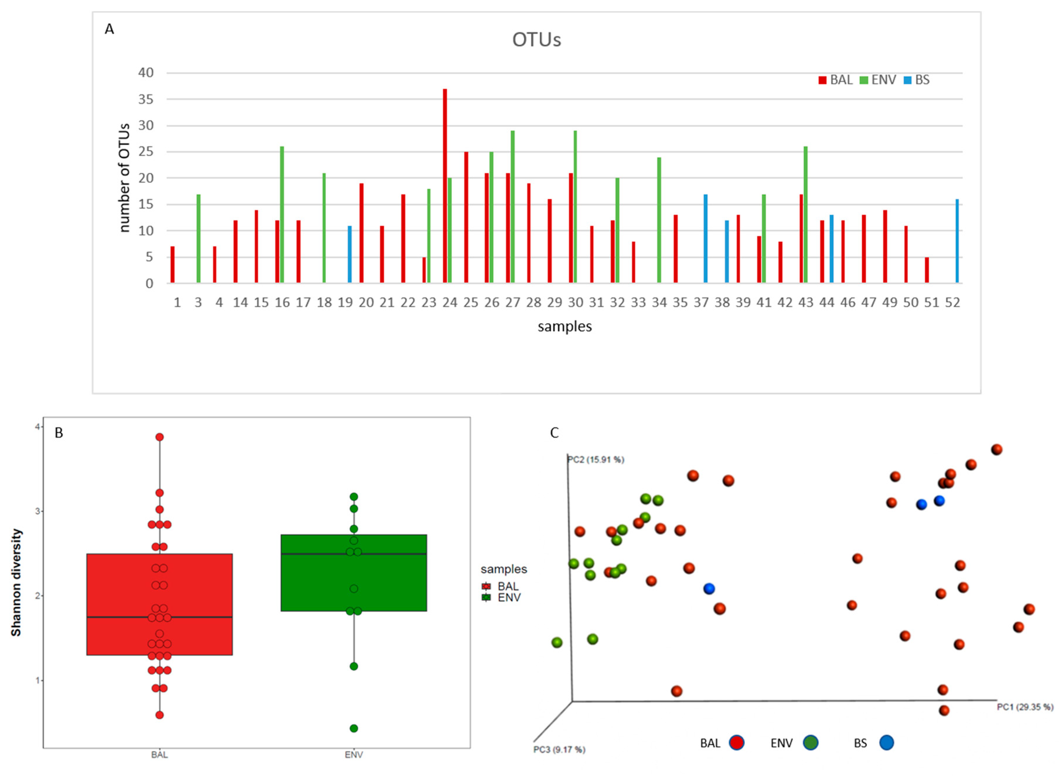
3.2.1. Richness and Diversity
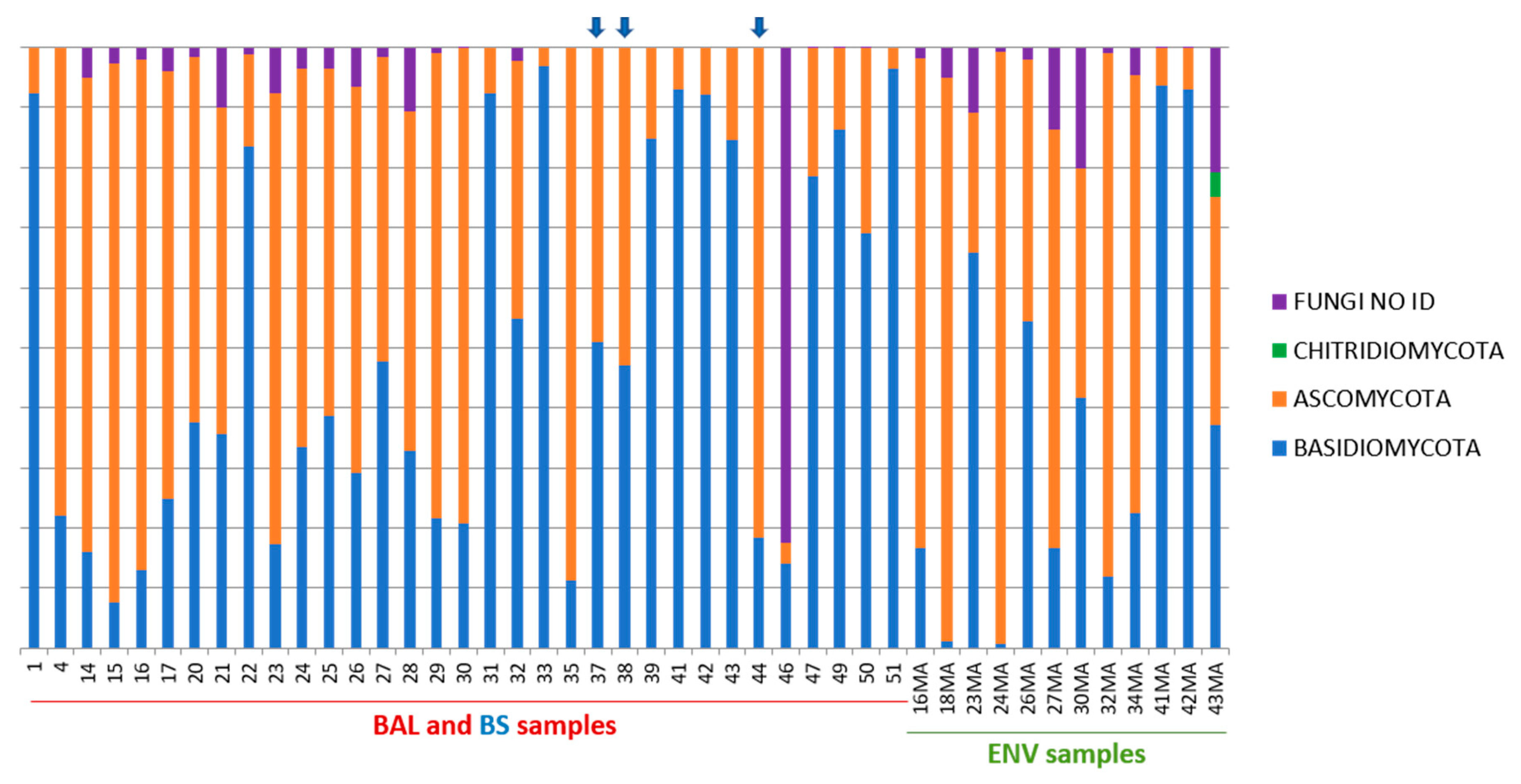
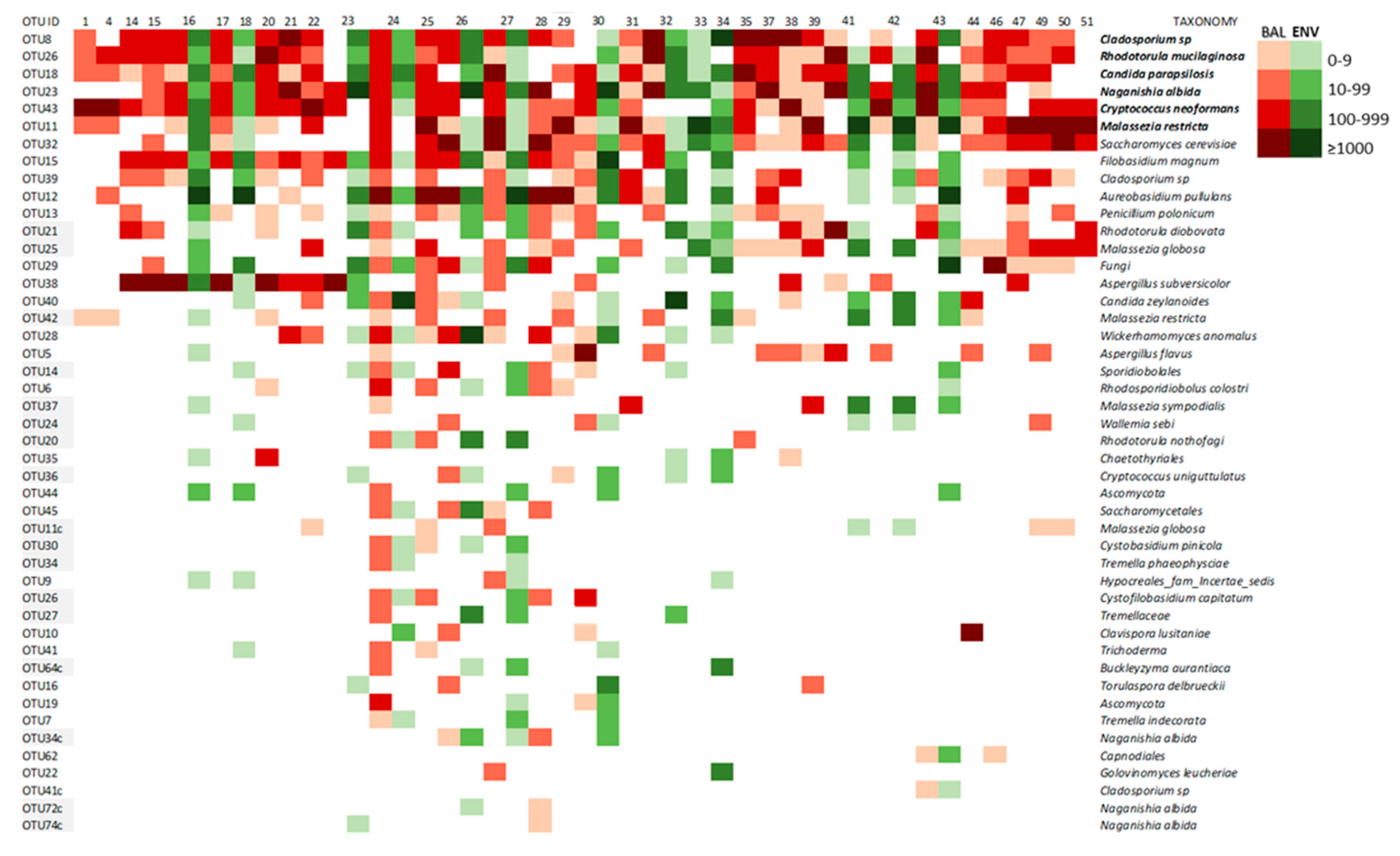
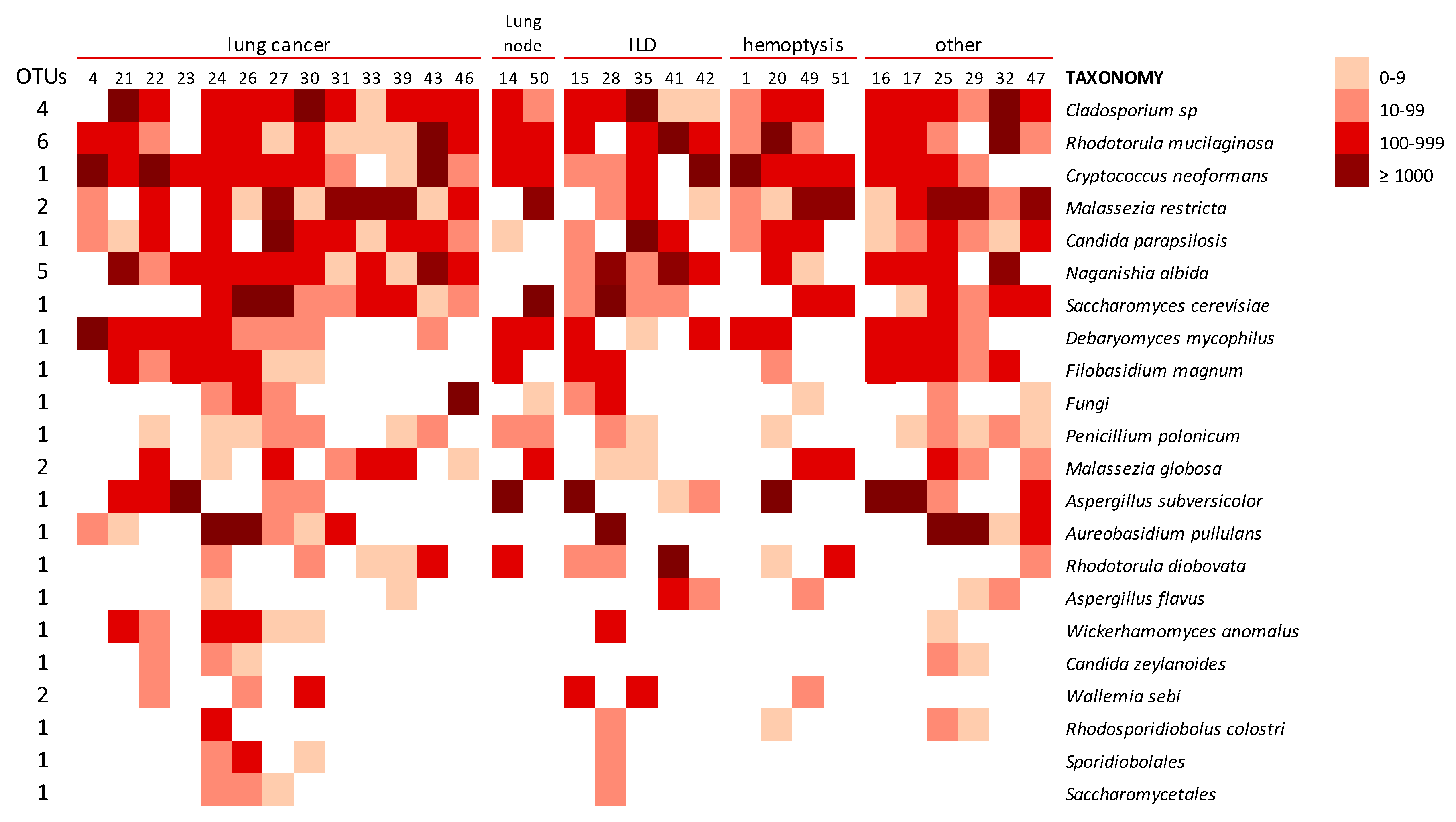
Forty-five BAL, 5 BS, and 20 ENV samples submitted to massive sequencing analysis yielded 7,015,877 sequences (5,046,424 from BAL, 1,861,275 from ENV samples, and 108,178 from BS). After chimeric detection and contaminant filtering, 4,012,543 ITS sequences were included in the subsequent analysis (2,826,988, 1,110,227, and 75,328 from BAL, ENV, and BS samples, respectively) with a mean value around 60,000 for BAL and ENV samples and 15,000 for bronchoscopes (Table 2). After sample rarefaction, 77 fungal Operational Taxonomic Units (OTUs) at 97% identity were identified, ranging from 5 to 37 in BAL samples, from 17 to 29 in ENV samples, and from 11 to 17 in BS samples (Figure 2A). Shannon-Wiener index ranged from 0.6 to 3.9 in BAL samples and from 0.4 to 3.2 in ENV samples (Figure 2B, Table 3). The similarity among different microbial communities (beta diversity) analysed by the Bray-Curtis index showed a very high similarity among domestic environmental samples (average 21.88) and a high diversity in BAL samples (average similarity 9.93). Accordingly, all ENV samples clustered together while BAL and BS samples were distributed in two different clusters (Figure 2C).

3.2.2. Taxonomy

Basidiomycota and Ascomycota phyla were detected in all types of samples with a relatively higher presence of Basidiomycota in the lungs (BAL samples 49.4%) than in environmental samples (38.2% in ENV) that had a slightly higher percentage of Ascomycota (54.65% in ENV) (Figure 3). The phylum Chytridiomycota was very poorly represented and only detected as 0.33% of the sequences in ENV samples (Table 3 and Figure 3). Bronchoscopes were shown to be contaminated by some of the fungi detected in lungs even though the number of sequences in these samples was very low (Table 2).
In our study, 1.1% of the detected sequences could belong to new species or genera as they could not be identified below family level (Supplementary material. Table S2: Taxonomy). These corresponded to 13 OTUs (16.9 %) that remained identified at different taxonomic levels. One of these OTUs (OTU29) identified as “unknown fungi” had no match under the kingdom level and was detected in many BAL and ENV samples. Other OTUs could only be classified at phylum Ascomycota (3.9%) or at different orders level (10.4%), according to the UNITE database (Table 4).

3.2.3. Core Mycobiome

OTUs that were present with sufficient relative abundance in more than 85% of the samples were considered a core mycobiome. For BAL samples, OTU8 that was present in 90.9% of samples. OTU26 and OTU43 were both present in 86.7% of them and were considered as the core mycobiome of the lungs. These three OTUs presented hits with Cladosporium sp., Rhodotorula mucilaginosa, and Cryptococcus neoformans, respectively (Figure 4). For the environmental samples, five OTUs (8, 12, 15, 18, and 23) that matched with Cladosporium sp., Aureobasidium pullulans, Filobasidium magnum, Candida parapsilosis, and Naganishia diffuens, respectively, were detected in 100% of the ENV samples studied and, therefore, considered as the core mycobiome of the houses. Five of these OTUs have an important presence in both kinds of samples from patients and environments (Table 5). Additionally, the OTU11 that matched with the species Malassezia restricta was detected in 83.3% of lungs and houses and was equally highly abundant in both of them (BAL and ENV) (Table 5 and Figure 5).

3.2.4. Lung Mycobiome

The fungal lineages constituting the BAL core mycobiome look very highly micro-diverse, as indicated by the presence of several OTUs being less abundant than the ones in Figure 5 but still matching to the same species. The number of these additional OTUs for Cladosporium sp. and Rh. mucilaginosa was four and six, respectively. These results reinforce the important presence of these fungi in the lungs. Other taxa with several OTUs detected in BAL samples corresponded to N. albida (5 OTUs), M. restricta (2 OTUs), Malassezia globose (2 OTUs), and Wallemia sebi (2 OTUs) (Figure 6). The distribution of different taxa was heterogeneous among the patients with different symptoms and diseases, and every analyzed individual had a specific mycobiome profile, which could not be related to the diagnosis nor clinical status (Figure 6).

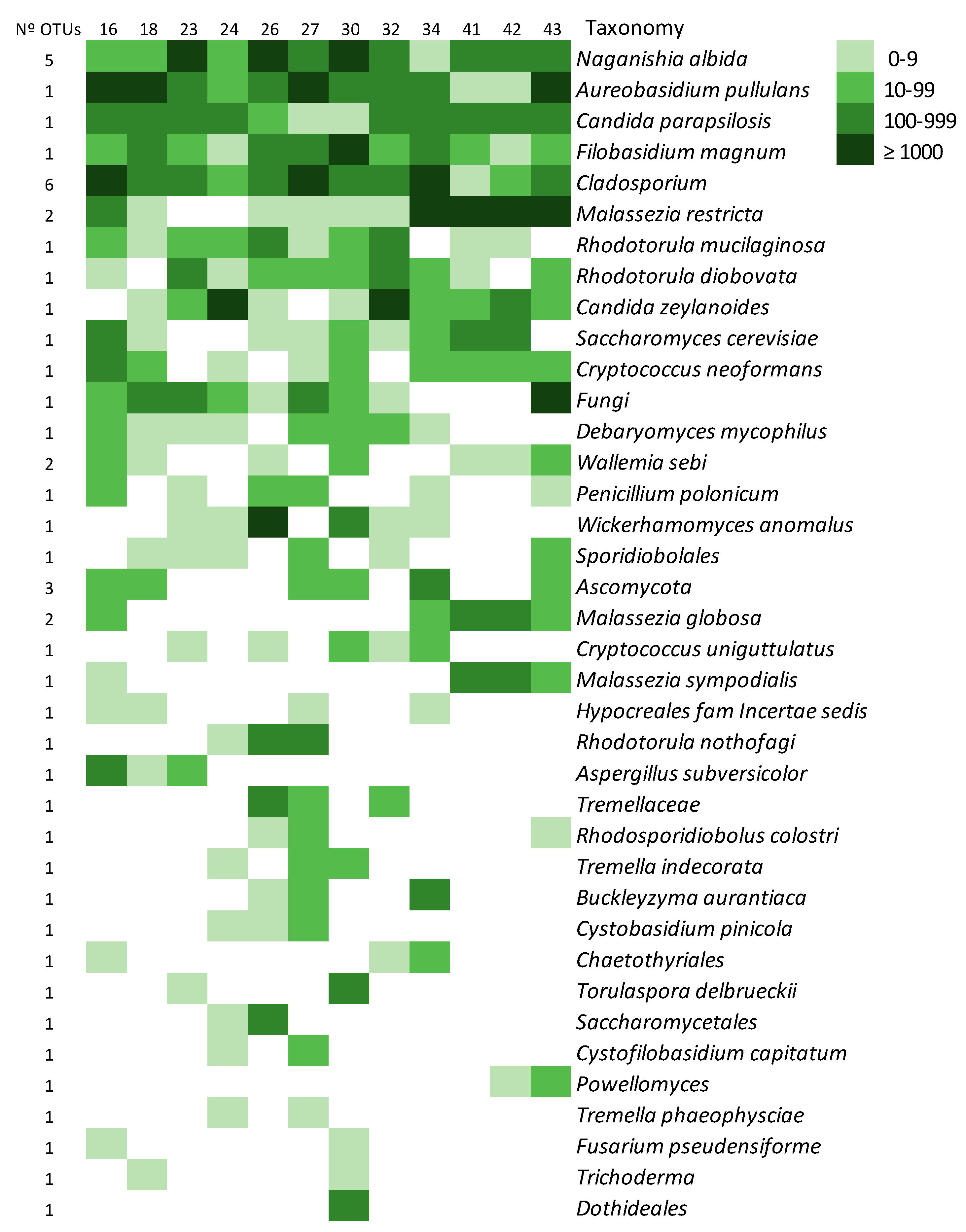
3.2.5. The Environmental Mycobiome

In samples from houses (ENV), three species were detected in all of them: Naganishia albida, Aureobasidium pullulans, Filobasidium magnum, Cladosporium sp., and Candida parapsilosis, which had 947, 1323, 565, 191, and 174 sequences per sample, respectively. These species constitute the core mycobiome of the domestic environment (Figure 7). Other species like Malassezia restricta, Rh. mucilaginosa, Rh. diobovata, and Candida zeylanoides were present in 83.3% of the samples with a high number of sequences. As with the lungs, each domestic environment had its specific profile, which was not related to the type of house, area of the location, or presence of pets (Figure 7).

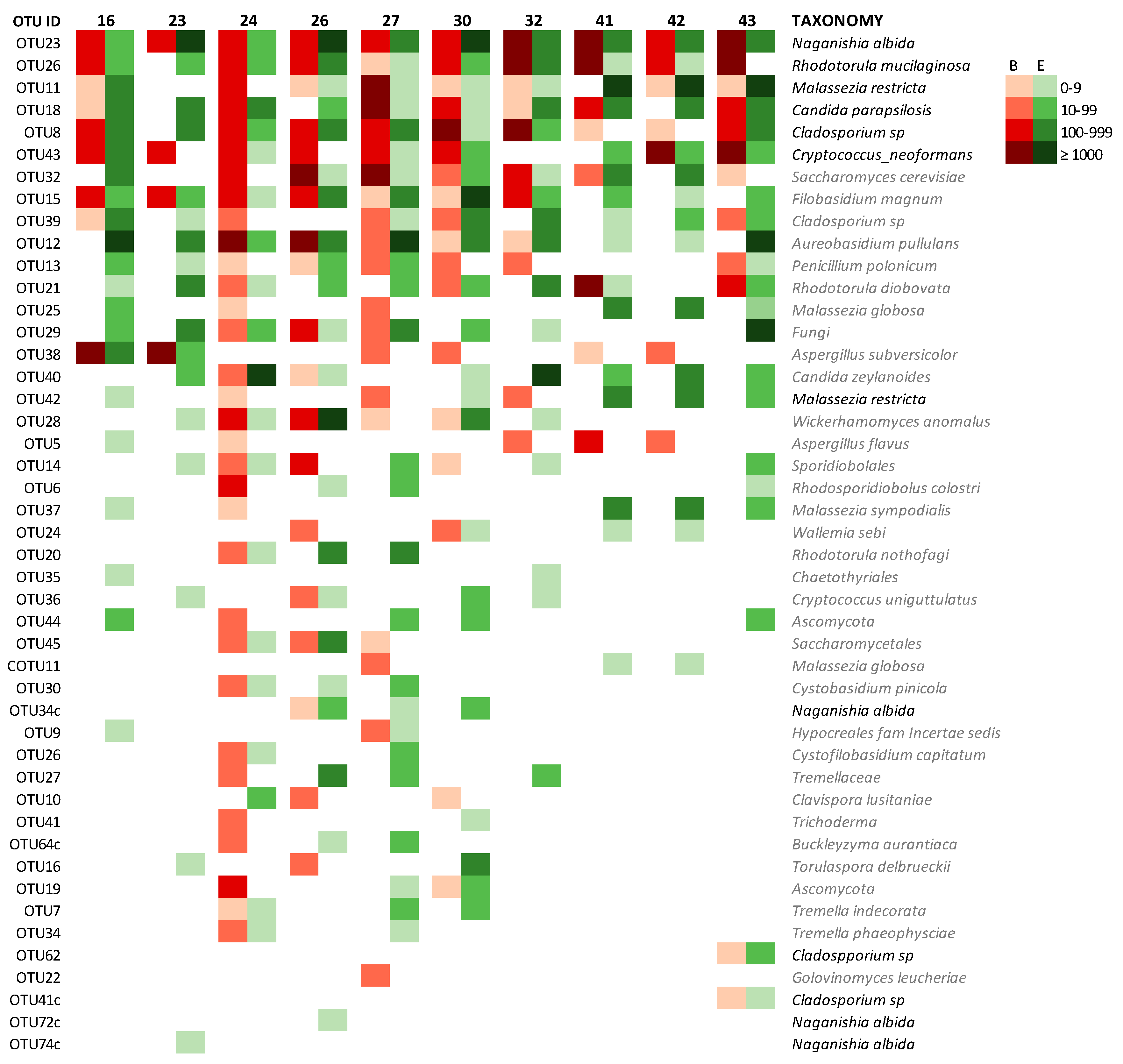
3.2.6. Mycobiome of Lungs and Environments

The comparison of the lung and the house mycobiome of the same patient is represented on the heatmap of Figure 8. Results obtained from BAL and ENV of patients that offered the two kinds of samples were analyzed and the similarity of the two profiles (lung and house) for each patient was assessed as the percentage of coincidence in the presence/absence of each OTU in both environments. The coincidence ranged from 71.4% to 81.8% with a mean value around 75% (Table 6). In general, lungs and houses mostly shared the OTUs corresponding to the fungi described as core mycobiome. In patients 16, 27, 30, and 32, these OTUs are 100% coincident (Figure 8). The similarity is also demonstrated by the presence of minority fungal OTUs like the one corresponding to A. subversicolor (OTU38), which is present in BAL and ENV of patients 16 and 23, or OTU21 (Rh. diobovata) coincident in BAL and ENV of patients 24, 30, 41, and 43 and the very rare OTUs 62 and 41, that were only detected in patient 43 in both samples BAL and ENV (Figure 8). The similarity of the fungal profiles is more evident for patients whose samples had higher fungal richness. In this regard, patients 24, 26, and 30 had mycobiomes highly diverse in the number of different OTUs that also were highly abundant in sequences (Table 6) and these three patients show around 50% of coincidence in the OTUs present in their two samples and from 75.3% to 79.2% of coincidence in the global profile. The situation of OTU12, which corresponds to A. pullulans, is of interest. This fungus had been described as highly prevalent in indoor samples [40] and we detected it in all ENV samples studied, and also in 43.3% of the BAL samples and in one bronchoscope (33.3%). The presence/absence of this species (A. pullulans) was the only variable that could explain the distribution of BAL and BS samples into two different clusters by the Bray-Curtis index (Figure 2C).

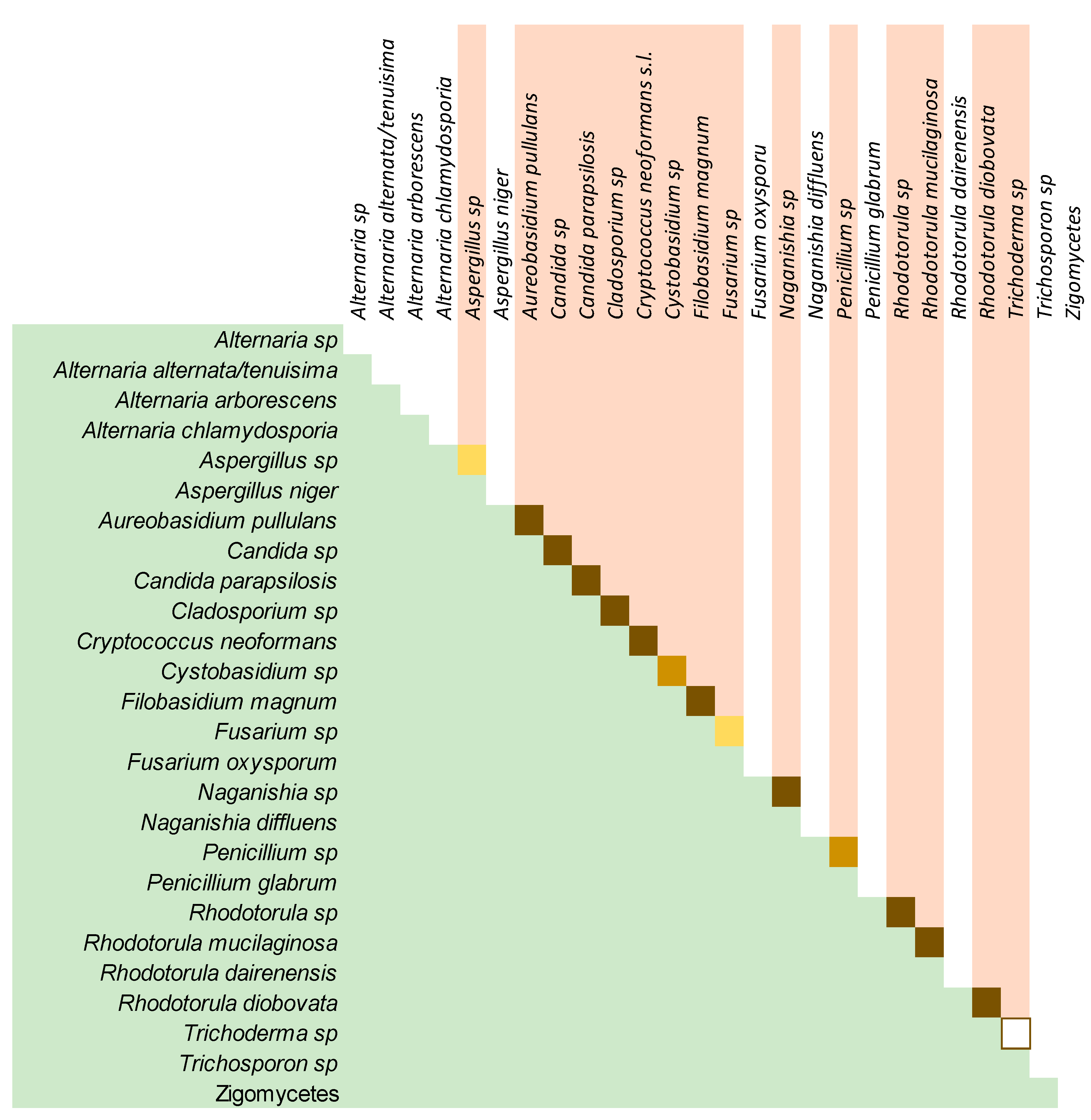
3.3. The Culturable Mycobiome

Cultures of BAL samples were mainly negative. However, after the incubation period, one or a few fungal colonies were detected in 46.7% (21) of the plates. The phenotypic identification of these fungi showed species of the genus Rhodotorula in the samples of five patients: Penicillium spp. and Aspergillus spp. in the BAL of four and three patients, respectively, Naganishia sp. in three, Cryptococcus neoformans sensu lato in two of the samples, and Candida sp and Cystobasidium sp. one of each in the sample of the same patient (patient 3). The molecular identification of some of these colonies identified them as Rh. mucilaginosa, P. glabrum, and Cr. neoformans sensu stricto. Most of the fungi isolated in cultures were also detected in the HTS study of the same patients. Samples 4 and 41 had N. albida in their HTS study and their cultures showed one CFU of a morphotype of Naganishia spp. in each sample. Rh. mucilaginosa was detected by HTS in the lungs of patients 33 and 37 and their cultures shown one and six CFU, respectively, of a yeast with morphotype corresponding to Rhodotorula sp. The culture of the BAL sample of patient 22 had two filamentous colonies corresponding with morphotypes of Penicillium spp. and Aspergillus spp. and the HTS analysis showed the presence of Penicillium polonicum and Aspergillus subversicolor in these samples. Finally, from the BAL sample of patient 23, P. polonicum was also identified by massive sequencing and a colony of a fungus morphologically corresponding to Penicillium spp. was obtained. Other isolates were obtained from cultures of BAL samples that were discarded for the HTS analysis after the procedure of cleaning the sequences and, therefore, we cannot compare the two methods. This is the case of patient 3 in whose sample the growth of two CFU corresponding to Candida sp., two to Naganishia sp., and one to Cystobasidium sp. were detected (Supplementary materials. Table S3: cultures).
In contrast with the BAL samples, cultures of environmental samples produced larger numbers of fungal colonies. Growth was detected in almost all the inoculated plates and a total of 12,994 CFU were detected. These included 387 morphotypes (188 filamentous fungi and 199 yeast-like isolates). Two colonies of each morphotype were selected for further study, except in cases in which only one colony of a specific morphotype was available. That selection gave 718 isolates that were submitted to phenotypical and molecular procedures for the identification at genus or species level and 168 of which were successfully identified (43.4%). The most represented genera in cultures for the yeast-like colonies were Rhodotorula, isolated from 80% of the houses, followed by Trichosporon, Naganishia, and Candida in 40% to 55% of houses, Filobasidium in only 10%, and Cryptococcus in 25% of the 20 domestic environments but with the highest number of colonies in the plates (1340 CFU). Among the filamentous fungi, Aureobasidium was isolated in 70% of the houses, and Penicillium, Aspergillus, Alternaria, and Cladosporium were present in 40% to 60% of the 20 houses and Cladosporium showed the highest number of colonies detected (Supplemental material cultures). All these genera were also detected by HTS in the same samples except for Trichosporon, Alternaria, and Aspergillus. For Aspergillus, the phenotypic characterization of the isolates obtained in ENV samples of patients 26, 27, and 30 showed A. niger and, in samples 27 and 30, an additional unidentified species of Aspergillus was isolated. The HTS analysis did not detect A. niger in any of the studied samples and the OTUs corresponding to this genus (OTU5 and 38) were not detected in the houses of these three patients. For Trichosporon and Alternaria, although both were isolated from almost half of the ENV samples (55% and 45%, respectively), the HTS did not detect these genera. For the genus Cladosporium, it was detected in all ENV samples by HTS and cultured from 40% (eight houses) of them (Figure 9).

4. Discussion

The composition of the indoor environment is presumably strongly influenced by the air and dust outdoors [41]. Accordingly, Ascomycota have been described as highly predominant in outdoor and indoor environments by previous culture-based studies [20,21,22,42]. In these studies, the house environment is mainly explored in relation to asthma patients and the analysis of fungal components in the environment is mostly focused in the detection of fungi, which are potently allergenic, such as the genera Aspergillus, Cladosporium, Penicillium, or Alternaria. Most of these belong to the phylum Ascomycota. In these works, yeasts were almost absent or may not be reported as they are not among the allergenic fungi. Therefore, the phylum Basidiomycota, which harbors many important human-related yeasts, was a rare minority in most of these studies. However, our results, together with other recent molecular studies, show a more significant presence of members of Basidiomycota and describe yeast of the genus Cryptococcus and Malassezia as highly present in indoor environments [23,24,40,43]. The work of Green et al. [23] specifically described 41% of the fungal microbiota inside a building as members of Basidiomycota and 55% of Ascomycota species, which is almost the same distribution detected in the houses of our patients.
The lung mycobiome. The analysis of the results of the HTS study of the BAL samples offered 30 different profiles in fungal composition. That means each patient had a specific distribution of fungal OTUs in their lung mycobiome (Figure 6), which is in agreement with the findings of other authors [8,44]. Nevertheless, the group of OTUs considered as a core mycobiome were repeatedly detected in these samples and the presence of genus like Malassezia, Candida, and Cryptococcus coincide with previous reports [11,13,14,45,46,47,48]. However, in these studies, a major presence of Candida spp. is described and Rhodotorula species were rarely reported previously in the lung mycobiome. The predominance of Candida spp., including C. albicans, which is absent in our study, would be explained by the frequent use of sputum as the sample representative of LRT mycobiome [7,11,47,49]. That kind of sample would carry fungi from the upper respiratory tract, where Candida species are highly prevalent [13,27,50]. Moreover, these studies mainly aim to correlate the mycobiome composition of the lungs with the clinical status of patients suffering from chronic inflammatory pulmonary diseases, such as CF, COPD, asthma, and bronchiectasis [11,49,51,52,53]. Although no specific correlation has been established between disease and mycobiome composition so far, the severity of the inflammatory process seems to have an inverse relationship to the diversity of the microbiome [3,4,8,11]. According to what is described as inflammatory diseases of the lung, such as pneumonia, acute respiratory distress syndrome (ARDS), asthma, and chronic obstructive pulmonary disease (COPD) [54], most of our patients had non-inflammatory disorders (Figure 1). Thus, inflammatory processes may correspond to a more similar mycobiome, less diverse than one of the persons with a “non-inflammatory” status. Among our patients, no significant relationship could be established between fungal profiles and any of their underlying pathologies or clinical situations. However, the high number of sequences of Malassezia species in cancer patients should be pointed out (Figure 6).
Apart from the underlying disease, the possible influence of clinical and demographic variables such as age, sex, profession, travel history, type of house, and lifestyle did not show any significant relationship with the richness, diversity, or fungal composition of the BAL samples of these groups of patients.
Relationship between lungs and dwellings. Comparing OTUs instead of species or higher taxa gives higher sensitivity to the analysis. The profiles of samples of the 10 patients that had the two kinds of samples showed that all lungs and houses harbor OTU23, which corresponds to Naganishia albida, a fungus that was not frequently described previously as a member of mycobiome. It was a former member of the genus Cryptococcus (Cr. albidus = N. albida) [54] and has been reported in the environment and also as an infrequent opportunistic pathogen [25,55]. Perhaps its presence in other studies is somehow hidden under the Cryptococcus spp. Group, which is described in different works [13,23,24,40].
The high similarity of the fungal composition of the LRT of our patients and their dwellings is in agreement with the hypothesis that the environment is one of the most significant determinants of the composition of the human microbiome [9]. Previous culture-based studies showed a higher prevalence of Aspergillus and Penicillium [20,21,22,42], which are traditionally the most expected genus in indoors environments. It seems that this is a bias of culture-based procedures as our results are coincident with other molecular approaches [24,43,56]. Nevertheless, Penicillium polonicum and two species of Aspergillus (A. subversicolor and A. flavus) were detected in 50% (6/12) and 33.3% (4/12) of the samples from houses and the genera were also cultured. Surprisingly, these two genera were more prevalent among the samples from the lower respiratory tract of the patients than from their environment (Figure 8).
Some authors have attempted to establish a difference between what is considered real human microbiota, and the microorganisms that can be detected in the lungs as a result of a transient colonization that most likely come from the environment [57,58]. This distinction is not easy to establish. Although, in our results, the lungs and houses showed different members of the core mycobiome. The species related to the most abundant and prevalent OTUs in the two kinds of samples tend to overlap and there is not enough evidence to declare which ones had evolved to be adapted to the lungs in contrast with the ones that are frequently inhaled and belong to the external environment. The study by Kramer et al. [57] concluded that the mycobiome is dominated by transient species from inhaled air rather than colonization. This situation is estimated by the analysis of sequential sputum samples from patients of cystic fibrosis that were frequently under antibiotic treatment. Sputum carries many microorganisms from the upper area via mouth, and these areas are more exposed to the external environment than the LRT. Therefore, presumably, these samples would reflect a higher variability due to external influence than the BAL ones. Considering that this situation can be extrapolated to the lower area via only speculation. Neither the previously mentioned works nor ours present enough evidence to make a clear distinction between the resident and transient biota of the lower airway.
Cultures confirmed the presence of most of the fungi that were prevalent in the samples studied by HTS. The three species detected as environmental core mycobiome in the molecular study were also detected by culture (A. pullulans, C. parapsilosis and Naganishia sp.). Other species and genera with a significant presence in the molecular approach like Rhodotorula and Cryptococcus and the filamentous fungi Aspergillus, Penicillium, and Cladosporium were also cultured from many environmental samples but also from some of the BAL samples in which Candida spp. was also detected. Only the genus Malassezia, which had a high relative presence in most of the lungs and houses studied, was not detected by culture, which can be explained by the special requirements of the members of this genus for in vitro growth [59]. The scarce fungal growth obtained from BAL samples compared to one of the same species from ENV samples leads us to consider the vital status of the fungi detected by HTS methods. The very low rate of isolation of fungi whose DNA is detected and that can easily grow in common media led us to conclude that most of them are not viable and they may not even be cells but rather debris of nucleic acids partially destroyed by the local immune system.
The high rate of detection of unidentified fungal sequences seems to be a common fact and has also been reported in other works [41,43,60]. We consider this a significant target for future studies.

5. Conclusions

Indoor environmental mycobiome is the result of the mixed outdoor fungal components and human (and pet) mycobiome. Humans exchange fungi with their environment and the proportions of different species or subspecies (OTUs) in both sites are the result of specific factors that the fungi find in each of these environments (temperature, nutrients, pH, immune response, and others). Therefore, some fungi are more prevalent in lungs than in the environment, like Rh. mucilaginosa, Cr. neformans, or Aspergillus spp., while others are more abundant in the air or dust of houses (e.g., M. restricta and Capnodiales). In any case, it seems that there is a reciprocal influence of the two environments and fungi inhaled from indoor air contribute to the composition of the human mycobiome and, in the same way, the human mycobiome may influence the indoor environmental mycobiota, which also receives most of its members from the outdoors.
Fungal species, such as Rh. mucilaginosa, C. parapsilosis, N. albida, M. restricta, A. pullulans, Cr. neoformans sl., and Cladosporium spp., are the most prevalent in both environments studied. This agrees with previous studies but also adds more information about species that were less frequently described, such as Naganishia spp. Nevertheless, we have to assume that the study of a unique molecular target obviates the presence of fungi-like Pneumocystis jirovecii, which is a well-known commensal of the LRT of different groups of patients and the normal population [18] but cannot be detected by this approach due to its low number of copies of the ITS region [61]. Perhaps other unknown fungi may remain hidden in the mycobiome when the study is only based on ITS amplicon sequencing. In addition, in this study as well as in many others performed with the same molecular approach, a significant number of OTUs could not be related to any specific fungal taxon through the UNITE database and other databases. That means that much remains to be explored regarding the lung mycobiome and the house environment, and their role in human health and disease.
Our results confirm that the environment could be the main determinant of the lung mycobiome, and that the health condition of individuals, specifically their inflammatory status, may influence the diversity and richness of the mycobiota more than its composition. However, it is important to note that mycobiome analysis offers only a partial view of the state of the microbiome of an environment, and its relationship to other microbial communities (bacteria, protozoa, viruses, and archaea) should always be considered for a robust global assessment.

Supplementary Materials

The following are available online at https://www.mdpi.com/2076-2607/8/11/1717/s1. Table S1: patients & houses. Table S2: taxonomy of the identified OTUs. Table S3: cultures. Figure S1: Project work flow. Figure S2: Rarefaction curves. Document S1: patient data sheet.

Author Contributions

Conceptualization, E.L., J.A. (Josefa Antón), B.A., and M.F.C. Data curation, B.G. and M.F.C. Formal analysis, E.R.-P., C.F. (Consuelo Ferrer), and M.F.C. Funding acquisition, J.A. (Josefa Antón), B.A., and M.F.C. Investigation, E.R.-P., B.G., V.E., E.R., C.P.-M., E.G.-I., J.A. (Jorge Adsuar), P.P., and M.F.C. Methodology, E.R.-P., D.O., E.L., C.F. (Cleofé Fernández), C.F. (Consuelo Ferrer), B.G., V.E., J.F., and M.F.C. Project administration, M.F.C. Resources, E.L., C.F. (Cleofé Fernández), J.A. (Josefa Antón), B.A., J.F., and M.F.C. Supervision, J.A. (Josefa Antón), B.A., and M.F.C. Validation, J.A. (Josefa Antón), B.A., and M.F.C. Visualization, E.R.-P. and M.F.C. Writing—original draft, M.F.C. Writing—review & editing, E.R.-P., J.A. (Josefa Antón), B.A., and M.F.C. All authors have read and agreed to the published version of the manuscript.

Funding

This research was funded by the Spanish Association for Respiratory Diseases (SEPAR) through their grant program RESPIRA with number PII026/2016. E. Revelles (2015-16) and J. Adsuar (2018) received a grant from the Spanish Ministry of Education for their contribution in this work. E. Gómez-Imbernon received a grant from the University Miguel Hernandez for his contribution to this work (2017).

Acknowledgments

The authors want to give special thanks to the BIOBANK at the General University Hospital of Alicante (BIOBANCO-ISABIAL) for its essential contribution to the management of LBA samples. In addition, we want to acknowledge the generosity of the patients whose samples were studied with special mention to the ones who permitted the environmental sampling inside their homes. Finally, we thank Karen Neller for her professional English editing.

Conflicts of Interest

The authors declare no conflict of interest. The funders had no role in the design of the study, in the collection, analyses, or interpretation of data, in the writing of the manuscript, or in the decision to publish the results.

References

  1. Lai, G.C.; Tan, T.G.; Pavelka, N. The mammalian mycobiome: A complex system in a dynamic relationship with the host. Wiley Interdiscip. Rev. Syst. Biol. Med. 2019, 11, e1438. [Google Scholar] [CrossRef] [Green Version]
  2. Thomas, S.; Izard, J.; Walsh, E.; Batich, K.; Chongsathidkiet, P.; Clarke, G.; Sela, D.A.; Muller, A.J.; Mullin, J.M.; Albert, K.; et al. The Host Microbiome Regulates and Maintains Human Health: A Primer and Perspective for Non-Microbiologists. Cancer Res. 2017, 77, 1783–1812. [Google Scholar] [CrossRef] [Green Version]
  3. Enaud, R.; Vandenborght, L.E.; Coron, N.; Bazin, T.; Prevel, R.; Schaeverbeke, T.; Berger, P.; Fayon, M.; Lamireau, T.; Delhaes, L. The Mycobiome: A Neglected Component in the M Microbiota-Gut-Brain Axis. Microorganisms 2018, 6, 22. [Google Scholar] [CrossRef] [Green Version]
  4. Huffnagle, G.B.; Noverr, M.C. The emerging world of the fungal microbiome. Trends Microbiol. 2013, 21, 334–341. [Google Scholar] [CrossRef] [PubMed] [Green Version]
  5. Limon, J.J.; Skalski, J.H.; Underhill, D.M. Commensal Fungi in Health and Disease. Cell Host Microbe 2017, 22, 156–165. [Google Scholar] [CrossRef] [PubMed]
  6. Seed, P.C. The Human Mycobiome. Cold Spring Harb. Perspect Med. 2015, 5, a019810. [Google Scholar] [CrossRef] [PubMed] [Green Version]
  7. Aliouat-Denis, C.M.; Chabéa, M.; Delhaes, L.; Dei-Cas, E. Aerially transmitted human fungal pathogens: What can we learn from metagenomics and comparative genomics? Rev. Iberoam. Micol. 2014, 31, 54–61. [Google Scholar] [CrossRef]
  8. Richardson, M.; Bowyer, P.; Sabino, R. The human lung and Aspergillus: You are what you breathe in? Med. Mycol. 2019, 57, S145–S154. [Google Scholar] [CrossRef] [Green Version]
  9. Rothschild, D.; Weissbrod, O.; Barkan, E.; Kurilshikov, A.; Korem, T.; Zeevi, D. Environment dominates over host genetics in shaping human gut microbiota. Nature 2018, 555, 7695. [Google Scholar] [CrossRef]
  10. Beck, J.M.; Young, V.B.; Huffnagle, G.B. The Microbiome of the Lung. Transl. Res. 2012, 160, 258–266. [Google Scholar] [CrossRef] [Green Version]
  11. Soret, P.; Vandenborght, L.E.; Francis, F.; Coron, N.; Enaud, R.; Avalos, M.; Schaeverbeke, T.; Berger, P.; Fayon, M.; Thiebaut, R.; et al. Respiratory mycobiome and suggestion of inter-kingdom network during acute pulmonary exacerbation in cystic fibrosis. Sci. Rep. 2020, 10, 3589. [Google Scholar] [CrossRef] [PubMed] [Green Version]
  12. Ward, T.L.; Dominguez-Bello, M.G.; Heisel, T.; Al-Ghalith, G.; Knights, D.; Gale, C.A. Development of the human mycobiome over the first month of life and across body sites. mSystems 2018, 3, e00140-17. [Google Scholar] [CrossRef] [PubMed] [Green Version]
  13. Bittinger, K.; Charlson, E.S.; Loy, E.; Shirley, D.J.; Haas, A.R.; Laughlin, A.; Yi, Y.; Wu, G.D.; Lewis, J.D.; Frank, I.; et al. Improved characterization of medically relevant fungi in the human respiratory tract using next-generation sequencing. Genome Biol. 2014, 15, 487. [Google Scholar] [CrossRef]
  14. Cui, L.; Morris, A.; Huang, L.; Beck, J.M.; Twigg, H.L.; von Mutius, E.; Ghedin, E. The Microbiome and the Lung. Ann. Am. Thorac. Soc. 2014, 11, S227–S232. [Google Scholar] [CrossRef] [Green Version]
  15. Hyytiäinen, H.K.; Jayaprakash, B.; Kirjavainen, P.V.; Saari, S.E.; Holopainen, R.; Keskinen, J.; Hämeri, K.; Hyvärinen, A.; Boor, B.E.; Täubel, M. Crawling-induced floor dust resuspension affects the microbiota of the infant breathing zone. Microbiome 2018, 6, 25. [Google Scholar] [CrossRef] [Green Version]
  16. Respaldiza, N.; Medrano, F.J.; Medrano, A.C.; Varela, J.M.; de la Horra, C.; Montes-Cano, M.; Ferrer, S.; Wichmann, I.; Gargallo-Viola, D.; Calderon, E.J. High seroprevalence of Pneumocystis infection in Spanish children. Clin. Microbiol. Infect 2004, 10, 1029–1031. [Google Scholar] [CrossRef] [PubMed] [Green Version]
  17. Rojas, P.; Friaza, V.; García, E.; de la Horra, C.; Vargas, S.; Calderón, E.; Pavón, A. Early Acquisition of Pneumocystis jirovecii Colonization and Potential Association with Respiratory Distress Syndrome in Preterm Newborn Infants. Clin. Infect. Dis. 2017, 65, 976–981. [Google Scholar] [CrossRef] [Green Version]
  18. Morris, A.; Norris, K.A. Colonization by Pneumocystis jirovecii and its role in disease. Clin. Microbiol. Rev. 2012, 25, 297–317. [Google Scholar] [CrossRef] [Green Version]
  19. Iliev, I.D.; Underhill, D.M. Striking a Balance: Fungal Commensalism versus Pathogenesis. Curr. Opin. Microbiol. 2013, 16, 366–373. [Google Scholar] [CrossRef] [Green Version]
  20. Dannemiller, K.C.; Gent, J.F.; Leaderer, B.P.; Peccia, J. Indoor microbial communities: Influence on asthma severity in atopic and non-atopic children. J. Allergy Clin. Immunol. 2016, 138, 76–83. [Google Scholar] [CrossRef] [Green Version]
  21. Pakarinen, J.; Hyvarinen, A.; Salkinoja-Salonen, M.; Laitinen, S.; Nevalainen, A.; Makela, M.J.; Haahtela, T.; von Hertzen, L. Predominance of Gram positive bacteria in house dust in the low-allergy risk Russian Karelia. Environ. Microbiol. 2008, 10, 3317–3325. [Google Scholar] [CrossRef] [PubMed]
  22. Von Hertzen, L.; Hyvarinen, A.; Laatikainen, T.; Makela, M.J.; Nevalainen, A.; Vartiainen, E.; Haahtela, T. Risk of atopy associated with microbial components in house dust. Ann. Allergy Asthma. Immunol. 2010, 104, 269–270. [Google Scholar] [CrossRef] [PubMed]
  23. Green, B.J.; Lemonsa, A.R.; Parkb, Y.; Cox-Ganserb, J.M.; Parkb, J.H. Assessment of fungal diversity in a water-damaged office building. J. Occup. Environ. Hyg. 2017, 14, 285–293. [Google Scholar] [CrossRef] [PubMed]
  24. Green, B.J. Emerging Insights into the Occupational Mycobiome. Curr. Allergy Asthma. Rep. 2018, 18, 62. [Google Scholar] [CrossRef]
  25. Yashiro, E.; Savova-Bianchi, D.; Niculita-Hirzel, H. Major Differences in the Diversity of Mycobiomes Associated with Wheat Processing and Domestic Environments: Significant Findings from High-Throughput Sequencing of Fungal Barcode ITS1. Int. J. Environ. Res. Public Health 2019, 16, 2335. [Google Scholar] [CrossRef] [Green Version]
  26. Krause, R.; Moissl-Eichinger, C.; Halwachs, B.; Gorkiewicz, G.; Berg, G.; Valentin, T.; Prattes, J.; Högenauer, C.; Zollner-Schwetz, I. Mycobiome in the lower respiratory tract—A clinical perspective. Front. Microbiol. 2017, 7, 2169. [Google Scholar] [CrossRef] [Green Version]
  27. Cui, L.; Lucht, L.; Tipton, L.; Rogers, M.B.; Fitch, A.; Kessinger, C.; Camp, D.; Kingsley, L.; Leo, N.; Greenblatt, R.M.; et al. Topographic Diversity of the Respiratory Tract Mycobiome and Alteration in HIV and Lung Disease. Am. J. Respir. Crit. Care Med. 2015, 191, 932–942. [Google Scholar] [CrossRef]
  28. Du Rand, I.A.; Blaikley, J.; Booton, R.; Chaudhuri, N.; Gupta, V.; Khalid, S.; Mandal, S.; Martin, J.; Mills, J.; Navani, N.; et al. British Thoracic Society Guideline for Diagnostic Flexible Bronchoscopy in Adults. Thorax 2013, 68, i1–i44. [Google Scholar] [CrossRef] [Green Version]
  29. Google Maps. Available online: https://www.google.es/maps (accessed on 19 November 2018).
  30. Colom, M.F.; Hagen, F.; Gonzalez, A.; Mellado, A.; Morera, N.; Linares, C.; García, D.F.; Peñataro, J.S.; Boekhout, T.; Sánchez, M. Ceratonia siliqua (carob) trees as natural habitat and source of infection by Cryptococcus gattii in the Mediterranean environment. Med. Mycol. 2012, 50, 67–73. [Google Scholar] [CrossRef] [Green Version]
  31. Pemán, J.; Martín Mazuelos, E.; Rubio, M.C. Guia Práctica de identificación y Diagnóstico en Micología Clínica, 2nd ed.; Revista Iberoamericana de Micologia-Elsevier: Bilbao, Spain, 2007. [Google Scholar]
  32. De Hoog, G.S.; Guarro, J.; Gené, J.M.; Figueras, M.J. Atlas of clinical fungi, 2nd ed.; ©Centraalbureau voor Schimmelcultures: Utrech, The Netherlands; Universitat Rovira I Virgili: Reus, Spain, 2000; p. 1126. [Google Scholar]
  33. White, T.J.; Bruns, T.; Lee, S.; Tailor, S. Amplification and direct sequencing of fungal ribosomal RNA genes for phylogenetics. In PCR Protocols: A Guide to Methods and Applications; Innins, M.A., Gelfand, D.H., Sninsky, J.J., White, T.J., Eds.; Academic Press: San Diego, CA, USA, 1990; pp. 315–322. [Google Scholar]
  34. Ferrer, C.; Colom, M.F.; Frasés, S.; Mulet, E.; Alió, J. Detection and Identification of fungal pathogens by PCR and by ITS2 5.8S rDNA Typing in ocular infections. J. Clin. Microbiol. 2001, 39, 2873–2879. [Google Scholar] [CrossRef] [Green Version]
  35. Caporaso, J.G.; Kuczynski, J.; Stombaugh, J.; Bittinger, K.; Bushman, F.D.; Costello, E.K.; Fierer, N.; Peña, A.G.; Goodrich, J.K.; Gordon, J.I.; et al. QIIME allows analysis of high-throughput community sequencing data. Nat. Methods 2010, 7, 335–336. [Google Scholar] [CrossRef] [PubMed] [Green Version]
  36. Edgar, R.C.; Haas, B.J.; Clemente, J.C.; Quince, C.; Knight, R. UCHIME improves sensitivity and speed of chimera detection. Bioinformatics 2011, 27, 2194–2200. [Google Scholar] [CrossRef] [Green Version]
  37. Aho, V.T.E.; Pereira, P.A.B.; Haahtela, T.; Pawankar, R.; Auvinen, P.; Koskinen, K. The microbiome of the human lower airways: A next generation sequencing perspective. World Allergy Organ. J. 2015, 8, 23. [Google Scholar] [CrossRef] [PubMed] [Green Version]
  38. Weiss, S.J.; Xu, Z.; Amir, A.; Peddada, S.; Bittinger, K.; Gonzalez, A.; Lozupone, C.; Zaneveld, J.R.; Vazquez-Baeza, Y.; Birmingham, A.; et al. Effects of library size variance, sparsity, and compositionality on the analysis of microbiome data. PeerJ PrePrints 2015, 3, e1157v1. [Google Scholar] [CrossRef]
  39. Lozupone, C.; Knight, R. UniFrac: A New Phylogenetic Method for Comparing Microbial Communities. Appl. Environ. Microbiol. 2005, 71, 8228–8235. [Google Scholar] [CrossRef] [Green Version]
  40. Hanson, B.; Zhoua, Y.; Bautista, E.J.; Urch, B.; Speck, M.; Silverman, F.; Muilenberg, M.; Phipatanakul, W.; Weinstock, G.; Sodergren, E.; et al. Characterization of the bacterial and fungal microbiome in indoor dust and outdoor air samples: A pilot study. Environ. Sci. Process Impacts 2016, 18, 713–724. [Google Scholar] [CrossRef] [Green Version]
  41. Adams, R.I.; Miletto, M.; Taylor, J.W.; Bruns, T.D. Dispersal in microbes: Fungi in indoor air are dominated by outdoor air and show dispersal limitation at short distances. ISME J. 2013, 7, 1262–1273. [Google Scholar] [CrossRef] [Green Version]
  42. Summerbell, R.C.; Saib, F.; Dales, R.; Nolard, N.; Kane, J.; Zwanenburg, H.; Burnett, R.; Krajden, S.; Fung, D.; Leong, D. Ecology of fungi in human dwellings. J. Med. Vet. Mycol. 1992, 30, 279–285. [Google Scholar] [CrossRef] [Green Version]
  43. Pitkaranta, M.; Meklin, T.; Hyvarinen, A.; Paulin, L.; Auvinen, P.; Nevalainen, A.; Rintala, H. Analysis of Fungal Flora in Indoor Dust by Ribosomal DNA Sequence Analysis, Quantitative PCR, and Culture. Appl. Environ. Microbiol. 2008, 74, 233–244. [Google Scholar] [CrossRef] [Green Version]
  44. Tipton, L.; Müller, C.L.; Kurtz, Z.D.; Huang, L.; Kleerup, E.; Morris, A.; Bonneau, R.; Ghedin, E. Fungi stabilize connectivity in the lung and skin microbial ecosystems. Microbiome 2018, 6, 12. [Google Scholar] [CrossRef] [PubMed] [Green Version]
  45. Mac Aogáin, M.; Chandrasekaran, R.; Lim, A.Y.H.; Low, T.B.; Tan, G.L.; Hassan, T.; Ong, T.H.; Ng, A.H.Q.; Bertrand, D.; Koh, J.Y.; et al. Immunological corollary of the pulmonary mycobiome in bronchiectasis: The CAMEB study. Eur. Respir. J. 2018, 52, 1800766. [Google Scholar] [CrossRef] [PubMed]
  46. Van Woerden, H.C.; Gregory, C.; Brown, R.; Marchesi, J.R.; Hoogendoorn, B.; Matthews, I.P. Differences in fungi present in induced sputum samples from asthma patients and non-atopic controls: A community-based case control study. BMC Infect. Dis. 2013, 13, 69. [Google Scholar] [CrossRef] [Green Version]
  47. Willger, S.D.; Grim, S.L.; Dolben, E.L.; Shipunova, A.; Hampton, T.H.; Morrison, H.G.; Filkins, L.M.; O’Toole, G.A.; Moulton, L.A.; Ashare, A.; et al. Characterization and quantification of the fungal microbiome in serial samples from individuals with cystic fibrosis. Microbiome 2014, 2, 40. [Google Scholar] [CrossRef] [Green Version]
  48. Zinter, M.S.; Dvorak, C.C.; Mayday, M.Y.; Iwanaga, K.; Ly, N.P.; McGarry, M.E.; Church, G.D.; Faricy, L.E.; Rowan, C.M.; Hume, J.R.; et al. Pulmonary Metagenomic Sequencing Suggests Missed Infections in Immunocompromised Children. Clin. Infect. Dis. 2019, 68, 1847–1855. [Google Scholar] [CrossRef] [PubMed]
  49. Delhaes, L.; Monchy, S.; Frealle, E.; Hubans, C.; Salleron, J.; Leroy, S.; Prevotat, A.; Wallet, F.; Wallaert, B.; Dei-Cas, E.; et al. The Airway Microbiota in Cystic Fibrosis: A Complex Fungal and Bacterial Community—Implications for Therapeutic Management. PLoS ONE 2012, 7, e36313. [Google Scholar] [CrossRef] [PubMed]
  50. Ghannoum, M.A.; Jurevic, R.J.; Mukherjee, P.K.; Cui, F.; Sikaroodi, M.; Naqvi, A.; Gillevet, P.M. Characterization of the Oral Fungal Microbiome (Mycobiome) in Healthy Individuals. PLoS Pathog. 2010, 6, e1000713. [Google Scholar] [CrossRef] [Green Version]
  51. Boutin, S.; Graeber, S.Y.; Weitnauer, M.; Panitz, J.; Stahl, M.; Clausznitzer, D.; Kaderali, L.; Einarsson, G.; Tunney, M.M.; Elborn, J.S.; et al. Comparison of Microbiomes from Different Niches of Upper and Lower Airways in Children and Adolescents with Cystic Fibrosis. PLoS ONE 2015, 10, e0116029. [Google Scholar] [CrossRef] [Green Version]
  52. Chambers, D.C.; Gellatly, S.L.; Hugenholtz, P.; Hansbro, P.M. JTD special edition ‘Hot Topics in COPD’—The microbiome in COPD. J. Thorac. Dis. 2014, 6, 1525–1531. [Google Scholar] [CrossRef]
  53. Krause, R.; Halwachs, B.; Thallinger, G.G.; Klymiuk, I.; Gorkiewicz, G.; Hoenigl, M.; Prattes, J.; Valentin, T.; Heidrich, K.; Buzina, W.; et al. Characterisation of Candida within the Mycobiome/Microbiome of the Lower Respiratory Tract of ICU Patients. PLoS ONE 2016, 11, e0155033. [Google Scholar] [CrossRef]
  54. Moldoveanu, B.; Otmishi, P.; Jani, P.; Walker, J.; Sarmiento, X.; Guardiola, J.; Saad, M.; Yu, J. Inflammatory mechanisms in the lung. J. Inflamm. Res. 2009, 2, 1–11. [Google Scholar]
  55. Liu, X.Z.; Wang, Q.M.; Göker, M.; Groenewald, M.; Kachalkin, A.V.; Lumbsch, H.T.; Millanes, A.M.; Wedin, M.; Yurkov, A.M.; Boekhout, T.; et al. Towards an integrated phylogenetic classification of the Tremellomycetes. Stud. Mycol. 2015, 81, 85–147. [Google Scholar] [CrossRef] [Green Version]
  56. Choe, Y.Z.; Blatt, D.B.; Yalcindag, A.; Geffert, S.F.; Bobenchik, A.M.; Michelow, I.C. Cryptococcus albidus Fungemia in an Immunosuppressed Child: Case Report and Systematic Literature Review. J. Pediatric Infect. Dis. Soc. 2020, 9, 100–105. [Google Scholar] [CrossRef]
  57. Kramer, R.; Sauer-Heilborn, A.; Welte, T.; Guzman, C.A.; Abraham, W.-R.; Höfle, M.G. Cohort study of airway mycobiome in adult cystic fibrosis patients: Differences in community structure between fungi and bacteria reveal predominance of transient fungal elements. J. Clin. Microbiol. 2015, 53, 2900–2907. [Google Scholar] [CrossRef] [Green Version]
  58. Hamm, P.S.; Taylor, J.W.; Cook, J.A.; Natvig, D.O. Decades-old studies of fungi associated with mammalian lungs and modern DNA sequencing approaches help define the nature of the lung mycobiome. PLoS Pathog. 2020, 16, e1008684. [Google Scholar] [CrossRef] [PubMed]
  59. Barberán, A.; Ladaub, J.; Leffa, J.W.; Pollard, K.S.; Menningerd, H.L.; Dunnd, R.R.; Fierera, N. Continental-scale distributions of dust-associated bacteria and fungi. Proc. Natl. Acad. Sci. USA 2015, 112, 5756–5761. [Google Scholar] [CrossRef] [PubMed] [Green Version]
  60. Kaneko, T.; Makimura, K.; Abe, M.; Shiota, R.; Nakamura, Y.; Kano, R.; Hasegawa, A.; Sugita, T.; Shibuya, S.; Watanabe, S.; et al. Revised Culture-Based System for Identification of Malassezia Species. J. Clin. Microbiol. 2007, 45, 3737–3742. [Google Scholar] [CrossRef] [Green Version]
  61. Guintoli, D.; Stringer, S.L.; Stringer, J.R. Extraordinarily low number of ribosomal RNA genes in P. carinii. J. Eukaryot. Microbiol. 1994, 41, 88S. [Google Scholar]
Figure 1. Distribution of patients according to the clinical situation that indicated the bronchoscopy. Others included: Wegener syndrome, dysnea, thymoma, TBC sequels, atelectasis, lung infiltrate, tracheal stenosis, Hodking and and Non-Hodgkin lymphoma, COPD, and cough.
Microorganisms 08 01717 g001
Figure 2. Fungal diversity in the studied simples. (A) Representation of the number of different OTUs detected in each sample. (B) Shannon-Wiener value for BAL and ENV samples. (C) Beta diversity. Bray-Curtis dissimilarity coefficient scores PCoA.
Microorganisms 08 01717 g002
Figure 3. Distribution of OTUs at a phylum level in each BAL, BS, and ENV samples. Upper blue arrows point at BS samples.
Microorganisms 08 01717 g003
Figure 4. Global distribution of putative fungal species detected in BAL, ENV, and BS samples. The intensity of the color corresponds with the number of sequences in a logarithmic scale. The left column shows the identification number of each specific OTU and their correspondent species are in the right column.
Microorganisms 08 01717 g004
Figure 5. Presence of the six most prevalent OTUs in BAL and ENV samples. The relative presence is represented by the percentage of samples in which they were detected and the abundance as the mean value of sequences per sample.
Microorganisms 08 01717 g005
Figure 6. Distribution of fungal species detected in BAL samples clustered by the underlying disease or symptoms (others corresponded to: Thymoma, TBC sequels, Atelectasis, lung infiltrate, tracheal stenosis, and COPD, respectively). The intensity of the color corresponds with the number of sequences that match with the putative species in the UNITE database at 97% similarity (logarithmic scale). The column at the left identifies the number of OTUs that showed a hit with the same species shown in the left column. Species present in less than 10% of the samples and with less than 100 sequences are not shown.
Microorganisms 08 01717 g006
Figure 7. Distribution of fungal putative species detected in ENV samples. The intensity of the color corresponds with the number of sequences that showed a hit with the species in the UNITE database at 97% similarity (logarithmic scale). The column at the left shows the number of OTUs that corresponded to each of the putative species. The ones present in less than 10% of the samples and with less than 100 sequences are not shown.
Microorganisms 08 01717 g007
Figure 8. Comparison between lung and environmental mycobiome. BAL and ENV profile of OTUs of each patient who permitted the analysis of both samples. The intensity of the color corresponds with the number of sequences that match with the species in the UNITE database at 97% (logarithmic scale). The column at the left shows the identification number of the OTU. The correspondent species are in the right column.
Microorganisms 08 01717 g008
Figure 9. Concordance between the detection of fungi by culture and by the HTS method. Green background: Genera and species obtained by the culture. Pink background: same genera and species in the HTS analysis. The co-occurrence of detection in the same samples is represented by squares at the intersection. Dark squares: 100% of positive samples by culture were detected by HTS. Golden squares: 40% and 70% of coincidence in culture and HTS (from left to right and up to down). Yellow squares: from 17% to 25% of coincidence. Empty squares: Detection in different samples (0% coincidence).
Microorganisms 08 01717 g009
Table 1. Patient data. Summary of demographic data of patients (complete information is submitted as supplementary material Table S1).
AGE RANGEPROFESSION
40–492Shoes industry9
50–5915Building7
60–6913Administrative4
70–7910House wife3
80–895Grocery4
SEX25 MEN (55.6%)Other (less than 2 each)20
Table 2. Number of sequences by type of sample before and after cleaning.
SampleTotal Before CleaningTotal CleanMean/Sample
BAL5,046,4242,826,98862,822
ENV1,861,2751,110,22758,433
BS108,17875,32815,066
TOTAL7,015,8774,012,543
Table 3. Distribution of sequences of the three classes of fungi and alpha diversity (mean value of Shannon-Wiener index) by the type of sample after rarefaction.
Fungal ClassBALENVBS
TotalMean%TotalMean%TotalMean%
ASCOMYCETES85,242275045.8539,348327954.655256262843.8
BASIDIOMYCETES91,948296649.4427,521229338.226744337256.2
CHYTRIDIOMYCETES00024720.60.33000
FUNGI UNIDENTIFIED87742834.748844076.8000
TOTAL185,964 99.9972,000 10012,000 100
alpha diversity 1.9016007652.1448060551.693532242
Table 4. OTUs that did not produce hits identified at a genus level by 97% similarity in the UNITE database.
OTU IDTaxonomy UNITELBAENV
Samples%SEQSamples%SEQ
OTU29Fungi1032.354701083.32588
OTU19Fungi; Ascomycota26.4301216.715
OTU44Fungi; Ascomycota13.234541.7228
OTU35Fungi; Ascomycota; Eurotiomycetes; Chaetothyriales26.413032576
OTU45Fungi; Ascomycota; Saccharomycetes; Saccharomycetales412.9131216.7351
OTU14Fungi; Basidiomycota; Microbotryomycetes; Sporidiobolales412.925765060
OTU27Fungi; Basidiomycota; Tremellomycetes; Tremellales; Tremellaceae13.264325283
OTU9Fungi; Ascomycota; Sordariomycetes; Hypocreales; Hypocreales fam Incertae sedis13.275433.39
Table 5. OTUs that were the most prevalent in both kinds of samples. Percentage of samples in which they were detected and abundance in the number of sequences.
OTU N°PRESENCE IN
SAMPLES (%)
N° OF SEQUENCES
(Mean Value)
Taxonomy
BAL MATOTALBALMATOTAL
OTU890.983.388.10%526.3447563Cladosporium spp.
OTU2686.783.385.70%326.343.3244Rhodotorula mucilaginosa
OTU4386.77583.30%794.585.6611Cryptococcus neoformans
OTU188010085.70%241.6174.4224Candida parapsilosis
OTU2376.710083.30%566.8947.4650Naganishia albida
OTU1183.383.383.30%1189.19051023Malassezia restricta
Table 6. Degree of co-occurrence of OTUs in the mycobiome of the lung and the house of 10 patients who offered both kinds of samples.
PATIENT ID16232426273032414243
coincident OTUs
(present + absent)
61635861556163616056
% coincidence
(present + absent OTUs)
79.2%81.8%75.3%79.2%71.4%79.2%81.8%79.2%77.9%72.7%
OTUs present27193831363323212132
Total sequences
(BAL + ENV)
141,606183,668761,132214,95385,412234,499140,07534,00217,15883,962
coincident OTUs
(present)
1141915141795411
% coincidence
(present OTUs)
40.7%21.1%46.2%48.4%38.9%51.5%39.1%23.8%19%34.4%
Publisher’s Note: MDPI stays neutral with regard to jurisdictional claims in published maps and institutional affiliations.

