Communication

Transcriptomic Analysis of Shiga Toxin-Producing Escherichia coli during Initial Contact with Cattle Colonic Explants

by
Zachary R. Stromberg
1,
Rick E. Masonbrink
2 and
Melha Mellata
1,*
1
Department of Food Science and Human Nutrition, Iowa State University, Ames, IA 50011, USA
2
Genome Informatics Facility, Iowa State University, Ames, IA 50011, USA
*
Author to whom correspondence should be addressed.
Microorganisms 2020, 8(11), 1662; https://doi.org/10.3390/microorganisms8111662
Submission received: 7 October 2020 / Revised: 23 October 2020 / Accepted: 25 October 2020 / Published: 27 October 2020

Abstract

:
Foodborne pathogens are a public health threat globally. Shiga toxin-producing Escherichia coli (STEC), particularly O26, O111, and O157 STEC, are often associated with foodborne illness in humans. To create effective preharvest interventions, it is critical to understand which factors STEC strains use to colonize the gastrointestinal tract of cattle, which serves as the reservoir for these pathogens. Several colonization factors are known, but little is understood about initial STEC colonization factors. Our objective was to identify these factors via contrasting gene expression between nonpathogenic E. coli and STEC. Colonic explants were inoculated with nonpathogenic E. coli strain MG1655 or STEC strains (O26, O111, or O157), bacterial colonization levels were determined, and RNA was isolated and sequenced. STEC strains adhered to colonic explants at numerically but not significantly higher levels compared to MG1655. After incubation with colonic explants, flagellin (fliC) was upregulated (log2 fold-change = 4.0, p < 0.0001) in O157 STEC, and collectively, Lon protease (lon) was upregulated (log2 fold-change = 3.6, p = 0.0009) in STEC strains compared to MG1655. These results demonstrate that H7 flagellum and Lon protease may play roles in early colonization and could be potential targets to reduce colonization in cattle.

1. Introduction

Shiga toxin-producing Escherichia coli (STEC) strains are gastrointestinal pathogens that cause mild to severe bloody diarrhea in humans [1]. Shiga toxin is the main virulence factor in STEC, encoded by the stx gene, and exists in two major types as stx1 and stx2 [2]. Clinical disease caused by STEC infections can occur in humans at any age, but children less than five years old are particularly susceptible [3]. Approximately 15% of STEC infections will progress to the severe disease, hemolytic uremic syndrome [4]. Although not all STEC strains cause severe disease, some STEC serogroups are predominantly found in patients with O26, O111, and O157 STEC, collectively accounting for an estimated 86–89% of STEC illnesses in the U.S. [5,6]. Common sources of human infection include contaminated foods such as beef, dairy, and produce [7]. The reduction of these pathogens in animals, food processing plants, and elimination from food products is important for protecting public health [8].
Ruminants, specifically cattle, constitute a significant reservoir of STEC and often carry this human pathogen asymptomatically, although a few clinical cases in cattle have been reported [9,10]. STEC strains use several factors to survive and colonize the gastrointestinal tract [11]. The adherence factor intimin (eae), and the type III secretion of the translocated intimin receptor and other effectors, play a major role in colonizing the bovine ileum and colon and causing attaching/effacing lesions [12,13,14]. Other colonization factors, such as flagella [15] and fimbria [16], have been identified as well. Some cattle colonized with STEC shed this bacterium in feces, but shedding frequencies and concentrations can greatly vary across herds [17]. Cattle shedding O157 STEC at high levels (≥104 CFU/g of feces) can considerably spread this pathogen to other animals in the feedlot and subsequently contaminate carcasses and beef [18,19]. Recently, some non-O157 STEC serogroups have also been shown to be shed at high levels in fecal samples [20,21]. Thus, reducing colonization in cattle could reduce the spread between animals and contamination during processing.
Though it is well known that intimin and the translocated intimin receptor are involved in adherence and attaching/effacing lesion formation [22], our knowledge of initial colonization factors is limited. Therefore, understanding which factors are essential for initial colonization of the bovine intestine will facilitate improved interventions. The overall goals of this study were to compare the abilities of O26, O111, and O157 STEC to colonize cattle colonic explants and use bacterial RNA-seq to determine which genes are differentially regulated in STEC compared with E. coli K-12 during colonization of cattle colonic explants. Detection of conserved genes found in all strains that are differentially regulated in STEC compared to E. coli K-12 could lead to improved interventions that reduce STEC colonization of the gastrointestinal tract while maintaining resident E. coli strains of the microbiota.

2. Materials and Methods

2.1. Bacterial Strains

The STEC strains DEC10E (O26:H11, stx1, eae) isolated from a calf [23], DEC8B (O111:H8, stx1, stx2, eae) isolated from a human patient [23], and EDL933 (O157:H7, stx1, stx2, eae) isolated from ground beef [24] were obtained from Dr. Shannon Manning (Michigan State University STEC Center, Lansing, MI, USA). The E. coli K-12 strain MG1655 (OR:H48) was from our laboratory collection.

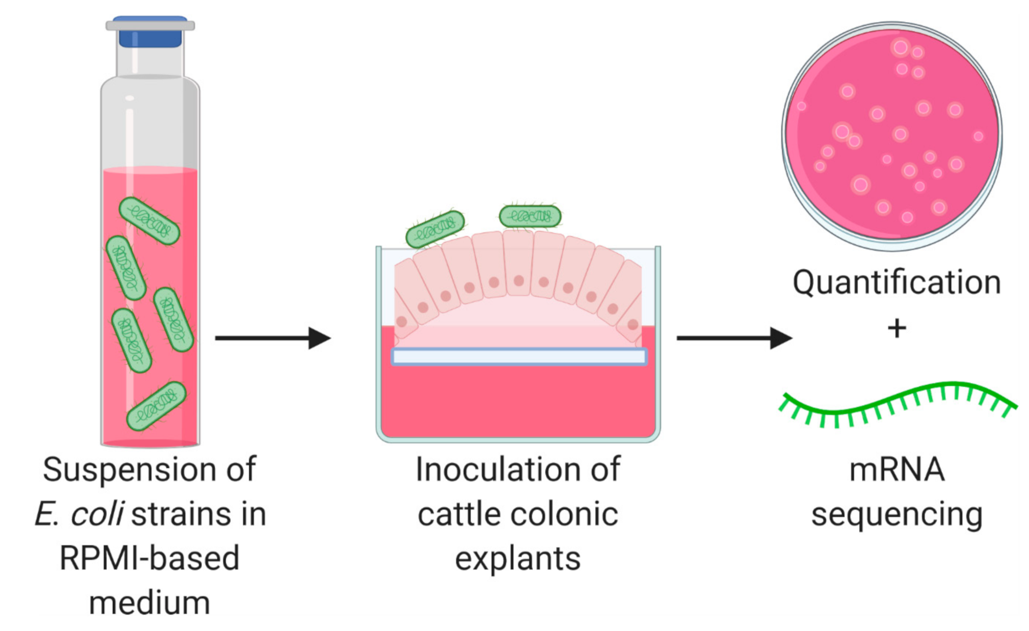
2.2. Cattle Colonic Explants

Bovine colonic explants and bacterial strains were prepared using methods previously described [13] with minor modifications. In 2017, tissues were obtained from three 18-month-old steers of approximately 1200 lb on three separate days (June 14, June 30, and July 24) at a meat processor with each animal representing a single experiment. A 0.3 m section of the centripetal coil was taken near the junction of the centrifugal and centripetal coils of the spiral colon following Iowa State University and USDA guidelines for collecting tissues from postmortem animals. The time-lapse between death and initiation of explant cultures was approximately 30 min. Collected tissues were washed with cold saline, immersed in CO2-independent medium (Gibco, Waltham, MA, USA) with gentamicin (50 µg/mL), penicillin (100 U/mL), and streptomycin (100 µg/mL), and transported to the laboratory on ice. Tissues were removed from media, thoroughly washed in saline, cut into 4 mm2 explants, and placed mucosal side up onto biopsy foam pads (4 explants per pad). The foam pads containing explants were placed one per well in six-well polystyrene tissue culture plates containing RPMI 1640 complete medium (RPMI 1640 supplemented with 10% fetal bovine serum, 0.25% lactalbumin hydrolysate, 0.10 μg/mL human insulin, and 1% D- + -mannose) (Figure 1). For the inoculation of explants, bacterial strains were grown statically overnight in trypticase soy broth (TSB) at 37 °C and then transferred 1:10 into fresh TSB for 6 h. Strains were washed and resuspended in RPMI 1640 complete medium and explants were inoculated with 2 × 10⁷ CFU in accordance with protocols approved by Iowa State University (protocol number 16-D/I-0015-A/H). The inoculum concentration was confirmed by serial dilution and plating on MacConkey agar. After inoculation, six-well plates were incubated for 2 h at 37 °C in 5% CO2 on a rocker (16 cycles/min).

2.3. Bacterial Adherence to Explants

Bacterial adherence to explants was determined using one explant per bacterial strain for each experiment (3 per strain) after the 2 h incubation. The explant was first weighed and then placed in a 2 oz. Whirl-Pak bag and homogenized in PBS. Samples were serially diluted in PBS, plated on MacConkey agar, and incubated at 37 °C for 18 h. To quantify the E. coli associated per explant, CFUs were recorded and divided by the weight of the explant (CFU/g).

2.4. RNA Extraction, Library Generation, and Sequencing

After the 2 h incubation, the remaining explants were snap-frozen in liquid nitrogen and stored at –80 °C until needed. For RNA extraction, samples were lysed and homogenized in 2 mL lysing matrix bead-beating tubes (MP Biomedicals, Santa Ana, CA, USA). The remaining extraction was performed using a PureLink RNA Mini Kit (Life Technologies, Carlsbad, CA, USA) with on-column DNase treatment. RNA quantity was determined using a Qubit fluorometer (Thermo Fisher, Waltham, MA, USA). RNA quality was determined using a 2100 Bioanalyzer (Agilent, Santa Clara, CA, USA) and samples with an RNA integrity number greater than 7 were used for mRNA-seq. Bacterial rRNA was depleted using Ribo-Zero (Illumina, San Diego, CA, USA) and samples were sequenced on a HiSeq 3000 (Illumina, San Diego, CA, USA) generating 150 bp single-end reads. RNA-seq data were deposited in the NCBI Sequence Read Archive (SRA) under accession number PRJNA667523.

2.5. RNA-seq Analysis and Detection of Differential Gene Expression

Genomes, genomic gff’s, and protein fastas were downloaded from NCBI for E. coli strains (accessions): DEC10E (GCF_000250075.1), DEC8B (GCF_000249815.1), MG1655 (GCF_000005845.2), and EDL933 (GCF_000732965.1) [25,26]. Raw reads were trimmed for adapters and quality using trim_galore version 0.4.5 (Babraham Institute, Babraham, UK). Reads were mapped to their respective E. coli genome using Hisat2 version 2.1.0. Read counts were obtained using featureCounts from the Subread package version 1.6.0 (WEHI, Melbourne, Australia). Orthologous gene families were identified using Orthofinder version 2.2.0 [27]. Single-copy orthologous genes were subjected to a differential gene expression analysis using DESeq2 (1.20.0) with a cutoff of adjusted p < 0.05 [28]. In the differential gene expression analysis, STEC strains were compared to MG1655 both as individual strains and as a collective group.

2.6. Statistical Analysis

Statistical analyses were performed using GraphPad Prism version 7 (San Diego, CA, USA). An ANOVA followed by Tukey’s method for multiple means comparison was used to compare E. coli strains in adherence assays. p < 0.05 was considered significant.

3. Results and Discussion

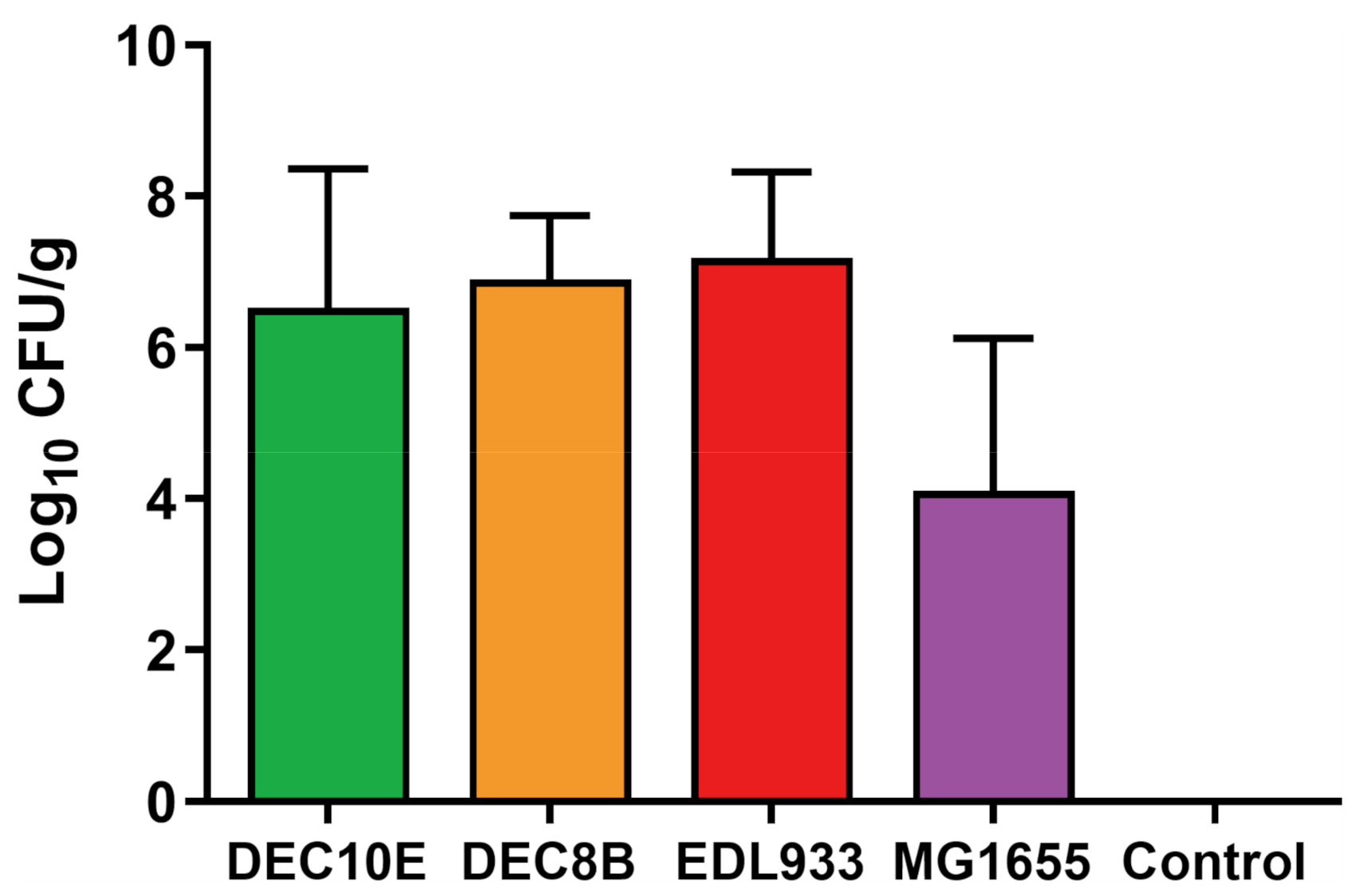
3.1. STEC Strains and MG1655 Adhered to Colonic Explants

STEC adherence to the colonic epithelium is a crucial step for colonizing cattle [29]. The proposed timeline of STEC adherence begins with initial interaction with the extracellular membrane [30], which was the focus of our study. To quantify this early interaction, a total of three bovine colonic explants were inoculated with E. coli strains and bacterial adherence was evaluated after 2 h. Previous studies demonstrated that 2 h is sufficient to produce initial adherence factors such as fimbriae and pili in E. coli and to allow for attachment to cell cultures [30,31]. Uninoculated explants were used as a control to ensure that no background E. coli or other Enterobacteriaceae organisms remained on the tissues. No growth was observed on MacConkey agar from plating the uninoculated colonic explant samples. The mean adherence level of the STEC strains ranged from 6.5 to 7.2 log CFU/g, while for MG1655 it was 4.2 log CFU/g (Figure 2). Although STEC strains adhered at numerically higher levels, no significant differences were detected between the strains. Variation in adherence levels between experiments was observed, likely explained by the variability in host tissues collected from three different steers. Adherence assays that use cell lines have less variation [32]; however, there is no commercial source of cattle colonic epithelial cells. Thus, researchers have relied upon explant cultures to assess STEC colonization in the laboratory [13,33,34,35]. Development of stable, immortalized intestinal epithelial cell lines such as that recently reported from the ileum of a calf [36] are needed and would help standardize results for several gastrointestinal pathogens in cattle.
In cattle, STEC organisms can be found throughout the gastrointestinal tract [37]. The bovine internal temperature is 39 °C; however, explants were cultured and infected at 37 °C based on established conditions in a previous study [13], which may be a limitation of our study in mimicking physiological conditions. In a previous study, calf colonic explants were inoculated with 107 CFU of O157 STEC strains, incubated for 3 h, and displayed similar adherence levels compared with our current study [35]. Most studies have only explored O157 STEC adherence to colonic explants, while we tested both O157 and non-O157 STEC strains. In one of the few studies that did test both O157 and non-O157 STEC strains, it was demonstrated that O26, O111, and O157 adhered to the terminal ileum, colon, and rectum from calves after an 8 h incubation and formed attaching/effacing lesions [34]. Demonstration of STEC adherence to bovine tissues is critical for understanding tissue tropism and adherence factors, but could also be used as a model system to evaluate interventions. Previously, the treatment of colonic explants with TNF-α reduced adhesion of O157 STEC [38]. An intervention was not evaluated in the current study; however, future assessments could evaluate the use of interventions to inhibit O157 and non-O157 STEC attachment in this model system.

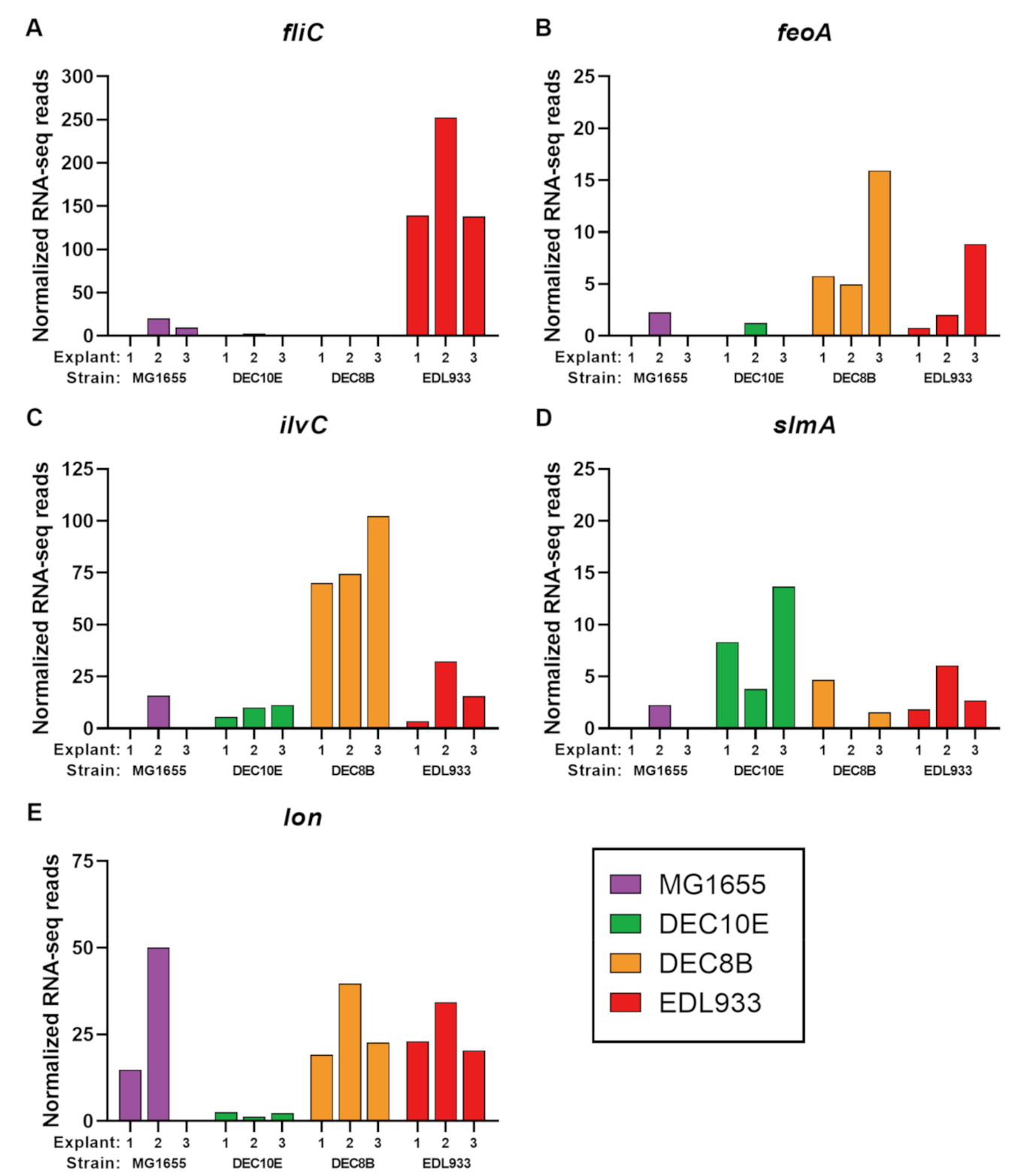
3.2. H7 Flagellin (fliC) and Lon Protease (lon) Upregulated during Incubation with Colonic Explants

We sought to identify differentially regulated genes between STEC strains and the E. coli K-12 strain MG1655 using RNA-seq reads. We first compared gene expression from individual STEC strains to MG1655 (i.e., DEC10E vs. MG1655, DEC8B vs. MG1655, and EDL933 vs. MG1655). Single-copy orthogroups were identified among strain comparisons with 3452 orthogroups found in DEC10E vs. MG1655, 3473 orthogroups in DEC8B vs. MG1655, and 3460 orthogroups in EDL933 vs. MG1655 (Table S1). No significant changes in gene expression were observed in DEC10E nor DEC8B when compared with MG1655. This suggests that the genes shared between these strains responded in a similar manner during the 2 h incubation with bovine explants. For strain EDL933, flagellin (fliC) was significantly upregulated (log2 fold-change = 4.0, adjusted p < 0.0001) in O157 STEC compared to MG1655 (Table S1). The RNA-seq count data was consistent between the three explants used with EDL933 having higher counts compared to MG1655 (Figure 3A). Flagellin is the main component of the bacterial flagellum and forms the filament [39]. These results support an earlier finding that H7 flagellin is expressed during contact with cattle intestinal epithelial cells [15]. In addition, the deletion of fliCH7 in EDL933 was previously shown to reduce adherence to bovine explants [40]. In vivo, cattle inoculated with an O157 STEC strain lacking flagellar expression had reduced levels of fecal shedding compared to the wild-type strain [41]. The induced production of flagellin-specific IgG and IgA antibodies and reducing colonization rates was previously demonstrated in cattle immunized intramuscularly with H7 flagellum [42]. Overall, these findings demonstrate that H7 flagellin functions as an adhesin in the gastrointestinal tract of cattle and a potential vaccine candidate to reduce STEC colonization. In addition to results found in cattle, fliCH7 in EDL933 has also been shown to play an important role in initial attachment to spinach leaves and glass [43], suggesting that H7 flagellum is a critical adherence factor for both the attachment to biotic and abiotic material. Other flagellin H-types such as fliCH12 have been shown to facilitate attachment to glass and stainless steel surfaces [44], but involvement in colonization of cattle is unclear. In the current study, we demonstrated that only fliCH7 in EDL933 was significantly upregulated, while no significant differences were found with fliCH11 in DEC10E and fliCH8 in DEC8B when compared to fliCH48 in MG1655. Thus, H7 flagellin appears to be a strong intervention candidate to reduce O157 STEC colonization, though few candidates exist that can reduce non-O157 STEC colonization in cattle.
Gene expression from STEC strains collectively was compared to MG1655 (i.e., STEC vs. MG1655). There were 3198 single-copy orthogroups identified among the STEC vs. MG1655 comparison. In this comparison, flagellin (fliC), ferrous iron transport protein A (feoA), ketol-acid reductoisomerase (ilvC), and nucleoid occlusion protein (slmA) were > 5-fold upregulated with a p < 0.05, but had an adjusted p > 0.05 likely due to variability between explants and between STEC strains. Interestingly, flagellin (Figure 3A) was identified in the EDL933 vs MG1655 comparison, but the relatively low reads from the other STEC strains DEC10E and DEC8B lend evidence that not all flagellin types are initial adherence factors. The ferrous iron transport protein A (feoA, Figure 3B) has a potential role in iron transport, which is an important nutrient for microorganisms [45]. However, relatively low levels of reads for DEC10E may have contributed to the adjusted p > 0.05. A relatively high level of reads were identified in some STEC, especially DEC8B, for the oxidoreductase enzyme ketol-acid reductoisomerase (ilvC, Figure 3C). However, in the individual comparison of DEC8B vs MG1655, the adjusted p = 0.051 and did not pass our cutoff. In addition, the normalized RNA-seq reads for nucleoid occlusion factor (slmA), which is involved in regulating cell division [46], was 6.2-fold higher in STEC compared to MG1655 (Figure 3D), but was not significant (adjusted p > 0.05).
Collectively, Lon protease (lon) was significantly upregulated (log2 fold-change = 3.6, adjusted p = 0.0009) in STEC strains compared to MG1655 (Table S1). However, there was variation between the explant samples (Figure 3E). For the RNA-seq counts, DEC10E remained relatively low across all three explants, DEC8B and EDL933 had higher levels than MG1655 for Explants 1 and 3, while numerically higher counts were observed for MG1655 from explant 2 (Figure 3A). As with the adherence levels, variation in this data may be due to the variability in the tissue collected from three different steers. The role of lon in colonization remains unclear; future studies are needed to compare colonization with wild-type and lon knockout mutants to determine whether lon is involved in colonization. In general, Lon protease is a highly conserved ATP-dependent protease responsible for the degradation of abnormal and misfolded proteins [47]. In E. coli, there are several regulatory functions of Lon protease that are related to stress response, including roles in amino acid starvation [48], defense against reactive oxygen species [49], and heat shock [50,51]. Thus, lon may have been upregulated in STEC strains to help survive stress encountered after the inoculation of bovine explants. Interestingly in STEC, Lon protease degrades the regulatory protein Ler, which activates genes in the locus of enterocyte effacement [52,53], which suggests that it contributes to regulation and timing of attaching/effacing lesions.
Unlike many studies that have evaluated gene expression changes under different conditions, the current study evaluated differences between strains. This approach was used to explore whether conserved genes found in all strains were differentially regulated in STEC compared to E. coli K-12. This approach may account for why we found only a few differentially regulated genes. In addition, the use of the complex RPMI 1640 medium during incubation may have contributed to the homogenous response observed by RNA-seq. The evaluation of only one incubation time may have resulted in similar responses between strains. To our knowledge, no study has systemically evaluated a time-series experiment to determine STEC adherence and gene expression over time during contact with bovine explants, which warrants future investigation. Other studies have used RNA-seq to explore the host response to STEC infection. Bando et al. found that a STEC strain from a human patient induced a distinct response in the Caco-2 colonic cell line compared with a strain isolated from bovine feces [54]. In another study, 351 differential regulated genes were identified throughout the bovine intestine of cattle shedding high levels of O157 STEC compared to cattle negative for O157 STEC [55]. In future studies, dual RNA-seq of the host and STEC could be used as a powerful approach to simultaneously understand both host and bacterial changes in gene expression, evaluate host–STEC interactions, and potentially combat STEC colonization of the bovine gastrointestinal tract.

4. Conclusions

In this study, cattle colonic explants were inoculated with STEC and E. coli K-12 strains and evaluated for colonization and bacterial gene expression. STEC strains adhered at numerically but not significantly higher levels compared to MG1655. Flagellin (fliC) was upregulated (log2 fold-change = 4.0, adjusted p < 0.0001) in O157 STEC compared to MG1655. Collectively, Lon protease (lon) was upregulated (log2 fold-change = 3.6, adjusted p = 0.0009) in STEC strains compared to MG1655. These results demonstrate that H7 flagellum and Lon protease potentially play roles in early colonization and could be intervention targets to reduce colonization in cattle.

Supplementary Materials

The following are available online at https://www.mdpi.com/2076-2607/8/11/1662/s1, Table S1: Comparison of normalized RNA-seq reads for single-copy orthogroups.

Author Contributions

Conceptualization, Z.R.S. and M.M.; methodology, Z.R.S. and M.M.; validation, Z.R.S. and M.M.; formal analysis, Z.R.S., R.E.M. and M.M.; investigation, Z.R.S.; resources, Z.R.S. and M.M.; data curation, Z.R.S., R.E.M., and M.M.; writing—original draft preparation, Z.R.S.; writing—review and editing, Z.R.S., R.E.M. and M.M.; visualization, Z.R.S. and M.M.; supervision, M.M.; project administration, Z.R.S. and M.M.; funding acquisition, Z.R.S. and M.M. All authors have read and agreed to the published version of the manuscript.

Funding

This work is supported by Grant No. 2017-67012-26120 from the USDA National Institute of Food and Agriculture.

Acknowledgments

The authors thank Graham Redweik and Catherine Spivak for technical support and the processing plant for collection of tissues.

Conflicts of Interest

The authors declare no conflict of interest.

References

  1. Croxen, M.A.; Law, R.J.; Scholz, R.; Keeney, K.M.; Wlodarska, M.; Finlay, B.B. Recent advances in understanding enteric pathogenic Escherichia coli. Clin. Microbiol. Rev. 2013, 26, 822–880. [Google Scholar] [CrossRef] [PubMed] [Green Version]
  2. Joseph, A.; Cointe, A.; Mariani Kurkdjian, P.; Rafat, C.; Hertig, A. Shiga toxin-associated hemolytic uremic syndrome: A narrative review. Toxins 2020, 12, 67. [Google Scholar] [CrossRef] [PubMed] [Green Version]
  3. Spinale, J.M.; Ruebner, R.L.; Copelovitch, L.; Kaplan, B.S. Long-term outcomes of Shiga toxin hemolytic uremic syndrome. Pediatr. Nephrol. 2013, 28, 2097–2105. [Google Scholar] [CrossRef] [PubMed]
  4. Tarr, P.I.; Gordon, C.A.; Chandler, W.L. Shiga-toxin-producing Escherichia coli and haemolytic uraemic syndrome. Lancet 2005, 365, 1073–1086. [Google Scholar] [CrossRef]
  5. Gould, L.H.; Mody, R.K.; Ong, K.L.; Clogher, P.; Cronquist, A.B.; Garman, K.N.; Lathrop, S.; Medus, C.; Spina, N.L.; Webb, T.H. Increased recognition of non-O157 Shiga toxin–producing Escherichia coli infections in the United States during 2000–2010: Epidemiologic features and comparison with E. coli O157 infections. Foodborne Pathog. Dis. 2013, 10, 453–460. [Google Scholar] [CrossRef]
  6. Crim, S.M.; Griffin, P.M.; Tauxe, R.; Marder, E.P.; Gilliss, D.; Cronquist, A.B.; Cartter, M.; Tobin-D’Angelo, M.; Blythe, D.; Smith, K. Preliminary incidence and trends of infection with pathogens transmitted commonly through food—Foodborne Diseases Active Surveillance Network, 10 US sites, 2006–2014. MMWR Morb. Mortal. Wkly Rep. 2015, 64, 495. [Google Scholar]
  7. Pires, S.M.; Majowicz, S.; Gill, A.; Devleesschauwer, B. Global and regional source attribution of Shiga toxin-producing Escherichia coli infections using analysis of outbreak surveillance data. Epidemiol. Infect. 2019, 147, e236. [Google Scholar] [CrossRef] [Green Version]
  8. Scallan, E.; Hoekstra, R.; Mahon, B.; Jones, T.; Griffin, P. An assessment of the human health impact of seven leading foodborne pathogens in the United States using disability adjusted life years. Epidemiol. Infect. 2015, 143, 2795–2804. [Google Scholar] [CrossRef] [Green Version]
  9. Moxley, R.A.; Stromberg, Z.R.; Lewis, G.L.; Loy, J.D.; Brodersen, B.W.; Patel, I.R.; Gangiredla, J. Haemorrhagic colitis associated with enterohaemorrhagic Escherichia coli O165: H25 infection in a yearling feedlot heifer. JMM Case Rep. 2015, 2, e005004. [Google Scholar] [CrossRef]
  10. Moxley, R.A.; Smith, D.R. Attaching-effacing Escherichia coli infections in cattle. Vet. Clin. N. Am. Food Anim. Pract. 2010, 26, 29–56. [Google Scholar] [CrossRef]
  11. Sapountzis, P.; Segura, A.; Desvaux, M.; Forano, E. An overview of the elusive passenger in the gastrointestinal tract of cattle: The Shiga toxin producing Escherichia coli. Microorganisms 2020, 8, 877. [Google Scholar] [CrossRef] [PubMed]
  12. Gaytán, M.O.; Martínez-Santos, V.I.; Soto, E.; González-Pedrajo, B. Type three secretion system in attaching and effacing pathogens. Front. Cell. Infect. Microbiol. 2016, 6, 129. [Google Scholar] [CrossRef] [Green Version]
  13. Baehler, A.A.; Moxley, R.A. Escherichia coli O157:H7 induces attaching-effacing lesions in large intestinal mucosal explants from adult cattle. FEMS Microbiol. Lett. 2000, 185, 239–242. [Google Scholar] [CrossRef]
  14. Phillips, A.; Navabpour, S.; Hicks, S.; Dougan, G.; Wallis, T.; Frankel, G. Enterohaemorrhagic Escherichia coli O157: H7 target Peyer’s patches in humans and cause attaching/effacing lesions in both human and bovine intestine. Gut 2000, 47, 377–381. [Google Scholar] [CrossRef] [Green Version]
  15. Mahajan, A.; Currie, C.G.; Mackie, S.; Tree, J.; McAteer, S.; McKendrick, I.; McNeilly, T.N.; Roe, A.; La Ragione, R.M.; Woodward, M.J. An investigation of the expression and adhesin function of H7 flagella in the interaction of Escherichia coli O157: H7 with bovine intestinal epithelium. Cell. Microbiol. 2009, 11, 121–137. [Google Scholar] [CrossRef] [Green Version]
  16. Gonyar, L.A.; Kendall, M.M. Ethanolamine and choline promote expression of putative and characterized fimbriae in enterohemorrhagic Escherichia coli O157: H7. Infect. Immun. 2014, 82, 193–201. [Google Scholar] [CrossRef] [PubMed] [Green Version]
  17. Venegas-Vargas, C.; Henderson, S.; Khare, A.; Mosci, R.E.; Lehnert, J.D.; Singh, P.; Ouellette, L.M.; Norby, B.; Funk, J.A.; Rust, S. Factors associated with Shiga toxin-producing Escherichia coli shedding by dairy and beef cattle. Appl. Environ. Microbiol. 2016, 82, 5049–5056. [Google Scholar] [CrossRef] [Green Version]
  18. Chase-Topping, M.; Gally, D.; Low, C.; Matthews, L.; Woolhouse, M. Super-shedding and the link between human infection and livestock carriage of Escherichia coli O157. Nat. Rev. Microbiol. 2008, 6, 904–912. [Google Scholar] [CrossRef] [PubMed]
  19. Arthur, T.M.; Brichta-Harhay, D.M.; Bosilevac, J.M.; Kalchayanand, N.; Shackelford, S.D.; Wheeler, T.L.; Koohmaraie, M. Super shedding of Escherichia coli O157:H7 by cattle and the impact on beef carcass contamination. Meat Sci. 2010, 86, 32–37. [Google Scholar] [CrossRef] [PubMed] [Green Version]
  20. Stromberg, Z.R.; Lewis, G.L.; Schneider, L.G.; Erickson, G.E.; Patel, I.R.; Smith, D.R.; Moxley, R.A. Culture-based quantification with molecular characterization of non-O157 and O157 enterohemorrhagic Escherichia coli isolates from rectoanal mucosal swabs of feedlot cattle. Foodborne Pathog. Dis. 2018, 15, 26–32. [Google Scholar] [CrossRef]
  21. Shridhar, P.B.; Noll, L.W.; Cull, C.A.; Shi, X.; Cernicchiaro, N.; Renter, D.G.; Bai, J.; Nagaraja, T. Spiral plating method to quantify the six major non-O157 Escherichia coli serogroups in cattle feces. J. Food Prot. 2017, 80, 848–856. [Google Scholar] [CrossRef] [PubMed]
  22. Vlisidou, I.; Dziva, F.; La Ragione, R.M.; Best, A.; Garmendia, J.; Hawes, P.; Monaghan, P.; Cawthraw, S.A.; Frankel, G.; Woodward, M.J. Role of intimin-tir interactions and the tir-cytoskeleton coupling protein in the colonization of calves and lambs by Escherichia coli O157: H7. Infect. Immun. 2006, 74, 758–764. [Google Scholar] [CrossRef] [PubMed] [Green Version]
  23. Reid, S.D.; Betting, D.J.; Whittam, T.S. Molecular detection and identification of intimin alleles in pathogenic Escherichia coli by multiplex PCR. J. Clin. Microbiol. 1999, 37, 2719–2722. [Google Scholar] [CrossRef] [Green Version]
  24. Riley, L.W.; Remis, R.S.; Helgerson, S.D.; McGee, H.B.; Wells, J.G.; Davis, B.R.; Hebert, R.J.; Olcott, E.S.; Johnson, L.M.; Hargrett, N.T. Hemorrhagic colitis associated with a rare Escherichia coli serotype. N. Engl. J. Med. 1983, 308, 681–685. [Google Scholar] [CrossRef] [PubMed]
  25. Hazen, T.H.; Sahl, J.W.; Redman, J.C.; Morris, C.R.; Daugherty, S.C.; Chibucos, M.C.; Sengamalay, N.A.; Fraser-Liggett, C.M.; Steinsland, H.; Whittam, T.S.; et al. Draft genome sequences of the diarrheagenic Escherichia coli collection. J. Bacteriol. 2012, 194, 3026–3027. [Google Scholar] [CrossRef] [Green Version]
  26. Latif, H.; Li, H.J.; Charusanti, P.; Palsson, B.Ø.; Aziz, R.K. A gapless, unambiguous genome sequence of the enterohemorrhagic Escherichia coli O157:H7 strain EDL933. Genome Announc. 2014, 2, e00821-14. [Google Scholar] [CrossRef] [Green Version]
  27. Emms, D.M.; Kelly, S. OrthoFinder: Solving fundamental biases in whole genome comparisons dramatically improves orthogroup inference accuracy. Genome Biol. 2015, 16, 157. [Google Scholar] [CrossRef] [Green Version]
  28. Love, M.I.; Huber, W.; Anders, S. Moderated estimation of fold change and dispersion for RNA-seq data with DESeq2. Genome Biol. 2014, 15, 550. [Google Scholar] [CrossRef] [Green Version]
  29. Farfan, M.J.; Torres, A.G. Molecular mechanisms that mediate colonization of Shiga toxin-producing Escherichia coli strains. Infect. Immun. 2012, 80, 903–913. [Google Scholar] [CrossRef] [Green Version]
  30. McWilliams, B.D.; Torres, A.G. Enterohemorrhagic Escherichia coli adhesins. Microbiol. Spectr. 2014, 2, EHEC00032013. [Google Scholar] [CrossRef] [Green Version]
  31. Ross, B.N.; Rojas-Lopez, M.; Cieza, R.J.; McWilliams, B.D.; Torres, A.G. The role of long polar fimbriae in Escherichia coli O104: H4 adhesion and colonization. PLoS ONE 2015, 10, e0141845. [Google Scholar] [CrossRef] [PubMed] [Green Version]
  32. Letourneau, J.; Levesque, C.; Berthiaume, F.; Jacques, M.; Mourez, M. In vitro assay of bacterial adhesion onto mammalian epithelial cells. J. Vis. Exp. 2011, e2783. [Google Scholar] [CrossRef] [PubMed] [Green Version]
  33. Hu, J.; Zhu, M.-J. Defects in polynucleotide phosphorylase impairs virulence in Escherichia coli O157: H7. Front. Microbiol. 2015, 6, 806. [Google Scholar] [CrossRef] [PubMed]
  34. Girard, F.; Dziva, F.; van Diemen, P.; Phillips, A.D.; Stevens, M.P.; Frankel, G. Adherence of enterohemorrhagic Escherichia coli O157, O26, and O111 strains to bovine intestinal explants ex vivo. Appl. Environ. Microbiol. 2007, 73, 3084–3090. [Google Scholar] [CrossRef] [Green Version]
  35. Cookson, A.L.; Woodward, M.J. The role of intimin in the adherence of enterohaemorrhagic Escherichia coli (EHEC) O157:H7 to HEp-2 tissue culture cells and to bovine gut explant tissues. Int. J. Med. Microbiol. 2003, 292, 547–553. [Google Scholar] [CrossRef]
  36. Katwal, P.; Thomas, M.; Uprety, T.; Hildreth, M.B.; Kaushik, R.S. Development and biochemical and immunological characterization of early passage and immortalized bovine intestinal epithelial cell lines from the ileum of a young calf. Cytotechnology 2019, 71, 127–148. [Google Scholar] [CrossRef] [Green Version]
  37. Keen, J.E.; Laegreid, W.W.; Chitko-McKown, C.G.; Durso, L.M.; Bono, J.L. Distribution of Shiga-toxigenic Escherichia coli O157 in the gastrointestinal tract of naturally O157-shedding cattle at necropsy. Appl. Environ. Microbiol. 2010, 76, 5278–5281. [Google Scholar] [CrossRef] [PubMed] [Green Version]
  38. Xue, Y.; Zhang, H.; Wang, H.; Hu, J.; Du, M.; Zhu, M.-J. Host inflammatory response inhibits Escherichia coli O157:H7 adhesion to gut epithelium through augmentation of mucin expression. Infect. Immun. 2014, 82, 1921–1930. [Google Scholar] [CrossRef] [Green Version]
  39. Rossez, Y.; Wolfson, E.B.; Holmes, A.; Gally, D.L.; Holden, N.J. Bacterial flagella: Twist and stick, or dodge across the kingdoms. PLoS Pathog. 2015, 11, e1004483. [Google Scholar] [CrossRef] [PubMed]
  40. Erdem, A.L.; Avelino, F.; Xicohtencatl-Cortes, J.; Girón, J.A. Host protein binding and adhesive properties of H6 and H7 flagella of attaching and effacing Escherichia coli. J. Bacteriol. 2007, 189, 7426–7435. [Google Scholar] [CrossRef] [PubMed] [Green Version]
  41. Bretschneider, G.; Berberov, E.M.; Moxley, R.A. Reduced intestinal colonization of adult beef cattle by Escherichia coli O157:H7 tir deletion and nalidixic-acid-resistant mutants lacking flagellar expression. Vet. Microbiol. 2007, 125, 381–386. [Google Scholar] [CrossRef] [PubMed] [Green Version]
  42. McNeilly, T.N.; Naylor, S.W.; Mahajan, A.; Mitchell, M.C.; McAteer, S.; Deane, D.; Smith, D.G.; Low, J.C.; Gally, D.L.; Huntley, J.F. Escherichia coli O157:H7 colonization in cattle following systemic and mucosal immunization with purified H7 flagellin. Infect. Immun. 2008, 76, 2594–2602. [Google Scholar] [CrossRef] [Green Version]
  43. Nagy, A.; Mowery, J.; Bauchan, G.R.; Wang, L.; Nichols-Russell, L.; Nou, X. Role of extracellular structures of Escherichia coli O157:H7 in initial attachment to biotic and abiotic surfaces. Appl. Environ. Microbiol. 2015, 81, 4720–4727. [Google Scholar] [CrossRef] [Green Version]
  44. Goulter, R.M.; Taran, E.; Gentle, I.R.; Gobius, K.S.; Dykes, G.A. Escherichia coli strains expressing H12 antigens demonstrate an increased ability to attach to abiotic surfaces as compared with E. coli strains expressing H7 antigens. Colloids Surf. B Biointerfaces 2014, 119, 90–98. [Google Scholar] [CrossRef] [PubMed]
  45. Lau, C.K.; Ishida, H.; Liu, Z.; Vogel, H.J. Solution structure of Escherichia coli FeoA and its potential role in bacterial ferrous iron transport. J. Bacteriol. 2013, 195, 46–55. [Google Scholar] [CrossRef] [Green Version]
  46. Bernhardt, T.G.; De Boer, P.A. SlmA, a nucleoid-associated, FtsZ binding protein required for blocking septal ring assembly over chromosomes in E. coli. Mol. Cell 2005, 18, 555–564. [Google Scholar] [CrossRef] [Green Version]
  47. Rosen, R.; Biran, D.; Gur, E.; Becher, D.; Hecker, M.; Ron, E.Z. Protein aggregation in Escherichia coli: Role of proteases. FEMS Microbiol. Lett. 2002, 207, 9–12. [Google Scholar] [CrossRef]
  48. Christensen, S.K.; Mikkelsen, M.; Pedersen, K.; Gerdes, K. RelE, a global inhibitor of translation, is activated during nutritional stress. Proc. Natl. Acad. Sci. USA 2001, 98, 14328–14333. [Google Scholar] [CrossRef] [PubMed] [Green Version]
  49. Griffith, K.L.; Shah, I.M.; Richard, E.W., Jr. Proteolytic degradation of Escherichia coli transcription activators SoxS and MarA as the mechanism for reversing the induction of the superoxide (SoxRS) and multiple antibiotic resistance (Mar) regulons. Mol. Microbiol. 2004, 51, 1801–1816. [Google Scholar] [CrossRef] [PubMed]
  50. Bissonnette, S.A.; Rivera-Rivera, I.; Sauer, R.T.; Baker, T.A. The IbpA and IbpB small heat-shock proteins are substrates of the AAA+ Lon protease. Mol. Microbiol. 2010, 75, 1539–1549. [Google Scholar] [CrossRef] [Green Version]
  51. Babin, B.M.; Kasperkiewicz, P.; Janiszewski, T.; Yoo, E.; Dra̧g, M.; Bogyo, M. Leveraging peptide substrate libraries to design inhibitors of bacterial Lon protease. ACS Chem. Biol. 2019, 14, 2453–2462. [Google Scholar] [CrossRef] [PubMed]
  52. Mellies, J.L.; Elliott, S.J.; Sperandio, V.; Donnenberg, M.S.; Kaper, J.B. The Per regulon of enteropathogenic Escherichia coli: Identification of a regulatory cascade and a novel transcriptional activator, the locus of enterocyte effacement (LEE)-encoded regulator (Ler). Mol. Microbiol. 1999, 33, 296–306. [Google Scholar] [CrossRef] [PubMed]
  53. De la Cruz, M.A.; Morgan, J.K.; Ares, M.A.; Yáñez-Santos, J.A.; Riordan, J.T.; Girón, J.A. The two-component system CpxRA negatively regulates the locus of enterocyte effacement of enterohemorrhagic Escherichia coli involving σ32 and Lon protease. Front. Cell. Infect. Microbiol. 2016, 6, 11. [Google Scholar] [CrossRef] [PubMed] [Green Version]
  54. Bando, S.Y.; Iamashita, P.; Silva, F.N.; Costa, L.d.F.; Abe, C.M.; Bertonha, F.B.; Guth, B.E.; Fujita, A.; Moreira-Filho, C.A. Dynamic gene network analysis of Caco-2 cell response to Shiga toxin-producing Escherichia coli-associated hemolytic–uremic syndrome. Microorganisms 2019, 7, 195. [Google Scholar] [CrossRef] [Green Version]
  55. Wang, O.; McAllister, T.A.; Plastow, G.; Stanford, K.; Selinger, B. Host mechanisms involved in cattle Escherichia coli O157 shedding: A fundamental understanding for reducing foodborne pathogen in food animal production. Sci. Rep. 2017, 7, 1–15. [Google Scholar] [CrossRef] [Green Version]
Figure 1. Experimental overview of bovine colonic explants inoculated with Escherichia coli. After a 2 h incubation, explants were either homogenized for bacterial enumeration on MacConkey agar or frozen for RNA extraction and sequencing.
Microorganisms 08 01662 g001
Figure 2. Bacterial adherence to cattle colonic explants. Bacterial counts of Shiga toxin-producing Escherichia coli (STEC) strains DEC10E (green), DEC8B (orange), and EDL933 (red), E. coli K-12 MG1655 (purple), and a control (uninoculated) were obtained after 2 h of incubation with explant cultures. Bars represent the mean of three individual experiments, and error bars represent standard deviation.
Microorganisms 08 01662 g002
Figure 3. Normalized RNA-seq reads. (A) Significantly upregulated flagellin gene (fliC) in the EDL933 vs. MG1655 comparison, (B) ferrous iron transport protein A gene (feoA), (C) ketol-acid reductoisomerase gene (ilvC), (D) nucleoid occlusion gene (slmA), and (E) significantly upregulated Lon protease gene (lon) in the Shiga toxin-producing Escherichia coli (STEC) vs. MG1655 comparison. Three biological replicates (Explants 1, 2, and 3) were inoculated with STEC strains DEC10E (green), DEC8B (orange), and EDL933 (red), and E. coli K-12 strain MG1655 (purple).
Microorganisms 08 01662 g003
Publisher’s Note: MDPI stays neutral with regard to jurisdictional claims in published maps and institutional affiliations.

