Article

Culturomics Discloses Anti-Tubercular Enterococci Exclusive of Pulmonary Tuberculosis: A Preliminary Report

1
IHU Méditerranée Infection, 13005 Marseille, France
2
Aix-Marseille Univ., IRD, MEPHI, IHU Méditerranée Infection, 13005 Marseille, France
3
UFR-ST Département Sciences Biologiques, Université Nazi Boni, Bobo-Dioulasso 01 BP 1091, Burkina Faso
4
Institut de Recherche en Sciences de la Santé, Direction Régionale de l’Ouest, Bobo Dioulasso 01 BP 545, Burkina Faso
5
Centre Muraz, Bobo-Doulasso, Institut National de Santé Publique, Bobo-Dioulasso 01 BP 390, Burkina Faso
*
Author to whom correspondence should be addressed.
These authors contributed equally to this work.
Microorganisms 2020, 8(10), 1544; https://doi.org/10.3390/microorganisms8101544
Submission received: 3 September 2020 / Revised: 25 September 2020 / Accepted: 4 October 2020 / Published: 7 October 2020
(This article belongs to the Section Gut Microbiota)

Abstract

:
Mycobacterium tuberculosis causes pulmonary tuberculosis, a deadly infection of which the clinical expression and prognosis are not fully understood at the individual level, apart from genetic susceptibility traits. We investigated whether individual gut microbiota may correlate with pulmonary tuberculosis status. Culturomics investigations of gut microbiota in two pulmonary tuberculosis patients and two controls in Burkina Faso found 60 different bacterial species in patients and 97 in controls, including 45 in common. Further analysis of the results at the individual level indicated seven bacteria, including Enterococcus mundtii and Enterococcus casseliflavus, which were exclusively cultured in controls. Blind quantitative PCR-based exploration of faeces samples in two cohorts in Burkina Faso and in France confirmed a nonsignificant association of E. mundtii and E. casseliflavus with controls. Further in vitro explorations found four E. mundtii and E. casseliflavus strains inhibiting the growth of M. tuberculosis strains representative of four different lineages as well as Mycobacterium africanum, Mycobacterium canettii, and Mycobacterium bovis, in an inoculum-dependent manner. Heat-killed E. mundtii or E. casseliflavus were ineffective. These unprecedented observations of direct interactions between gut E. mundtii and E. casseliflavus with M. tuberculosis complex mycobacteria suggest that gut microbiota may modulate the expression of pulmonary tuberculosis.

1. Introduction

Tuberculosis is a deadly bacterial infection due to mycobacteria forming the Mycobacterium tuberculosis complex (MTBC), and is responsible for a high burden of morbidity and mortality in the most countries worldwide [1]. In 2018, the World Health Organization (WHO) reported 10 million new cases and 1.5 million deaths [2].
The interplay between host and pathogen determining the clinical expression and prognosis of tuberculosis is not fully understood at the individual level. Indisputably, the M. tuberculosis inoculum, genotype and antibiotic susceptibility profile may play in part the clinical expression and prognosis of pulmonary tuberculosis [3,4]. Host traits mostly related to genetic alterations of the interferon gamma pathway have also been elucidated in highly susceptible tuberculosis individuals and families [5]. Such genetic traits have been significantly associated with certain clinical expressions of tuberculosis, such as onset in the early years of life [6,7]. Accordingly, at the population level, discrete co-evolution of certain M. tuberculosis genotypes with certain populations has been clearly established in some geographic areas [3,7,8,9].
However, the factors mention above may not explain all the variations observed in the clinical spectrum of tuberculosis and the hypothesis that individual microbiota may also govern part of the clinical expression of tuberculosis, has emerged [10]. A few studies recently indicated significant correlations between the repertoire of microorganisms in the digestive tract microbiota and pulmonary tuberculosis [11,12,13], yet these studies relied upon metagenomics, leaving it unknown whether living organisms were indeed significantly associated with tuberculosis.
In order to advance this knowledge, we herein embarked on a preliminary study of the digestive tract microbiota in individuals with and without pulmonary tuberculosis using culturomics, in order to discover whether some living microorganisms detected in the digestive tract microbiota, including bacteria and archaea, were significantly correlated and anticorrelated with pulmonary tuberculosis prior to anti-TB treatment.

2. Materials and Methods

2.1. Ethics Statement and Population Information

A total of 150 stool samples collected from 150 individuals were included in this study. Fifty stools specimens from fifty individuals were collected as part of a tuberculosis diagnosis kit in patients being routinely investigated for medical suspicion of pulmonary tuberculosis in the IHU Méditerranée Infection, Marseille, France [14]. Microbiological diagnosis using sputum Ziehl-Neelsen staining, culture, and molecular biology (GeneXpert) identified 14 samples belonging to tuberculosis patients and 36 belonging to non-tuberculosis patients. Ethical approval was obtained from the Institutional Ethics Committee of the Institut Hospitalier Universitaire—Méditerranée Infection (Marseille, France) under number 2012-A01598-35. One hundred individuals were sampled in Burkina Faso. The participants were recruited in the tuberculosis Diagnosis and Treatment Centre at Bobo-Dioulasso. Among 100 samples collected in Burkina Faso, 50 were collected from patients with confirmed pulmonary tuberculosis and 50 from individuals without pulmonary tuberculosis based on microscopy test using sputum Ziehl-Neelsen staining. The ethics statement was approved by the Research Ethics Committee in Health of Science, Comité d’Éthique Institutionnel pour la Recherche en Science de la Santé (CEIRS) (N/Ref.002-2018-CEIRS) (Bobo-Dioulasso, Burkina Faso). After written consent was obtained from all participants for sample collection and subsequent analyses, stool and sputum samples were collected from patients and were placed in sterile containers. Stool and sputum samples were stored at −80 °C and sent for further analyses to the partner laboratory at the IHU at Marseille, France. All stool samples were collected prior to diagnosis and prior to starting treatment for tuberculosis.

2.2. Culturomics

To analyze the differences between the digestive microbiota of patients with pulmonary tuberculosis and subjects without pulmonary tuberculosis, culturomics was performed on 4 stool samples collected in Burkina Faso, 2 stool samples randomly selected from samples collected from individuals positive for pulmonary tuberculosis and 2 stool samples randomly selected from samples collected from individuals negative for pulmonary tuberculosis (Table 1).
The culture was performed as previously described with modifications [15]. In brief, one gram of stool was diluted into 1 mL of Dulbecco’s phosphate-buffered saline (DPBS 1X, Thermo Fisher Scientific, Illkirch, France) and the suspension was inoculated into each of the two blood culture bottles (BD Bactec Plus; Becton Dickinson, Le Pont-de-Claix, France), one for anaerobic culture and the other for aerobic culture. At the same time, one-gram stool suspensions were inoculated on YCFA medium modified for solid and liquid medium, prepared as described on the web site (https://www.dsmz.de/microorganisms/medium/pdf/DSMZ_Medium1611.pdf) (Deutsche Sammlung von Mikroorganismen und Zellkulturen GmbH, Braunschweig, Germany). The blood culture bottles and YCFA medium were previously supplemented with 3 mL of filtered rumen fluid and 3 mL of sheep blood (Becton Dickinson). The inoculated blood culture bottles and negative controls, represented by blood culture bottle inoculated with DPBS, were incubated at 37 °C. Inoculations on solid agar were performed at three hours, six hours, nine hours, twenty-four hours, three days, seven days, and 14 days post-incubation. For each inoculation, 1 mL of supernatant was collected and diluted in 900 µL of DPBS in serial dilutions from 1/10 to 1/1010. These dilutions were inoculated on 5% sheep blood agar (bioMérieux, La Balme-les-Grottes, France) incubated at 37°C for 48 h, respecting the strict atmospheric conditions of aerobic and anaerobic culture. All colonies were sub-cultured and identified by matrix-assisted laser desorption ionisation-time of flight mass spectrometry (MALDI-TOF-MS), as previously described [16]. Unidentified colonies by MALDI-TOF-MS were identified using 16S rRNA gene sequencing as previously described [17]. The bacterial species isolated from four stools of each person were compared by preparing a Venn diagram (http://bioinformatics.psb.ugent.be/webtools/Venn/).

2.3. Real-Time Quantitative PCR

In order to further explore the prevalence of two bacteria of interest (E. casseliflavus and E. mundtii) in the stools of TB-positive and TB-negative patients, 150 stools specimens were tested with molecular PCR-based assays as previously described [18]. For each sample, DNA were extracted from 200 mg of stool with a semi-automated method using an EZ1 DNA Tissue Kit (Qiagen, Hilden, Germany) and EZ1 Advanced XL extractor (Qiagen). Real-time qPCR reactions incorporated a total volume of 20 µL including 10 µL LightCycler® 480 Probes Master (Roche Diagnostics GmbH, Mannheim, Germany), 0.5 µL of each primer, 0.5 µL of probe, 3 µL of distilled water, 0.5 µL of Uracil-DNA glycosylase (UDG) and 5 µL of DNA. Analyses were performed using the thermal cycler CFX96 Touch™ (BIO-RAD, Marnes-la-Coquette, France). DNA extraction, PCR inhibitors and total bacterial richness was measured using real-time PCR targeting the universal bacterial 16S rRNA gene in the presence of negative controls (Supplementary Table S1). Two quantitative PCR systems were designated for Enterococcus casseliflavus and Enterococcus mundtii. Finally, the presence of Methanobrevibacter smithii was detected using real-time PCR, as previously described [19] (Supplementary Table S1). qPCR results were interpreted as positive when the cycle threshold (Ct) value was less than or equal to 39.

2.4. Anti-Tuberculosis Activity Assays

The antituberculosis activity of seven bacteria specifically found in the gut microbiota of two tuberculosis-free patients was assessed using the agar well diffusion method, with modifications [20,21]. Bacteria were cultured on COS agar medium according to the conditions necessary for the growth of each bacterium. Fresh culture colonies were suspended in DPBS, adjusted to 15 McFarland and serial dilution was performed in DPBS. The anti-tuberculosis activity of these seven bacteria was then tested against eight MTBC clinical strains including M. tuberculosis lineage 1 strain (CSURP9458), M. tuberculosis lineage 2 strain Beijing CSUR Q1316 [18] M. tuberculosis lineage 3 strain CSURP7204, M. tuberculosis lineage 4 strain CSUR P7739 [22], M. africanum strain CSURP7201, M. canettii strain CIPT140010059, and M. bovis strain CSURP7222 [23]. All strains were cultured in Middlebrook 7H10 culture medium and calibrated to 107 CFU/mL, as previously described [24]. This suspension was inoculated in Middlebrook 7H10 agar with Middlebrook OADC enrichment (Becton Dickinson) at 37 °C for one day, in Petri dishes (Ø × H = 90 × 13 mm, Dutscher, 67170 Brumath, France) or in square Petri dish plates (W × L × H = 120 × 120 × 15 mm Dutscher) after 200 µL wells had been made using a sterile tip. Each bacterial suspension was then deposited in each well alongside sterile DPBS in the negative control well and the plates were incubated at 37 °C and observed every three days up to 15 days, to monitor and enumerate any zones of MTBC growth inhibition around the wells. To measure any potential pH changes in the wells, a 200 μL Middlebrook 7H10 agar disk was incubated for 48 h at 37 °C with a 10 MacFarland suspension of each bacterial species (after growth on Middlebrook 7H10 agar had been verified); and the pH was measured. In order to further assess the potential M. tuberculosis growth inhibition by dead bacteria, the bacterial suspensions were inactivated by one-hour incubation at 100°C and the potential inhibitory effect of inactivated bacteria was tested as described above. All these manipulations were carried out three times in the presence of DPBS as a negative control.

2.5. Statistical Analyses

Data was entered into Microsoft Excel for Office 365 to calculate median, average and standard deviation. A clustering heatmap was used to visualise the potential clustering of cultured gut bacterial repertoires in positive and negative pulmonary tuberculosis patients. The heatmap and the principal component analysis were performed using XLSTAT v2019.1 statistical and data analysis solution (Long Island, NY, USA (https://www.xlstat.com). Statistical tests were performed on the website biostaTGV (https://biostatgv.sentiweb.frS) including Student’s T test to compare DPBS and the bacteria inhibition diameter and the ANOVA test was used to compare the susceptibility of mycobacteria to enterococci. Differences were considered to be statistically significant when p < 0.05.

3. Results

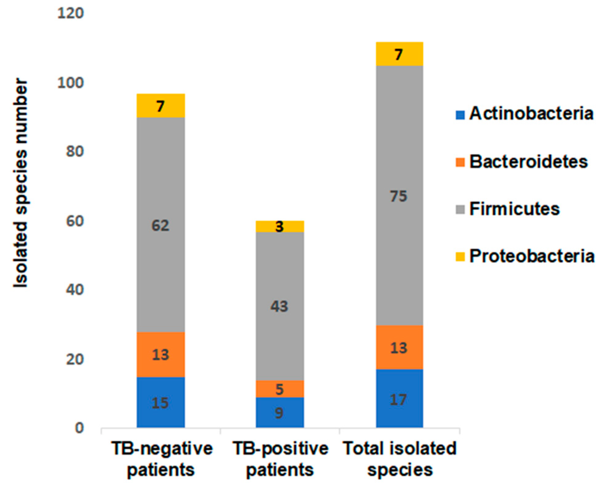
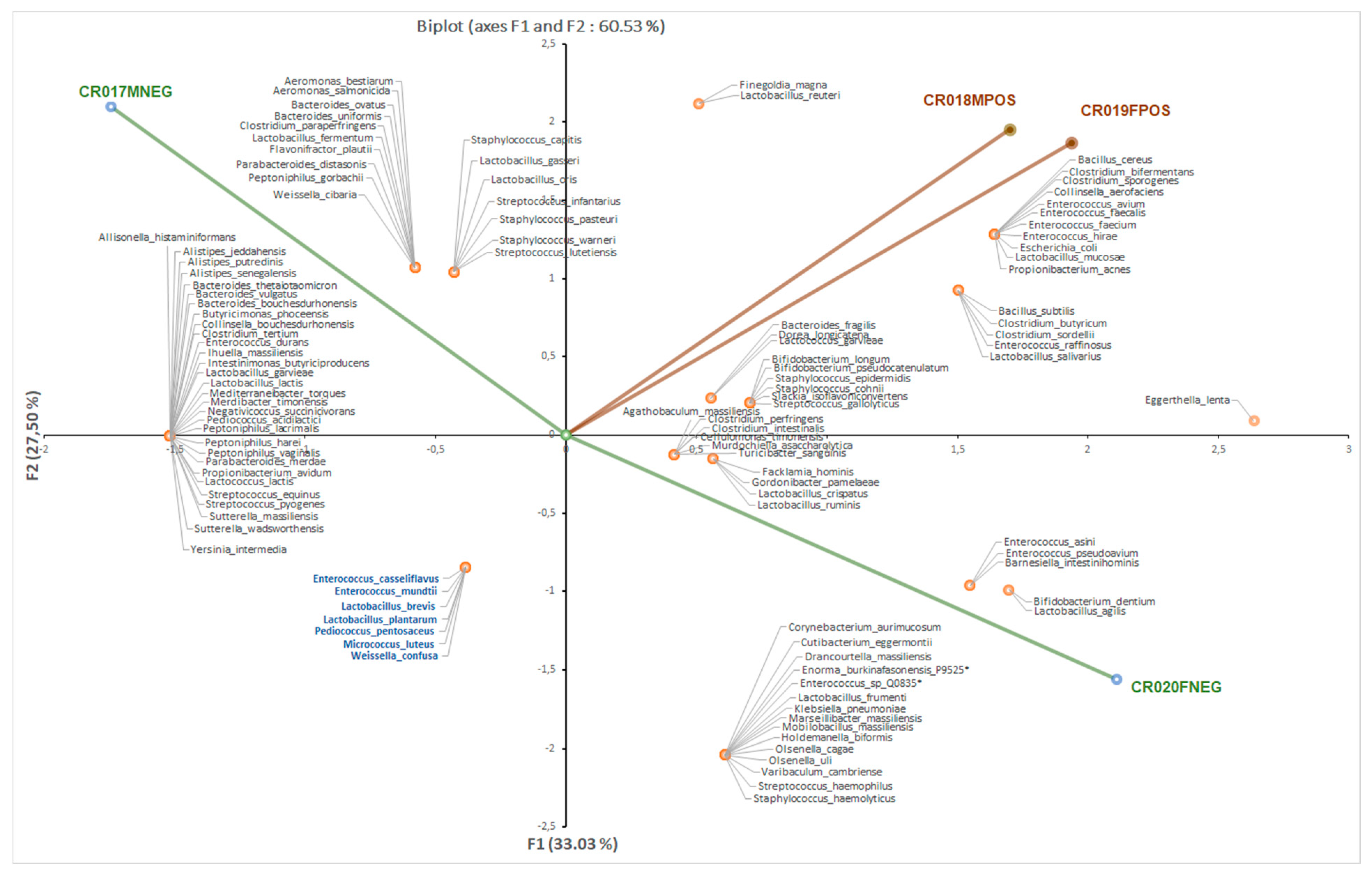
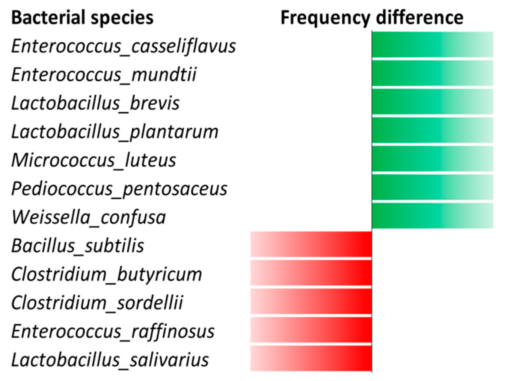
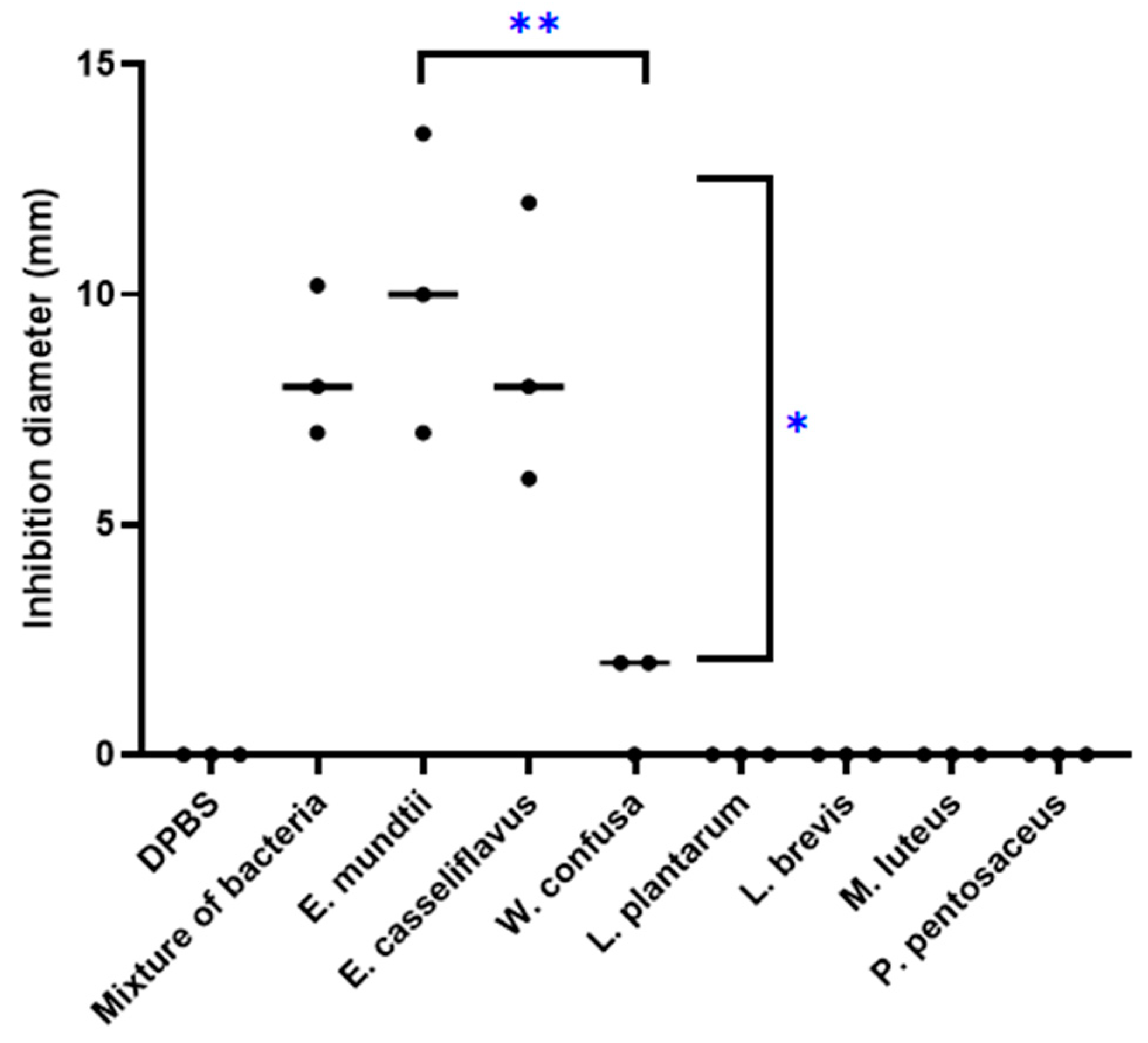
3.1. Bacterial Culturomics

We investigated faeces samples collected from two TB-positive and two TB-negative patients in Burkina Faso. The MALDI-TOF MS identification yielded a total of 112 different bacteria species, divided into 49 genera and 34 families including one unclassified family and four phyla Actinobacteria, Bacteroides, Firmicutes and Proteobacteria. A total of 60 bacterial species scattered across 28 genera were identified in the stools collected from two TB-positive patients and 97 species scattered across 43 genera were identified in the stools collected in two TB-negative patients. At the level of bacterial phyla, Firmicutes were more prevalent in the TB-positive patients, whereas Bacteroidetes were more prevalent in the TB-negative patients (Figure 1).
At the level of bacterial genus, culturomics performed for four individuals showed that the Clostridium bacteria were more abundant in the TB-positive patients (seven in 28; 25%) than in the TB-negative patients (four in 43; 9%). Furthermore, we observed a reduction in the number of Bacteroides, Enterococcus, Streptococcus and Staphylococcus members in the TB-positive patients compared to the TB-negative patients and saw that the number of bacterial species was lower in the TB-positive patients. Preparing out a heatmap (Supplementary Figure S1) and a principal component analysis (Figure 2) showed a clustering of the bacteria isolated from the two TB patients.
The Venn diagram highlighted 15 bacterial species isolated only in the TB-positive patients, 52 bacterial species isolated only in TB-negative patients and 45 bacterial species that were isolated in both the TB-positive and TB-negative patients (Supplementary Figure S2). More precisely, seven bacterial species (Enterococcus casseliflavus, Enterococcus mundtii, Lactobacillus brevis, Lactobacillus plantarum, Micrococcus luteus, Pediococcus pentosaceus, and Weissella confusa) were cultured in both the two TB-negative patients and were not cultured in both the two TB-positive patients, while five bacterial species (Bacillus subtilis, Clostridium butyricum, Clostridium sordellii, Enterococcus raffinosus and Lactobacillus salivarius) were shared by the two TB-positive patients and were absent from the two TB-negative patients (Figure 3). M. smithii was detected by qPCR in all four of these stool samples. Finally, two new species were isolated from one TB-negative patient [25,26,27].

3.2. Real-Time PCR Assays

In order to further explore the prevalence of E. casseliflavus and E. mundtii in the faeces of TB-positive and TB-negative patients, 50 stools were collected from 50 patients being routinely investigated for medical suspicion of pulmonary tuberculosis in the IHU Méditerranée Infection, Marseille, France [28] and 100 specimens were collected from 100 patients routinely investigated for medical suspicion of pulmonary tuberculosis in Burkina Faso (97 specimens collected from individuals in Bobo-Dioulasso and three from individuals in Houndé). In the presence of negative controls (which remained negative), quantitative PCR analysis targeting the 16S rRNA gene of all bacteria within these 150 faeces samples yielded a median Ct of 18.44 and an average Ct average of 18.32 (Ct standard deviation, 3.08; range 11.5–24.4). In the subset of 64 TB-positive patients, the same analysis yielded a median Ct of 18.18 and an average Ct of 18.31 (Ct standard deviation, 2.62; range 13.5–23.8). In the subset of 86 TB-negative patients, the same analysis yielded a median Ct of 17.5 and an average Ct of 18.32 (Ct standard deviation, 3.39; range 11.5–24.4). There were no significant differences in the Ct values between TB-positive and TB-negative patients, indicating an absence of PCR inhibitors in any of the faeces samples investigated here. This allowed us to further investigate the negative results in further quantitative real-time PCR assays, regardless of the TB status of the patient. M. smithii was detected by qPCR in 139/150 (92.7%) faeces samples: five faeces collected from TB-positive patients and six from TB-negative patients were negative. Accordingly, in the very same 150 individuals, blind quantitative real-time PCR targeting E. mundtii yielded 28 (18.22%) positive individuals with a median Ct of 35.47, an average Ct of 34.8, and Ct standard deviation of 2.61, including nine out of 64 (14%) TB-positive patients and 19 in 86 (22%) TB-negative patients (p = 0.21). All 28 E. mundtii-positive faeces specimens had been collected from Burkinabe individuals and none from French individuals. In TB-positive patients, E. mundtii was detected with a median Ct of 34.5, an average Ct of 34.2, and standard deviation of Ct 2.5 (Ct value range, 30.6–38.8), no different from Ct values obtained in TB-negative patients with, respectively, a median Ct of 34.48, an average Ct of 34.23, and standard deviation of Ct 2.6 (Ct value range, 28.8–38.44). Quantitative real-time PCR targeting E. casseliflavus yielded 78 in 150 (52%) positive individuals with a median Ct of 34.8, an average Ct of 34.1, and a Ct standard deviation of 2.85, including 28 in 64 (43%) TB-positive patients and 50 in 86 (58%) TB-negative patients (p = 0.08). In TB-positive patients, E. casseliflavus was detected with a median Ct of 34.45, an average Ct of 35.06, and a standard deviation Ct 2.06 (Ct value range, 28.44–37.19), no different from Ct values obtained in TB-negative patients with, respectively, a median Ct of 34.11, an average Ct of 34.9, and a standard deviation Ct 2.85 (Ct value range, 22.67–38.85).

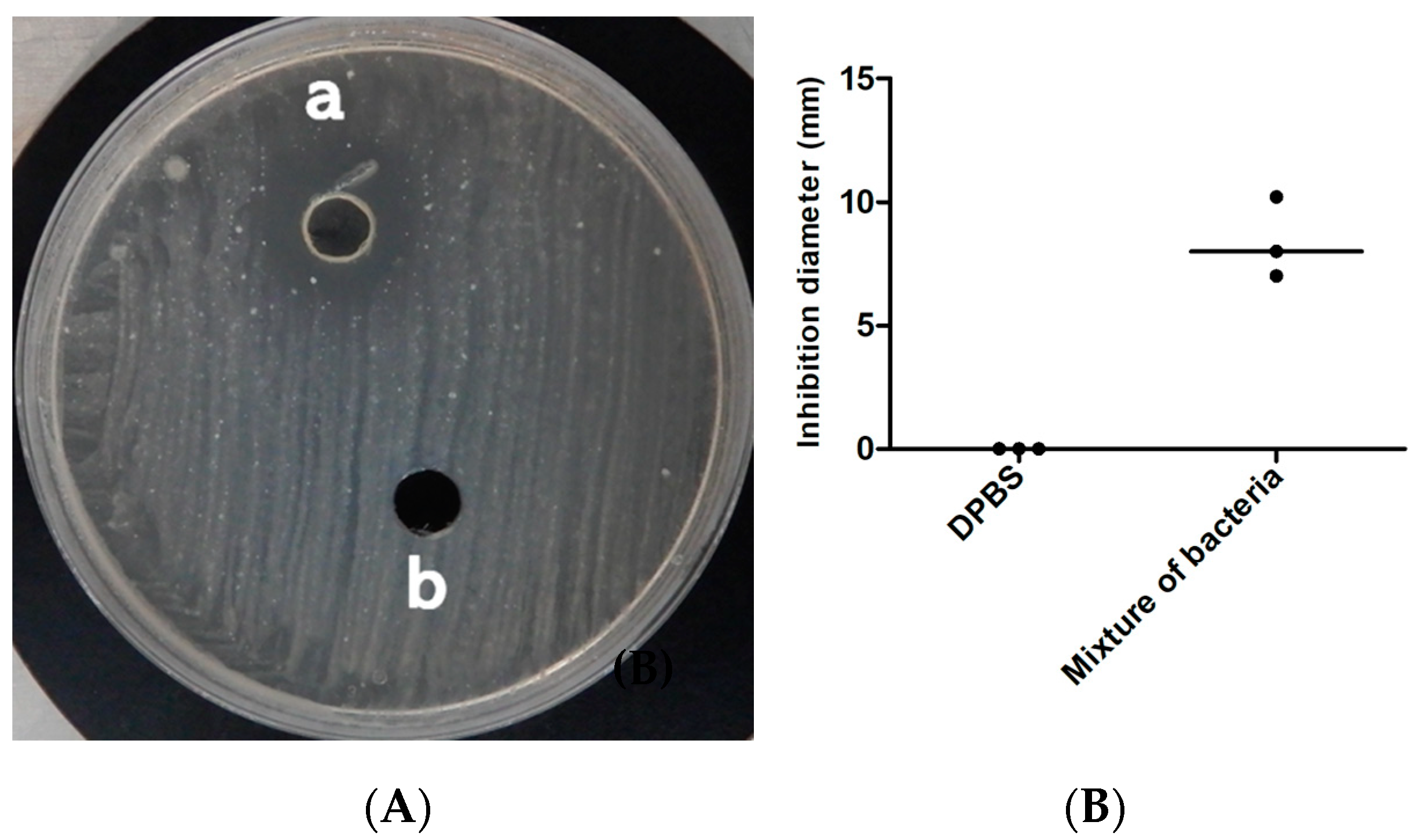
3.3. Antitubercular Activity

The mixture of these seven bacteria lowered the local pH of the Middlebrook culture medium to 5 and inhibited growth of the M. tuberculosis strain Beijing CSUR Q1316 (Figure 4). Further experiments aimed to determine which of these seven bacteria were individually inducing the inhibition of growth of M. tuberculosis, first indicated that the local pH of the Middlebrook culture medium varied from pH 5 for Lactobacillus species to pH 6 for M. tuberculosis and pH 7 for M. luteus. Further, inhibition of growing M. tuberculosis was reproducibly observed only for E. mundtii and E. casseliflavus but not for Lactobacillus brevis, Lactobacillus plantarum, Micrococcus luteus, Pediococcus pentosaceus and W. confusa (Figure 5). Inhibition growth of M. tuberculosis was observed for E. mundtii and E. casseliflavus with bacterial inoculum of 15 McFarland but not for inoculum of 5 McFarland. (data not shown). The M. tuberculosis growth inhibition effect was observed on all tested strains of the MTBC including Mycobacterium canettii, Mycobacterium bovis and Mycobacterium africanum with diameters of growth inhibition being on average three times larger for M. canettii than for the other species (Table 2) (Figure 6). E. mundtii and E. casseliflavus have a similar inhibitory effect against all tested MTBC, except for M. canettii which is significantly more susceptible than the other species of the MTBC (p = 2.14 × 10⁻⁷). The M. tuberculosis growth inhibition effect was not merely due to changes in the pH, as the pH of the suspension was increased from 5 to 6, however M. tuberculosis also reduced the pH of the suspension to 6. However, a few colonies growing in the M. tuberculosis growth inhibition zone around either E. mundtii or E. casseliflavus were confirmed to be colonies of M. tuberculosis by whole genome sequencing. Genome sequences have been deposited in the European Molecular Biology Laboratory (EMBL).

4. Discussion

Previous metagenomics studies all indicated that the repertoire of bacteria in the gut microbiota significantly differed between pulmonary tuberculosis patients and controls, suggesting biological relationships between pulmonary tuberculosis and gut microbiota [10]. However, contradictory data from these studies complicated the interpretation of these differences. Decreases in the number of bacterial species and bacterial diversity reported in TB-positive patients [29] including children [30], contrasted with the reported increased diversity in TB-patients in another study [12]. In children, microbial richness was not found to significantly differ between TB-positive children and controls, although the Simpson index was found to be significantly lower in TB-positive children [30] with five different bacterial species found to be significantly balanced between the two groups [30]. The interpretation of metagenomics studies data was limited by the inherent profounder bias of the method, lack of accurate bacterial species identification [10], and the fact that detected organisms may not be viable, thus rendering them to be of limited physiological interest.
In this study, we used culturomics (in addition to PCR-based detection of the methanogen M. smithii) to overcome some limitations of the metagenomics studies, and confirmed the decreased prevalence in Bacteroidetes in TB-positive patients compared to TB-negative ones [12] and the increase of Clostridium members. This was in line with observations made in experimental severe tuberculosis in macaques [31]. Unexpectedly, culturomics yielded two Firmicutes E. casseliflavus and E. mundtii exclusively in the faeces of TB-negative patients. These two Firmicutes were further qPCR-detected with an increased prevalence in the faeces of TB-negative patients in two independent cohorts. These observations were extended by experimental observations showing these two Firmicutes inhibited the growth of M. canettii, regarded as the MTBC species most closely related to the common ancestor of the complex and a pathogen responsible for tuberculosis in exposed individuals in the Horn of Africa [1], and that of M. tuberculosis and M. bovis responsible for tuberculosis in humans and animals [32]. These observations, all reproduced at least three times, were attested to by the negativity of the negative controls. We also observed that heat-killed bacteria no longer inhibited MTBC growth which was inoculum-dependent and not merely related to pH variation. All observations suggest an active process which remains to be elucidated. The two Firmicutes are known to produce bacteriocins, yet none of these bacteriocins has ever been demonstrated to be active against M. tuberculosis [33,34,35]. As for E. mundtii, all previous studies have dealt with the antibiotic activity of environmental isolates against opportunistic pathogens, mainly Listeria monocytogenes, such as E. mundtii fish isolates LP17 and LP18 [36]. Identifying the mechanism of action of E. mundtii and E. casseliflavus against MTBC was beyond the scope of the present study, yet the E. mundtii and E. casseliflavus strains here investigated all encode for bacteriocin, as anticipated by their whole genome sequence.
Interactions between M. tuberculosis and inhibitory enterococci are likely to occur in the gut. M. tuberculosis is routinely cultured from the faeces collected from TB-positive patients but not from TB-negative ones [28]. In the “Lübeck Disaster 1929–1933”, oral inoculation of the BCG vaccine inadvertently contaminated 251 neonates with M. tuberculosis, causing clinical tuberculosis in 228 (90.83%) cases, while five in 251 (1.99%) of the children died of unrelated causes. Although 18 neonates showed no signs of tuberculosis, 72 died. Follow-up of the 174 surviving children revealed only six sick children, while the others’ symptoms were spontaneously resolved. Such a heterogeneous natural history of tuberculosis in neonates in the Lübeck series of cases may partly be explained by different gut microbiota maturation in the neonates [37]. Indeed, translocating M. canettii and M. tuberculosis has been shown to lead to both disseminating in the lymphatic and lung tissues after experimental gavage in mice [18,38]. It would be interesting to further document E. mundtii and E. casseliflavus interactions with M. tuberculosis in a mouse model, to observe interference with M. tuberculosis gut survival, translocation, and recirculation in the lungs. Complementary, indirect interactions may be mediated by inflammation and immune cells in the exact model of what is now established in immune cancerology [39]. As an example, gut Prevotella, the prevalence of which was increased in TB-positive children and adults [12,30], were shown to correlate with blood CD4-positive lymphocytes and have been suggested to boost the inflammatory response to TB [12]. Likewise, Clostridium members that were found to be more abundant in TB-positive patients, induce Treg production in mice guts and an increased number of Venus+CD4+ cells in the liver and lungs [40]. Clostridium members are short-chain fatty acid (SCFA) producers inducing anti-inflammatory production (IL-10) [41], and SCFAs metabolites are acknowledged to modulate immune and proinflammatory responses to pulmonary TB [42].
This is a preliminary study involving by culturomics only in four individuals, so that the generalized of data here reported are not possible today and will required additional studies in larger population. Also, this study disclosed potential limitations in the enterococci interactions with M. tuberculosis. First, E. mundtii was detected only in faeces samples collected from Burkinabe individuals and not from French ones, suggesting that gut carriage of E. mundtii is population-dependant and is probably linked to diet, given that E. mundtii has been found in several plants and meat entering the human food chain [43,44]. Second, we did observe a few M. tuberculosis colonies that were resistant to E. mundtii-inhibition, suggesting that the mechanism of growth inhibition is not absolute in one M. tuberculosis population.

5. Conclusions

In conclusion, the data reported here indicate a higher bacterial diversity in non-tuberculous individuals versus pulmonary tuberculosis patients. In addition, we confirm a protective effect of certain gut microbiota and, particularly, of the Firmicutes E. casseliflavus and E. mundtii against pulmonary tuberculosis. Further studies may evaluate the relative recombination of innate immunity in these observations such as macrophage and cytokine expression induced by enterococci and E. mundtii, in particular during the intracellular phase of tuberculosis. Depending on the elucidation of the underlying mechanisms and topography of the inhibition and extent of M. tuberculosis complex, some of these Firmicutes may be considered as probiotic adjuvants to conventional antibiotics in the fight against deadly tuberculosis.

Supplementary Materials

The following are available online at https://www.mdpi.com/2076-2607/8/10/1544/s1. Figure S1: Venn diagram analysis perform on the bacterial species isolated in the two groups (tuberculous and nontuberculous patients) showing the differential distribution and the common bacteria, Figure S2: Heatmap clustering of bacterial species cultured in the four individuals included in this study showing the bacterial species distribution between tuberculous and nontuberculous patients. Red and green scores indicate the absence and presence of bacterial species, respectively in tuberculosis patients (CR019FPOS and CR018MPOS) and control individuals (CR020FNEG and CR017MNEG), Table S1: Primers and probes used in this study.

Author Contributions

M.F. performed culturomics, molecular biology, anti-tuberculous activity tests and draft the manuscript; N.G. performed stool collection, culturomics and drafted the manuscript; M.B. designed and set-up specific qPCR assays; M.S., D.Z., and Z.T. collected stools; M.M. performed data analyses; M.D. conceived the study and draft the manuscript. All authors have read and agreed to the published version of the manuscript.

Funding

This study was supported by the French Government under the Investissements d’Avenir (Investments for the Future) program managed by the Agence Nationale de la Recherche (ANR, fr: National Agency for Research), [reference: Méditerranée Infection 10-IAHU-03]. This study was also supported by Région Le Sud (Provence Alpes Côte d’Azur) and a European funding agency [FEDER PA 0000320 PRIMI]. Mustapha FELLAG is supported by a PhD grant from IHU Méditerranée Infection, Marseille, France.

Acknowledgments

The authors acknowledge technical contributions from Odilon Kabore, Mbonging Fonkou and Olivier Sombié, along with staff from the Centre de Diagnostic et de Traitement de la Tuberculose in Bobo-Dioulasso for their collaboration regarding the collection of faeces samples in Burkina Faso. The authors acknowledge technical contributions from Ahmed Loukil for statistical analyses.

Conflicts of Interest

The authors declare no conflict of interest.

References

  1. Aboubaker, O.D.; Bouzid, F.; Canaan, S.; Drancourt, M. Smooth Tubercle Bacilli: Neglected Opportunistic Tropical Pathogens. Front. Public Health 2015, 3, 283. [Google Scholar] [CrossRef] [PubMed] [Green Version]
  2. WHO. Global Tuberculosis Report, Geneva. World Health Organization 2019. Available online: https://apps.who.int/iris/bitstream/handle/10665/329368/9789241565714-eng.pdf?ua=1 (accessed on 19 July 2020).
  3. Brites, D.; Gagneux, S. The Nature and Evolution of Genomic Diversity in the Mycobacterium tuberculosis Complex. Adv. Exp. Med. Biol. 2017, 1019, 1–26. [Google Scholar] [PubMed]
  4. Copin, R.; Wang, X.; Louie, E.; Escuyer, V.; Coscolla, M.; Gagneux, S.; Palmer, G.H.; Ernst, J.D. Within Host Evolution Selects for a Dominant Genotype of Mycobacterium tuberculosis while T Cells Increase Pathogen Genetic Diversity. PLoS Pathog. 2016, 12, e1006111. [Google Scholar] [CrossRef] [PubMed] [Green Version]
  5. Turner, R.D.; Chiu, C.; Churchyard, G.J.; Esmail, H.; Lewinsohn, D.M.; Gandhi, N.R.; Fennelly, K.P. Tuberculosis Infectiousness and Host Susceptibility. J. Infect. Dis. 2017, 216, S636–S643. [Google Scholar] [CrossRef] [Green Version]
  6. Bustamante, J.; Aksu, G.; Vogt, G.; de Beaucoudrey, L.; Genel, F.; Chapgier, A.; Orchidée, F.S.; Jacqueline Feinberg, J.; Emile, J.F.; Kutukculer, N.; et al. BCG-osis and tuberculosis in a child with chronic granulomatous disease. J. Allergy Clin. Immunol. 2007, 120, 32–38. [Google Scholar] [CrossRef]
  7. Abel, L.; El-Baghdadi, J.; Bousfiha, A.A.; Casanova, J.-L.; Schurr, E. Human genetics of tuberculosis: A long and winding road. Philos. Trans. R. Soc. Lond. B Biol. Sci. 2015, 369, 20130428. [Google Scholar] [CrossRef] [Green Version]
  8. Casanova, J.-L.; Abel, L. Genetic Dissection of Immunity to Mycobacteria: The Human Model. Annu. Rev. Immunol. 2002, 20, 581–620. [Google Scholar] [CrossRef]
  9. Zhang, S.; Wang, X.; Han, Y.; Wang, C.; Zhou, Y.; Zheng, F. Certain Polymorphisms in SP110 Gene Confer Susceptibility to Tuberculosis: A Comprehensive Review and Updated Meta-Analysis. Yonsei Med. J. 2017, 58, 165–173. [Google Scholar] [CrossRef] [Green Version]
  10. Sekyere, J.O.; Maningi, N.E.; Fourie, P.B. Mycobacterium tuberculosis, antimicrobials, immunity, and lung–gut microbiota crosstalk: Current updates and emerging advances. Ann. N. Y. Acad. Sci. 2020, 1–27. [Google Scholar] [CrossRef]
  11. Winglee, K.; Eloe-Fadrosh, E.; Gupta, S.; Guo, H.; Fraser, C.; Bishai, W. Aerosol Mycobacterium tuberculosis Infection Causes Rapid Loss of Diversity in Gut Microbiota. PLoS ONE 2014, 9, e97048. [Google Scholar] [CrossRef]
  12. Luo, M.; Liu, Y.; Wu, P.; Luo, D.-X.; Sun, Q.; Zheng, H.; Hu, R.; Pandol, S.J.; Li, Q.-F.; Han, Y.-P.; et al. Alternation of Gut Microbiota in Patients with Pulmonary Tuberculosis. Front. Physiol. 2017, 8, 822. [Google Scholar] [CrossRef] [PubMed]
  13. Hu, Y.; Feng, Y.; Wu, J.; Liu, F.; Zhang, Z.; Hao, Y.; Liang, S.; Li, B.; Li, J.; Lv, N.; et al. The Gut Microbiome Signatures Discriminate Healthy From Pulmonary Tuberculosis Patients. Front. Cell. Infect. Microbiol. 2019, 9, 90. [Google Scholar] [CrossRef] [PubMed] [Green Version]
  14. Asmar, S.; Chatellier, S.; Mirande, C.; van Belkum, A.; Canard, I.; Raoult, D.; Drancourt, M. A Novel Solid Medium for Culturing Mycobacterium tuberculosis Isolates from Clinical Specimens. J. Clin. Microbiol. 2015, 53, 2566–2569. [Google Scholar] [CrossRef] [PubMed] [Green Version]
  15. Lagier, J.-C.; Hugon, P.; Khelaifia, S.; Fournier, P.-E.; Scola, B.L.; Raoult, D. The Rebirth of Culture in Microbiology through the Example of Culturomics To Study Human Gut Microbiota. Clin. Microbiol. Rev. 2015, 28, 237–264. [Google Scholar] [CrossRef] [PubMed] [Green Version]
  16. Seng, P.; Drancourt, M.; Gouriet, F.; La Scola, B.; Fournier, P.-E.; Rolain, J.M.; Raout, D. Ongoing revolution in bacteriology: Routine identification of bacteria by matrix-assisted laser desorption ionization time-of-flight mass spectrometry. Clin. Infect. Dis. 2009, 49, 543–551. [Google Scholar] [CrossRef] [PubMed]
  17. Lagier, J.-C.; Khelaifia, S.; Alou, M.T.; Ndongo, S.; Dione, N.; Hugon, P.; Caputo, A.; Cadoret, F.; Traore, I.S.; Seck, E.S.; et al. Culture of previously uncultured members of the human gut microbiota by culturomics. Nat. Microbiol. 2016, 1, 1–8. [Google Scholar] [CrossRef] [PubMed]
  18. Fellag, M.; Loukil, A.; Saad, J.; Lepidi, H.; Bouzid, F.; Brégeon, F.; Drancourt, M. Translocation of Mycobacterium tuberculosis after experimental ingestion. PLoS ONE 2019, 14, e0227005. [Google Scholar] [CrossRef] [Green Version]
  19. Dridi, B.; Henry, M.; El Khéchine, A.; Raoult, D.; Drancourt, M. High prevalence of Methanobrevibacter smithii and Methanosphaera stadtmanae detected in the human gut using an improved DNA detection protocol. PLoS ONE 2009, 4, e7063. [Google Scholar] [CrossRef]
  20. Lagier, J.-C.; Million, M.; Togo, A.H.; Khelaifia, S.; Raoult, D. Culturomics provides critical prokaryote strains for anti-Listeria and anti-cancer probiotics. Int. J. Antimicrob. Agents 2019, 54, 407–409. [Google Scholar] [CrossRef]
  21. Balouiri, M.; Sadiki, M.; Ibnsouda, S.K. Methods for in vitro evaluating antimicrobial activity: A review. J. Pharm. Anal. 2016, 6, 71–79. [Google Scholar] [CrossRef] [Green Version]
  22. Fellag, M.; Saad, J.; Armstrong, N.; Chabrière, E.; Eldin, C.; Lagier, J.-C.; Drancourt, M. Routine Culture-Resistant Mycobacterium tuberculosis Rescue and Shell-Vial Assay, France. Emerging Infect. Dis. 2019, 25, 2131–2133. [Google Scholar] [CrossRef] [PubMed]
  23. Lapierre, S.G.; Fellag, M.; Magan, C.; Drancourt, M. Mycobacterium malmoense pulmonary infection in France: A case report. BMC Res. Notes 2017, 10, 436. [Google Scholar] [CrossRef] [PubMed] [Green Version]
  24. Asmar, S.; Drancourt, M. Chlorhexidine decontamination of sputum for culturing Mycobacterium tuberculosis. BMC Microbiol. 2015, 15, 155. [Google Scholar] [CrossRef] [PubMed] [Green Version]
  25. Gouba, N.; Saad, J.; Drancourt, M.; Fellag, M. Genome Sequence of Enorma sp. Strain Marseille-P9525T, a Member of a Human Gut Microbiome. Microbiol. Resour. Announc. 2019, 8, e00785-19. [Google Scholar] [CrossRef] [Green Version]
  26. Gouba, N.; Hassani, Y.; Saad, J.; Drancourt, M.; Mbogning Fonkou, M.D.; Fellag, M. Enorma burkinafasonensis sp. nov., a new bacterium isolated from a human gut microbiota. New Microbes New Infect. 2020, 36, 100687. [Google Scholar]
  27. Gouba, N.; Yimagou, E.K.; Hassani, Y.; Drancourt, M.; Fellag, M.; Mbogning Fonkou, M.D. Enterococcus burkinafasonensis sp. nov. isolated from human gut microbiota. New Microbes New Infect. 2020, 36, 100702. [Google Scholar] [CrossRef]
  28. Drancourt, M. Culturing Stools to Detect Mycobacterium tuberculosis. J. Clin. Microbiol. 2018, 56, e02033-17. [Google Scholar] [CrossRef] [Green Version]
  29. Hu, Y.; Yang, Q.; Liu, B.; Dong, J.; Sun, L.; Zhu, Y.; Su, H.; Liu, B.; Yang, J.; Yang, F.; et al. Gut microbiota associated with pulmonary tuberculosis and dysbiosis caused by anti-tuberculosis drugs. J. Infect. 2019, 78, 317–322. [Google Scholar] [CrossRef]
  30. Li, W.; Zhu, Y.; Liao, Q.; Wang, Z.; Wan, C. Characterization of gut microbiota in children with pulmonary tuberculosis. BMC Pediatr. 2019, 19, 445. [Google Scholar] [CrossRef]
  31. Namasivayam, S.; Kauffman, K.D.; McCulloch, J.A.; Yuan, W.; Thovarai, V.; Mittereder, L.R.; Trinchieri, G.; Barber, D.L.; Sheret, A. Correlation between Disease Severity and the Intestinal Microbiome in Mycobacterium tuberculosis-Infected Rhesus Macaques. mBio 2019, 10, 3. [Google Scholar] [CrossRef] [Green Version]
  32. Behr, M.A.; Waters, W.R. Is tuberculosis a lymphatic disease with a pulmonary portal? Lancet Infect. Dis. 2014, 14, 250–255. [Google Scholar] [CrossRef]
  33. Bravo, M.; Combes, T.; Martinez, F.O.; Cerrato, R.; Rey, J.; Garcia-Jimenez, W.; Fernandez-Llario, P.; Risco, D.; Gutierrez-Merinet, J. Lactobacilli Isolated From Wild Boar (Sus scrofa) Antagonize Mycobacterium bovis Bacille Calmette-Guerin (BCG) in a Species-Dependent Manner. Front. Microbiol. 2019, 10, 1663. [Google Scholar] [CrossRef] [PubMed] [Green Version]
  34. Sivaraj, A.; Sundar, R.; Manikkam, R.; Parthasarathy, K.; Rani, U.; Kumar, V. Potential applications of lactic acid bacteria and bacteriocins in anti-mycobacterial therapy. Asian Pac. J. Trop. Med. 2018, 11, 453. [Google Scholar] [CrossRef]
  35. Sosunov, V.; Mischenko, V.; Eruslanov, B.; Svetoch, E.; Shakina, Y.; Stern, N.; Majorov, K.; Sorokoumova, G.; Selishcheva, A.; Apt, A. Antimycobacterial activity of bacteriocins and their complexes with liposomes. J. Antimicrob. Chemother. 2007, 59, 919–925. [Google Scholar] [CrossRef] [PubMed] [Green Version]
  36. Iseppi, R.; Stefani, S.; de Niederhausern, S.; Bondi, M.; Sabia, C.; Messi, P. Characterization of Anti-Listeria monocytogenes Properties of two Bacteriocin-Producing Enterococcus mundtii Isolated from Fresh Fish and Seafood. Curr. Microbiol. 2019, 76, 1010–1019. [Google Scholar] [CrossRef]
  37. Fox, G.J.; Orlova, M.; Schurr, E. Tuberculosis in Newborns: The Lessons of the “Lübeck Disaster” (1929–1933). PLoS Pathog. 2016, 12, e1005271. [Google Scholar] [CrossRef] [Green Version]
  38. Bouzid, F.; Brégeon, F.; Lepidi, H.; Donoghue, H.D.; Minnikin, D.E.; Drancourt, M. Ready Experimental Translocation of Mycobacterium canettii Yields Pulmonary Tuberculosis. Infect. Immun. 2017, 85. [Google Scholar] [CrossRef] [Green Version]
  39. Zitvogel, L.; Kroemer, G. Immunostimulatory gut bacteria. Science 2019, 366, 1077–1078. [Google Scholar] [CrossRef]
  40. Atarashi, K.; Tanoue, T.; Shima, T.; Imaoka, A.; Kuwahara, T.; Momose, Y.; Cheng, G.; Yamasaki, S.; Saito, T.; Ohba, Y.; et al. Induction of Colonic Regulatory T Cells by Indigenous Clostridium Species. Science 2011, 331, 337–341. [Google Scholar] [CrossRef] [Green Version]
  41. Naidoo, C.C.; Nyawo, G.R.; Wu, B.G.; Walzl, G.; Warren, R.M.; Segal, L.N.; Theron, G. The microbiome and tuberculosis: State of the art, potential applications, and defining the clinical research agenda. Lancet Respir. Med. 2019, 7, 892–906. [Google Scholar] [CrossRef]
  42. Lachmandas, E.; van den Heuvel, C.N.A.M.; Damen, M.S.M.A.; Cleophas, M.C.P.; Netea, M.G.; van Crevel, R. Diabetes Mellitus and Increased Tuberculosis Susceptibility: The Role of Short-Chain Fatty Acids. J. Diabetes Res. 2016, 2016, e6014631. [Google Scholar] [CrossRef] [PubMed]
  43. Chingwaru, W.; Mpuchane, S.F.; Gashe, B.A. Enterococcus faecalis and Enterococcus faecium Isolates from Milk, Beef, and Chicken and Their Antibiotic Resistance. J. Food Prot. 2003, 66, 931–936. [Google Scholar] [CrossRef] [PubMed]
  44. Devriese, L.A.; Pot, B.; Van Damme, L.; Kersters, K.; Haesebrouck, F. Identification of Enterococcus species isolated from foods of animal origin. Int. J. Food Microbiol. 1995, 26, 187–197. [Google Scholar] [CrossRef]
Figure 1. Cultured gut bacteria distribution between tuberculous and nontuberculous patients. Phyla and number of isolated bacterial species per group included tuberculosis patients and controls.
Microorganisms 08 01544 g001
Figure 2. Principal component analysis (PCA) of 112 bacterial species isolated from the guts of the four individuals included in this study. PCA revealed a clustering of bacterial composition in the tuberculosis patients between the bacteria isolated in the two tuberculosis patients.
Microorganisms 08 01544 g002
Figure 3. Frequency analysis performed on bacteria isolated from the four individuals reveals seven bacteria isolated only in the non-tuberculous patients shown in green and five bacteria isolated only in the tuberculous patients shown in red.
Microorganisms 08 01544 g003
Figure 4. Effect of mixture bacteria on growth of M. tuberculosis lineage 2. (A) Observation of the inhibition spectrum caused by the mixture bacteria (a) on M. tuberculosis and DPBS (b) as negative controls. The picture was prepared 12 days after incubation of M. tuberculosis at 37 °C. (B) The inhibition test was carried out three times, the diameter of inhibition was measured on the three cultures. The image indicates the median.
Microorganisms 08 01544 g004
Figure 5. In vitro activity effect of the seven bacteria isolated only from non-tuberculosis patients against M. tuberculosis lineage 2. For each bacterium and DPBS negative control tests were carried out in triplicate. “**” Comparison of the mean inhibition diameter of M. tuberculosis by E. mundtii vs. the mean diameter inhibition of M. tuberculosis by W. confusa (p Value = 0.0156). “*” Comparison of the mean inhibition diameter of M. tuberculosis by E. casseliflavus vs. the mean diameter inhibition of M. tuberculosis by W. confusa (p Value = 0.0345). Means inhibition diameters were compared using Tukey’s test (p < 0.05).
Microorganisms 08 01544 g005
Figure 6. In vitro activity of E. mundtii against M. tuberculosis lineage 2 growth. (A) Picture showing the inhibitory effect of E. mundtii (a) on M. tuberculosis, DPBS buffer (b) and M. luteus (c) as a negative control. (B) Triplicate test inhibition of E. mundtii against M. tuberculosis.
Microorganisms 08 01544 g006
Table 1. Characteristics of the four individuals studied by culturomics.
PatientsSexAgeWeightSizeBMISmear ResultSputum CultureGeneXpert
CR017M45901.8127negativenegativenegative
CR018M65561.6819positive+++positivepositive
CR019F28501.6019.53positive+++positivepositive
CR020F22731.6626.54negativenegativenegative
Table 2. Inhibition effect of E. mundtii and E. casseliflavus against M. tuberculosis complex growth, DPBS as a negative control.
MTBC StrainDPBSMixture E. mundtiip-Value * E. casseliflavusp-Value *
M. tuberculosis L 208.4 ± 1.210.83 ± 1.770.0159.1 ± 1.880.023
M. tuberculosis L10-10.66 ± 3.400.0329.83 ± 1.750.0104
M. tuberculosis L 30-8.16 ± 1.040.0056.33 ± 1.520.01884
M. tuberculosis L 40-9.66 ± 3.050.0318.83 ± 30.000516
M. africanum0-9.66 ± 1.520.0088.33 ± 1.520.011015
M. canettii0-29.33 ± 1.150.000527.33 ± 2.08 0.00192
M. bovis0-11.33 ± 2.080.01110.66 ± 2.510.0180
* p value was obtained using student test. Experiments were triplicate. The growth inhibition diameters are expressed in mm. Values are expressed as mean ± standard deviation.

