Article

Antibacterial Activity of Four Plant Extracts Extracted from Traditional Chinese Medicinal Plants against Listeria monocytogenes, Escherichia coli, and Salmonella enterica subsp. enterica serovar Enteritidis

1
School of Biological Sciences, Queen’s University Belfast, Belfast BT9 5DL, Northern Ireland, UK
2
Agri-Food and Biosciences Institute, Hillsborough BT26 6DR, Northern Ireland, UK
3
School of Pharmacy, Queen’s University Belfast, Belfast BT9 7BL, Northern Ireland, UK
4
Agri-Food and Biosciences Institute, Veterinary Sciences Division, Belfast BT4 3SD, Northern Ireland, UK
5
Institute for Global Food Security, Queen’s University Belfast, Belfast BT9 5DL, Northern Ireland, UK
*
Author to whom correspondence should be addressed.
Microorganisms 2020, 8(6), 962; https://doi.org/10.3390/microorganisms8060962
Submission received: 10 June 2020 / Revised: 25 June 2020 / Accepted: 25 June 2020 / Published: 26 June 2020

Abstract

:
The worldwide ethnobotanical use of four investigated plants indicates antibacterial properties. The aim of this study was to screen and determine significant antibacterial activity of four plant extracts in vitro and in a poultry digest model. Using broth microdilution, the concentrations at which four plant extracts inhibited Listeria monocytogenes, Salmonella enteritidis, and Escherichia coli over 24 h was determined. Agrimonia pilosa Ledeb, Iris domestica (L.) Goldblatt and Mabb, Anemone chinensis Bunge, and Smilax glabra Roxb all exhibited a minimum inhibitory concentration (MIC) of 62.5 mg/L and a minimum bactericidal concentration (MBC) of 500 mg/L against one pathogen. A. pilosa Ledeb was the most effective against L. monocytogenes and E. coli with the exception of S. enteritidis, for which A. chinensis Bunge was the most effective. Time–kills of A. pilosa Ledeb and A. chinensis Bunge against L. monocytogenes, E. coli and S. enteritidis incubated in poultry cecum were used to determine bactericidal activity of these plant extracts. A. chinensis Bunge, significantly reduced S. enteritidis by ≥ 99.99% within 6 h. A. pilosa Ledeb exhibited effective significant bactericidal activity within 4 h against L. monocytogenes and E. coli. This paper highlights the potential of these plant extracts to control pathogens commonly found in the poultry gastrointestinal tract.

Graphical Abstract

1. Introduction

Antibiotic resistance is currently a global concern and significant research is taking place to tackle this issue [1,2], including research into the use of antibiotics in animal production [3]. Poultry farming is a rapidly growing global industry3. In many countries outside of Europe antibiotics are used as poultry feed additives at subtherapeutic levels over prolonged periods to promote growth and control gastrointestinal infections in the flock [4,5]. While there are legitimate therapeutic reasons for antibiotic use in poultry farming there are concerns about the overuse and misuse of antibiotics [3]. A link between subtherapeutic application of broad-spectrum tetracyclines in agriculture and the development of acquired-resistant human isolates led to a ban on the use of tetracyclines for growth promotion in Europe in the early 1970s [6]. Acquired resistance is the major contributor to baseline resistance and is a major threat to the continued success of antibiotics [7]. Due to the alarming rate of increased acquired antibiotic resistance in pathogens it is imperative that researchers explore other safe and sustainable alternatives to antibiotics. Alternatives to antibiotics could be used in livestock production to maintain production performance and control infections caused by pathogens commonly found in the poultry gastrointestinal tract. For example, Escherichia, Salmonella, and Listeria [8,9,10].
Numerous medicinal plants have been utilized as traditional medicines globally [11] for human therapeutic use to treat diseases of pathogenic origin [12]. Plant extracts consist of numerous bioactive compounds including polyphenols, terpenes, and phytosterols [13] and exhibit multiple modes of action to inhibit or kill bacteria [14]. An accumulating quantity of research has demonstrated that many plants used in traditional Chinese medicine have antibacterial activity both in vitro and in vivo in poultry production [15]. More specifically, the antibacterial properties of solvent extracts of Agrimonia pilosa Ledeb, Smilax glabra Roxb, Anemone chinensis Bunge, and Iris domestica (L.) Goldblatt and Mabb have been documented through in vitro screening [16,17,18,19]. Aqueous extraction methods are inexpensive and the bioactive compounds are less toxic and therefore more suitable for use as a poultry feed supplement. Furthermore, the antibacterial activity of the aqueous extract of Anemone chinensis Bunge exhibited antibacterial activity against Staphylococcus aureus during disc diffusion, highlighting its antibacterial properties [20]. A. pilosa Ledeb, S. glabra Roxb, A. chinensis Bunge, and I. domestica (L.) Goldblatt and Mabb are listed in the Chinese Pharmacopoeia [21] and are used to treat infections in many parts of the world. However, to our knowledge, no report exists on the bactericidal activity of these aqueous plant extracts in an in vitro poultry digest model. It is necessary to establish scientific evidence for the bactericidal activity of plant extracts as they may provide a source for the development of novel antibiotics.
Agrimonia pilosa Ledeb, S. glabra Roxb, A. chinensis Bunge, and I. domestica (L.) Goldblatt and Mabb are hardy, perennial plants and can therefore be grown in abundance in Europe, the UK, and Asia [22,23,24,25] with minimum maintenance. This makes them suitable as accessible sources of novel antibiotics. These plants are native to different regions. For example, A. pilosa Ledeb is native to Eastern Europe, China, Korea, and Japan. A. chinensis Bunge is native to East Russia, China, and Korea. I. domestica (L.) Goldblatt and Mabb is native across regions from Himalaya to Japan and Philippines and has been successfully introduced to the USA. S. glabra Roxb is native to Southeast Asia.
The antibacterial properties of plant extracts of A. pilosa Ledeb, S. glabra Roxb, A. chinensis Bunge, and I. domestica (L.) Goldblatt and Mabb, commonly used in traditional Chinese medicine to treat diseases of pathogenic origin are presented in this paper against three pathogens commonly found in the poultry gastrointestinal tract: Escherichia coli, Salmonella enteritidis, and Listeria monocytogenes. The aim of this research was to determine the antibacterial properties of these plant extracts and to evaluate the most effective bactericidal effect using an in vitro poultry digest model.

2. Materials and Methods

2.1. Preparation of Reference Strains and Clinical Isolates

Reference strains and clinical isolates were obtained as frozen stocks from Agri-Food and Biosciences Institute (AFBI), the National Collection of Type Cultures (NCTC), the American Type Culture Collection (ATCC), and Queen’s University Belfast (QUB) from several sources (Table 1). Each bacterium was identified using 16S PCR, Sanger sequencing, and BLAST analysis as per manufacturer’s instructions for using MyTaq™ Red Mix [26]. Gram staining was also used to confirm the identity of bacteria [27]. L. monocytogenes, Salmonella enterica subsp. enterica serovar Enteritidis, and E. coli were selected for antimicrobial susceptibility testing because they are pathogens that cause foodborne diseases and are commonly found in the intestinal tract of poultry [8,9,10].

2.2. Preparation of Plant Extracts

Four dry plant samples were purchased from Tong Ren Tang (Beijing). These included: the herb of Agrimonia pilosa Ledeb, the tuber of Smilax glabra Roxb, the rhizome of Iris domestica (L.) Goldblatt and Mabb, and the root of Anemone chinensis Bunge. The accepted names of these plants are in accordance with The Plant List [28]. Ten milligrams of each plant were powdered using a rotary ball mill (Retch PM 100 planetary ball mill, QUB) according to manufacturer’s instructions resulting in a yield of 9 ± 0.8 mg of each plant. Two milligrams of powder were dissolved with deionised water (1:1), placed in an ultrasonic bath for 15 min then boiled in a water bath for 20 min. Solutions were stored at <4 °C prior to analysis.

2.3. Determination of Antibacterial Activity In Vitro

Plant extracts were screened using the broth microdilution method [29] to determine the minimum inhibitory concentration (MIC) against L. monocytogenes, S. enteritidis, and E. coli. Bacterial cultures were incubated overnight under the following conditions, optimizing them for bacterial growth: L. monocytogenes was incubated in tryptone soy broth (Oxoid, UK) with 5% lysed horse blood (TCS Biosciences Ltd., UK) at 35 ± 1 °C, 5% CO2; E. coli, and S. enteritidis were incubated in Mueller Hinton broth (Oxoid, UK) at 35 ± 1°C, ambient air. Two-fold serial dilutions were made up of antibiotics and each plant extract. One hundred microlitres of each antibiotic and plant extract concentration (2000 mg/L to 0.06 mg/L) were each added to individual wells of a 96-well plate. The bacterial culture was adjusted to an optical density equivalent to 1 × 10⁸ CFUmL−1 then diluted to 1 × 10⁶ CFUmL−1. Optical density values were confirmed with a bacterial count and were all within optical density ±0.02. One hundred microlitres of bacterial suspension was added to each well. A negative control included broth only. Ampicillin was used as a quality control for L. monocytogenes, S. enteritidis, and E. coli. The dilutions were set up in triplicate. The MIC was determined by the well with the lowest concentration of antibacterial agent that had no visible growth after incubation for 24 h under the following conditions: L. monocytogenes was incubated in tryptone soy agar with 5% lysed horse blood at 35 ± 1 °C, 5% CO2; E. coli and S. enteritidis were incubated in Mueller Hinton agar at 35 ± 1 °C, ambient air.

2.4. Determination of Bactericidal Activity in an In Vitro Cecum Model

Cecum contents were obtained from 3-week male Ross 308 broilers (n = 45) offered a commercial cereal-based diet (12.9 MJ/kg apparent metabolisable energy and 200 g/kg crude protein) at AFBI and stored at −80 °C. The trial was approved by the Animal Welfare Ethical Review Body at AFBI and conducted under the confines of the Animals Scientific Act 1986. One milliliter of cecum sample was mixed with 1mL selective broth to eliminate bacteria that was not the genus being studied. This was a modification to a previous method used by Johny et al. [30]. This was incubated overnight under the following conditions: L. monocytogenes was incubated in PALCAM broth (Sigma, UK) at 35 ± 1 °C, 5% CO2; E. coli was incubated in MacConkey broth (Sigma, UK) at 35 ± 1 °C, ambient air; and S. enteritidis was incubated in Tetrathionate Brilliant Green (Sigma, UK) under at 35 ± 1 °C, ambient air. Time–kill assays were based on approved methods by the Clinical and Laboratory Standards Institute [31] and modified for use in an in vitro model using cecum content as the broth. Time–kill assays were used to quantify the inhibition and killing of E. coli, L. monocytogenes, and S. enteritidis with plant extracts from A. pilosa Ledeb and A. chinensis Bunge (31.25 mg/L to 4000 mg/L). These plant extracts were selected for time–kill assays because they exhibited comparatively low MIC (≤ 62.5 mg/L). To conduct the time–kill assay using the in vitro model, each plant extract was added to 1mL cecum solution to obtain 1/2, 1, 2, 3, and 4 × MIC of each plant extract. Concentrations were based on the broth microdilution results which identified the minimum concentrations of plant extracts that inhibit bacteria in broth after 24 h. Separate mixtures of L. monocytogenes, S. enteritidis, and E. coli inoculum were prepared. Each mixture contained three strains of the same pathogen: L. monocytogenes strains QA1018, LS12519, and CP102; S. enteritidis strains QA60, LE103, and QA76; and E. coli strains UM004, UM012, and UM011. Five different bacteria colonies were selected from each of three clinical isolates per genus. These were incubated in broth overnight under the following conditions: L. monocytogenes was incubated in PALCAM broth under 35 ± 1 °C; 5% CO2; E. coli was incubated in MacConkey broth at 35 ± 1 °C, ambient air; and S. enteritidis was incubated in Tetrathionate Brilliant Green at 35 ± 1 °C, ambient air. The three isolates were each sedimented by centrifugation and the pellet of each was suspended in phosphate buffered saline (PBS) [31]. This was diluted in selective broth then added to plant extracts in cecum solution (final inoculation 1 × 10⁵ CFU/mL) and incubated overnight under the following conditions: L. monocytogenes was incubated in PALCAM broth at 35 ± 1 °C, 5% CO2; E. coli, was incubated in MacConkey broth at 35 ± 1 °C, ambient air; and S. enteritidis was incubated in Tetrathionate Brilliant Green at 35 ± 1 °C, ambient air. Replicates (n = 5) of bacterial dilutions were plated at 0, 0.5, 1, 2, 4, 6, and 24 h. Total viable count of bacteria was recorded. Distilled water was used as a negative control. Ampicillin was the quality control for E. coli, L. monocytogenes, and S. enteritidis [32].

2.5. Statistical Analysis

Minimum inhibitory concentration (MIC) and minimum bactericidal concentration (MBC) values were expressed as the mean of triplicate measurements rounded to the nearest well. MIC values were observed independently by two researchers who agreed on the value. For the percentage reduction results statistical significance in differences was measured using ANOVA completed using Prism 5.0 (Prism software available at QUB). For the time–kill results a bactericidal effect was defined as a 3-log reduction of the total viable count [31].

3. Results

3.1. Antibacterial Activity In Vitro

The MIC values of extracts of A. pilosa Ledeb, S. glabra Roxb, A. chinensis Bunge, and I. domestica (L.) Goldblatt and Mabb obtained from antibacterial testing using the broth microdilution method are presented (Table 2). The most potent antibacterial activity was exhibited by the extract of A. pilosa Ledeb against all E. coli isolates at 7.81 mg/L. The extract of A. pilosa Ledeb exhibited the lowest MIC and was most effective in the inhibition of L. monocytogenes. The extract of A. chinensis Bunge inhibited S. enteritidis at the lowest MIC of 62.5 mg/L. The extracts of A. pilosa Ledeb, S. glabra Roxb, A. chinensis Bunge, and I. domestica (L.) Goldblatt and Mabb all demonstrated considerably low MIC values ≤1000 mg/L and were therefore chosen for further investigation to determine their effect on the inhibition and killing of L. monocytogenes, E. coli, and S. enteritidis in an in vitro digest model.

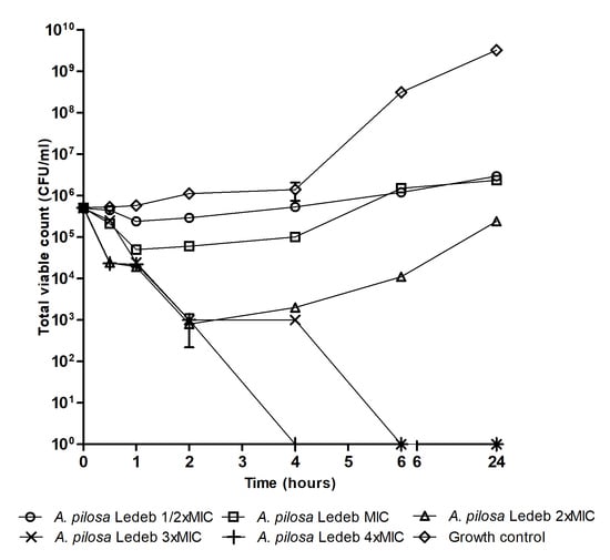
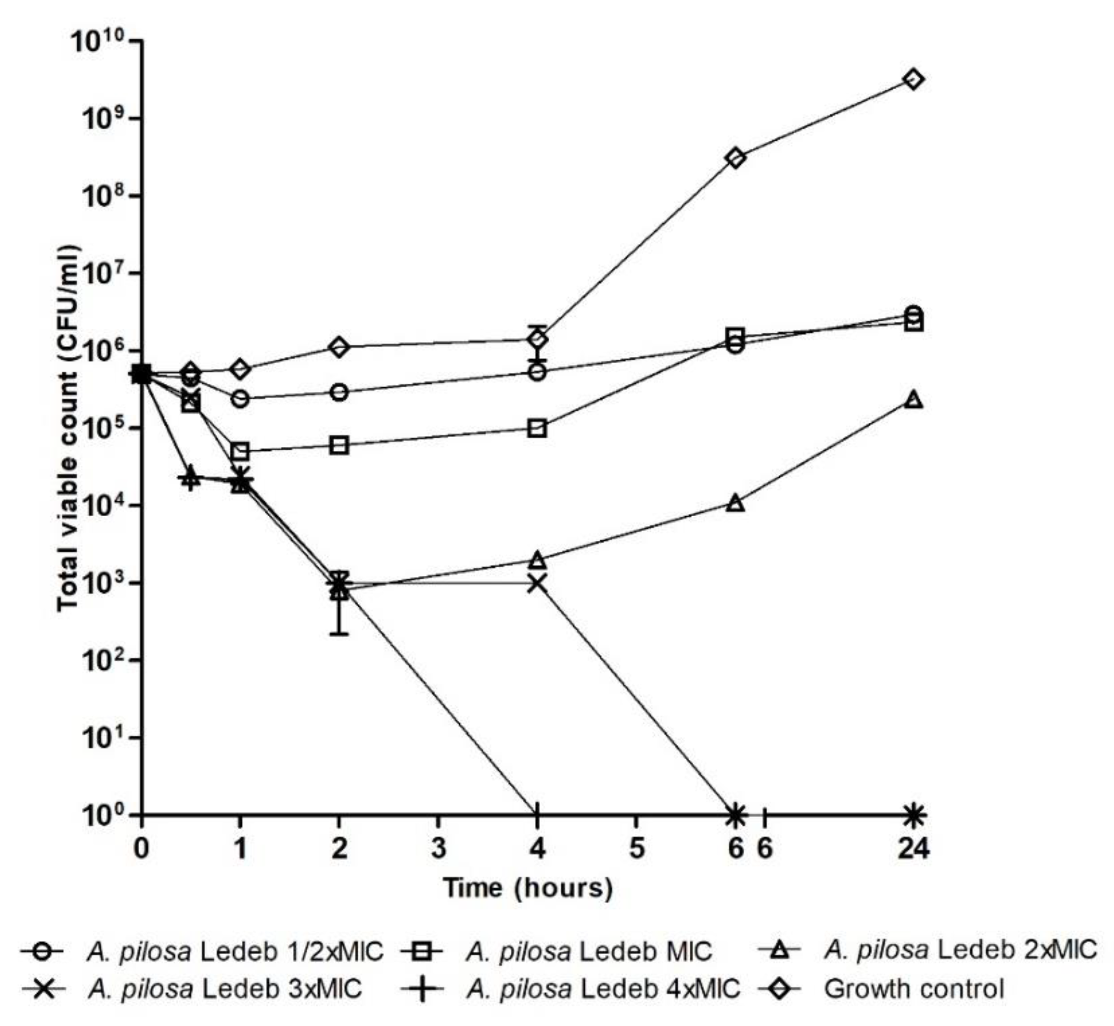
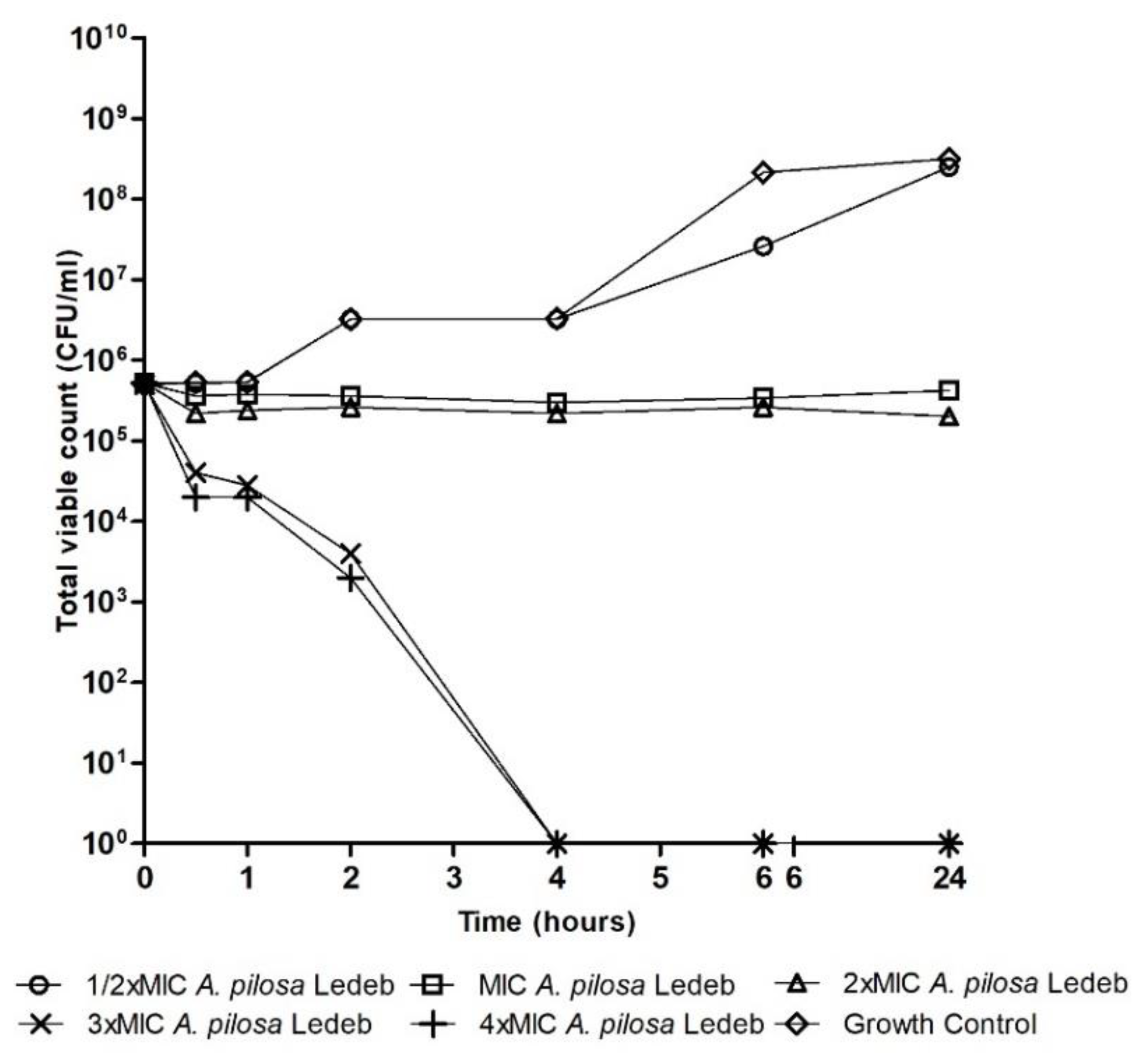
3.2. Bactericidal Activity in an In Vitro Cecum Model

Next, we have investigated the bactericidal effect using an in vitro cecum model. In Table 3, Table 4 and Table 5 we present the percentage kill of bacteria incubated in cecum content over 24 h using 4 x MIC of A. pilosa Ledeb, A. chinensis Bunge, S. glabra Roxb, I. domestica Goldblatt and Mabb, and Ampicillin. All plant extracts reduced ≥ 99.9% of viable E. coli in ≤ 6 h (Table 3). A. pilosa Ledeb and Ampicillin reduced (p < 0.001) E. coli cells in 0.5 h (Table 1). I. domestica Goldblatt and Mabb exhibited bactericidal activity at 6 h and demonstrated a significantly (p < 0.001) lower percentage kill of E. coli than the other plant extracts at two hours. All plant extracts reduced ≥99.9% of viable L. monocytogenes in 24 h (Table 4). All plant extracts reduced (p < 0.001) L. monocytogenes cells in 0.5 h (Table 4). I. domestica Goldblatt and Mabb exhibited bactericidal activity at 24 h and demonstrated a significantly (p < 0.001) lower percentage kill of L. monocytogenes than the other plant extracts from 0.5 to 6 h. A. chinensis Bunge and I. domestica Goldblatt and Mabb reduced ≥ 99.9% of viable S. enteritidis in 6 h (Table 5). All plant extracts reduced (p < 0.001) S. enteritidis cells in two hours (Table 5). I. domestica Goldblatt and Mabb and A. chinensis Bunge reduced S. enteritidis by ≥99.9%. S. glabra Roxb and A. pilosa Ledeb exhibited the lowest reduction (p < 0.001) in total viable count of S. enteritidis (99.61% and 99.52% reduction). A. pilosa Ledeb was the most effective against E. coli and L. monocytogenes, while A. chinensis Bunge was the most effective against S. enteritidis.
Figure 1, Figure 2 and Figure 3 are time–kill graphs which present the total viable count of surviving populations of bacteria in the presence of extracts of A. pilosa Ledeb and A. chinensis Bunge incubated in cecum content for 24 h. At concentrations of 4 × MIC the extract of A. pilosa Ledeb exhibited bactericidal activity by 4 h against L. monocytogenes and E. coli (Figure 1 and Figure 2). The extract of A. chinensis Bunge exhibited bactericidal activity against S. enteritidis by 6 h (Figure 3). The MIC of these plant extracts all exhibited bacteriostatic activity and inhibited the population of pathogens over 24 h (Figure 1, Figure 2 and Figure 3).

4. Discussion

The aim of this research was to determine the antibacterial properties of plant extracts and evaluate the bactericidal activity of the most effective plant extracts using an in vitro poultry digest model. The broth microdilution method [29] was used to screen for antibacterial activity of A. pilosa Ledeb, S. glabra Roxb, A. chinensis Bunge, and I. domestica (L.) Goldblatt and Mabb against L. monocytogenes, E. coli, and S. enteritidis. The selection of plants was based on longstanding traditional claims [12] and current knowledge [20,33,34,35] of their antibacterial activities. A review of the literature highlighted that there is a lack of research investigating the antibacterial properties of the aqueous extracts of A. pilosa Ledeb, S. glabra Roxb, A. chinensis Bunge, and I. domestica (L.) Goldblatt and Mabb. However, the four plants chosen for this study are used traditionally as formulations or individual extracts to treat diseases of pathogenic origin such as bacterial infections and this indicates that they may exhibit antibacterial activity [12].
Agrimonia pilosa Ledeb exhibited the most effective antibacterial activity of all four plant extracts against both E. coli and L. monocytogenes with a comparatively low and potent MIC of 7.81 mg/L and 31.25 to 250 mg/L, respectively. A. chinensis Bunge exhibited the most effective antibacterial activity of all four plant extracts against S. enteritidis, with a comparatively-low MIC of 62.5 mg/L. Furthermore, A. pilosa Ledeb, S. glabra Roxb, A. chinensis Bunge, and I. domestica (L.) Goldblatt and Mabb demonstrated considerable antibacterial activity against Gram-positive bacteria (L. monocytogenes) and Gram-negative bacteria (E. coli S. enteritidis) with MIC values ≤ 500 mg/L. In a review of the literature Ríos and Recio [36] summarize that MIC values of ≤1000 mg/L exhibited by plant extracts are considered to demonstrate significant antibacterial activity. Concentrations above this might indicate that the bioactive compounds responsible for the antibacterial activity need to be isolated further to be effective. These results demonstrate effective antibacterial potency or a high concentration of bioactive components in these plant extracts. This is the first paper to demonstrate the potent broad-spectrum antibacterial activity of the aqueous extracts of A. pilosa Ledeb, S. glabra Roxb, A. chinensis Bunge, and I. domestica (L.) Goldblatt and Mabb against L. monocytogenes, E. coli, and S. enteritidis.
The broth microdilution results support the antibacterial properties of A. pilosa Ledeb, S. glabra Roxb, A. chinensis Bunge, and I. domestica (L.) Goldblatt and Mabb and provide preliminary scientific validation for the use of these plant extracts in traditional Chinese medicine to treat diseases of pathogenic origin, such as infections. This remarkable broad-spectrum antibiotic activity of A. pilosa Ledeb, S. glabra Roxb, A. chinensis Bunge, and I. domestica (L.) Goldblatt and Mabb provides compelling scientific evidence that these extracts possess antibacterial activity which may be representative of multiple bioactive compounds that inhibit bacteria. Often when single compounds are isolated from plant extracts the same bioactivity can no longer be detected [37]. Therefore, the synergistic interactions of several bioactive compounds in these plant extracts are likely to be responsible for this antibacterial activity. For example, A. pilosa Ledeb contains catechin and phenol derivatives which exhibit antibacterial activity against Staphylococcus aureus [34,38]. Catechins and phenols have been found to exert antibacterial properties against various pathogens including E. coli and L. monocytogenes [39,40,41]. These bioactive compounds could have been responsible for the antibacterial activity exhibited by the extract of A. pilosa Ledeb against E. coli and L. monocytogenes in this study. For example, A. chinensis Bunge contains quercetin and saponins [42] which exhibit antibacterial activity against various pathogens including S. enteritidis [43,44]. These bioactive compounds could have been responsible for the antibacterial activity exhibited by the extract of A. chinensis Bunge against S. enteritidis in this study.
The most potent antibacterial activity demonstrated by the broth microdilution results was observed using aqueous extracts of A. pilosa Ledeb against E. coli and L. monocytogenes, and A. chinensis Bunge against S. enteritidis using the broth microdilution method. These were selected to investigate the killing properties over time and concentration of these plant extracts whilst incubated in poultry cecum content. Research by Johny et al. [30] used autoclaved cecum content to study the effects of natural plant extracts on Salmonella and Campylobacter as the cecum content medium resembled conditions in live broilers more closely than other synthetic laboratory mediums. For this reason, poultry cecum content was used in this study. In the research by Johny et al. [30] chicken cecum contents were autoclaved to eliminate inhibitory effects of endogenous bacteria. However, the current study used the following significant modification to the model—selective broth was used to maintain the populations of existing endogenous bacteria in the ceca of poultry belonging to the genus being studied. This is more reflective of the natural environment in the poultry gastrointestinal tract because it maintains some of the background bacteria in the poultry cecum content—proteins are not denatured because autoclaving was avoided. These bacteria and proteins would be intact in live chickens and maintaining the environment of the natural content of ceca of poultry provides a model which can be used to provide results which are predictive of what might happen in the chicken gastrointestinal tract. The results of this study can therefore be used to justify further experiments and hypothesize that these plant extracts may exert an antibacterial effect against pathogenic microorganisms in vivo.
The results from the in vitro cecum model strongly support and confirm the antibacterial activity found in the broth microdilution experiment of A. pilosa Ledeb against L. monocytogenes and E. coli, and A. chinensis Bunge against S. enteritidis. In particular, 4 × MIC (250 mg/L and 62.5 mg/L, respectively) A. pilosa Ledeb significantly reduced the total viable count of L. monocytogenes and E. coli by ≥99.99% within 4 h (p < 0.001). A. chinensis Bunge significantly reduced the total viable count of S. enteritidis by ≥99.99% within 6 h (p < 0.001). This indicated rapid bactericidal activity against endogenous and inoculated bacteria cultures. At lower concentrations A. chinensis Bunge and A. pilosa Ledeb inhibited and reduced L. monocytogenes, E. coli, and S. enteritidis. This is the first study to report the rapid bactericidal activity of A. pilosa Ledeb and A. chinensis Bunge. This significant finding demonstrates the potent bactericidal efficacy of these plant extracts in poultry cecum content. This highlights the potential for these plant extracts to be used as alternatives to antibiotics in poultry feed to maintain pathogen populations in the poultry gastrointestinal tract. The bactericidal activity exhibited by extracts of A. pilosa Ledeb and A. chinensis Bunge indicates that caution should be taken to determine the correct concentration for supplementation in poultry feed during in vivo trials. High concentrations could potentially kill E. coli, L. monocytogenes, and S. enteritidis in poultry cecum content. Low population counts of E. coli, L. monocytogenes, and S. enteritidis have been found to be natural inhabitants of poultry cecum content without causing clinical manifestations in poultry [8,9,10]. Killing the entire E. coli, L. monocytogenes, and S. enteritidis population could lead to disruption of the poultry gastrointestinal tract microbiota. Modulation of the poultry gastrointestinal microbiota can therefore lead to improved health.

5. Conclusions

This study demonstrates broad-spectrum antibacterial activity of four plant extracts, namely, A. pilosa Ledeb, S. glabra Roxb, A. chinensis Bunge, and I. domestica (L.) Goldblatt and Mabb against S. enteritidis, L. monocytogenes, and E. coli. This study also provided evidence that A. pilosa Ledeb and A. chinensis Bunge reduced cecum colonization of E. coli, L. monocytogenes, and S. enteritidis in vitro. These initial results justify further in vivo trials of these plant extracts to determine their efficacy against S. enteritidis, L. monocytogenes, and E. coli in broiler chickens. This would also provide an opportunity to investigate the effects of A. pilosa Ledeb, S. glabra Roxb, A. chinensis Bunge, and I. domestica (L.) Goldblatt and Mabb on poultry health, performance, and microbiota. The results of this study contribute to research into the use of plant extracts in poultry feed and provide new information on the antibacterial activity of A. chinensis Bunge and A. pilosa Ledeb. They also highlight the potential of plant extracts used in traditional Chinese medicine as possible alternatives to antibiotics for use as poultry feed additives.

Author Contributions

Conceptualization, R.L.M. and C.S.; methodology, R.L.M.; validation, R.L.M.; formal analysis, R.L.M., and M.E.E.B.; investigation, R.L.M.; data curation, R.L.M.; visualization, R.L.M.; project administration, R.L.M. and C.S.; funding acquisition, R.L.M., and C.S.; writing—original draft, R.L.M.; writing—editing, R.L.M.; writing—review, M.E.E.B., N.C., C.S., and M.M.T.; supervision, M.E.E.B., C.S., and M.M.T.; resources, M.E.E.B., M.M.T., N.C., and C.S. All authors have read and agreed to the published version of the manuscript.

Funding

This work was supported by the Department of Agriculture, Environment and Rural Affairs.

Conflicts of Interest

The authors declare no conflict of interest. The funders had no role in the design of the study; in the collection, analyses, or interpretation of data; in the writing of the manuscript; or in the decision to publish the results.

References

  1. World Health Organisation. Antimicrobial Resistance Global Report on Surveillance 2014; World Health Organisation: Geneva, Switzerland, 2014. [Google Scholar]
  2. Choski, A.; Sifri, Z.; Cennimo, D.; Horng, H. Global Contributors to Antibiotic Resistance. J. Glob. Infect. Dis. 2019, 11, 36–42. [Google Scholar]
  3. Muaz, K.; Riaz, M.; Akhtar, S.; Park, S.; Ismail, A. Antibiotic Residues in Chicken Meat: Global Prevalence, Threats, and Decontamination Strategies: A Review. J. Food Prot. 2018, 81, 619–627. [Google Scholar] [CrossRef] [PubMed]
  4. Dibner, J.J.; Richards, J.D. Antibiotic Growth Promoters in Agriculture: History and Mode of Action. Poult. Sci. 2005, 84, 634–643. [Google Scholar] [CrossRef]
  5. Mehdi, Y.; Létourneau-Montminy, M.P.; Gaucher, M.L.; Chorfi, Y.; Suresh, G.; Rouissi, T.; Brar, S.K.; Côté, C.; Ramirez, A.A.; Godbout, S. Use of Antibiotics in Broiler Production: Global Impacts and Alternatives. Anim. Nutr. 2018, 4, 170–178. [Google Scholar] [CrossRef]
  6. Chopra, I.; Marilyn, R. Tetracycline Antibiotics: Mode of Action, Applications, Molecular Biology, and Epidemiology of Bacterial Resistance. Microbiol. Mol. Biol. Rev. 2001, 65, 232–260. [Google Scholar] [CrossRef] [Green Version]
  7. Silver, L.L.; Bostian, K.A. Minireview Discovery and Development of New Antibiotics: The Problem of Antibiotic Resistance. Antimicrob. Agents Chemother. 1993, 37, 377–383. [Google Scholar] [CrossRef] [Green Version]
  8. Antunes, P.; Mourão, J.; Campos, J.; Peixe, L. Salmonellosis: The Role of Poultry Meat. Clin. Microbiol. Infect. 2016, 22, 110–121. [Google Scholar] [CrossRef] [Green Version]
  9. Umaraw, P.; Prajapati, A.; Verma, A.K.; Pathak, V.; Singh, V.P. Control of Campylobacter in Poultry Industry from Farm to Poultry Processing Unit: A Review. Crit. Rev. Food Sci. Nutr. 2017, 57, 659–665. [Google Scholar] [CrossRef]
  10. Davis, G.S.; Waits, K.; Nordstrom, L.; Grande, H.; Weaver, B.; Papp, K.; Horwinski, J.; Koch, B.; Hungate, B.A.; Liu, C.M.; et al. Antibiotic-Resistant Escherichia Coli from Retail Poultry Meat with Different Antibiotic Use Claims. BMC Microbiol. 2018, 18, 1–7. [Google Scholar] [CrossRef]
  11. Van Galen, E. Traditional Herbal Medicines Worldwide, from Reappraisal to Assessment in Europe. J. Ethnopharmacol. 2014, 158, 498–502. [Google Scholar] [CrossRef] [PubMed]
  12. Bensky, D.; Clavey, S.; Stoger, E.; Gamble, A.; Bensky, L.L. Chinese Herbal Medicine Materia Medica, 3rd ed.; Eastland Press: Seattle, WA, USA, 2004. [Google Scholar] [CrossRef]
  13. Lillehoj, H.; Liu, Y.; Calsamiglia, S.; Fernandez-Miyakawa, M.E.; Chi, F.; Cravens, R.L.; Oh, S.; Gay, C.G. Phytochemicals as Antibiotic Alternatives to Promote Growth and Enhance Host Health. Vet. Res. 2018, 49, 1–18. [Google Scholar] [CrossRef] [PubMed] [Green Version]
  14. Chitemerere, T.A.; Mukanganyama, S. Evaluation of Cell Membrane Integrity as a Potential Antimicrobial Target for Plant Products. BMC Complement. Altern. Med. 2014, 14, 278. [Google Scholar] [CrossRef] [PubMed] [Green Version]
  15. Mohammadi Gheisar, M.; Kim, I.H. Phytobiotics in Poultry and Swine Nutrition—A Review. Ital. J. Anim. Sci. 2018, 17, 92–99. [Google Scholar] [CrossRef]
  16. Dhami, D.S.; Shah, G.C.; Kumar, V.; Joshi, Y.; Tripathi, M.; Bisht, M. Essential Oil Composition and Antibacterial Activity of Agrimonia Pilosa Ledeb (Rosaceae). Chem. Sci. Trans. 2018, 7, 499–505. [Google Scholar] [CrossRef] [Green Version]
  17. Joung, D.K.; Mun, S.H.; Lee, K.S.; Kang, O.H.; Choi, J.G.; Kim, S.B.; Gong, R.; Chong, M.S.; Kim, Y.C.; Lee, D.S.; et al. The Antibacterial Assay of Tectorigenin with Detergents or ATPase Inhibitors Against Methicillin-Resistant Staphylococcus Aureus. Evidence-Based Complement. Altern. Med. 2014, 2014, 1–7. [Google Scholar] [CrossRef] [Green Version]
  18. Fang, X.; Liu, J.; Yang, L.; Li, X. Study on the Optimization of Extraction Technology of Anemonin from Pulsatilla Chinensis and Its Inhibitory Effect on Alternaria Panax. J. Dis. Med. Plants 2019, 5, 94–102. [Google Scholar] [CrossRef]
  19. Hua, S.; Zhang, Y.; Liu, J.; Dong, L.; Huang, J.; Lin, D.; Fu, X. Ethnomedicine, Phytochemistry and Pharmacology of Smilax Glabra: An Important Traditional Chinese Medicine. Am. J. Chin. Med. 2018, 46, 261–297. [Google Scholar] [CrossRef]
  20. Fong, S.C.; Mulyana, Y.; Girawan, D. Antibacterial Effect of Pulsatilla Chinensis towards Staphylococcus Aureus, Shigella Dysenteriae, and Salmonella Typhi. Althea Med. J. 2016, 3, 292–295. [Google Scholar] [CrossRef] [Green Version]
  21. Chinese Pharmacopoeia Commission. Pharmacopoeia of the People’s Republic of China, 1st ed.; China Medical Science Press: Beijing, China, 2015. [Google Scholar]
  22. Plants for a Future. Plants for a Future: Belamcanda Chinensis - (L.) DC. Available online: https://pfaf.org/user/Plant.aspx?LatinName=Belamcanda+chinensis (accessed on 3 May 2020).
  23. Plants for a Future. Plants for a Future: Agrimonia Pilosa – Ledeb. Available online: https://pfaf.org/user/Plant.aspx?LatinName=Agrimonia+pilosa (accessed on 3 May 2020).
  24. Plants for a Future. Plants for a Future: Pulsatilla Chinensis - (Bunge.)Regel. Available online: https://pfaf.org/user/Plant.aspx?LatinName=Pulsatilla+chinensis (accessed on 3 May 2020).
  25. Plants for a Future. Plants for a Future: Smilax China C- L. Available online: https://pfaf.org/user/plant.aspx?latinname=Smilax+china (accessed on 3 May 2020).
  26. Bioline. Available online: https://www.bioline.com/uk/downloads/dl/file/id/2697/mytaq_red_mix_product_manual.pdf (accessed on 16 April 2020).
  27. Principi, N.; Esposito, S. Antibiotic Administration and the Development of Obesity in Children. Int. J. Antimicrob. Agents 2016, 47, 171–177. [Google Scholar] [CrossRef]
  28. The Plant List. Available online: http://www.theplantlist.org/ (accessed on 24 January 2020).
  29. Clinical and Laboratory Standards Institute. Methods for Dilution: Antimicrobial Susceptibility Tests for Bacteria That Grow Aerobically; Approved Standard, 8th ed.; CLSI: Wayne, PA, USA, 2009; Volume 29. [Google Scholar]
  30. Johny, A.K.; Darre, M.J.; Donoghue, A.M.; Donoghue, D.J.; Venkitanarayanan, K. Antibacterial Effect of Trans-Cinnamaldehyde, Eugenol, Carvacrol, and Thymol on Salmonella Enteritidis and Campylobacter Jejuni in Chicken Cecal Contents in Vitro. J. Appl. Poult. Res. 2010, 19, 237–244. [Google Scholar] [CrossRef]
  31. Clinical and Laboratory Standards Institute. Methods for Determining Bactericidal Activity of Antimicrobial Agents; Approved Guideline; CLSI: Wayne, PA, USA, 2009; Volume 19. [Google Scholar]
  32. European Committee on Antimicrobial Susceptibility Testing. Breakpoint Tables for Interpretation of MICs and Zone Diameters. Version 8.1. 2018. Available online: http://www.eucast.org/fileadmin/src/media/PDFs/EUCAST_files/Breakpoint_tables/v_8.1_Breakpoint_Tables.pdf (accessed on 3 May 2020).
  33. Franzblau, S.G.; Cross, C. Comparative in Vitro Antimicrobial Activity of Chinese Medicinal Herbs. J. Ethnopharmacol. 1986, 15, 279–288. [Google Scholar] [CrossRef]
  34. Kasai, S.; Watanabe, S.; Kawabata, J.; Tahara, S.; Mizuntani, J. Antimicrobial Catechin Derivatives of Agrimonia Pilosa. Phytochemistry 1992, 31, 787–789. [Google Scholar] [CrossRef]
  35. Xu, S.; Shang, M.; Liu, G.; Xu, F.; Wang, X.; Shou, C.; Cai, S. Chemical Constituents from the Rhizomes of Smilax Glabra and Their Antimicrobial Activity. Molecules 2013, 18, 5265–5287. [Google Scholar] [CrossRef] [PubMed] [Green Version]
  36. Ríos, J.L.; Recio, M.C. Medicinal Plants and Antimicrobial Activity. J. Ethnopharmacol. 2005, 100, 80–84. [Google Scholar] [CrossRef] [PubMed]
  37. Wink, M. Modes of Action of Herbal Medicines and Plant Secondary Metabolites. Medicines 2015, 2, 251–286. [Google Scholar] [CrossRef] [PubMed]
  38. Yamaki, M.; Kashihara, M.; Ishiguro, K.; Takagi, S. Antimicrobial Principles of Xian He Cao (Agrimonia Pilosa). Planta Med. 1989, 55, 169–170. [Google Scholar] [CrossRef] [PubMed]
  39. Skroza, D.; Šimat, V.; Smole Možina, S.; Katalinić, V.; Boban, N.; Generalić Mekinić, I. Interactions of Resveratrol with Other Phenolics and Activity against Food-Borne Pathogens. Food Sci. Nutr. 2019, 7, 2312–2318. [Google Scholar] [CrossRef] [Green Version]
  40. Nakayama, M.; Shimatani, K.; Ozawa, T.; Shigemune, N.; Tsugukuni, T.; Tomiyama, D.; Kurahachi, M.; Nonaka, A.; Miyamoto, T. A Study of the Antibacterial Mechanism of Catechins: Isolation and Identification of Escherichia Coli Cell Surface Proteins That Interact with Epigallocatechin Gallate. Food Control. 2013, 33, 433–439. [Google Scholar] [CrossRef]
  41. Diarra, M.S.; Hassan, Y.I.; Block, G.S.; Drover, J.C.G.; Delaquis, P.; Oomah, B.D. Antibacterial Activities of a Polyphenolic-Rich Extract Prepared from American Cranberry (Vaccinium Macrocarpon) Fruit Pomace Against Listeria Spp. LWT - Food Sci. Technol. 2020, 123. [Google Scholar] [CrossRef]
  42. Kumar, S.; Madaan, R.; Farooq, A.; Sharma, A. Plant Review The Genus Pulsatilla: A Review. Pharmacogn. Rev. 2008, 2, 116–124. [Google Scholar]
  43. Khan, M.I.; Ahhmed, A.; Shin, J.H.; Baek, J.S.; Kim, M.Y.; Kim, J.D. Green Tea Seed Isolated Saponins Exerts Antibacterial Effects against Various Strains of Gram Positive and Gram Negative Bacteria, a Comprehensive Study in Vitro and in Vivo. Evid. Based Complement. Alternat. Med. 2018, 2018, 3486106. [Google Scholar] [CrossRef] [PubMed] [Green Version]
  44. Lee, N.K.; Jung, B.S.; Na, D.S.; Yu, H.H.; Kim, J.S.; Paik, H.D. The Impact of Antimicrobial Effect of Chestnut Inner Shell Extracts against Campylobacter Jejuni in Chicken Meat. LWT - Food Sci. Technol. 2016, 65, 746–750. [Google Scholar] [CrossRef]
Figure 1. Total viable count of endogenous and inoculated Listeria monocytogenes QA1018, LS12519, and CP102 mixture in the presence of Agrimonia pilosa Ledeb.
Microorganisms 08 00962 g001
Figure 2. Total viable count of endogenous and inoculated E. coli UM004, UM011, and UM012 mixture in the presence of Agrimonia pilosa Ledeb.
Microorganisms 08 00962 g002
Figure 3. Total viable count of endogenous and inoculated Salmonella enteritidis QA0419, LE103, and 1F6144 mixture in the presence of Anemone chinensis Bunge.
Microorganisms 08 00962 g003
Table 1. Reference strains, clinical isolates, and their sources.
Species Isolate Source
Listeria monocytogenesNCTC 11994 Reference Queen’s University Belfast (QUB)
LS12519 Retail ready to eat sliced meat Agri-Food and Biosciences Institute (AFBI)
OT11230 Retail chopping board (AFBI)
CP102 Retail ham and cheese filling (AFBI)
CP1132 Retail cooked chicken breast (AFBI)
QA1018 Quality assurance sample (AFBI)
Salmonella enteritidisNCTC 0074 Reference (QUB)
1F6144 Quality assurance sample (AFBI)
LE103 Egg filter (AFBI)
QA04/19 Quality assurance sample (AFBI)
Escherichia coliATCC 25922 Reference (QUB)
UM004 Urinary tract infection (QUB)
UM011 Urinary tract infection (QUB)
UM012 Urinary tract infection (QUB)
Table 2. Minimum inhibitory concentration (MIC, mg/L) of four plant extracts relative to ampicillin against Listeria monocytogenes, Salmonella enteritidis, and E. coli.
PathogenPlant Extract
Agrimonia pilosa LedebSmilax glabra RoxbAnemone chinensis BungeIris domestica (L.) Goldblatt and MabbAmpicillin
L. monocytogenesNCTC 1199431.2562.51251250.25
LS1251931.2531.251251250.5
OT1123062.51251251250.5
CP10231.2531.251251250.25
CP113212531.251251250.25
QA101831.2531.251251251
S. enteritidisNCTC 007450025062.52504
IF614412512562.51252
LE10312512562.51254
QA041912512562.51258
E. coliATCC 259227.8112512562.58
UM0047.8112512562.54
UM0117.8112512562.54
UM0127.8112512562.54
Table 3. Average percentage kill of E. coli ATCC 25922, UM004, UM011, and UM012 over 24 h in the presence of 4 × minimum inhibitory concentration (MIC) of plant extracts and ampicillin over 24 h.
Treatment
Time (Hours) A. pilosa LedebA. chinensis BungeS. glabra Roxb I. domestica (L.) Goldblatt and MabbAmpicillin SEM p
0000000.000NS
0.596.24 b0.76 a0.25 a1.92 a95.10 b0.940< 0.001
196.16 b0.76 a1.49 a2.31 a95.92 b0.890< 0.001
299.60 b99.24 b99.27 b31.01 a99.66 b0.278< 0.001
499.99 c99.62 b99.99 c56.59 a99.99 c0.480< 0.001
699.9999.9999.9999.9999.990.000NS
2499.9999.9999.9999.9999.990.000NS
a,b,c superscripts indicate significance. Means with differing superscripts are significantly different (p < 0.001).
Table 4. Average percentage kill of Listeria monocytogenes NCTC 11994, QA1018, LS12519, and CP102 in the presence of 4 × minimum inhibitory concentration (MIC) of plant extracts and ampicillin over 24 h.
Treatment
Time (Hours) A. pilosa LedebA. chinensis Bunge S. glabra Roxb I. domestica (L.) Goldblatt and MabbAmpicillin SEM p
0000000.000NS
0.595.47 c96.27 c93.64 b47.81 a95.97 c1.019< 0.001
195.67 c99.63 d95.65 c43.83 a96.47 b0.358< 0.001
299.80 b99.63 b99.61 b44.23 a99.62 b0.483< 0.001
499.99 b99.99 b99.80 b42.63 a99.62 b0.410< 0.001
699.99 b99.99 b99.99 b99.18 a99.99 b0.020< 0.001
2499.9999.9999.9999.9999.990.000NS
a,b,c superscripts indicate significance. Means with differing superscripts are significantly different (p < 0.001).
Table 5. Average percentage kill of Salmonrlla enteritidis NCTC 0074, QA0419, LE103, and 1F6144 in the presence of 4 × minimum inhibitory concentration (MIC) of plant extracts and ampicillin over 24 h.
Treatment
Time (Hours) A. pilosa LedebA. chinensis BungeS. glabra Roxb I. domestica (L.) Goldblatt and MabbAmpicillin SEM p
0000000.000NS
0.51.18 a2.26 a1.15 a0.77 a95.75 b2.559< 0.001
11.58 a3.02 a2.32 a0.77 a96.03 b2.174< 0.001
299.13 b99.25 b99.22 b33.59 a99.60 b0.761< 0.001
499.29 b99.62 b99.22 b61.25 a99.80 b2.692< 0.001
699.60 b99.99 c99.22 a99.99 c99.99 c0.132< 0.001
2499.52 b99.99c99.61a99.99 c99.99 c0.206< 0.001
a,b,c superscripts indicate significance. Means with differing superscripts are significantly different (p < 0.001).

