Article

Genomic Sequence Analysis of the Multidrug-Resistance Region of Avian Salmonella enterica serovar Indiana Strain MHYL

1
National Demonstration Center for Animal Experimental Education, Beijing University of Agriculture, Beijing 102206, China
2
Beijing Key Laboratory of Traditional Chinese Veterinary Medicine, Beijing University of Agriculture, Beijing 102206, China
3
United States Department of Agriculture, Agricultural Research Service, Southern Plains Agricultural Research Center, Food and Feed Safety Research Unit, College Station, TX 77845–4988, USA
*
Author to whom correspondence should be addressed.
They contributed equally to this work.
Microorganisms 2019, 7(8), 248; https://doi.org/10.3390/microorganisms7080248
Submission received: 30 June 2019 / Revised: 6 August 2019 / Accepted: 7 August 2019 / Published: 9 August 2019

Abstract

:
A series of human and animal diseases that are caused by Salmonella infections pose a serious threat to human health and huge economic losses to the livestock industry. We found antibiotic resistance (AR) genes in the genome of 133 strains of S. Indiana from a poultry production site in Shandong Province, China. Salmonella enterica subsp. enterica serovar Indiana strain MHYL had multidrug-resistance (MDR) genes on its genome. Southern blot analysis was used to locate genes on the genomic DNA. High-throughput sequencing technology was used to determine the gene sequence of the MHYL genome. Areas containing MDR genes were mapped based on the results of gene annotation. The AR genes blaTEM, strA, tetA, and aac(6′)-Ib-cr were found on the MHYL genome. The resistance genes were located in two separate MDR regions, RR1 and RR2, containing type I integrons, and Tn7 transposons and multiple IS26 complex transposons with transposable functions. Portions of the MDR regions were determined to be highly homologous to the structure of plasmid pAKU_1 in S. enterica serovar Paratyphi A (accession number: AM412236), SGI11 in S. enterica serovar Typhimurium (accession number: KM023773), and plasmid pS414 in S. Indiana (accession No.: KC237285).

1. Introduction

Salmonella species are a major worldwide bacterial cause of acute gastroenteritis [1,2]. Salmonellosis is commonly caused by non-typhoidal Salmonella enterica serotypes [2]. The Centers for Disease Control and Prevention (CDC) has estimated that in the United States, foodborne Salmonella causes about 1 million illnesses, 19,000 hospitalizations, and 380 deaths each year [3]. Children under the age of 5, the elderly, and people with weakened immune systems are the most likely to have severe infections [4]. Recently, there has been a shift in the Salmonella serotypes found in poultry production, especially in diverse geographical regions of the world [2]. Salmonella enterica serovar Indiana was first reported in China during 1984 and is now commonly found in animals, animal processing facilities, food, and people [5]. Salmonella Indiana has become an important human foodborne pathogen found in poultry [2]. In recent years, there have been reports of human infection with diarrhea caused by multidrug-resistant S. Indiana in many provinces in China [6]. Antibiotics are commonly used for the treatment of bacteremia caused by Salmonella [3]. However, with the overuse of antibacterial drugs, drug-resistant strains with multidrug-resistance (MDR) phenotypes have appeared in large numbers, making the treatment of salmonellosis progressively more difficult [7]. Studies have shown that acquired resistance genes may be carried on the genomic DNA of Salmonella [8]. Through a study of multiple-chromosomal-type resistant Salmonella, it was determined that most of these resistance genes were concentrated in a small genomic island called SGI1 (Salmonella genomic island 1), and the area on the genome where these resistance genes are located is called the MDR region [9]. Due to the mobility of the resistance genes in SGI1, it is of great significance when studying the acquisition and transmission of bacterial MDR. The horizontal gene transfer of antimicrobial resistance (AMR) determinants located on plasmids was thought to be the main cause of the rapid proliferation and spread of drug resistance [10]. However, strains harboring SGI1 tend to rapidly disseminate and be more virulent [11]. It has been shown that SGI1 has the same general chromosomal location in different Salmonella serotypes, indicating that its insertion occurs through site-specific recombination [12]. Gene replacement in the integron structure is another way to contribute to the variability of the SGI1 antibiotic resistance (AR) gene cluster, which may result in stronger virulence and the further exchange of AR genes between other multidrug-resistant bacteria [12].
In the early stages of this study, we initially did drug testing and found MDR phenotypes in Salmonella and wanted to diagnose the role of drug-resistance genes in these bacteria. PCR was thought to be the best method to use but we were unable to determine whether the resistance genes were located on one or two fragments of the genome. Ultimately, the result led to sequencing the whole genome. This study carried out a comparative analysis of the gene sequences in the MDR regions of the genome of a S. enterica subsp. enterica serovar Indiana strain MHYL isolate and explored possible sources and routes for obtaining AR genes. The work here may help understand the emergence and spread of bacterial resistance and may be useful for guiding the prevention and management of clinically utilized drugs for the control of S. Indiana with the MDR phenotype.

2. Materials and Methods

2.1. Bacterial Strains and Drugs Tested

In this study, 133 strains of S. Indiana were isolated from a poultry food production site in Shandong Province, China. For each of the isolates, the broth microdilution method was used to determine the minimum inhibitory concentration (MIC) of 19 commonly used clinical antibacterial drugs: amikacin, amoxicillin, ampicillin, cefazolin, ceftiofur, chloramphenicol, danofloxacin, doxycycline, enrofloxacin, florfenicol, gentamicin, kanamycin, nalidixic acid, norfloxacin, polymyxin E, sulfamethoxazole, sulfisoxazole, tetracycline, and trimethoprim. The MIC values were determined according to methods of the American Clinical Laboratory Standards Committee (CLSI) [13]. The strain MHYL isolate that we selected had resistance to 17 drugs, and was only susceptible to amikacin and polymyxin E, and can be used as a representative strain for later studies. Escherichia coli ATCC 25922 was used as a quality control for antimicrobial susceptibility testing [14].

2.2. Kits and Reagents

The bacterial genomic DNA extraction kit, DNA MarkerII, was purchased from Tiangen Biotechnology Co., Ltd., Beijing, China; the 2×Trans Taq-T PCR Super Mix was purchased from the Beijing Quanjin Biotechnology Co., Ltd., Beijing, China; the lambda DNA Marker was purchased from the Beijing Dingguo Changsheng Biotech Co., Ltd., Beijing, China; the DIG High Prime DNA Labeling Detection Starter Kit was purchased from Sigma-Aldrich Chemie Gmbh, Munich, Germany; and the HindIII endonuclease, and the 10× NEB Buffer was purchased from New England Biolabs, Ipswich, MA, USA.

2.3. Concentration and Purification

The bacterial genomic DNA was extracted according to the DNA Marker II bacterial genomic DNA extraction kit instructions. The genomic DNA was then digested with the HindIII endonuclease.
To produce the sample size needed for electrophoresis and Southern blot hybridization, the enzyme digestion DNA gel electrophoresis loading conditions and the optimization of the Southern blot hybridization method were carried out. The digested genomic DNA was reduced in volume using phenol-chloroform-isoamyl alcohol and ethanol precipitation.

2.4. Amplification of Drug-Resistance Genes and Southern Blot Restriction Mapping

Amplification of the β-lactam resistance gene blaTEM, aminoglycoside resistance gene strA, tetracycline drug-resistance gene tetA, amido alcohol drug-resistance gene floR, and the quinolone-resistance gene aac(6′)-Ib-cr were carried out. The primer sequences are shown in Table 1, and the PCR reaction parameters are shown in Table 2 [15].
Specific probes for each drug-resistance gene were prepared, and the primers were synthesized by the Beijing Dingguo Changsheng Biotechnology Co., Ltd., Beijing, China. Southern blot experiments were performed according to the manufacturer’s directions included with the DIG High Prime DNA Labeling Detection Starter Kit.

2.5. Sequence Analysis of the MDR Region of the Strain MHYL Genome

The avian S. Indiana strain MHYL was sent to the Beijing Baimaike Biotechnology Co., Ltd., Beijing, China, for genome-wide sequencing, assembly, and gene function annotation. Based on the results of gene function annotation, the structure map showing the drug-resistance gene regions on the strain MHYL genome was mapped using Vector NTI software (version 11.5.1; ThermoFisher Scientific, Waltham, MA, USA), which is a suite of sequence analysis and design tools.

2.6. Prediction of Strain MHYL Genome-Encoding Genes and Non-Coding Genes

The Glimmer software (version 3.02; Institute for Genomic Research, Rockville, MD, USA) was used to identify the coding sequences of the genome; the rRNA, tRNA, and miRNA of the sequenced strain; and the sequenced genes were compared with Clusters of Orthologous Groups (COGs), Kyoto Encyclopedia of Genes and Genomes (KEGG), Gene Ontology (GO), Swiss-Prot, and NR Protein databases. The data were subjected to BLAST alignment, and the gene function annotation was performed.

3. Results

3.1. Southern Blot Hybridization of Drug-Resistance Genes

Southern blot methodology was used to hybridize the resistance genes blaTEM, strA, tetA, floR, and aac(6′)-Ib-cr corresponding to the observed drug-resistance phenotypes before and after digestion of the strain MHYL genome. The extracted genomic DNA of strain MHYL remained intact as was shown using electrophoresis of the hybridization results before and after digestion of the MHYL genome, which had no plasmid DNA interference and was successfully treated with the HindIII endonuclease.
Specific probes for each drug-resistance gene were hybridized with the strain MHYL genome before and after digestion. The results showed that each drug-resistance gene was successfully hybridized and mapped to the strain MHYL genomic DNA both before and after digestion. The electrophoresis map of the strain MHYL genomic DNA and the hybridization results for each drug-resistance gene were compared, and the band sizes were consistent. The presence of each drug-resistance gene on the MHYL genomic DNA was determined. Subsequently, the positional relationship of the restriction fragments for each drug-resistance gene was compared and analyzed. The resistance genes from the strain MHYL genome were concentrated in three regions of different fragment sizes, 20 kb (tetA and aac(6′)-Ib-cr), 10 kb (blaTEM, floR, aac(6′)-Ib-cr), and 9.4 kb (strA).

3.2. Statistics of the Strain MHYL Genome Sequencing Data

Based on the data obtained using sequencing, a MHYL genome library was constructed. The genomic size of the sequenced strain was estimated to be 5.3 Mb, as determined using sequencing data and sequencing depth.

3.3. Screening and Clustering Statistics of Genes Related to Drug Resistance

The annotation information of the genome-encoding genes of the sequenced strains was screened, the genes related to drug resistance were sorted, and the drug-resistance genes were classified and counted as shown in Table 3. There are a variety of genes on the genome related to the observed drug-resistance phenotypes of the multidrug-resistant S. Indiana strain MHYL obtained from poultry. These genes encode for proteins that result in resistance to multiple drugs.

3.4. Assembly and Sequencing of the Strain MHYL Genome Gene Sequence

Since the genome-wide gene sequence of S. Indiana was not found in NCBI, the genomic gene sequence of S. Typhimurium was selected as a reference [16]. In the S. Indiana strain MHYL genome, MDR genes and mobile elements were mainly located in two non-adjacent regions of the genome, resistance region 1 (RR1) and resistance region 2 (RR2). The GC content of the whole MHYL genome was 51.62%, while the GC content of RR1 and RR2 was higher than that of the whole MHYL genome at 69.85% and 63.41%, respectively. The strain MHYL whole genome sequence data was uploaded to NCBI (SUBID: SUB805704; BioSample: SAMN03287656; BioProject: PRJNA273784).

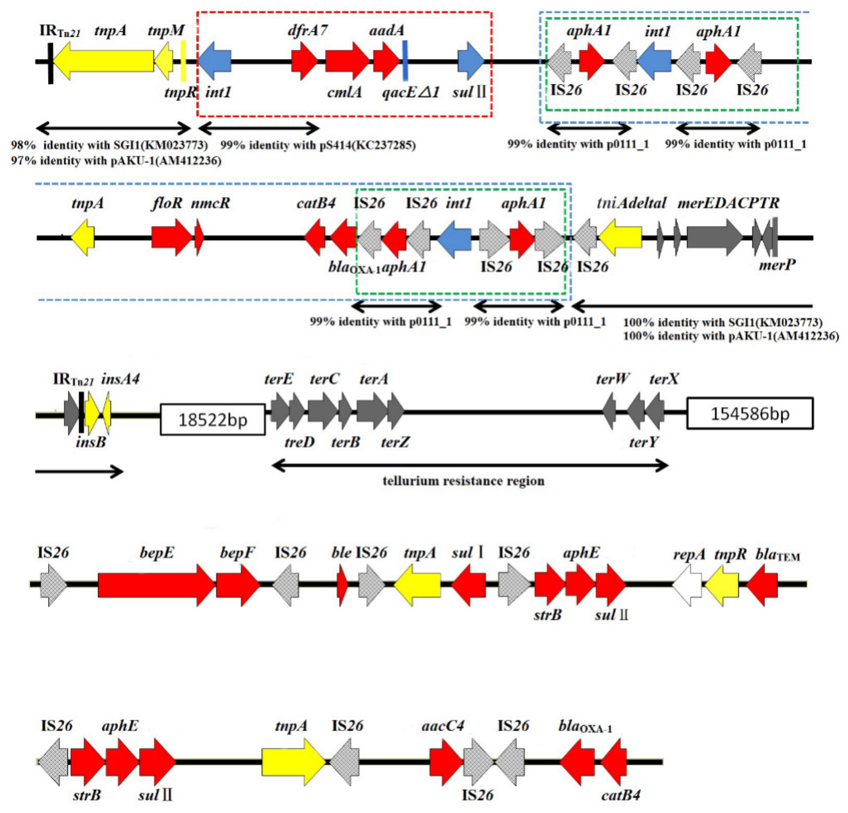
3.5. Mapping of the MDR Region of the Strain MHYL Genome

The gene structure of the RR1 and RR2 sequences on the MHYL genome were mapped and are shown in Figure 1 and Figure 2. By comparing and analyzing the genetic structure of each drug-resistance area, the possible sources and modes of transmission of drug-resistance genes may be inferred.
As shown in Figure 1, the RR1 sequence contains genes conferring resistance to β-lactams, amide alcohols, aminoglycosides, sulfonamides, and heavy metals such as mercury and other non-metallic substances. According to the gene structure map, RR1 can be roughly divided into two parts: an upstream region and a downstream region.
In the upstream region of RR1, the Tn21 skeleton structure was observed, and on the end was an incomplete IS1 (insAB) fragment. The inverted repeat sequence IRTn21 with a size of 9 bp was detected at both ends of the upstream region, and the integrase gene int1 and trimethoprim resistance gene dfrA7 were detected therein. The chloramphenicol resistance genes cmlA and floR, aminoglycoside nontransferable encoding gene aadA, quaternary ammonium compound resistance gene qacE△1, sulfonamide resistance gene sulII, aminoglycoside phosphotransferase encoding gene aphA1, carbapenemase encoding gene nmcR, chloramphenicol acetyltransferase encoding gene catB4, extended spectrum β-lactamases (ESBLs) encoding gene blaOXA-1, truncated tniA transposase, heavy metal mercury resistance gene merP and the mercury resistance operon merEDACPTR, and the IS26 element was inserted therein to form a complete Tn21 transposon structure. Among them, the int1 gene and the IS26 element constitute a complete integron structure, and the front-end of the int1-dfrA7 gene had a 99% homology with the integron sequence of the S. Indiana carrying plasmid pS414 in GenBank (accession number: KC237285) [17]. A BLAST comparison of the upstream gene sequence of RR1 revealed that it had a structure similar to SGI11 (accession number: KM023773) in GenBank [18]. Both had incomplete IS1 fragments, a Tn21 skeleton structure, type I integrons, and resistance genes related to the heavy metal mercury. This structure is also like that found in the MDR plasmid IncHI1 of S. Paratyphi A [19]. The aminoglycoside phosphotransferase encoding gene aphA1 was flanked by two isotropic or inverted IS26 sequences, forming the IS26-aphA1-IS26 structure. This structure was first discovered on an Escherichia coli plasmid p0111_1 reported in 2001 (GenBank registration, No.: AP010961). It was shown that the aphA1 gene is inserted between two transposable IS26 sequences to form part of the transposon Tn5715 [20], which mediates the insertion and transposition of MDR genes in a variety of Gram-negative bacteria. The presence of this structure was detected in the plasmid and MDR region of the Corynebacterium urealyticum genome [21]. Compared with SGI11, there are four IS26-aphA1-IS26 structures upstream of RR1, and the integrase int1 gene is inserted between two IS26-aphA-IS26 structures in forward and reverse repeats to form two IS26-aphA-IS26-int1-IS26-aphA-IS26 structures. The resistance-related genes floR, nmcR, catB4, and blaOXA-1 were inserted within the two four-IS26 repeat structures, and all the IS26s were preceded by a 14 bp inverted repeat (GGCACTGTTGCAAA), forming a set of transposons. The complex transposon structure, which integrates functions, may be conducive to capturing exogenous resistance genes.
There are many IS26 mobile elements with reverse repeats at both ends in the downstream region of RR1. The bleomycin resistance gene ble, sulfonamide resistance genes sulI and sulII, aminoglycoside resistance gene strB, ESBL encoding gene blaTEM, and the aminoglycoside acetyltransferase encoding gene aacC4 were inserted therein, and these mobile elements were part of a variety of composite transposons. It was suggested that the composite transposon structure consisting of two IS mobile elements with inverted repeats at both ends can mediate the movement of genes inserted between the IS elements [21]. Also, some IS mobile elements may cause the homologous recombination of the bacterial genome to occur, resulting in variability in the genetic structure of the MDR region [22].
As can be seen from Figure 2, RR2 contains the chloramphenicol acetyltransferase-encoding gene cat, IS1 (insAB), IS21 (istAB), the transposon Tn7 encoding for at least three transposition genes (tnsABC), the gene cluster pcoABCDR providing tolerance to the metal element copper, and an incomplete Tn1721-like transposon encoding tetracycline resistance. The IS1 and IS21 gene sequences were compared and found to lack direct repeats at both ends. It has been shown that IS elements lacking direct repeats at both ends are more likely to be derived from unconventional recombination rather than through a transposition mechanism [23]. The copper metal tolerance efflux system encoding genes pcoA, pcoB, pcoR, and pcoC may be activated when Cu2+ is in excess, and the protein pcoC can bind to bacterial intracellular Cu2+ together with the proteins pcoA, pcoB, and pcoD is an inner membrane-spanning protein that is required for complete Cu2+ removal from the bacteria, thereby, exerting resistance to metallic copper [24,25]. In a study of the genomic gene sequence of Acinetobacter baumannii, a truncated MDR Tn1721 transposon was found comprised of the tetracycline drug-resistance genes tetA and tetR, together with the perM gene and transposase gene tnpA [26]. The incomplete Tn1721-like transposon (accession number: CT025832) had 99% homology with the incomplete Tn1721 transposon gene sequence obtained in this study with S. Indiana strain MHYL. By comparison analysis, multiple strains of Salmonella were found to have consistent genomes with strain MHYL.

4. Discussion

By comparing and analyzing the gene sequences of the two MDR regions on the MHYL genome, it was found that: (i) part of the MHYL genome sequence and the S. Paratyphi A strain carrying the plasmid pAKU_1 (accession number: AM412236) had the same skeleton structure, and with high homology; (ii) the type I integron skeletal structure of the MHYL genome had high homology with the plasmid pS414 (accession number: KC237285) carried by S. Indiana; (iii) the multiple repeating sequences with the IS26 mobile element at both ends could mediate the integration of drug-resistance genes into the genome of a new strain through the transposition mechanism, and may also cause homologous recombination of the strain MHYL genome; (iv) two IS26-aphA-IS26-int1-IS26-aphA-IS26 structures may be a novel complex integron structure, which is conducive to capturing foreign drug-resistance genes; (v) the IS1 and IS21 lacking direct repeat sequences found at both ends of RR2 may be derived from unconventional recombination; and (vi) tetA and tetR genes may be derived from the Tn1721-mediated transposition mechanism and be transmitted horizontally between strains. The results of the comprehensive comparative analysis indicated that multiple acquired resistance genes on the genome of the multidrug-resistant S. Indiana strain MHYL may be derived from homologous or non-homologous strain genomes or their resistance plasmids. Mediated by IS26 elements and a variety of transposons, resistance genes may be integrated into the genome via transposition or homologous recombination by a stable propagation mechanism independent of drug selective pressure. At the same time, however, the exogenous genes on the genome of some strains may remain in the multiple transposon structures that mediate their movement. The MDR region on the MHYL genome is composed of IS26 elements and is speculated to be highly transferable. With the help of transposons, there may be movement of resistance genes within the genome of the same strain and the horizontal transmission of resistance genes between genomes of different strains and plasmids, but the possibility of this mechanism requires further study.
On the other hand, it was found using BLAST analysis that the upstream gene sequence of RR1 and the SGI11 (accession number: KM023773) sequence carried on the genome of S. Typhi had the same skeleton structure with high homology, and the yidC gene was present downstream of RR1. It has been shown that SGI1 and its variants are usually located in the upstream region of the yidC gene [9]. Therefore, it is speculated that the upstream gene structure of RR1 is an amplified SGI11-like structure, and multiple integration and transposition elements exist on it. This structure may be more conducive to capturing exogenous resistance genes, thereby enhancing the tolerance of strains to various antimicrobial agents, effectively demonstrating MDR. Whether this structure is a new type of SGI1 subtype remains to be seen following further study.

5. Conclusions

In this study, a whole genomic gene sequence of avian MDR Salmonella serrata was obtained, which confirmed that the S. Indiana strain MHYL genome carries the exogenous AMR genes blaTEM, strA, tetA, floR, and aac(6′)-Ib-cr related to the drug-resistance phenotype. Also, the MHYL genome carried the heavy metal mercury resistance gene merP and the mer operon merEDACPTR. The merR gene is the universal regulator for the mer operon [27]. Either merT or merP is sufficient to specify a mercury transport system [28]. The merA gene encodes for a reductase that reduces Hg2+ to Hg0 [27]. The tniA transposase gene is usually present in integrons and composite transposons conferring antibiotic resistance [29]. The S. enterica Indiana strain MHYL genome carries genes conferring resistance to β-lactams, amide alcohols, tetracyclines, aminoglycosides, and sulfonamides. The drug-resistance genes are concentrated in two MDR regions, and there is an MDR region gene structure composed of type I integrons and with multiple complex transposons.

Author Contributions

Conceptualization, data curation, formal analysis, methodology, and validation: Y.L. (Yan Lu) and Y.W. Project administration, supervision, and resources: G.H. and X.H Writing—original draft: Y.L. (Yan Lu) and R.C.B. Writing—review and editing: Y.L. (Yan Lu), Y.W., G.H., Y.L. (Yuqi Liu), R.C.B., and X.H.

Funding

This research was funded by the National Key Research and Development Program of China (2016YFD0501307), and by the Da Bei Nong young teachers’ scientific research program (14ZK004) and the 2017 Research and Development Fund for experimental technology systems of the Beijing University of Agriculture.

Acknowledgments

This study was also supported by the 2019 discipline construction and graduate quality improvement program of the Beijing University of Agriculture.

Conflicts of Interest

The authors declare no conflict of interest.

References

  1. Majowicz, S.E.; Musto, J.; Scallan, E.; Angulo, F.J.; Kirk, M.; O’Brien, S.J.; Jones, T.F.; Fazil, A.; Hoekstra, R.M. The global burden of nontyphoidal Salmonella gastroenteritis. Clin. Infect. Dis. 2010, 50, 882–889. [Google Scholar] [CrossRef] [PubMed]
  2. Antunes, P.; Mourão, J.; Campos, J.; Peixe, L. Salmonellosis: The role of poultry meat. Clin. Microbial. Infect. 2016, 22, 110–121. [Google Scholar] [CrossRef]
  3. Scallan, E.; Hoekstra, R.M.; Angulo, F.J.; Tauxe, R.V.; Widdowson, M.-A.; Roy, S.L.; Jones, J.L.; Griffin, P.M. Foodborne illness acquired in the United States—Major pathogens. Emerg. Infect. Dis. 2011, 17, 7–15. [Google Scholar] [CrossRef]
  4. Centers for Disease Control and Prevention (CDC). Foodborne Diseases Active Surveillance Network (FoodNet): FoodNet Surveillance Report for 2012 (Final Report); Department of Health and Human Services: Atlanta, GA, USA, 2014; p. 9. Available online: https://www.cdc.gov/foodnet/PDFs/2012_annual_report_508c.pdf (accessed on 11 October 2018).
  5. Gong, J.; Kelly, P.; Wang, C. Prevalence and antimicrobial resistance of Salmonella enterica serovar Indiana in China (1984–2016). Zoonoses Public Health 2017, 64, 239–251. [Google Scholar] [CrossRef] [PubMed]
  6. Li, G.-S.; Wang, Y.-Y.; Zhu, Z.-Y.; Wang, H.-X.; Liu, D.-H.; Qiu, W.; Zheng, Y.; Zhang, F.-Q.; Fan, Q.-S. Identification and drug resistance of the first Salmonella Indiana in Yunnan military garrison. Chin. Trop. Med. 2013, 13, 1051–1053. [Google Scholar]
  7. Mead, P.S.; Slutsker, L.; Dietz, V.; McCaig, L.F.; Bresee, J.S.; Shapiro, C.; Griffin, P.M.; Tauxe, R.V. Food-related illness and death in the United States. Emerg. Infect. Dis. 1999, 5, 607–625. [Google Scholar] [CrossRef]
  8. Shahada, F.; Sekizuka, T.; Kuroda, M.; Kusumoto, M.; Ohishi, D.; Matsumoto, A.; Okazaki, H.; Tanaka, K.; Uchida, I.; Izumiya, H.; et al. Characterization of Salmonella enterica serovar Typhimurium isolates harboring a chromosomally encoded CMY-2 β-lactamase gene located on a multidrug resistance genomic island. Antimicrob. Agents Chemother. 2011, 55, 4114–4121. [Google Scholar] [CrossRef] [PubMed]
  9. Boyd, D.A.; Peters, G.A.; Ng, L.-K.; Mulvey, M.R. Partial characterization of a genomic island associated with the multidrug resistance region of Salmonella enterica Typhymurium DT104. FEMS Microbiol. Lett. 2000, 189, 285–291. [Google Scholar] [CrossRef] [PubMed]
  10. Pornsukarom, S.; Thakur, S. Horizontal dissemination of antimicrobial resistance determinants in multiple Salmonella serotypes following isolation from the commercial swine operation environment after manure application. Appl. Environ. Microbiol. 2017, 83, e01503-17. [Google Scholar] [CrossRef]
  11. Mulvey, M.R.; Boyd, D.A.; Olson, A.B.; Doublet, B.; Cloeckaert, A. The genetics of Salmonella genomic island 1. Microb. Infect. 2006, 8, 1915–1922. [Google Scholar] [CrossRef]
  12. Doublet, B.; Lailler, R.; Meunier, D.; Brisabois, A.; Boyd, D.; Mulvey, M.R.; Chasius-Dancia, E.; Cloeckaert, A. Variant Salmonella genomic island 1 antibiotic resistance gene cluster in Salmonella enterica serovar Albany. Emerg. Infect. Dis. 2003, 9, 585–591. [Google Scholar] [CrossRef] [PubMed]
  13. Clinical and Laboratory Standards Institute (CLSI). Performance Standards for Antimicrobial Disk and Dilution Susceptibility Tests for Bacteria Isolated from Animals; Approved Standard Third Edition. Informational Supplement; CLSI document M31-A3; Clinical and Laboratory Standards Institute: Wayne, PA, USA, 2012. [Google Scholar]
  14. Lu, Y.; Zhao, H.; Sun, J.; Liu, Y.; Zhou, X.; Beier, R.C.; Wu, G.; Hou, X. Characterization of multidrug-resistant Salmonella enterica serovars Indiana and Enteritidis from chickens in Eastern China. PLoS ONE 2014, 9, e96050. [Google Scholar] [CrossRef]
  15. Lu, Y.; Wu, C.-M.; Wu, G.-J.; Zhao, H.-Y.; He, T.; Cao, X.-Y.; Dai, L.; Xia, L.-N.; Qin, S.-S.; Shen, J.-Z. Prevalence of antimicrobial resistance among Salmonella isolates from chicken in China. Foodborne Pathog. Dis. 2011, 8, 45–53. [Google Scholar] [CrossRef] [PubMed]
  16. Zhao, E.-Y.; Bao, H.-X.; Tang, L.; Zou, Q.-H.; Liu, W.-Q.; Zhu, D.-L.; Chin, J.; Dong, Y.-Y.; Li, Y.-G.; Cao, F.-L.; et al. Genomic comparison of Salmonella typhimurium DT104 with non-DT104 strains. Mol. Genet. Genom. 2013, 288, 549–557. [Google Scholar] [CrossRef]
  17. Lai, J.; Wang, Y.; Shen, J.; Li, R.; Han, J.; Foley, S.L.; Wu, C. Unique class 1 integron and multiple resistance genes co-located on IncHI2 plasmid is associated with the emerging multidrug resistance of Salmonella Indiana isolated from chicken in China. Foodborne Pathog. Dis. 2013, 10, 581–588. [Google Scholar] [CrossRef] [PubMed]
  18. Chiou, C.-S.; Alam, M.; Kuo, J.-C.; Liu, Y.-Y.; Wang, P.-J. Chromosome-mediated multidrug resistance in Salmonella enterica serovar Typhi. Antimicrob. Agents Chemother. 2015, 59, 721–723. [Google Scholar] [CrossRef] [PubMed]
  19. Holt, K.E.; Thomson, N.R.; Wain, J.; Phan, M.D.; Nair, S.; Hasan, R.; Bhutta, Z.A.; Quail, M.A.; Norbeertczak, H.; Walker, D.; et al. Multidrug-resistant Salmonella enterica serovar Paratyphi A harbors IncHI1 plasmids similar to those found in serovar Typhi. J. Bacteriol. 2007, 189, 4257–4264. [Google Scholar] [CrossRef] [PubMed]
  20. Tauch, A.; Krieft, S.; Kalinowski, J.; Pühler, A. The 51,409-bp R-plasmid pTP10 from the multiresistant clinical isolate Corynebacterium striatum M82B is composed of DNA segments initially identified in soil bacteria and in plant, animal, and human pathogens. Mol. Gen. Genet. 2000, 263, 1–11. [Google Scholar] [CrossRef] [PubMed]
  21. Tauch, A.; Trost, E.; Tilker, A.; Ludewig, U.; Schneiker, S.; Goesmann, A.; Arnold, W.; Bekel, T.; Brinkrolf, K.; Brune, I.; et al. The lifestyle of Corynebacterium urealyticum derived from its complete genome sequence established by pyrosequencing. J. Biotechnol. 2008, 136, 11–21. [Google Scholar] [CrossRef] [PubMed]
  22. Berg, D.E.; Howe, M.M. Mobile DNA; ASM Press: Washington, DC, USA, 1989. [Google Scholar]
  23. Doublet, B.; Praud, K.; Bertrand, S.; Collard, J.-M.; Weill, F.-X.; Cloeckaert, A. Novel insertion sequence- and transposon-mediated genetic rearrangements in genomic island SGI1 of Salmonella enterica serovar Kentucky. Antimicrob. Agents Chemother. 2008, 52, 3745–3754. [Google Scholar] [CrossRef] [PubMed]
  24. Argüello, J.M.; Raimunda, D.; Padilla-Benavides, T. Mechanisms of copper homeostasis in bacteria. Front. Cell. Infect. Microbiol. 2013, 3, 73. [Google Scholar] [CrossRef] [Green Version]
  25. Hobman, J.L.; Crossman, L.C. Bacterial antimicrobial metal ion resistance. J. Med. Microbiol. 2014, 64, 471–497. [Google Scholar] [CrossRef] [PubMed]
  26. Fournier, P.-E.; Vallenet, D.; Barbe, V.; Audic, S.; Ogata, H.; Poirel, L.; Richet, H.; Robert, C.; Mangenot, S.; Abergel, C.; et al. Comparative genomics of multidrug resistance in Acinetobacter baumannii. PloS Genet. 2006, 2, 62–72. [Google Scholar] [CrossRef]
  27. Barkay, T.; Miller, S.M.; Summers, A.O. Bacterial mercury resistance from atoms to ecosystems. FEMS Microbiol. Rev. 2003, 27, 355–384. [Google Scholar] [CrossRef] [PubMed] [Green Version]
  28. Hamlett, N.V.; Landale, E.C.; Davis, B.H.; Summers, A.O. Roles of the Tn21 merT, merP, and merC gene products in mercury resistance and mercury binding. J. Bacteriol. 1992, 174, 6377–6385. [Google Scholar] [CrossRef] [PubMed]
  29. Balabanov, V.P.; Kotova, V.Y.; Kholodii, G.Y.; Mindlin, S.Z.; Zavilgelsky, G.B. A novel gene, ardD, determines antirestriction activity of the non-conjugative transposon Tn5053 and is located antisense within the tniA gene. FEMS Microbiol. Lett. 2012, 337, 55–60. [Google Scholar] [CrossRef]
Figure 1. Schematic diagram of RR1. The red arrows in the figure represent drug-resistance gene cassettes, yellow arrows represent genes related to a transposition function, blue arrows represent the type I integrase genes and the integron-gene cassette 3′ conserved region gene structures, light gray arrows represent movable IS26 elements, dark gray arrows represent heavy-metal- and non-metal-resistance genes, the red dashed box is a type I integron structure, the green dashed boxes are assumed to be a movable element structure, and the blue dashed boxes are assumed to be compound recombination. The structure of the arrow: the direction of the arrow shows the direction of gene coding, and the size of the arrow is drawn according to the size of the coding gene and the length of the gene sequence of the RR1 region.
Microorganisms 07 00248 g001
Figure 2. Schematic diagram of RR2. The red arrows in the figure represent the drug-resistance gene cassettes, the yellow arrows represent the genes associated with the transposition function, and the dark gray arrows represent the heavy metal resistance genes. The size of the arrow is in proportion to the size of the coding gene and the length of the RR2 gene sequence.
Microorganisms 07 00248 g002
Table 1. Primer sequences of resistance genes.
Antibacterial Drug TypeGeneUpstream primer (5′–3′)
Downstream primer (5′–3′)
Registration No.Fragment Size (bp)
β-lactamblaTEMCAGCGGTAAGATCCTTGAGA
ACTCCCCGTCGTGTAGATAA
AY463797643
AminoglycosidesstrACGACTTCTTACCGGACGAGGAC
ACAGGTTGCGAAACGTGCCAAT
NC_009981422
TetracyclinetetAGCTACATCCTGCTTGCCTTC
CATAGATCGCCGTGAAGAGG
X75761210
Amide alcoholfloRTCCTGAACACGACGCCCGCTAT
TCACCGCCAATGTCCCGACGAT
AJ251806962
Quinolonesaac(6’)-Ib-crTTGCGATGCTCTATGAGTGGCTA
CTCGAATGCCTGGCGTGTTT
EU543272482
Table 2. PCR reaction parameters.
GeneInitial DenaturationDenaturationAnnealingExtensionCyclesFinal Extension
blaTEM95 °C, 4 min94 °C, 30 s48 °C, 30 s72 °C, 1 min3072 °C, 10 min
strA95 °C, 4 min94 °C, 30 s57 °C, 30 s72 °C, 1 min3072 °C, 10 min
tetA95 °C, 4 min94 °C, 30 s57 °C, 30 s72 °C, 1 min3072 °C, 10 min
floR95 °C, 4 min94 °C, 1 min63 °C, 1.5 min72 °C, 1.5 min3072 °C, 10 min
aac(6’)-Ib-cr95 °C, 4 min94 °C, 45 s55 °C, 45 s72 °C, 45 s3072 °C, 10 min
Table 3. Classification of genes detected by PCR and BLAST on the strain MHYL genome.
CategoriesResistance GenesResistance Phenotypes
Beta lactamsblaTEM, blaOXA, blaCTX-M-65, AmpC, nmcRAmpicillin, amoxicillin-clavulanic acid, cefazolin, ceftiofur
AminoglycosidesstrA, aphA, aphE, strB, aacC4, aadA, aadA1Trimethoprim, gentamicin, kanamycin, amikacin
TetracyclinestetA, tetRTetracycline, doxycycline
Amide alcoholscmlA, floR, catB4Chloramphenicol, florfenicol
Quinolonesaac(6’)-Ib-crNalidixic acid, enrofloxacin, norfloxacin, dafloxacin
TetracyclinesulI, sulIISulfisoxazole, sulfamethoxazole
Efflux pump membrane transport coding genesbepD, bepE, bepF
MDR protein-encoding genesmdtA, mdtB, mdtC, mdtE, mdtG, mdtH, mdtK, mdtL, marA, marB, marC, marR, emr
Fosfomycin resistance Protein-encoding genefsrFosfomycin
Acridine yellow resistance protein-encoding geneacrA, acrB, acre, acrFAcridine yellow
Macrolide efflux pump protein-encoding genesmacA, macBMacrolide
Metal ion resistance protein encoding genesmerA, merB, merC, merE, merP,merT, merR, TerA, TerB, TerC, TerD, pcoA, pcoB, pcoC, pcoD, TerE, TerW, TerX, TerY, TerZ, zraPMercury, antimony, copper, zinc
Bleomycin resistance protein-encoding genesBle, bcrBleomycin
Quaternary ammonium salt-resistance protein-encoding genesqacF, sugEQuaternary ammonium compound

