Article

Analysis of Microbial Diversity in South Shetland Islands and Antarctic Peninsula Soils Based on Illumina High-Throughput Sequencing and Cultivation-Dependent Techniques

1
Key Laboratory of Molecular Medicine and Biotherapy, School of Life Science, Beijing Institute of Technology, Beijing 100081, China
2
China General Microbiological Culture Collection Center, Institute of Microbiology, Chinese Academy of Sciences, Beijing 100101, China
*
Author to whom correspondence should be addressed.
Microorganisms 2023, 11(10), 2517; https://doi.org/10.3390/microorganisms11102517
Submission received: 12 September 2023 / Revised: 4 October 2023 / Accepted: 6 October 2023 / Published: 9 October 2023
(This article belongs to the Special Issue New Insights into the Diversity and Characterization of Extremophiles)

Abstract

:
To assess the diversity of bacterial taxa in Antarctic soils and obtain novel microbial resources, 15 samples from 3 sampling sites (DIS5, GWS7, FPS10) of South Shetland Islands and 2 sampling sites (APS18, CIS17) of Antarctic Peninsula were collected. High-throughput sequencing (HTS) of 16S rRNA genes within these samples was conducted on an Illumina Miseq platform. A total of 140,303 16S rRNA gene reads comprising 802 operational taxonomic units (OTUs) were obtained. After taxonomic classification, 25 phyla, 196 genera, and a high proportion of unidentified taxa were detected, among which seven phyla and 99 genera were firstly detected in Antarctica. The bacterial communities were dominated by Actinomycetota (40.40%), Pseudomonadota (17.14%), Bacteroidota (10.55%) and Chloroflexota (10.26%). Based on the HTS analyses, cultivation-dependent techniques were optimized to identify the cultivable members. A total of 30 different genera including 91 strains were obtained, the majority of which has previously been reported from Antarctica. However, for the genera Microterricola, Dyadobacter, Filibacter, Duganella, Ensifer, Antarcticirhabdus and Microvirga, this is the first report in Antarctica. In addition, seven strains represented novel taxa, two of which were psychropoilic and could be valuable resources for further research of cold-adaptability and their ecological significance in Antarctica.

Graphical Abstract

1. Introduction

Most of Antarctica is covered by snow and ice; only about 0.4% of land area is permanently ice-free [1]. As a result of unique climatic and geographical characteristics like severe cold, strong wind and high radiation, microorganisms have an absolute advantage in the Antarctic soil ecosystem compared with plants and animals, playing a major role in the physical and chemical cycle. The traditional view is that extreme environments generally support biological communities with low biomass and low diversity [2]. However, after years of exploration and scientific research, it has been discovered that Antarctica is a huge treasure house of microbial resources, and the microbial diversity in its soil is much higher than previously thought [2,3,4,5]. Furthermore, Antarctic microorganisms have formed unique biomolecular structures and special physiological and biochemical properties in the long-term natural selection evolution. Many strains can produce low-temperature (cold-resistant) enzymes, as well as anti-radiation, anti-bacterial, anti-cancer active substances, which are very important in many fields including environmental engineering, agriculture, food industry, pharmaceutical industry, enzyme industry, biofuel, etc.
The South Shetland Islands are composed of a series of islands, the largest of which is King George Island, followed by Livingston and Deception Island. More than 80% of the archipelago was covered by sea ice from early April to early December. As glaciers are retreating in recent years, the regions closer to the sea are free of snow and ice, submitted to rapid cycles of freeze/thaw, and may receive significant quantities of organic material from marine animals [6,7]. There are many scientific research stations on the Fildes Peninsula of King George Island. A large number of nitrogen-fixing bacteria, nitrifying bacteria, and denitrifying bacteria have been isolated via traditional culture methods [8]. Then, more taxa in the soil near the Great Wall Station including Pseudomonadota, Actinomycetota, Bacteroidota, Acidobacteriota, Cyanobacteriota, and Chloroflexota were found via molecular biology methods. The Deception Island is formed by the crater collapse after the Antarctic submarine volcano erupted during the ancient glacial period [9]. Most ice-free areas on the island are covered by volcanic rocks and volcanic ash. The island also experienced volcanic eruption in the 1960s, leading to a stagnation of research on its microbial community in the next 30 years. The Antarctic Peninsula harbored a location further south (63° S) and a correspondingly colder climate. Affected by climate characteristics, geographical factors, and ecological environment, these five sampling sites may have unique microbial diversity and resources.
Up to now, only a small fraction of all Antarctic microbes has been isolated and characterized, which was limited by available technology and difficulty in sampling. 16S rRNA gene cloning and denaturing gradient gel electrophoresis (DGGE) have been traditionally used to identify uncultured microbial community structures in the past [10]. With the rapid technological innovation in molecular biology, High-throughput sequencing precludes the need to build clone libraries, and the efficiency associated with HTS sequencing is increasing and the widespread adoption of the Illumina Miseq sequencing platform has accelerated analytical capabilities [11,12].
Conducting microbial diversity analysis on soil in the Antarctic region not only reveals the structure of microbial communities in the region and the impact of environmental factors on microbial communities, but also facilitates the discovery of new species and enriches the gene pool of species. Moreover, digging and developing some strains with special active functions has important theoretical and practical significance, which has broad application prospects in the development of new bioactive substances and bioremediation due to the abundant microbial resources in Antarctica. In this context, the study aimed to (1) obtain a systematic understanding of the community structure and diversity of microorganisms inhabiting the soils from South Shetland Islands and Antarctic Peninsula; (2) optimize the isolation and cultivation strategies to explore the bacterial strains and to assess their bio-characteristics and potential functions in depth; and (3) explore novel taxa and enrich the gene pool to lay the foundation for the protection and utilization of Antarctic environmental microbial resources.

2. Materials and Methods

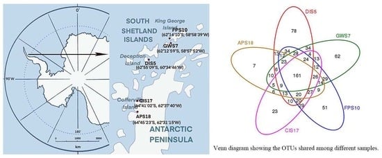
2.1. Sample Collection

Soil samples were collected from South Shetland Islands (Deception Island and King George Island) and Antarctic Peninsula in February, during the summer in Antarctica. The soil samples were collected at different latitudes and longitudes (Figure 1). According to the five-point sampling method, five sampling points were set up at different sites and three parallel samples were taken at each sampling point. Each sampling point was separated by 2 m. The surface debris was removed with a sterile spatula, and the samples were collected in sterile plastic tubes and shipped at −20 °C to laboratory. Once samples arrived at the laboratory, they were maintained at −80 °C until processing. Samples were selected from 5 sampling sites for this investigation. Samples from the same sampling site were mixed together for the experiment. The detailed information of the sampling sites is shown in Table 1 and the chemical and physical characteristics of the soil samples is shown in Table 2.

2.2. DNA Extraction, PCR Amplification and Sequencing

The soil DNA was extracted using the PowerSoil® DNA Isolation Kit (MoBio, Solana Beach, CA, USA). The extracted genomic DNA was used as the template to amplify the V3–V4 region of 16S rRNA genes with primers 338 forward (5′-ACTCCTACGGGAGGCAGCA-3′) and 806 reverse (5′-GGACTACHVGGGTWTCTAAT-3′) [13]. The reaction components were 12.5 µL of 2×Taq PCR Mastermix (Promega; Madison, WI, USA), 1 µL of dNTP (10 mM), 3 µL of BSA (2 ng/μL), 1 µL of Forward Primer (5 μM), 1 µL of Reverse Primer (5 μM), X µL of DNA (30 ng), and 6.5-X µL of ddH2O. The PCR was carried out under the following thermocyling conditions: 95 °C for 5 min, followed by 25 cycles of 95 °C for 45 s, 50 °C for 50 s, 72 °C for 45 s, with a final extension at 72 °C for 5 min. This was repeated 3 times for each sample. The PCR products of the same sample were mixed at the same concentration and detected by 1% agarose gel electrophoresis. The AxyPrepDNA gel recovery kit (AXYGEN) was used to recover bands of 400–500 bp. The PCR products were detected and quantified through QuantiFluor™ -ST (Promega). The double-end sequencing analysis was carried out on the Illumina MiSeq PE300.

2.3. Sequence Processing and Analyses

Overlapping reads were merged using the program FLASH with default parameters [14]. QIIME [15] processing and the UCHIME arithmetic [16] were used and then effective tags were obtained. Operational taxonomic units (OTUs) were clustered by UPARSE [17] according to an open-reference OTU picking protocol based on 97% nucleotide similarity. The Venn diagram [18] made by the R platform was used to count the number of common and unique OTUs in multiple samples. Taxonomic relative abundance profiles (such as at the phylum, family and genus levels) were generated based on OTU annotation. Sequences obtained from this research were deposited in the NCBI SRA database (http://www.ncbi.nlm.nih.gov/traces/sra/, accessed on 25 July 2023): Bioprojects PRJNA681991.

2.4. Statistical Analysis

Statistical analysis was implemented using the R platform. Dilution curves and Shannon–Wiener curves were obtained through MOTHUR [19] and the R platform. Shannon index, phylogenetic diversity, Chao1 index, and the observed number of species were used to evaluate alpha diversity [20], and the weighted and unweighted UniFrac distances were used to evaluate beta diversity with QIIME (Version 1.7.0) [21,22,23]. Chao1 index and Shannon index were calculated at the lowest sequencing depth by random sampling using QIIME [24]. The relationships between bacterial community structures were evaluated by principal coordinate analysis (PCoA) based on the UniFrac distances between samples.

2.5. Isolation, Purification and Preservation of Strains

Bacterial strains were isolated by serial dilution and plating techniques to calculate the number of colonies in the sample and facilitate the cultivation and growth of bacterial colonies to obtain a single colony. Nine modified media (beef extract peptone, Gauze’s synthetic medium No.1, LB, R2A, TSBA, 1/10 TSBA, improved glycerol asparagin medium, Modified Gauze’s No.2, and MA) were adopted to optimize the cultivation of a broader range of soil microorganisms [25,26,27]. One gram of each sample was suspended in 9 mL of sterile water, homogenized in an incubator with shaking at 16 °C and 150 rpm for 1 h, and serially diluted from 10−1 to 10−6. Next, 0.1 mL of each dilution was spread onto nine media, respectively, and incubated at 4 °C and 28 °C for 15–20 days and 7–10 days, three plates at each temperature. After incubation, bacterial colonies were obtained from suitably diluted plates and transferred onto freshly prepared plates until pure cultures were obtained. The pure strains were maintained at 4 °C as slant and glycerol stock (20%) at −80 °C for further use.

2.6. Psychrophilic Bacteria Screening

The isolates were screened for temperature tolerance by incubating the culture spot inoculated plates at different temperatures [28]. The cultures obtained at 4 °C were screened further by inoculating at −4 °C, 0 °C, 4 °C, 10 °C, 15 °C, 20 °C, 25 °C and 28 °C for 3–14 days. All the tested strains were screened in triplicate and depending on the temperature range for optimal growth.

2.7. 16S rRNA Gene Sequencing and Data Analysis of Isolated Strains

DNA extraction was performed following the protocol described by Kim et al. [27] and Rainey et al. [29] with some modifications. The PCR reaction components were 25 µL of 2×PCR Mastermix (Promega; Madison, USA), 1 µL of dNTP (10 mM), 1 µL of Forward Primer (27F), 1 µL of Reverse Primer (1525R), 2 µL of DNA, and 20 µL of ddH2O. The PCR program consisted of an initial denaturation of 95 °C for 5 min, 34 cycles of 95 °C for 35 s, 54 °C for 60 s and 72 °C for 90 s, and a final extension step of 72 °C for 7 min. The PCR products were purified via the Wizard PCR Purification System (Promega), and the operation method was performed according to the procedure recommended in the instructions. Finally, sequencing was performed at Nuosai Genome Research Center (Beijing, China). The 16S rRNA gene sequences were compared with the EzBioCloud database [30] and GenBank databases using BLAST. The phylogenetic tree of 16S rRNA gene sequences of target strains and similar strains was constructed using the software of MEGA 6.0 [31].

3. Results

3.1. OTU Clustering and Annotation

A total of 140,303 sequences were obtained from the Illumina Miseq sequencing platform and 118,261 clean tags were determined to be of high quality, resulting in 802 OTUs (stringency at 97%). The number of high-quality reads per sample ranged from 16,539 to 27,577 (400–440 bp) (Table 3). The FPS10 sample harbored the highest number of OTUs, followed by DIS5, GWS7, CIS17 and APS18 (Table 3). Rarefaction curves (Figure 2), combined with the estimated coverage values (Table 3), suggested that the libraries were sufficiently large to capture a large majority of the bacterial diversity in the samples used in this study. A Venn diagram of the between-group OTUs was generated using the ggplot2 (Figure 3). Of 802 OTUs, 161 were common to all samples. The numbers of OTUs exclusive to the DIS5, GWS7, FPS10, CIS17 and APS18 samples were 78, 62, 51, 23, and 7, respectively.
The Chao1 index was used to estimate bacterial community richness and the Shannon–Wiener index was used to estimate bacterial community diversity in the five different samples (Table 3). The results indicated that both richness and diversity of the bacterial communities followed the same trend. The highest was for the King George Island, followed by Antarctic Peninsula and Deception Island. Samples were scattered among the three quadrants in the Principal Component Analysis (PCA) analysis plot. As shown in Figure 4, three samples in South Shetland Islands clustered together on the right of the coordinate axis, and two samples in the Antarctic Peninsula gathered together on the left of the coordinate axis. Communities in DIS5 were obviously deviated from those in the other four samples.

3.2. Composition and Relative Abundance of Microbiota

A total of 25 identified phyla and 196 identified genera were detected in samples. The phyla Actinomycetota, Pseudomonadota, Chloroflexota, Bacteroidota, Bacillota, Nitrospirota, Cyanobacteriota, Saccharibacteria, Armatimonadota, Verrucomicrobiota, Gemmatimonadota and Acidobacteriota were detected in all samples. However, their relative abundances varied across different samples. In the DIS5 sample, Actinomycetota (60.71%) and Pseudomonadota (13.37%) accounted for 74.08% of all bacteria. In the GWS7 sample, Actinomycetota (52.66%), Pseudomonadota (17.80%) and Chloroflexota (14.95%) represented 85.41% of all bacterial species. In the FPS10 sample, Chloroflexota (23.14%), Gemmatimonadota (21.93%) and Actinomycetota (20.92%) comprised 65.99% of the total microbiota. In the CIS17 sample, Actinomycetota (25.11%), Pseudomonadota (22.61%) and Bacteroidota (22.09%) accounted for 69.81% of all bacteria. In the APS18 sample, Actinomycetota (40.76%), Pseudomonadota (27.56%) and Bacteroidota (16.40%) comprised 84.72% of the total microbiota. The relative abundances of the top 14 phyla are shown in Figure 5a.
Overall, Actinomycetota was the predominant phylum in the DIS5, GWS7, CIS17 and APS18 samples, whereas in the FPS10 sample, Chloroflexota representatives were most abundant. In addition, the phylum Chloroflexota was more distributed on King George Island, and Bacteroidota accounted for high proportion in the Antarctic Peninsula samples.
The distribution of the microbiota at the genus level is illustrated in Figure 5b. In the DIS5 sample, the dominant genera included Nocardioides, Bacillus, Fusobacterium and Pseudomonas, which accounted for 5.21, 2.31, 1.90, and 1.66% of the microbiota, respectively. In the GWS7 sample, Acidiphilium, Gaiella, Nocardioides and Prevotella were the dominant genera, representing 6.89, 3.36, 2.55, and 2.03% of the microbiota, respectively. The FPS10 sample was dominated by Gemmatimonas, Closteriopsisacicularis, Prasiolacrispa and Oryzihumus, which contributed 6.45, 2.29, 1.86 and 1.66% to the total bacterial species, respectively. In the CIS17 sample, the dominant genera included Luedemannella, Rhodanobacter, Gemmatimonas and Pseudomonas, which accounted for 12.64, 7.27, 4.69 and 2.40% of the microbiota, respectively. In the APS18 sample, Arthrobacter, Rhodanobacter, Gottschalkia and Sporosarcina species represented 16.52, 12.15, 4.65 and 2.02% of the microbiota, respectively. These data indicated that in samples from South Shetland Islands, Nocardioides, Acidiphilium and Gemmatimonas were the most represented genera. Rhodanobacter, Arthrobacter and Luedemannella were the predominant genera in samples from Antarctic Peninsula. Additionally, unidentified genera represented 65.10, 60.59, 63.05, 44.82 and 27.89% of the microbiota in DIS5, GWS7, FPS10, CIS17 and APS18 samples, respectively.
It is noteworthy that seven phyla including Gracilibacteria, Elusimicrobiota, Latescibacteria, Microgenomates, Parcubacteria, Saccharibacteria and Chlorobiota and 99 genera (Roseiflexus, Patulibacter, Perlucidibaca, Oceaniovalibus, Luteibacter, Rhizorhapis, Bryobacter, Frigoribacterium, Oryzihumus, Jatrophihabitans, etc.) were firstly detected in Antarctica. In addition, many unclassified groups were detected in all samples, including even the group at the phylum taxon level. These results point out the high level of microbial diversity in Antarctica still to be explored.

3.3. Isolation of Cultivable Microorganisms

Ninety-one strains were isolated and purified from all soil samples, belonging to 4 phyla distributed in 30 genera (Table 4). Among them, Actinomycetota accounted for the highest proportion (34.07%), followed by Bacillota (31.87%), Pseudomonadota (30.77%) and Bacteroidota (3.29%), which presented results that were highly similar to those of the cultivation-independent analyses. At the genus level, Bacillus accounted for 21.98%, followed by Pseudomonas (10.99%), Paeniglutamicibacter (7.69%), Sporosarcina (6.59%), Arthrobacter (6.59%) and Rhodococcus (6.59%).
A total of 41 strains in Actinomycetota, Bacillota and Pseudomonadota were obtained from DIS5, comprising 19 genera (Table 4). Paeniglutamicibacter and Paracoccus were the dominant genera, and they were not isolated from the other five samples. In comparison with previously reported results [2,3,4,5,32,33,34,35], four genera including Microterricola, Dyadobacter, Microvirga, Ensifer were firstly isolated from Antarctica. In addition, analysis based on 16S rRNA gene sequences revealed that strains 3F2, 3J3, 6E9, R10 and 3D7 shared less than 98.65% similarity to their most closely related species [36]. Then, we measured the full-length sequence of these strains and constructed phylogenetic trees, selected several standard strains for comparison based on the phylogenetic trees, measured various indicators such as physiological and biochemical characteristics, morphological characteristic, genetic analysis, and compared them with the standard strains. Based on phylogenetic, genotypic, chemotaxonomic, and phenotypic analyses, strains 3F2, 3J3, 6E9, and 3D7 represented novel species of the genus Hymenobacter, Dyadobacter, Sporosarcina and Microvirga, strain R10 represented a novel genus of the family Aurantimonadaceae (Figure 6).
Thirty strains were isolated from GWS7 and FPS10, comprising 4 phyla and 13 genera (Table 4). At the genus level, the dominant genera of GWS7 were Pseudomonas, Rhodococcus and Sporosarcina. The dominant bacteria in FPS10 were Bacillus and Microbacterium. The genus Duganella was isolated from Antarctica for the first time. Strain NJC23 represented a novel species of the genus Planococcus (Figure 6).
Twenty strains were isolated from the APS18 and CIS17, comprising three phyla and nine genera (Table 4). The dominant genera of CIS17 were Bacillus and Pseudomonas. Only five strains were isolated from APS18, among which the genus Filibacter was firstly obtained from the Antarctic. Strain Z5 represented a novel species of the genus Pseudarthrobacter (Figure 6).

3.4. Psychrophilic Members in Antarctic Soils

All 55 representative strains were obtained at 4 °C and screened further for tolerance to range of temperatures. Thirty-two strains were psychrophilic and exhibited an optimum temperature not above 20 °C. Most of the isolates recovered were pigmented and formed different -colored colonies (red, pink, orange, yellow, creamy-yellow and creamy white) on different media. Additionally, most strains grow well in relatively oligoltrophic media such as R2A and 1/10 strength TSB, indicating that there were many psychrophilic anatrophic-tolerant bacteria in Antarctic soils, which was consistent with our previous supposition. Importantly, among seven strains representing novel taxa, strains 3F2 and 3J3 could grow at −4 °C and exhibited the optimum growth temperature at 4 °C and 20 °C, respectively. These novel bacterial taxa are worthy of further bioprospecting studies.

4. Discussion

Over the last decade, rapid advances in molecular and cultural methodologies have started to realize some of the vast potential of Antarctic microbiology. The microbiota community of Grove Mountains soil Eastern Antarctica was analyzed previously by a traditional plate-culture technique, and 20 genera of Bacillota, Pseudomonadota, Actinomycetota, and Bacteroidota were isolated and identified. A large number of strains of Acidobacteriota, Actinomycetota and Bacteroidota were found in Dry Valleys soil of Antarctica, as well as a large proportion of unknown psychrophilic florac [2,32,35]. Despite continuing restrictions in spatial coverage, Antarctic microbiologists are now increasingly confident that Antarctic soil ecosystems harbor a rich bacterial community performing versatile ecological functions [32].
In this study, we performed Illumina high-throughput sequencing to determine microbiota in samples of South Shetland Islands and Antarctic Peninsula. A total of 25 phyla and 196 genera were detected though bioinformatics analysis. The major group identified were species of Actinomycetota, Pseudomonadota, Chloroflexota, and Bacteroidota, similar to previous studies in other geographic regions of Antarctica [2,4,5,35]. The widespread distribution of Actinomycetes in different Antarctic soils might be attribute to them being able to produce spores with high resistance to extreme environments such as dryness and extreme cold, and maintain a dynamic but dormant state in the form of spores for a long time. Unlike the previous studies, seven phyla and 99 genera including some rare genera like Microterricola and Chryseolinea were detected in Antarctica for the first time. Additionally, a fairly high proportion (27.89–65.10%) of unidentified genera were detected, supporting the idea that Antarctica is an excellent location to explore novel microorganisms, and very little is known about the huge treasure house [37].
Obvious differences in community structures were also observed among the samples. Nocardioides, Acidiphilium and Gemmatimonas represented the dominant genera in three samples from South Shetland islands, and Luedemannella and Arthrobacter dominated in two samples from Antarctic Peninsula. We found that the microbial diversity in the soils of King George Island and Deception Island was higher than the Antarctic Peninsula, which was consistent with the latter’s lower temperature due to its location further south. Recent studies demonstrated that the Antarctic soil microbial ecosystem is flexible and capable of rapid community adjustment in response to external environmental fluctuation [38,39]. The microbial groups in Deception Island soil samples were significantly different from other points, which may be related to its surface temperature, physical and chemical properties of the soil after the volcanic eruption and changes in the mineral elements. The Deception Island, as a special polar active volcanic island, was strongly influenced by the ocean, and had steep environmental gradients. Moreover, the soil of the Deception Island was a special case of extreme environments due to the geological factors and the volcanic eruption, where the microbial communities might have unique group compositions and survival strategies to resist nutritional deficiency and extreme temperatures. Its surface temperature, pH, salinity, and nutrient concentrations might together explain significant amounts of the variation in bacterial diversity. We speculated that the existence of these bacteria was related to the polar environment of the Deceptive Island and the geothermal activity during the volcanic period (volcanic activity, the marine environment, and the cryosphere). Furthermore, the Deception Island has high levels of heavy metals from its volcano, which might affect the diversity of microorganisms in this region. It was worth noting that most of the novel strains were isolated from here, indicating the huge potential for the development of microbial resources. Differences in microbial groups between soil samples at the Great Wall Station and the southern end of the Fildes Peninsula revealed the impact of human activities on microbial populations. Further studies will compare the diversity dynamics with seasonal changes as well as potentially identifying correlations with global warming.
Most microorganisms in nature are difficult to be cultivated in a laboratory. To tackle this problem, nine modified media were designed with the dilution plating method to isolate more strains in this study [40]. A total of 91 strains were obtained after culture separation, covering Actinomycetota, Bacteroidota, Bacillota and Pseudomonadota, which were highly similar to those of the cultivation-independent analyses. Among them, Actinomycetota mainly consisted of Arthrobacter and Rhodococcus, Bacillota mainly consisted of Bacillus, and Pseudomonadota mainly consisted of Pseudomonas. A considerable part of these genera were cold-resistant bacteria, including Pseudomonas sp., Micrococcus sp., Flavobacterium sp., Bacillus sp., etc. Seven new taxa belonging to Pseudarthrobacter, Rhodoferax, Microvirga, Sporosarcina, Hymenobacter, Dyadobacter, Planococcus and Aurantimonadaceae were obtained. Strain 3F2 grow at −4 °C–20 °C and exhibited the optimum growth temperature at 4 °C. Strain 3J3 grow at −4 °C–30 °C and optimally at 20 °C. They were highly resistant to ambient temperature and oligotrophic-tolerant.
In general, psychrophilic microorganisms exhibit higher growth yield and microbial activity at low temperatures compared to temperatures close to the maximum temperature of growth and has more often been put forth as an explanation to successful microbial adaptation to the natural cold environment [41]. Antarctica, represents cold deserts and a niche for cold-adapted microorganisms. Psychrophilic microorganisms have immense significance in the field of biotechnology because of their distinct metabolism from other organisms, which are also potential sources of novel pigments (as food additives), cold-active enzymes and antifreeze compounds.
Overall, this study provided valuable information regarding the diversity of bacteria inhabiting the Antarctic soils. Isolated strains of psychropoilic could not only constitute excellent models for the study of bacterial adaptation mechanisms to extremely cold conditions, but also could be used for future biotechnological applications.

5. Conclusions

In order to evaluate the diversity of bacterial communities in Antarctic soil and obtain new microbial resources, 15 samples were collected from three sampling sites (DIS5, GWS7, FPS10) in the South Shetland Islands and two sampling sites (APS18, CIS17) in the Antarctic Peninsula. High-throughput sequencing (HTS) of the 16S rRNA genes in these samples was performed on the Illumina Miseq platform. A total of 140,303 16S rRNA gene readings were obtained, including 802 operational taxonomic units (OTUs). Investigation showed that the dominant bacteria found in these samples belonged to Actinomycetota, Pseudomonadota, Chloroflexota and Bacteroidota. At the genus level, representatives of Nocardioides, Acidiphilium, Gaiella, Gemmatimonas, Luedemannella and Arthrobacter comprised most of the identified genera. Seven phyla and 99 genera were detected in Antarctica for the first time as well as a large number of unclassified groups. Ninety-one strains were isolated and identified from all samples using improved cultivation-dependent techniques. Most of them were cold-adapted and anatrophic-tolerant. Seven genera were isolated firstly from Antarctica. Six strains were identified as novel species of genera Pseudarthrobacter, Hymenobacter, Dyadobacter, Planococcus, Sporosarcina and Microvirga, and a strain was identified as novel genus of the family Aurantimonadaceae, two strains were psychrophilic. Most of the novel strains were isolated from the Deception Island, indicating the huge potential for the development of microbial resources. This study complemented the limited information available on the microbiology of Antarctic soils and helped attract attention on further development of Antarctic microbial resources. The isolated psychrophilic strains could also serve in future studies related to different fields of biotechnology.

Author Contributions

Conceptualization, S.C., J.D. and L.Z.; methodology, S.C., J.D. and L.Z.; formal analysis, S.C. and J.D.; investigation, L.Z. and D.X.; data curation, S.C. and L.Z.; writing—original draft preparation, S.C., J.D. and L.Z.; writing—review and editing, J.Z. and Y.X.; supervision, J.Z.; project administration, J.Z. All authors have read and agreed to the published version of the manuscript.

Funding

This research was funded by the Beijing Natural Science Foundation, grant number 5232017.

Data Availability Statement

The authors confirm that the data supporting the findings of this study are available within the article. The sequences generated during the current study are available in the NCBI SRA database (http://www.ncbi.nlm.nih.gov/traces/sra/, accessed on 25 July 2023): Bioprojects PRJNA681991. The 16S rRNA gene sequences of seven new strains (Z5, 3F2, 3J3, NJC23, 6E9, R10 and 3D7) were deposited in GenBank (https://www.ncbi.nlm.nih.gov/genbank/, accessed on 25 July 2023) under accession numbers KT715739, MH561857, MH561860, KP658431, MH561856, MK881160 and MH561859. Reference sequences used are noted in Table 4.

Conflicts of Interest

The authors declare no conflict of interest.

References

  1. Cary, S.C.; McDonald, I.R.; Barrett, J.E.; Cowan, D.A. On the rocks: The microbiology of Antarctic Dry Valley soils. Nat. Rev. Microbiol. 2010, 8, 129–138. [Google Scholar] [CrossRef]
  2. Niederberger, T.D.; McDonald, I.R.; Hacker, A.L.; Soo, R.M.; Barrett, J.E.; Wall, D.H.; Cary, S.C. Microbial community composition in soils of Northern Victoria Land, Antarctica. Environ. Microbiol. 2008, 10, 1713–1724. [Google Scholar] [CrossRef]
  3. de la Torre, J.R.; Goebel, B.M.; Friedmann, E.I.; Pace, N.R. Microbial diversity of cryptoendolithic communities from the McMurdo Dry Valleys, Antarctica. Appl. Environ. Microbiol. 2003, 69, 3858–3867. [Google Scholar] [CrossRef]
  4. Ganzert, L.; Lipski, A.; Hubberten, H.W.; Wagner, D. The impact of different soil parameters on the community structure of dominant bacteria from nine different soils located on Livingston Island, South Shetland Archipelago, Antarctica. FEMS Microbiol. Ecol. 2011, 76, 476–491. [Google Scholar] [CrossRef]
  5. Roesch, L.F.W.; Fulthorpe, R.R.; Pereira, A.B.; Pereira, C.K.; Lemos, L.N.; Barbosa, A.D.; Suleiman, A.K.A.; Gerber, A.L.; Pereira, M.G.; Loss, A.; et al. Soil bacterial community abundance and diversity in ice-free areas of Keller Peninsula, Antarctica. Appl. Soil Ecol. 2012, 61, 7–15. [Google Scholar] [CrossRef]
  6. Ruisi, S.; Barreca, D.; Selbmann, L.; Zucconi, L.; Onofri, S. Fungi in Antarctica. Rev. Environ. Sci. Bio/Technol. 2007, 6, 127–141. [Google Scholar] [CrossRef]
  7. Holdgate, M.W. Terrestrial Ecosystems in the Antarctic. Philos. Trans. R. Soc. Lond. Ser. B Biol. Sci. 1977, 279, 5–25. [Google Scholar]
  8. Xiao, C.; Dali, L.; Peijin, Z. Preliminary approach on the soil microbial ecological effect in the Great Wall Station area, Antarctic. Biodiv Sci. 1995, 3, 134–138. [Google Scholar]
  9. Baker, P.E.; McReath, I.; Harvey, M.R.; Roobol, M.J.; Davies, T.G. The Geology of the South. Shetland Islands: V. Volcanic Evolution of Deception Island; British Antarctic Survey Scientific Reports; British Antarctic survey: Cambridge, UK, 1975; No. 78. [Google Scholar]
  10. Dowd, S.E.; Sun, Y.; Secor, P.R.; Rhoads, D.D.; Wolcott, B.M.; James, G.A.; Wolcott, R.D. Survey of bacterial diversity in chronic wounds using pyrosequencing, DGGE, and full ribosome shotgun sequencing. BMC Microbiol. 2008, 8, 43. [Google Scholar] [CrossRef]
  11. Poulsen, P.H.B.; Abu Al-Soud, W.; Bergmark, L.; Magid, J.; Hansen, L.H.; Sorensen, S.J. Effects of fertilization with urban and agricultural organic wastes in a field trial—Prokaryotic diversity investigated by pyrosequencing. Soil Biol. Biochem. 2013, 57, 784–793. [Google Scholar] [CrossRef]
  12. Wu, R.N.; Yu, M.L.; Meng, L.S.; Xu, X.; Yue, X.Q.; Wu, R. PCR-DGGE analysis of the microbial diversity in naturally fermented suan-cai from northeast China. Mod. Food Sci. Technol. 2014, 30, 8–12. [Google Scholar]
  13. Boucher, D.; Jardillier, L.; Debroas, D. Succession of bacterial community composition over two consecutive years in two aquatic systems: A natural lake and a lake-reservoir. FEMS Microbiol. Ecol. 2006, 55, 79–97. [Google Scholar] [CrossRef] [PubMed]
  14. Magoč, T.; Salzberg, S.L. FLASH: Fast length adjustment of short reads to improve genome assemblies. Bioinformatics 2011, 27, 2957–2963. [Google Scholar] [CrossRef]
  15. Caporaso, J.G.; Kuczynski, J.; Stombaugh, J.; Bittinger, K.; Bushman, F.D.; Costello, E.K.; Fierer, N.; Peña, A.G.; Goodrich, J.K.; Gordon, J.I.; et al. QIIME allows analysis of high-throughput community sequencing data. Nat. Methods 2010, 7, 335–336. [Google Scholar] [CrossRef] [PubMed]
  16. Edgar, R.C.; Haas, B.J.; Clemente, J.C.; Quince, C.; Knight, R. UCHIME improves sensitivity and speed of chimera detection. Bioinformatics 2011, 27, 2194–2200. [Google Scholar] [CrossRef]
  17. Edgar, R.C. UPARSE: Highly accurate OTU sequences from microbial amplicon reads. Nat. Methods 2013, 10, 996–998. [Google Scholar] [CrossRef]
  18. Fouts, D.E.; Szpakowski, S.; Purushe, J.; Torralba, M.; Waterman, R.C.; MacNeil, M.D.; Alexander, L.J.; Nelson, K.E. Next generation sequencing to define prokaryotic and fungal diversity in the bovine rumen. PLoS ONE 2012, 7, e48289. [Google Scholar] [CrossRef]
  19. Wang, Q.; Garrity, G.M.; Tiedje, J.M.; Cole, J.R. Naive Bayesian classifier for rapid assignment of rRNA sequences into the new bacterial taxonomy. Appl. Environ. Microbiol. 2007, 73, 5261–5267. [Google Scholar] [CrossRef]
  20. Schloss, P.D.; Gevers, D.; Westcott, S.L. Reducing the effects of PCR amplification and sequencing artifacts on 16S rRNA-based studies. PLoS ONE 2011, 6, e27310. [Google Scholar] [CrossRef]
  21. Wang, Y.; Sheng, H.F.; He, Y.; Wu, J.Y.; Jiang, Y.X.; Tam, N.F.; Zhou, H.W. Comparison of the levels of bacterial diversity in freshwater, intertidal wetland, and marine sediments by using millions of illumina tags. Appl. Environ. Microbiol. 2012, 78, 8264–8271. [Google Scholar] [CrossRef]
  22. Jami, E.; Israel, A.; Kotser, A.; Mizrahi, I. Exploring the bovine rumen bacterial community from birth to adulthood. ISME J. 2013, 7, 1069–1079. [Google Scholar] [CrossRef] [PubMed]
  23. Jiang, X.T.; Peng, X.; Deng, G.H.; Sheng, H.F.; Wang, Y.; Zhou, H.W.; Tam, N.F. Illumina sequencing of 16S rRNA tag revealed spatial variations of bacterial communities in a mangrove wetland. Microb. Ecol. 2013, 66, 96–104. [Google Scholar] [CrossRef]
  24. Kuczynski, J.; Stombaugh, J.; Walters, W.A.; González, A.; Caporaso, J.G.; Knight, R. Using QIIME to analyze 16S rRNA gene sequences from microbial communities. Curr. Protoc. Bioinform. 2011, 27, 1E-5. [Google Scholar] [CrossRef] [PubMed]
  25. Köpke, B.; Wilms, R.; Engelen, B.; Cypionka, H.; Sass, H. Microbial Diversity in Coastal Subsurface Sediments: A Cultivation Approach Using Various Electron Acceptors and Substrate Gradients. Appl. Environ. Microbiol. 2005, 71, 7819–7830. [Google Scholar] [CrossRef] [PubMed]
  26. Alain, K.; Querellou, J. Cultivating the uncultured: Limits, advances and future challenges. Extremophiles 2009, 13, 583–594. [Google Scholar] [CrossRef]
  27. Kim, S.B.; Yoon, J.H.; Kim, H.; Lee, S.T.; Park, Y.H.; Goodfellow, M. A phylogenetic analysis of the genus Saccharomonospora conducted with 16S rRNA gene sequences. Int. J. Syst. Bacteriol. 1995, 45, 351–356. [Google Scholar] [CrossRef]
  28. Yadav, A.N.; Verma, P.; Kumar, M.; Pal, K.K.; Dey, R.; Gupta, A.; Padaria, J.C.; Gujar, G.T.; Kumar, S.; Suman, A.; et al. Diversity and phylogenetic profiling of niche-specific Bacilli from extreme environments of India. Ann. Microbiol. 2015, 65, 611–629. [Google Scholar] [CrossRef]
  29. Rainey, F.A.; Ward-Rainey, N.; Kroppenstedt, R.M.; Stackebrandt, E. The genus Nocardiopsis represents a phylogenetically coherent taxon and a distinct actinomycete lineage: Proposal of Nocardiopsaceae fam. nov. Int. J. Syst. Bacteriol. 1996, 46, 1088–1092. [Google Scholar] [CrossRef]
  30. Yoon, S.H.; Ha, S.M.; Kwon, S.; Lim, J.; Kim, Y.; Seo, H.; Chun, J. Introducing EzBioCloud: A taxonomically united database of 16S rRNA gene sequences and whole-genome assemblies. Int. J. Syst. Evol. Microbiol. 2017, 67, 1613–1617. [Google Scholar] [CrossRef]
  31. Tamura, K.; Stecher, G.; Peterson, D.; Filipski, A.; Kumar, S. MEGA6: Molecular Evolutionary Genetics Analysis version 6.0. Mol. Biol. Evol. 2013, 30, 2725–2729. [Google Scholar] [CrossRef]
  32. Cowan, D.A.; Russell, N.J.; Mamais, A.; Sheppard, D.M. Antarctic Dry Valley mineral soils contain unexpectedly high levels of microbial biomass. Extremophiles 2002, 6, 431–436. [Google Scholar] [CrossRef]
  33. Brinkmeyer, R.; Knittel, K.; Jürgens, J.; Weyland, H.D.; Amann, R.I.; Helmke, E. Diversity and Structure of Bacterial Communities in Arctic versus Antarctic Pack Ice. Appl. Environ. Microbiol. 2003, 69, 6610–6619. [Google Scholar] [CrossRef] [PubMed]
  34. Tindall, B.J. Prokaryotic Diversity in the Antarctic: The Tip of the Iceberg. Microb. Ecol. 2004, 47, 271–283. [Google Scholar] [CrossRef] [PubMed]
  35. Aislabie, J.; Chhour, K.-L.; Saul, D.J.; Miyauchi, S.; Ayton, J.; Paetzold, R.; Balks, M.R. Dominant bacteria in soils of Marble Point and Wright Valley, Victoria Land, Antarctica. Soil Biol. Biochem. 2006, 38, 3041–3056. [Google Scholar] [CrossRef]
  36. Kim, M.; Oh, H.S.; Park, S.C.; Chun, J. Towards a taxonomic coherence between average nucleotide identity and 16S rRNA gene sequence similarity for species demarcation of prokaryotes. Int. J. Syst. Evol. Microbiol. 2014, 64(Pt. 2), 346–351. [Google Scholar] [CrossRef]
  37. Ellis-Evans, J.C.; Laybourn-Parry, J.E.M.; Bayliss, P.; Perriss, S.J. Physical, chemical and microbial community characteristics of lakes of the Larsemann Hills, Continental Antarctica. Arch. Fur Hydrobiol. 1998, 141, 209–230. [Google Scholar] [CrossRef]
  38. Tiao, G.; Lee, C.K.; McDonald, I.R.; Cowan, D.A.; Cary, S.C. Rapid microbial response to the presence of an ancient relic in the Antarctic Dry Valleys. Nat. Commun. 2012, 3, 660. [Google Scholar] [CrossRef]
  39. Dennis, P.G.; Newsham, K.K.; Rushton, S.P.; Ord, V.J.; O’donnell, A.G.; Hopkins, D.W. Warming constrains bacterial community responses to nutrient inputs in a southern, but not northern, maritime Antarctic soil. Soil Biol. Biochem. 2013, 57, 248–255. [Google Scholar] [CrossRef]
  40. Lei, X.; Sheng-guo, Z.; Nan, Z.; Song-li, L.; Jia-qi, W. Advance in isolation and culture techniques of uncultured microbes: A review. Microbiol. China 2017, 44, 3053–3066. [Google Scholar]
  41. Prasad, S.; Manasa, P.; Buddhi, S.; Tirunagari, P.; Begum, Z.; Rajan, S.; Shivaji, S. Diversity and bioprospective potential (cold-active enzymes) of cultivable marine bacteria from the subarctic glacial Fjord, Kongsfjorden. Curr. Microbiol. 2014, 68, 233–238. [Google Scholar] [CrossRef]
Figure 1. Location of South Shetland Islands and Antarctic Peninsula and the sampling sites. Three parallel settings for each sampling site, each 2 m apart.
Microorganisms 11 02517 g001
Figure 2. Rarefaction curves based on the sequences of the V3–V4 region of the 16S rRNA gene from all samples.
Microorganisms 11 02517 g002
Figure 3. Venn diagram showing the OTUs shared among different samples.
Microorganisms 11 02517 g003
Figure 4. Hierarchical cluster analysis of communities in all samples.
Microorganisms 11 02517 g004
Figure 5. (a) Relative abundances of microbial communities at the phylum level in all samples. (b) Relative abundances of microbial communities at the genus level in all samples.
Microorganisms 11 02517 g005
Figure 6. Dendrogram based on 16S rRNA gene sequences showing the relationship between seven novel isolates and their related type strains. Bar indicates 0.05 substitutions per nucleotide position.
Microorganisms 11 02517 g006
Table 1. The information of 5 samples collected from Antarctica.
Sample NameSample ColorSoil Sampling LocationGeographical Coordinates S/W
DIS5blackDeception Island (pendulum bay)62°55′09″/60°34′46″
GWS7black brownKing George Island (Great Wall Station)62°12′59″/58°57′52″
FPS10yellowish-brownKing George Island (the southernmost tip of the Fildes Peninsula)62°14′10″/58°58′39″
CIS17black brownAntarctic Peninsula (Cofferville island)64°41′02″/62°37′40″
APS18brownAntarctic Peninsula64°45′23″/62°31′15″
Table 2. The chemical and physical characteristics of the soil samples.
Sample NameCarbon ContentsNitrogen ContentspHMain Chemical Elements
DIS51.19%0.16%6.8Fe, Mn
GWS70.73%0.09%6.3Al, Ca, Mg, Fe
FPS100.70%0.12%6.1Al, Cu, Fe
CIS1710.96%1.32%5.8Al, Fe, Cu, Zn
APS189.50%1.33%6.0Al, Cu, Zn
Table 3. Numbers of clean tags, Operational taxonomic unit (OTU) richness and diversity indices of different soil samples with a 97% similarity cut-off.
SampleLean TagsOTUsShannonChao1Coverage (%)
DIS527,5774896.1049898.5
GWS723,5424606.9754598.1
FPS1026,0325196.7760698.0
CIS1724,5713615.9642798.5
APS1816,5394026.5250698.3
Table 4. Bacteria recovered from different culture media from soil samples.
PhylumGenusStrain
Number
SiteClosest MatchGenBank IDSimilarity (%)
ActinobacteriaArthrobacterLB8DIS5Arthrobacter
alpinus
DSM 22274T
GQ22741398.98
T10DIS5Arthrobacter
oryzae
NRRL B-24478T
CLG_4853399.60
D3GWS7Arthrobacter
pascens
DSM 20545T
X8074099.58
LB13, LB14, K11DIS5Arthrobacter
psychrochitiniphilus
GP3T
AJ81089698.86
ActinobacteriaKocuriaLB4DIS5Kocuria
palustris
DSM 11925T
Y16263100.00
C21FPS10Kocuria
rosea
DSM 20447T
X8775699.73
LeifsoniaT9DIS5Leifsonia
kafniensis
KFC-22T
AM88913599.73
MicrobacteriumH3-2FPS10Microbacterium
aurum
KACC 15219T
CP01876299.86
O3GWS7Microbacterium
hydrocarbonoxydans
BNP48T
AJ69872699.74
C7, C8FPS10, CIS17Microbacterium
paraoxydans
NBRC 103076T
AJ49180699.86
B11FPS10Microbacterium
rhizomatis
DCY102T
KP16185198.88
MicrococcusH3-1FPS10Micrococcus
aloeverae
AE-6T
KF524364100.00
MicroterricolaLBNDIS5Microterricola
gilv
SSWW-21T
AM28641499.04
PaeniglutamicibacterONDIS5Paeniglutamicibacter
cryotolerans
LI3T
GQ40681299.59
T2, LB5DIS5Paeniglutamicibacter
sulfureus
DSM 20167T
X8340999.58
T3, T8, LB6DIS5Paeniglutamicibacter
sulfureus
DSM 20167T
X8340999.45
LB10DIS5Paeniglutamicibacter
sulfureus
DSM 20167T
X8340998.62
PseudarthrobacterZ5APS18Pseudarthrobacter
sulfonivorans
ALLT
AF23509198.07
H2FPS10Pseudarthrobacter
sulfonivorans
ALLT
AF23509199.59
RhodococcusL1GWS7Rhodococcus
fascians
LMG 3623T
X79186100.00
ActinobacteriaRhodococcusH4FPS10Rhodococcus
kyotonensis
JCM 23211T
AB26926199.18
P1, B18, B21, E1GWS7, CIS17, APS18Rhodococcus
qingshengii
JCM 15477T
DQ090961100.00
BacteroidetesFlavobacteriumNJA2GWS7Flavobacterium
circumlabens
CCM 8828T
AM177392100.00
Hymenobacter3F2DIS5Hymenobacter
sedentarius
DG5BT
CP01390997.03
Dyadobacter3J3DIS5Dyadobacter
koreensis
DSM19938T
jgi.105518098.08
FirmicutesBacillusH1FPS10Bacillus
cereus
ATCC 14579T
AE016877100.00
T4, LB3, K18, O8DIS5Bacillus
oceanisediminis
H2T
GQ29277299.30
B16CIS17Bacillus
paralicheniformis
KJ-16T
LBMN01100.00
B14, C12, B22FPS10, CIS17Bacillus
paramycoides
NH24A2T
KJ812444100.00
C8CIS17Bacillus
siamensis
KCTC 13613T
GQ28129999.46
C4, B24FPS10, CIS17Bacillus
siamensis
KCTC 13613T
GQ281299100.00
C19CIS17Bacillus
siamensis
KCTC 13613T
GQ28129999.86
C22FPS10Bacillus
subtilis subsp.
subtilis
NCIB 3610T
AJ27635199.89
B23, C14CIS17, FPS10Bacillus
wiedmannii
FSL W8-0169T
KU198626100.00
K20, C10, E2DIS5, CIS17, APS18Bacillus
zhangzhouensis
DW5-4T
JX680133100.00
C20FPS10Bacillus
zhangzhouensis
DW5-4T
JX68013399.86
FirmicutesFilibacterW6APS18Filibacter
limicola
ATCC 43646T
AJ29231698.77
PaenisporosarcinaL10DIS5Paenisporosarcina
indica
PN2TT
FN39765999.52
PlanococcusNJC23FPS10Planococcus
salinarum
DSM 23820T
FJ76541598.47
SporosarcinaK12,DIS5Sporosarcina
globispora
DSM 4T
X68415100.00
B30, B31, B32GWS7Sporosarcina
globispora
DSM 4T
X6841599.86
B7APS18Sporosarcina
globispora
DSM 4T
X6841599.15
6E9DIS5Sporosarcina
pasteurii
NCIMB 8841T
X6063197.61
ProteobacteriaAcinetobacterC24FPS10Acinetobacter
lwoffii
NCTC 5866T
X8166599.86
AurantimonasON4, ON5DIS5Aurantimonas
endophytica
EGI 6500337T
KM114215100.00
DuganellaL8GWS7Duganella
zoogloeoides
IAM 12670T
D1425698.65
EnsiferLB2DIS5Ensifer
meliloti
LMG 6133T
X6722299.88
JanthinobacteriumB28CIS17Janthinobacterium
lividum
DSM 1522T
Y0884699.73
JiellaR10DIS5Jiella
aquimaris
LZB041T
KJ62098496.35
MassiliaC9CIS17Massilia
varians
CCUG 35299T
AM77458799.34
MethylobacteriumR12DIS5Methylobacterium
hispanicum
GP34T
AJ63530499.86
Microvirga3D7DIS5Microvirga
subterranean
DSM 14364T
FR73370896.75
ProteobacteriaParacoccusP13, R15, R17, LB12, LB1DIS5Paracoccus
aerius
011410T
KX664462100.00
K15DIS5Paracoccus
marinus
KKL-A5T
AB18595798.72
PseudomonasP6, K3, C30GWS7Pseudomonas
caspiana
FBF102T
NR_15263999.76
P2GWS7Pseudomonas
frederiksbergensis
JAJ28T
AJ24938299.17
C15, B17CIS17Pseudomonas
gessardii
DSM 17152T
AF074384100.00
B13CIS17Pseudomonas
gessardii
DSM 17152T
AF07438499.88
R5DIS5Pseudomonas
mandelii
CIP 105273T
AF05828699.17
C29GWS7Pseudomonas
prosekii
LMG 26867T
LT62976298.86
C5FPS10Pseudomonas
prosekii
LMG 26867T
LT62976299.43
Rhodoferax3D4DIS5Rhodoferax
koreense
DCY110T
CP01923699.09
SphingomonasG1DIS5Sphingomonas
panni
C52T
AJ57581898.86
Disclaimer/Publisher’s Note: The statements, opinions and data contained in all publications are solely those of the individual author(s) and contributor(s) and not of MDPI and/or the editor(s). MDPI and/or the editor(s) disclaim responsibility for any injury to people or property resulting from any ideas, methods, instructions or products referred to in the content.

