Article

Detection of Fungal Diseases in Lettuce by VIR-NIR Spectroscopy in Aquaponics

1
Department of Animal Husbandry—Non-Ruminants and Other Animals, Faculty of Agriculture, Students Campus, Trakia University, 6000 Stara Zagora, Bulgaria
2
Department of Biological Sciences, Faculty of Agriculture, Students Campus, Trakia University, 6000 Stara Zagora, Bulgaria
3
Department of Botany and Agrometeorology, Faculty of Agronomy, Agricultural University, 12 Mendeleev blvd, 4000 Plovdiv, Bulgaria
4
Department of Agricultural Engineering, Faculty of Agriculture, Students Campus, Trakia University, 6000 Stara Zagora, Bulgaria
*
Author to whom correspondence should be addressed.
Microorganisms 2023, 11(9), 2348; https://doi.org/10.3390/microorganisms11092348
Submission received: 30 June 2023 / Revised: 14 September 2023 / Accepted: 17 September 2023 / Published: 20 September 2023
(This article belongs to the Section Veterinary Microbiology)

Abstract

:
One of the main challenges facing the development of aquaponics is disease control, due on one hand to the fact that plants cannot be treated with chemicals because they can lead to mortality in cultured fish. The aim of this study was to apply the visible–near-infrared spectroscopy and vegetation index approach to test aquaponically cultivated lettuce (Lactuca sativa L.) infected with different fungal pathogens (Aspergillus niger, Fusarium oxysporum, and Alternaria alternata). The lettuces on the third leaf formation were placed in tanks (with dimensions 1 m/0.50 m/0.35 m) filled up with water from the aquaponics system every second day. In this study, we included reference fungal strains Aspergillus niger NBIMCC 3252, Fusarium oxysporum NBIMCC 125, and Alternaria alternata NBIMCC 109. Diffuse reflectance spectra of the leaves of lettuce were measured directly on the plants using a USB4000 spectrometer in the 450–1100 nm wavelength range. In near-infrared spectral range, the reflectance values of infected leaves are lower than those of the control, which indicates that some changes in cell structures occurred as a result of the fungal infection. All three investigated pathogens had a statistically significant effect on leaf water content and water band index. Vegetative indices such as Chlorophyll Absorption in Reflectance Index (CARI), Modified chlorophyll absorption in reflectance index (MCARI), Plant Senescence Reflectance Index (PSRI), Red Edge Index (REI2), Red Edge Index (REI3), and Water band index (WBI) were found to be effective in distinguishing infected plants from healthy ones, with WBI demonstrating the greatest reliability.

1. Introduction

Aquaponics is a system where aquaculture and hydroponics are integrated into a recirculation system [1]. Aquaponics has a number of advantages such as sustainability, reduced resource consumption, and fewer environmental impacts compared to classical forms of aquaculture [2]. One of the main challenges facing the development of this type of technology is disease control, due on one hand to the fact that plants cannot be treated with chemicals because they can lead to mortality in cultured fish and, on the other hand, to the fact that therapeutic agents that can be used to treat diseases in fish can be absorbed and concentrated by the plants [3].
The absence of soil is also one of the advantages of aquaponic systems, as it excludes contamination and the development of so-called soil-borne diseases. Switching from soil to soilless cultivation in an aqueous solution increases the risk of the expansion of other groups of pathogens, the fungal ones, which can easily spread in the recirculating nutrient solution [4]. Typical examples of fungal pathogens that can lead to serious problems in aquaponics are Alternaria alternata, Aspergillus niger, and Fusarium oxysporum.
A. alternata is a fungus that produces mycotoxins and causes toxicity of plant parts and fruits. A. niger is the most common species of Aspergillus and characteristically causes black mold in fruits and vegetables. F. oxysporum is responsible for the wilting of the aerial part of a plant [5]. Early detection of fungal pathogens on lettuce plants using VIR-NIR spectroscopy in aquaponics is essential for minimizing crop losses and optimizing resource use, benefiting the economic viability of aquaponic systems. Additionally, this approach advances our scientific understanding of plant –pathogen interactions in controlled environments, enabling more effective disease management strategies.
In recent years, to avoid pesticide residues in food, intensive worldwide research has been conducted to find environmentally friendly and efficient agents that can combat phytopathogenic fungi [6].
The studies related to the control of phytopathogenic microorganisms in these types of innovative systems are very limited [3,4,7,8,9,10,11]. Most of these studies emphasize the possibility that the suppressive effect of these types of systems is due to naturally occurring beneficial microorganisms in the system [3,4,7,10]. One study investigated different methods (preventive measures—the use of certified disease-resistant plant varieties and cultural practices that keep the plants in optimum conditions to withstand pests and diseases—and ecological approach—maintenance of natural enemies and/or microbial competitors) of treating pathogens in this type of system [8].
An important factor in the combat of fungal pathogens in this system is the prompt detection of their appearance, which could prevent their spread. Prompt detection of fungal pathogens (latent infection) is a crucial factor in combating their presence in this system, as it effectively prevents their spread and subsequent damage. Crop conditions, including diseases, could be estimated based on plants’ spectral characteristics in the visible and near-infrared regions [12,13,14]. Spectral features of plants in the visible region from 400 to 700 nm are connected mainly with the content of pigments. The structure of leaves and their water content define the spectra in the near-infrared region from 700 to 1100 nm. Spectral vegetation indices are developed based on plants’ reflectance at two or more wavelengths designed to highlight a particular property of vegetation. They are used to analyze and to detect changes in plant physiology and chemistry [12,15,16].
The aim of this study was to apply the visible–near-infrared spectroscopy and vegetation indices approach to test aquaponically cultivated lettuce (Lactuca sativa L.) infected with different fungal pathogens (Aspergillus niger, Fusarium oxysporum, and Alternaria alternata).

2. Materials and Methods

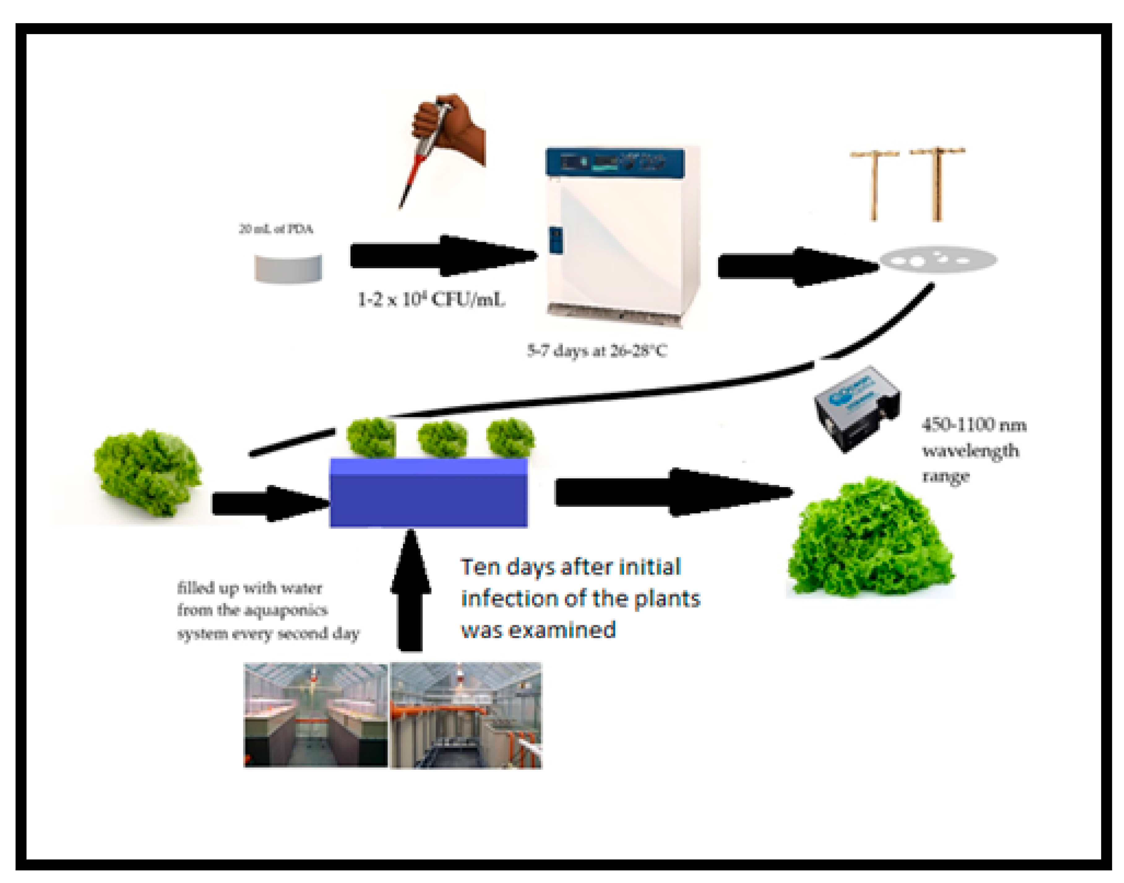
2.1. Experimental Design and Characterization of Aquaponic System and Vessels

The effect on vegetative indices obtained through NIR spectroscopy was tested during infection with three fungal pathogen species (A. alternata, A. niger, and F. oxysporum).
The lettuces on the third leaf formation were placed in tanks (with dimensions 1 m/0.50 m/0.35 m) filled up with water from the aquaponics system every second day. The aquaponics system was positioned in the aquaculture base at the Faculty of Agriculture at Trakia University (Stara Zagora, Bulgaria). The nutrient solution was taken from the plant section of the system itself, which had been in operation for 4 months at that point.
The tanks were stored at the Botanical laboratory of the Faculty of Agriculture, where appropriate climatic and light conditions were ensured. Excess water before each refill with a fresh nutrient solution from a 6-month-operating aquaponics system was removed by siphoning. A minimum distance of 20 cm between plants was maintained. In order to prevent anaerobic zones around the roots of the plant, the containers were oxygenated with an aerator.

2.2. Microorganisms Studied

This study included reference fungal strains Aspergillus niger NBIMCC 3252, Fusarium oxysporum NBIMCC 125, and Alternaria alternata NBIMCC 109, all of which were obtained from the National Bank for Industrial Microorganisms and Cell Cultures (NBIMCC) in Bulgaria. The fungal strains were stored at 0–4 °C. Prior to use, they were grown on potato dextrose agar (Biolife, Monza, Italy).
The fungal cultures were grown on potato dextrose agar (PDA). Every Petri dish was filled with 20 mL of PDA. After solidification, 0.1 mL of inoculum of the fungal strains (1–2 × 10⁴ CFU/mL) was introduced onto the agar plate surface. An incubation period of 5–7 days at 26–28 °C was maintained. Afterwards, 3–5 g of the infected PDA (hyphal disk) was introduced on the surface of every lettuce plant (L. sativa). Ten days later, initial infection with all three pathogens (A. niger, F. oxysporum, and A. alternata) was examined in the plants (Figure 1).
Before infection, another group was established to serve as a control, consisting of uninfected plants (control group). All experimental groups included 10 plants each.

2.3. NIR Spectroscopy Measurement

The spectral measurements of the lettuce plants were performed as follows (Figure 1).
The plants subject to spectral study were removed from the aquaponic tanks together with the pots in which they were rooted and placed in other tubs. In this condition, the plants were transferred to the spectral measurement laboratory. Every single plant was taken out from the tanks, measured during about 10–15 min, and put back in it.
Diffuse reflectance spectra of the leaves of lettuce were measured directly on the plants. The measurements were performed with a USB4000 spectrometer (Ocean Optics, Inc., Orlando, FL, USA) in the 450–1100 nm wavelength range. A fiber optics probe for diffuse reflection measurement was used. The probe consists of several optic fibers through which the illuminating light from a halogen lamp was fed to the area of interest and one optic fiber through which the light reflected from the measured surface was delivered to the spectrometer. The recorded spectra represent a dependence of the reflectivity of an area of about 6.5 mm in diameter on the wavelength. Every single spectrum corresponds to a different location on the lettuce leaves. As the plants were not large, all leaves large enough to reliably measure a circular area of about 6.5 mm in diameter were used for measurements. Further, 24 vegetation indices (shown in Table 1) were calculated from every spectrum and their values were analyzed.

2.4. Statistical Data Analysis

Statistical data analysis includes multivariate ANOVA, applied to calculate the significant differences between the plants infected with different fungal strains, based on the vegetative indices, most sensitive to the presence of investigated diseases. Significant differences were assumed based on the post hoc multiple comparisons with the Dunnett T3 or Tukey’s test (depending on Levene’s test of equality of error variances) at p-value ≤ 0.05. The IBM SPSS Statistics 26.0 (IBM, Chicago, IL, USA) package was used to process the data.

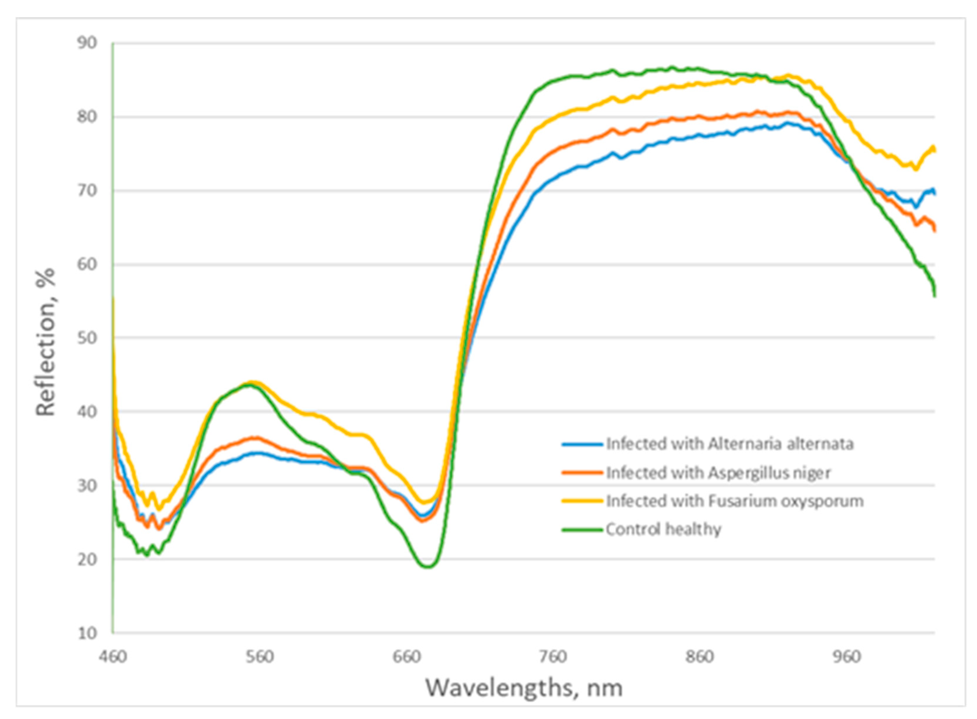
3. Results and Discussion

Healthy plants exhibited lower visible reflectance around 470 and 640–670 nm due to the chlorophyll absorption bands and higher near-infrared reflectance. In contrast to the healthy plants, infected plants showed higher reflectance in these areas because of their decreasing chlorophyll content. Changes were also observed in the green area of visible spectra. The reflection from plants infected with A. niger and A. alternata was lower than that of control plants, showing a change in the color of leaves. In the near-infrared spectral range, the reflectance values of infected leaves were lower than those of the control, which indicates that some changes in cell structures appeared as a result of the fungal infection (Figure 2).
The results of the multivariate ANOVA (Table 2) show that the vegetative indices CARI, MCARI, PSRI, REI2, REI3, and WBI could be used to distinguish plants infected by the pathogen A. alternata from the control variant. A. alternata is an important phytopathogen that is occasionally found in lettuce crops [33]. Only the vegetative indices PSRI and WBI could be used for distinguishing plants infected by the pathogen A. niger from the control variant. It was found that A. niger cause over 30% of fungal deterioration in lettuce from Yankaba and Sharada vegetable markets (situated in Nigeria), which made A. niger the most prevalent fungal pathogen in lettuce according to the samples studied [34]. The largest numbers of vegetative indices have the ability to distinguish plants infected by the pathogen F. oxysporum from the control variant as follows: CARI, Clred edge, Greenness index, MCARI, mNDVI, PI, REI 1, REI 2, REI 3, and WBI. On the basis of an analysis of variance, WBI was found to be the most suitable for finding reliable differences between plants infected with the studied pathogens and those of the control variant. The coefficient of determination of this index is R2 = 0.553 (Table 1), i.e., about 55.3% of the variations in the vegetation indices are due to the influence of the pathogen type. According to Mukhtar et al. [35], F. oxysporum causes moderate rot on 46.15% of the healthy lettuce samples inoculated with this fungal phytopathogen. This was the highest severity of rot, compared to Rhizopus spp., Rhizoctonia spp., A. flavus, A. fumigatus, A. niger, F. oxysporum, and Mucor sp. rot in sweet orange, cucumber, and lettuce. This is probably due at least in part to the structure of lettuce, since in cucumber, F. oxysporum caused mild rot in 20.63% of the healthy lettuce samples inoculated with this pathogen. These authors found that F. oxysporum caused rot in cucumber more frequently than A. niger—20.63% vs. 16.55%, respectively.
As a result of various diseases, including fungal ones, changes in the fluorescence of chlorophyll and, as a result, in the process of photosynthesis have been found [36]. The interaction of fungal pathogens and plants (method of obtaining nutrition, growth type, pigmentation, developmental stage, and disease severity) influenced the changes in spectral characteristics of infected plants. Changes in plant physiology during a pathogen attack can be linked to specific changes in plant reflectance patterns. The plants stop photosynthesizing and performing other assimilatory metabolism processes, and increase respiration and other processes required for defense from fungal pathogens. Although the suppression of photosynthesis and the induction of sink metabolism appear to be common responses to pathogen infections, the effect on sugar levels varies greatly between different plant–pathogen interactions [37]. For example, spectral changes in the region from 500 to 680 nm are observed if a pathogen has influenced the photosynthesis pigments. Pathogens changing the cellular structure of plants have a high influence in the near-infrared region from 700 to 1000 nm.
All three investigated pathogens had a statistically significant effect on leaf water content and water band index. Leaf symptoms of A. alternata include round, brown spots with concentric rings. This explains the statistical significance of the differences between the CARI, MCARI, Clred edge, RE2, and RE3 indices, related to chlorophyll content. Zarger et al. [38] found that A. alternata infection decreases the content of chlorophyll, carotenoids, ascorbic acid, proteins, and sugar in mango leaves. The same pathogen was found to decrease the lycopene, β-carotene, and total carotenoids but to increase proline content in tomato fruits [39]. The smallest changes in spectral characteristics were observed in plants infected with A. niger. Apart from the water band index, we only have statistically significant differences for PSRI. Studies of plant primary metabolism and plant–pathogen interactions based on quantitative imaging of chlorophyll fluorescence showed that photosynthesis was reduced both in cells directly below the fungal colonies and in adjacent cells [40].
F. oxysporum colonizes and metastasizes in xylem vessels, and causes systemic yellowing, wilting, and death of infected plants. These changes lead to changes in the spectral characteristics of infected plants both in the visible range and in the range above 700 nm. This explains the higher number of vegetative indices (CARI, MCARI, PI, G, PSRI, mNDVI, RE1, RE2, and RE3) that are statistically significantly different between infected and healthy plants.
Some of the indices allow for distinguishing the type of pathogen.
Expanding upon this point, the presence of the three pathogens—A. niger, A. alternata, and F. oxysporum—brings about different alterations in the spectral characteristics of infected lettuce plants, reflected in the various vegetative indices [41]. These differences suggest that different pathogens affect the physiological and biochemical processes of plants to different extents, leading to alterations in the reflectance spectra [42].
While A. alternata affects chlorophyll content and, consequently, coloration and reflectance in the visible range [28], F. oxysporum metastasizes in xylem vessels, affecting a broader range of physiological processes, including water content [43]. In contrast, A. niger appears to cause the least change in spectral characteristics, primarily influencing the water band index and photosynthesis, reflected in the PSRI [44].
The differences observed in the impact of each pathogen might be attributed to their distinct modes of infection and the corresponding plant responses [45]. For instance, A. niger, a common pathogen of many crops, may provoke a more localized response, primarily affecting photosynthesis and resulting in limited changes to spectral characteristics [40].
On the other hand, A. alternata, well-known for causing leaf spot diseases, can influence a broader range of physiological processes, as evidenced by the reduction in chlorophyll, carotenoids, ascorbic acid, proteins, and sugar in infected leaves [46]. This drastic change in physiological attributes could significantly alter the reflectance patterns, making it easier to detect this pathogen compared to others [44].
Similarly, F. oxysporum, a systemic pathogen known to colonize xylem vessels, can alter plant physiology extensively, leading to systemic yellowing, wilting, and death [35]. These physiological changes would be expected to dramatically alter the plant’s reflectance patterns, making it potentially easier to identify infection with this pathogen using spectral analysis [43].
Interestingly, the distinct spectral changes induced by each pathogen might potentially provide a way to not only detect infection but also identify the type of pathogen involved [40]. Indices like PSRI and WBI, which were significantly different for plants infected by A. niger and A. alternata, could serve as key indicators for these specific pathogens [30].
Meanwhile, several indices, including CARI, MCARI, PI, G, PSRI, mNDVI, RE1, RE2, and RE3, which were significantly different in plants infected with F. oxysporum, could help in the identification of this pathogen [41]. These findings highlight the potential of using spectral analysis and vegetative indices as non-invasive, real-time tools for disease detection and identification in crops [42].
Nonetheless, it is critical to note that these findings are based on controlled experimental conditions, and further research is needed to validate these indices under field conditions where multiple stress factors can influence the spectral characteristics of plants [43]. Also, disease progression stages may present varying spectral signatures and need to be considered in further studies [47].
Moreover, there are specific environmental and genetic factors that could influence these spectral changes and should be taken into account [48]. Changes in light conditions, soil properties, plant nutrient status, and even plant genetic variations could potentially affect plant reflectance patterns and thus the performance of these vegetative indices in disease detection [49].
While this study provides promising results in detecting and identifying specific pathogens based on their distinct impacts on plant spectral characteristics, it also raises several questions. Future research should focus on understanding how these indices behave under different environmental and disease conditions and whether it is possible to develop universal or crop-specific indices for disease detection [48].
Additionally, more advanced techniques, such as machine learning algorithms, could be explored to analyze spectral data and extract more reliable and precise disease indicators [50]. Such methodologies could potentially improve the sensitivity and specificity of disease detection using remote sensing techniques [49].
Finally, the practical application of these findings in real-time disease detection in fields should be explored. This would involve developing cost-effective, portable spectral sensors and integrating them with crop management systems to facilitate timely disease detection and treatment [18].
Our understanding of how different pathogens impact plant spectral characteristics has increased substantially. It has allowed us to leverage this knowledge in developing more precise disease detection and identification techniques. The future of crop disease management will likely heavily rely on the advancements in remote sensing technologies and machine learning algorithms, improving both the speed and accuracy of disease detection [48]. By continuing research in this area, we can potentially revolutionize disease management in agriculture, reducing crop losses and improving global food security.

4. Conclusions

In conclusion, this study of reflectance spectra in lettuce plants infected with different pathogens has provided valuable insights into the effects of fungal infections on plant physiology and reflectance patterns. Vegetative indices such as CARI, MCARI, PSRI, REI2, REI3, and WBI were found to be effective in distinguishing infected plants from healthy ones, with WBI demonstrating the greatest reliability. The distinct influences of Aspergillus niger, Alternaria alternata, and Fusarium oxysporum on the reflectance spectra emphasize the potential for spectral analysis in pathogen detection and crop health management. Furthermore, understanding the impact of these fungal pathogens on chlorophyll fluorescence and photosynthesis provides a crucial link to monitoring plant health and productivity. This study underscores the importance of further exploring the potential of remote sensing and spectral analysis in precision agriculture for optimal disease management and productivity.

Author Contributions

Conceptualization, I.S., K.V. and T.D.; Methodology, I.S., K.V. and T.D.; Software, P.V.; Validation, V.A. and S.A.; Formal analysis, D.Y.; Investigation, I.S., K.V., D.S.-S., E.V. and D.Y.; Resources, I.S.; Writing—original draft, I.S., T.D. and S.A.; Writing—review & editing, K.V.; Visualization, D.Y.; Supervision, I.S.; Project administration, S.A. All authors have read and agreed to the published version of the manuscript.

Funding

This research was funded by Bulgarian Ministry of Education and Science (MES) in the frames of Bulgarian National Recovery and Resilience Plan, Component “Innovative Bulgaria”, the Project No. BG-RRP-2.004-0006-C02 “Development of research and innovation at Trakia University in service of health and sustainable well-being” and National Research Program “Smart crop production”: No. 866/26.11.2020.

Data Availability Statement

All data are available from the corresponding author upon request.

Conflicts of Interest

The authors declare no conflict of interest.

References

  1. Graber, A.; Junge, R. Aquaponic Systems: Nutrient recycling from fish wastewater by vegetable production. Desalination 2009, 246, 147–156. [Google Scholar] [CrossRef]
  2. Colt, J.; Schuur, A.; Weaver, D.; Semmens, K. Engineering design of aquaponics systems. Rev. Fish. Sci. Aquac. 2021, 30, 33–80. [Google Scholar] [CrossRef]
  3. Sirakov, I.; Lutz, M.; Graber, A.; Mathis, A.; Staykov, Y.; Smits, T.; Junge, R. Potential for combined biocontrol activity against fungal fish and plant pathogens by bacterial isolates from a model aquaponic system. Water 2016, 8, 518. [Google Scholar] [CrossRef]
  4. Suárez-Cáceres, G.; Pérez-Urrestarazu, L.; Avilés, M.; Borrero, C.; Eguíbar, J.; Fernández-Cabanás, V. Susceptibility to water-borne plant diseases of hydroponic vs. aquaponics systems. Aquaculture 2021, 544, 737093. [Google Scholar] [CrossRef]
  5. Dean, R.; Van Kan, J.; Pretorius, Z.; Hammond-Kosack, K. The top 10 fungal pathogens in molecular plant pathology. Mol. Plant Pathol. 2012, 13, 414–430. [Google Scholar] [PubMed]
  6. Martinko, K.; Ivankovic, S.; Lazarevic, B.; Ðermic, E.; Ðermic, D. Control of early blight fungus (Alternaria alternata) in tomato by boric and phenylboronic acid. Antibiotics 2022, 11, 320. [Google Scholar] [CrossRef]
  7. Gravel, D.; Poisot, T.; Albouy, C.; Velez, L.; Mouillot, D. Inferring food web structure from predator–prey body size relationships. Methods Ecol. Evol. 2013, 4, 1083–1090. [Google Scholar] [CrossRef]
  8. Nemethy, S.; Bittsanszky, A.; Schmautz, Z.; Junge, R.; Komives, T. Protecting plants from pests and diseases in aquaponic systems. In Ecological Footprint in Central Europe, Multiauthorial Monograph; The University College of Tourism and Ecology Press: Sucha Beskidzka, Poland, 2016; pp. 166–179. [Google Scholar]
  9. Sirakov, I.; Velichkova, K.; Stoyanova, S.; Zhelyazkov, G.; Staykov, Y. The effect of diet supplemented with dandelion’s (Taraxacum officinale) extract on the productive and blood parameters of common carp (Cyprinus carpio L.), cultivated in the recirculation system. Maced. Vet. Rev. 2019, 42, 131–139. [Google Scholar] [CrossRef]
  10. Stouvenakers, G.; Massart, S.; Depireux, P.; Jijakli, M. Microbial origin of aquaponic water suppressiveness against Pythium aphanidermatum lettuce root rot disease. Microorganisms 2020, 8, 1683. [Google Scholar] [CrossRef]
  11. Rivas-García, S.; Bernal, J.; Bachiller-Corral, J. Rhabdomyolysis as the main manifestation of coronavirus disease 2019. Rheumatology 2020, 59, 2174–2176. [Google Scholar] [CrossRef]
  12. Mahlein, A.; Kuska, M.; Behmann, J.; Polder, G.; Walter, A. Hyperspectral sensors and imaging technologies in phytopathology: State of the art. Annu. Rev. Phytopathol. 2018, 56, 535–558. [Google Scholar] [CrossRef] [PubMed]
  13. Thomas, S.; Kuska, M.; Bohnenkamp, D.; Brugger, A.; Alisaac, E.; Wahabzada, M.; Behmann, J.; Mahlein, A. Benefits of hyperspectral imaging for plant disease detection and plant protection: A technical perspective. J. Plant Dis. Prot. 2018, 125, 5–20. [Google Scholar] [CrossRef]
  14. Terentev, A.; Dolzhenko, V.; Fedotov, A.; Eremenko, D. Current State of Hyperspectral Remote Sensing for Early Plant Disease Detection: A Review. Sensors 2022, 22, 757. [Google Scholar] [CrossRef] [PubMed]
  15. Sims, D.A.; Gamon, J.A. Relationships between leaf pigment content and spectral reflectance across a wide range of species, leaf structures and developmental stages. Remote Sens. Environ. 2002, 81, 337–354. [Google Scholar] [CrossRef]
  16. Wan, L.; Li, H.; Li, C.; Wang, A.; Yang, Y.; Wang, P. Hyperspectral Sensing of Plant Diseases: Principle and Methods. Agronomy 2022, 12, 1451. [Google Scholar] [CrossRef]
  17. Haboudane, D.; Miller, J.; Pattey, E.; Zarco-Tejada, P.; Strachan, I. Hyperspectral vegetation indices and novel algorithms for predicting green LAI of crop canopies: Modeling and validation in the context of precision agriculture. Remote Sens. Environ. 2004, 90, 337–352. [Google Scholar] [CrossRef]
  18. Gitelson, A.; Viña, A.; Ciganda, V.; Rundquist, D.; Arkebauer, T. Remote estimation of canopy chlorophyll content in crops. Geophys. Res. Lett. 2005, 32, L08403. [Google Scholar] [CrossRef]
  19. Mahlein, A.; Rumpf, T.; Welke, P. Development of spectral indices for detecting and identifying plant diseases. Remote Sens. Environ. 2013, 128, 21–30. [Google Scholar] [CrossRef]
  20. Gitelson, A.; Merzlyak, M.; Zur, Y.; Stark, R.; Gritz, U. Non-destructive and remote sensing techniques for estimation of vegetation status. In Proceedings of the Third European Conference on Precision Agriculture, Montpelier, France, 18–20 June 2001; Volume 1, pp. 301–306. [Google Scholar]
  21. Moshou, D.; Bravo, C.; Oberti, R.; West, J.; Bodria, L.; McCartney, A.; Ramon, H. Plant disease detection basedondata fusion of hyper-spectral and multi-spectral fluorescence imaging using Kohonen maps. Real-Time Imaging 2005, 1, 75–83. [Google Scholar] [CrossRef]
  22. Smith, R.; Adams, J.; Stephens, D.; Hick, P. Forecasting wheat yield in a Mediterranean-type environment from the NOAA satellite. Aust. J. Agric. Res. 1995, 46, 113–125. [Google Scholar] [CrossRef]
  23. Daughtry, C.; Walthall, C.; Kim, M.; Colstoun, E.; Murtrey, J. Estimating corn leaf chlorophyll concentration from leaf and canopy reflectance. Remote Sens. Environ. 2000, 74, 229–239. [Google Scholar] [CrossRef]
  24. Tucker, C.; Holben, B.; Elgin, J.; James, H.; McMurtrey, I.; James, E. Remote sensing of total dry-matter accumulation in winter wheat. Remote Sens. Environ. 1998, 11, 171–189. [Google Scholar] [CrossRef]
  25. Main, R.; Cho, M.; Moses, A.; Mathieu, R.; Kennedy, M.; Martha, M.; Ramoelo, A.; Koch, S. An investigation into robust spectral indices for leaf chlorophyll estimation. J. Photogramm. Remote Sens. 2011, 66, 751–761. [Google Scholar] [CrossRef]
  26. Brantley, S.; Zinnert, J.; Young, D. Application of hyperspectral vegetation indices to detect variations in high leaf area index temperate shrub thicket canopies. Remote Sens. Environ. 2011, 115, 514–523. [Google Scholar] [CrossRef]
  27. Datt, B. Remote sensing of chlorophyll a, chlorophyll b, chlorophyll a + b and total carotenoid content in eucalyptus leaves. Remote Sens. Environ. 1998, 66, 111–121. [Google Scholar] [CrossRef]
  28. Merzlyak, M.; Gitelson, A.; Chivkunova, O.; Rakitin, V. Non-destructive optical detection of pigment changes during leaf senescence and fruit ripening. Physiol. Plant. 1999, 106, 135–141. [Google Scholar] [CrossRef]
  29. Vogelmann, J.; Rock, B.; Moss, D. Red edge spectral measurements from sugar maple leaves. Int. J. Remote Sens. 1993, 14, 1563–1575. [Google Scholar] [CrossRef]
  30. Gitelson, A.; Merzlyak, M. Remote estimation of chlorophyll content in higher plant leaves. Int. J. Remote Sens. 1997, 18, 2691–2697. [Google Scholar] [CrossRef]
  31. Broge, N.; Leblanc, E. Comparing prediction power and stability of broadband and hyperspectral vegetation indices for estimation of green leaf area index and canopy chlorophyll density. Remote Sens. Environ. 2001, 76, 156–172. [Google Scholar] [CrossRef]
  32. Wang, L.; Qu, J. NMDI: A normalized multi-band drought index for monitoring soil and vegetation moisture with satellite remote sensing. Geophys. Res. Lett. 2007, 34, L20405. [Google Scholar] [CrossRef]
  33. Miranda-Apodaca, J.; Artetxe, U.; Aguado, I.; Martin-Souto, L.; Ramirez-Garcia, A.; Lacuesta, M.; Becerril, J.; Estonba, A.; Ortiz-Barredo, A.; Hernández, A.; et al. Stress response to climate change and postharvest handling in two differently pigmented lettuce genotypes: Impact on Alternaria alternata invasion and mycotoxin production. Plants 2023, 12, 1304. [Google Scholar] [CrossRef] [PubMed]
  34. Yahaya, S.; Fagwalawa, L.; Ali, M.; Lawan, M.; Mahmud, S. Isolation and identification of pathogenic fungi causing deterioration of lettuce plant (Lactuca sativa). A case study of Yankaba and Sharada vegetables markets. J. Plant Sci. Res. 2016, 3, 141. [Google Scholar]
  35. Mukhtar, Y.; Muhammad, M.; Zubairu, S.; Galalain, A.; Ahmad, U. Isolation, identification and pathogenicity of fungal organisms causing postharvest rot of sweet oranges, cucumber and lettuce in Sharada Market, Kano State-Nigeria. Asian J. Med. Biol. Res. 2019, 5, 286–291. [Google Scholar] [CrossRef]
  36. Swarbrick, P.; Schulze-Lefert, P.; Scholes, J. Metabolic consequences of susceptibility and resistance in barley leaves challenged with powdery mildew. Plant Cell Environ. 2006, 29, 1061–1076. [Google Scholar] [CrossRef]
  37. Berger, S.; Benediktyová, Z.; Matous, K.; Bonfig, K.; Mueller, M.; Nedbal, L.; Roitsch, T. Visualization of dynamics of plant-pathogen interaction by novel combination of chlorophyll fluorescence imaging and statistical analysis: Differential effects of virulent and avirulent strains of P. syringae and of oxylipins on A. thaliana. J. Exp. Bot. 2007, 58, 797–806. [Google Scholar] [CrossRef]
  38. Zarger, M.; Rizvi, S.; Bagga, P. Biochemical response of mango (Mangifera indica) to the infection of leaf spot pathogen (Alternaria alternata). J. Environ. Biol. 2014, 35, 529–534. [Google Scholar]
  39. Tiwari, J.; Upadhyay, D. Correlation and path-coefficient studies in tomato (Lycopersicon esculentum Mill.). J. Agri. Sci. 2011, 2, 63–68. [Google Scholar]
  40. Berger, S.; Sinha, A.; Roitsch, T. Plant physiology meets phytopathology: Plant primary metabolism and plant–pathogen interactions. J. Exp. Bot. 2007, 58, 4019–4026. [Google Scholar] [CrossRef]
  41. Jackson, R.; Huete, A. Interpreting vegetation indices. Prev. Vet. Med. 1991, 11, 185–200. [Google Scholar] [CrossRef]
  42. Jones, H.; Vaughan, R. Remote Sensing of Vegetation; Oxford University Press: New York, NY, USA, 2010. [Google Scholar]
  43. Penuelas, J.; Frederic, B.; Filella, I. Semi-Empirical Indices to Assess Carotenoids/Chlorophyll-a Ratio from Leaf Spectral Reflectance. Photosynthetica 1995, 31, 221–230. [Google Scholar]
  44. Gamon, J.; Penuelas, J.; Field, C. A narrow-waveband spectral index that tracks diurnal changes in photosynthetic efficiency. Remote Sens. Environ. 1992, 41, 35–44. [Google Scholar] [CrossRef]
  45. Agrios, G. Antifungal Activity of Selected Medicinal Plant Extract on Fusarium oxysporum Schlechtthe Causal Agent of Fusarium Wilt Disease in Tomato. Plant Pathol. 2005, 26–27, 398–401. [Google Scholar]
  46. Zarger, S.; Rizvi, G.; Parashar, R.; Paijwar, M. Impact of Alternaria alternata on organic components of mango leaves. Mycopath 2014, 12, 129–131. [Google Scholar]
  47. Zarco-Tejada, P.; González-Dugo, V.; Berni, J. Fluorescence, temperature and narrow-band indices acquired from a UAV platform for water stress detection using a micro-hyperspectral imager and a thermal camera. Remote Sens. Environ. 2012, 117, 322–337. [Google Scholar] [CrossRef]
  48. Furbank, R.; Tester, M. Phenomics–technologies to relieve the phenotyping bottleneck. Trends Plant Sci. 2011, 16, 635–644. [Google Scholar] [CrossRef]
  49. Thenkabail, P.; Lyon, J.; Huete, A. Hyperspectral Remote sensing of Vegetation; CRC Press: Boca Raton, FL, USA, 2012. [Google Scholar]
  50. Kira, K.; Rendell, L. A practical approach to feature selection. In Machine Learning Proceedings; Morgan Kaufmann: Burlington, MA, USA, 1992; pp. 249–256. [Google Scholar]
Figure 1. Scheme of experimental design.
Microorganisms 11 02348 g001
Figure 2. Reflectance spectra of control and infected lettuce plants.
Microorganisms 11 02348 g002
Table 1. Vegetation indices—definitions.
IndexDefinitionReferences
CARI
Chlorophyll Absorption in Reflectance Index
C A R I = ( R 700 R 670 ) 0.2 ( R 700 R 550 ) Haboudane et al. [17]
Cl green
Clorophyll Index
C l g r e e n = R 760 R 550 1 Gitelson et al. [18]
Cl red edge
Clorophyll Index at red edge
C l r e d   e d g e = R 760 R 714 1 Gitelson et al. [18]
CLSI
Cercospora leaf spot index
C L S I = ( R 698 R 570 ) ( R 698 + R 570 ) R 734 Mahlein et al. [19]
CRI
Carotenoids Reflectance index
C R I = 1 R 510 1 R 550 Gitelson et al. [20]
fD
Index of disease
f D = R 500 R 500 + R 570 Moshou et al. [21]
G
Greennes index
G = R 554 R 677 Smith et al. [22]
HI
Healthy index
H I = R 534 R 698 R 534 + R 698 0.5 R 704 Mahlein et al. [19]
MCARI
Modified chlorophyll absorption in reflectance index
M C A R I = [ ( R 700 R 670 ) 0.2 ( R 700 R 550 ) ] R 700 R 670 Daughtry et al. [23]
mNDVI
Modified normalized difference vegetation index
m N D V I = R 750 R 705 R 750 + R 705 Tucker et al. [24]
NDVI (1)
Normalized difference vegetation index
N D V I = R 845 R 665 R 845 + R 665 Tucker et al. [24]
NDVI (2)
Normalized difference vegetation index
N D V I = R 800 R 680 R 800 + R 680 Main et al. [25]
NDVI (3)
Normalized difference vegetation index
N D V I = R 800 R 670 R 800 + R 670 Brantley et al. [26]
PI
Pigment index
P I = R 677 R 554 Datt [27]
PMI
Powdery mildew index
P M I = R 520 R 584 R 520 + R 584 R 724 Mahlein et al. [19]
PRI
Photochemical reflectance index
P R I = R 531 R 570 R 531 + R 570 Sims and Gamon [15]
PSRI
Plant Senescence Reflectance Index
P S R I = R 680 R 500 R 750 Merzlyak et al. [28]
REI 1
Red Edge Index
R E I 1 = R 740 R 720 Vogelmann et al. [29]
REI 2
Red Edge Index
R E I 2 = R 734 R 747 R 715 R 720 Vogelmann et al. [29]
REI 3
Red Edge Index
R E I 3 = R 734 R 747 R 715 R 726 Vogelmann et al. [29]
SBRI
Sugar beet rust index
S B R I = R 520 R 513 R 570 + R 513 0.5 R 704 Mahlein et al. [19]
SR
Simple ratio
S R = R 760 R 695 Gitelson and Merzlyak [30]
TVI
triangular vegetation index
T V I = 0.5 [ 120 ( R 750 R 550 ) 200 ( R 670 R 550 ) ] Broge and Leblanc [31]
WBI
Water band index
W B I = R 900 R 970 Wang and Qu [32]
Table 2. Multivariate ANOVA of the observed plant infections based on the varied types of vegetation indices.
x ± SD
Vegetation IndicesClassesR2
Infected with Alternaria alternata
(n = 8)
Infected with Aspergillus niger
(n = 9)
Infected with Fusarium oxysporum
(n = 7)
Uninfected (n = 5)
CARI1.975 ± 0.676 a2.356 ± 0.3541.854 ± 0.251 c2.744 ± 0.535 a, c0.339
Clgreen5.146 ± 2.2283.909 ± 1.0515.195 ± 1.2603.457 ± 0.9650.212
Clred edge0.817 ± 0.2080.682 ± 0.1200.838 ± 0.010 c0.599 ± 0.133 c0.309
CLSI−10.957 ± 0.932−11.204 ± 0.393−11.408 ± 0.576−11.466 ± 0.4190.099
CRI−1.661 ± 3.670−2.825 ± 0.550−4.117 ± 3.463−2.604 ± 1.1100.116
fD8.038 ± 30.228−2.741 ± 6.6500.184 ± 2.914−2.362 ± 6.5100.077
G2.800 ± 0.7063.291 ± 0.5812.483 ± 0.783 c3.732 ± 0.580 c0.333
HI−2.168 ± 0.436−2.344 ± 0.174−2.184 ± 0.193−2.576 ± 0.3070.225
MCARI7.744 ± 3.630 a10.253 ± 2.6146.681 ± 3.039 c13.665 ± 4.427 a, c0.378
mNDVI0.486 ± 0.0750.446 ± 0.0400.497 ± 0.032 c0.412 ± 0.056 c0.285
NDVI (1)0.921 ± 0.0530.928 ± 0.0170.913 ± 0.0490.930 ± 0.0250.032
NDVI (2)0.844 ± 0.0430.849 ± 0.0120.837 ± 0.0390.852 ± 0.0180.032
NDVI (3)0.874 ± 0.0460.880 ± 0.0140.867 ± 0.0420.884 ± 0.0210.038
PI0.380 ± 0.1030.315 ± 0.0740.434 ± 0.119 c0.273 ± 0.041 c0.316
PMI−11.719 ± 4.662−10.247 ± 0.336−10.369 ± 0.292−10.669 ± 0.3900.066
PRI−0.825 ± 0.877−0.436 ± 0.158−0.619 ± 0.276−0.405 ± 0.1440.119
PSRI0.207 ± 0.014 a0.215 ± 0.009 b0.220 ± 0.0130.232 ± 0.009 a, b0.397
REI 11.382 ± 0.0901.324 ± 0.0521.393 ± 0.042 c1.287 ± 0.059 c0.314
REI 20.678 ± 0.090 a0.612 ± 0.0630.682 ± 0.053 c0.557 ± 0.063 a, c0.352
REI 30.336 ± 0.038 a0.308 ± 0.0260.338 ± 0.020 c0.282 ± 0.029 a, c0.377
SBRI1.383 ± 9.3155.111 ± 16.361−4.683 ± 6.5220.840 ± 3.9520.110
SR7.739 ± 2.6226.532 ± 0.9127.506 ± 1.4135.999 ± 1.1590.154
TVI738.660 ± 46.810762.604 ± 23.518757.503 ± 19.201785.200 ± 25.6460.218
WBI1.882 ± 0.063 a1.929 ± 0.044 b1.920 ± 0.058 c2.049 ± 0.046 a, b, c0.553
Same superscripts within the same row represent significant differences at the level of significance p < 0.05, as follows: a—between the group infected with Alternaria alternata and all other groups; b—between the group infected with Aspergillus niger and all other groups; c—between the group infected with Fusarium oxysporum and all other groups; no statistically significant differences existed if there are no superscript letters in the row. R2—coefficients of determination based on observed means.
Disclaimer/Publisher’s Note: The statements, opinions and data contained in all publications are solely those of the individual author(s) and contributor(s) and not of MDPI and/or the editor(s). MDPI and/or the editor(s) disclaim responsibility for any injury to people or property resulting from any ideas, methods, instructions or products referred to in the content.

