
Concurrent and Subsequent Co-Infections of Clostridioides difficile Colitis in the Era of Gut Microbiota and Expanding Treatment Options

 , ,
, , 
Abstract
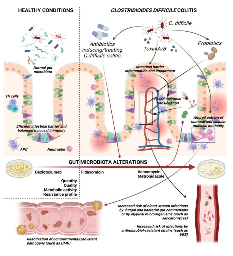
:1. Introduction
2. Bacterial Blood-Stream Infections and Clostridioides difficile
3. Candida spp. and Clostridioides difficile
4. Cytomegalovirus and Clostridioides difficile
5. Other Co-Infections in Clostridioides difficile Colitis
6. Probiotics and Clostridioides difficile
7. Fecal Microbiota Transplantation in Clostridioides difficile Colitis
8. Practical Considerations
9. Conclusions
Author Contributions
Funding
Conflicts of Interest
References
- Mylonakis, E.; Ryan, E.T.; Calderwood, S.B. Clostridium Difficile—Associated Diarrhea: A Review. Arch. Intern. Med. 2001, 161, 525–533. [Google Scholar] [CrossRef] [PubMed]
- Guh, A.Y.; Mu, Y.; Winston, L.G.; Johnston, H.; Olson, D.; Farley, M.M.; Wilson, L.E.; Holzbauer, S.M.; Phipps, E.C.; Dumyati, G.K.; et al. Trends in U.S. Burden of Clostridioides difficile Infection and Outcomes. N. Engl. J. Med. 2020, 382, 1320–1330. [Google Scholar] [CrossRef] [PubMed]
- Plachouras, D.; Kärki, T.; Hansen, S.; Hopkins, S.; Lyytikäinen, O.; Moro, M.L.; Reilly, J.; Zarb, P.; Zingg, W.; Kinross, P.; et al. Antimicrobial Use in European Acute Care Hospitals: Results from the Second Point Prevalence Survey (PPS) of Healthcare-Associated Infections and Antimicrobial Use, 2016 to 2017. Eurosurveillance 2018, 23, 1800393. [Google Scholar] [CrossRef] [PubMed] [Green Version]
- Ponce-Alonso, M.; Sáez de la Fuente, J.; Rincón-Carlavilla, A.; Moreno-Nunez, P.; Martínez-García, L.; Escudero-Sánchez, R.; Pintor, R.; García-Fernández, S.; Cobo, J. Impact of the Coronavirus Disease 2019 (COVID-19) Pandemic on Nosocomial Clostridioides difficile Infection. Infect. Control Hosp. Epidemiol. 2021, 42, 406–410. [Google Scholar] [CrossRef] [PubMed]
- Ofori, E.; Ramai, D.; Dhawan, M.; Mustafa, F.; Gasperino, J.; Reddy, M. Community-Acquired Clostridium Difficile: Epidemiology, Ribotype, Risk Factors, Hospital and Intensive Care Unit Outcomes, and Current and Emerging Therapies. J. Hosp. Infect. 2018, 99, 436–442. [Google Scholar] [CrossRef]
- Chu, Y.-M.; Lee, C.-L.; Chen, H.-Y.; Hung, C.-S. Predictors of Mortality in Patients with Clostridium Difficile Infection. Adv. Dig. Med. 2020, 7, 77–82. [Google Scholar] [CrossRef] [Green Version]
- Roo, A.C.D.; Regenbogen, S.E. Clostridium Difficile Infection: An Epidemiology Update. Clin. Colon Rectal Surg. 2020, 33, 49–57. [Google Scholar] [CrossRef]
- Czepiel, J.; Dróżdż, M.; Pituch, H.; Kuijper, E.J.; Perucki, W.; Mielimonka, A.; Goldman, S.; Wultańska, D.; Garlicki, A.; Biesiada, G. Clostridium Difficile Infection: Review. Eur. J. Clin. Microbiol. Infect. Dis. 2019, 38, 1211–1221. [Google Scholar] [CrossRef] [Green Version]
- Tsai, C.-S.; Hung, Y.-P.; Lee, J.-C.; Syue, L.-S.; Hsueh, P.-R.; Ko, W.-C. Clostridioides difficile Infection: An Emerging Zoonosis? Expert Rev. Anti-Infect. Ther. 2021, 19, 1543–1552. [Google Scholar] [CrossRef]
- Suchartlikitwong, S.; Laoveeravat, P.; Teerakanok, J.; Mingbunjerdsuk, T.; Thavaraputta, S.; Vutthikraivit, W.; Thongprayoon, C.; Nugent, K.; Cheungpasitporn, W. Meta-Analysis Comparing the Effects of Statins on the Risk of Clostridium Difficile Diarrhea. Bayl. Univ. Med. Cent. Proc. 2018, 31, 447–452. [Google Scholar] [CrossRef]
- Hudson, S.L.; Arnoczy, G.; Gibson, H.; Thurber, C.; Lee, J.; Kessell, A. Probiotic Use as Prophylaxis for Clostridium Difficile-Associated Diarrhea in a Community Hospital. Am. J. Infect. Control 2019, 47, 1028–1029. [Google Scholar] [CrossRef] [PubMed]
- Cunningham, R.; Dial, S. Is Over-Use of Proton Pump Inhibitors Fuelling the Current Epidemic of Clostridium Difficile-Associated Diarrhoea? J. Hosp. Infect. 2008, 70, 1–6. [Google Scholar] [CrossRef] [PubMed]
- O’Keefe, S.J. Tube Feeding, the Microbiota, and Clostridium Difficile Infection. World J. Gastroenterol. 2010, 16, 139–142. [Google Scholar] [CrossRef] [PubMed]
- Trunfio, M.; Savoldi, A.; Viganò, O.; d’Arminio Monforte, A. Bacterial Coinfections in Dengue Virus Disease: What We Know and What Is Still Obscure about an Emerging Concern. Infection 2017, 45, 1–10. [Google Scholar] [CrossRef]
- Wee, L.E.; Hnin, S.W.K.; Xu, Z.; Lee, L.S.-U. Strongyloides Hyperinfection Associated with Enterococcus Faecalis Bacteremia, Meningitis, Ventriculitis and Gas-Forming Spondylodiscitis: A Case Report. Trop. Med. Infect. Dis. 2020, 5, 44. [Google Scholar] [CrossRef] [Green Version]
- Chumbler, N.M.; Farrow, M.A.; Lapierre, L.A.; Franklin, J.L.; Haslam, D.B.; Haslam, D.; Goldenring, J.R.; Lacy, D.B. Clostridium Difficile Toxin B Causes Epithelial Cell Necrosis through an Autoprocessing-Independent Mechanism. PLoS Pathog. 2012, 8, e1003072. [Google Scholar] [CrossRef]
- Shen, A. Clostridium Difficile Toxins: Mediators of Inflammation. J. Innate Immun. 2012, 4, 149–158. [Google Scholar] [CrossRef] [Green Version]
- Hasegawa, M.; Yada, S.; Liu, M.Z.; Kamada, N.; Muñoz-Planillo, R.; Do, N.; Núñez, G.; Inohara, N. Interleukin-22 Regulates the Complement System to Promote Resistance against Pathobionts after Pathogen-Induced Intestinal Damage. Immunity 2014, 41, 620–632. [Google Scholar] [CrossRef] [Green Version]
- Abbas, A.; Zackular, J.P. Microbe–Microbe Interactions during Clostridioides difficile Infection. Curr. Opin. Microbiol. 2020, 53, 19–25. [Google Scholar] [CrossRef]
- Langdon, A.; Schwartz, D.J.; Bulow, C.; Sun, X.; Hink, T.; Reske, K.A.; Jones, C.; Burnham, C.-A.D.; Dubberke, E.R.; Dantas, G.; et al. Microbiota Restoration Reduces Antibiotic-Resistant Bacteria Gut Colonization in Patients with Recurrent Clostridioides difficile Infection from the Open-Label PUNCH CD Study. Genome Med. 2021, 13, 28. [Google Scholar] [CrossRef]
- Louie, T.J.; Cannon, K.; Byrne, B.; Emery, J.; Ward, L.; Eyben, M.; Krulicki, W. Fidaxomicin Preserves the Intestinal Microbiome during and after Treatment of Clostridium Difficile Infection (CDI) and Reduces Both Toxin Reexpression and Recurrence of CDI. Clin. Infect. Dis. 2012, 55 (Suppl. 2), S132–S142. [Google Scholar] [CrossRef] [PubMed]
- Oliva, A.; Aversano, L.; De Angelis, M.; Mascellino, M.T.; Miele, M.C.; Morelli, S.; Battaglia, R.; Iera, J.; Bruno, G.; Corazziari, E.S.; et al. Persistent Systemic Microbial Translocation, Inflammation, and Intestinal Damage During Clostridioides difficile Infection. Open Forum Infect. Dis. 2020, 7, ofz507. [Google Scholar] [CrossRef] [PubMed]
- Falcone, M.; Russo, A.; Iraci, F.; Carfagna, P.; Goldoni, P.; Vullo, V.; Venditti, M. Risk Factors and Outcomes for Bloodstream Infections Secondary to Clostridium Difficile Infection. Antimicrob. Agents Chemother. 2016, 60, 252–257. [Google Scholar] [CrossRef] [PubMed] [Green Version]
- Vallabhaneni, S.; Almendares, O.M.; Farley, M.M.; Reno, J.; Smith, Z.; Stein, B.; Magill, S.S.; Smith, R.M.; Cleveland, A.; Lessa, F.C. Candida Co-Infection Among Adults With Clostridium Difficile Infection in Metropolitan Atlanta, 2009–2013. Open Forum Infect. Dis. 2015, 2, 921. [Google Scholar] [CrossRef]
- Thomas, J.A.; Newman, K.C.; Doshi, S.; Logan, N.; Musher, D.M. Bacteraemia from an Unrecognized Source (Occult Bacteraemia) Occurring during Clostridium Difficile Infection. Scand. J. Infect. Dis. 2011, 43, 269–274. [Google Scholar] [CrossRef]
- Ulrich, R.J.; Santhosh, K.; Mogle, J.A.; Young, V.B.; Rao, K. Is Clostridium Difficile Infection a Risk Factor for Subsequent Bloodstream Infection? Anaerobe 2017, 48, 27–33. [Google Scholar] [CrossRef]
- Roghmann, M.-C.; McCarter, R.J., Jr.; Brewrink, J.; Cross, A.S.; Morris, J.G., Jr. Clostridium Difficile Infection Is a Risk Factor for Bacteremia Due to Vancomycin-Resistant Enterococci (VRE) in VRE-Colonized Patients with Acute Leukemia. Clin. Infect. Dis. 1997, 25, 1056–1059. [Google Scholar] [CrossRef]
- Poduval, R.D.; Kamath, R.P.; Corpuz, M.; Norkus, E.P.; Pitchumoni, C.S. Clostridium Difficile and Vancomycin-Resistant Enterococcus: The New Nosocomial Alliance. Am. J. Gastroenterol. 2000, 95, 3513–3515. [Google Scholar] [CrossRef]
- Axelrad, J.E.; Lebwohl, B.; Cuaresma, E.; Cadwell, K.; Green, P.H.R.; Freedberg, D.E. Gut Colonization with Vancomycin-Resistant Enterococcus and Risk for Subsequent Enteric Infection. Gut Pathog. 2018, 10, 28. [Google Scholar] [CrossRef]
- Lee, N.-Y.; Huang, Y.-T.; Hsueh, P.-R.; Ko, W.-C. Clostridium Difficile Bacteremia, Taiwan. Emerg. Infect. Dis. 2010, 16, 1204–1210. [Google Scholar] [CrossRef]
- Libby, D.B.; Bearman, G. Bacteremia Due to Clostridium Difficile—Review of the Literature. Int. J. Infect. Dis. 2009, 13, e305–e309. [Google Scholar] [CrossRef] [PubMed] [Green Version]
- van Prehn, J.; Reigadas, E.; Vogelzang, E.H.; Bouza, E.; Hristea, A.; Guery, B.; Krutova, M.; Norén, T.; Allerberger, F.; Coia, J.E.; et al. European Society of Clinical Microbiology and Infectious Diseases: 2021 Update on the Treatment Guidance Document for Clostridioides difficile Infection in Adults. Clin. Microbiol. Infect. 2021, 27 (Suppl. 2), S1–S21. [Google Scholar] [CrossRef] [PubMed]
- Johnson, S.; Lavergne, V.; Skinner, A.M.; Gonzales-Luna, A.J.; Garey, K.W.; Kelly, C.P.; Wilcox, M.H. Clinical Practice Guideline by the Infectious Diseases Society of America (IDSA) and Society for Healthcare Epidemiology of America (SHEA): 2021 Focused Update Guidelines on Management of Clostridioides difficile Infection in Adults. Clin. Infect. Dis. 2021, 73, e1029–e1044. [Google Scholar] [CrossRef] [PubMed]
- Raponi, G.; Visconti, V.; Brunetti, G.; Ghezzi, M.C. Clostridium Difficile Infection and Candida Colonization of the Gut: Is There a Correlation? Clin. Infect. Dis. 2014, 59, 1648–1649. [Google Scholar] [CrossRef] [Green Version]
- Nerandzic, M.M.; Mullane, K.; Miller, M.A.; Babakhani, F.; Donskey, C.J. Reduced Acquisition and Overgrowth of Vancomycin-Resistant Enterococci and Candida Species in Patients Treated With Fidaxomicin Versus Vancomycin for Clostridium Difficile Infection. Clin. Infect. Dis. 2012, 55, S121–S126. [Google Scholar] [CrossRef]
- Markey, L.; Shaban, L.; Green, E.R.; Lemon, K.P.; Mecsas, J.; Kumamoto, C.A. Pre-Colonization with the Commensal Fungus Candida albicans Reduces Murine Susceptibility to Clostridium Difficile Infection. Gut Microbes 2018, 9, 497–509. [Google Scholar] [CrossRef] [Green Version]
- Leelahavanichkul, A.; Panpetch, W.; Worasilchai, N.; Somparn, P.; Chancharoenthana, W.; Nilgate, S.; Finkelman, M.; Chindamporn, A.; Tumwasorn, S. Evaluation of Gastrointestinal Leakage Using Serum (1 → 3)-β-D-Glucan in a Clostridium Difficile Murine Model. FEMS Microbiol. Lett. 2016, 363, fnw204. [Google Scholar] [CrossRef] [Green Version]
- van Leeuwen, P.T.; van der Peet, J.M.; Bikker, F.J.; Hoogenkamp, M.A.; Oliveira Paiva, A.M.; Kostidis, S.; Mayboroda, O.A.; Smits, W.K.; Krom, B.P. Interspecies Interactions between Clostridium Difficile and Candida albicans. Msphere 2016, 1, e00187-16. [Google Scholar] [CrossRef] [Green Version]
- Falcone, M.; Venditti, M.; Sanguinetti, M.; Posteraro, B. Management of Candidemia in Patients with Clostridium Difficile Infection. Expert Rev. Anti-Infect. Ther. 2016, 14, 679–685. [Google Scholar] [CrossRef]
- Russo, A.; Falcone, M.; Fantoni, M.; Murri, R.; Masucci, L.; Carfagna, P.; Ghezzi, M.C.; Posteraro, B.; Sanguinetti, M.; Venditti, M. Risk Factors and Clinical Outcomes of Candidaemia in Patients Treated for Clostridium Difficile Infection. Clin. Microbiol. Infect. 2015, 21, e1–e493. [Google Scholar] [CrossRef] [Green Version]
- Kocak, E.D.; Sherwin, J.C.; Hall, A.J. Cytomegalovirus Disease in Immunocompetent Adults. Med. J. Aust. 2015, 202, 419. [Google Scholar] [CrossRef] [PubMed]
- Chan, K.-S.; Lee, W.-Y.; Yu, W.-L. Coexisting Cytomegalovirus Infection in Immunocompetent Patients with Clostridium Difficile Colitis. J.Microbiol. Immunol. Infect. 2016, 49, 829–836. [Google Scholar] [CrossRef] [PubMed] [Green Version]
- Jentzer, A.; Veyrard, P.; Roblin, X.; Saint-Sardos, P.; Rochereau, N.; Paul, S.; Bourlet, T.; Pozzetto, B.; Pillet, S. Cytomegalovirus and Inflammatory Bowel Diseases (IBD) with a Special Focus on the Link with Ulcerative Colitis (UC). Microorganisms 2020, 8, 1078. [Google Scholar] [CrossRef] [PubMed]
- John, S.G.; Dominguez, C.; Chandiramani, V.; Vemulappalli, T. A Rare Case Intractable Diarrhea Secondary to Clostridium Difficile and Cytomegalovirus Coinfection. Am. J. Case Rep. 2013, 14, 498–501. [Google Scholar] [CrossRef] [PubMed] [Green Version]
- Florescu, D.F.; Mindru, C.; Chambers, H.E.; Kalil, A.C. Clostridium Difficile and Cytomegalovirus Colitis Co-Infection: Search for the Hidden “Bug”. Transpl. Infect. Dis. 2011, 13, 411–415. [Google Scholar] [CrossRef] [PubMed]
- Lachance, P.; Chen, J.; Featherstone, R.; Sligl, W.I. Association Between Cytomegalovirus Reactivation and Clinical Outcomes in Immunocompetent Critically Ill Patients: A Systematic Review and Meta-Analysis. Open Forum Infect. Dis. 2017, 4, ofx029. [Google Scholar] [CrossRef] [PubMed] [Green Version]
- Limaye, A.P.; Boeckh, M. CMV in Critically Ill Patients: Pathogen or Bystander? Rev. Med. Virol. 2010, 20, 372–379. [Google Scholar] [CrossRef] [Green Version]
- Chan, K.-S.; Yang, C.-C.; Chen, C.-M.; Yang, H.-H.; Lee, C.-C.; Chuang, Y.-C.; Yu, W.-L. Cytomegalovirus Colitis in Intensive Care Unit Patients: Difficulties in Clinical Diagnosis. J. Crit. Care 2014, 29, e1–e474. [Google Scholar] [CrossRef]
- Taori, S.K.; Wroe, A.; Hardie, A.; Gibb, A.P.; Poxton, I.R. A Prospective Study of Community-Associated Clostridium Difficile Infections: The Role of Antibiotics and Co-Infections. J. Infect. 2014, 69, 134–144. [Google Scholar] [CrossRef]
- Wang, L.; Xiao, L.; Duan, L.; Ye, J.; Guo, Y.; Guo, M.; Liu, L.; Feng, Y. Concurrent Infections of Giardia Duodenalis, Enterocytozoon Bieneusi, and Clostridium Difficile in Children during a Cryptosporidiosis Outbreak in a Pediatric Hospital in China. PLoS Negl. Trop. Dis. 2013, 7, e2437. [Google Scholar] [CrossRef] [Green Version]
- Warren, C.A.; Labio, E.; Destura, R.; Sevilleja, J.E.; Jamias, J.D.; Daez, M.L.O. Clostridium Difficile and Entamoeba Histolytica Infections in Patients with Colitis in the Philippines. Trans. R. Soc. Trop. Med. Hyg. 2012, 106, 424–428. [Google Scholar] [CrossRef] [PubMed]
- Roldan, G.A.; Cui, A.X.; Pollock, N.R. Assessing the Burden of Clostridium Difficile Infection in Low- and Middle-Income Countries. J. Clin. Microbiol. 2018, 56, e01747-17. [Google Scholar] [CrossRef] [PubMed] [Green Version]
- de Graaf, H.; Pai, S.; Burns, D.A.; Karas, J.A.; Enoch, D.A.; Faust, S.N. Co-Infection as a Confounder for the Role of Clostridium Difficile Infection in Children with Diarrhoea: A Summary of the Literature. Eur. J. Clin. Microbiol. Infect. Dis. 2015, 34, 1281–1287. [Google Scholar] [CrossRef] [PubMed]
- Khatami, S.S.; Ravakhah, K.; Mukunda, B. Coinfection with Giardia Lamblia and Clostridium Difficile after Use of Ranitidine. Am. J. Med. Sci. 2004, 327, 91–93. [Google Scholar] [CrossRef]
- Lugito, N.H.; Susanti, K.A.; Yanto, T.A.; Tjiang, M.M.; Setiadinata, R.E.S.A.; Wijaya, I.A. A 21 Year-Old Male Colorectal Cancer Patient with Clostridium Difficile and Intestinal Amebiasis Infection. Indones. J. Cancer 2014, 8, 71. [Google Scholar] [CrossRef]
- Mills, J.P.; Rao, K.; Young, V.B. Probiotics for Prevention of Clostridium Difficile Infection. Curr. Opin. Gastroenterol. 2018, 34, 3–10. [Google Scholar] [CrossRef]
- Goldenberg, J.Z.; Yap, C.; Lytvyn, L.; Lo, C.K.-F.; Beardsley, J.; Mertz, D.; Johnston, B.C. Probiotics for the Prevention of Clostridium Difficile-Associated Diarrhea in Adults and Children. Cochrane Database Syst. Rev. 2017, 12, CD006095. [Google Scholar] [CrossRef]
- Saltzman, T.; Fazzari, M.; Chung, S.; Cunha, B.A.; Blum, S. The Effect of Probiotics on the Incidence of Clostridioides difficile: Retrospective Cohort Analysis. Am. J. Infect. Control 2020, 48, 184–188. [Google Scholar] [CrossRef]
- Heil, E.L.; Harris, A.D.; Brown, C.; Seung, H.; Thom, K.A.; von Rosenvinge, E.; Sorongon, S.; Pineles, L.; Goodman, K.E.; Leekha, S. A Multicenter Evaluation of Probiotic Use for the Primary Prevention of Clostridioides difficile Infection. Clin. Infect. Dis. 2021, 73, 1330–1337. [Google Scholar] [CrossRef]
- Fadhel, M.; Patel, S.; Liu, E.; Levitt, M.; Asif, A. Saccharomyces Cerevisiae Fungemia in a Critically Ill Patient with Acute Cholangitis and Long Term Probiotic Use. Med. Mycol. Case Rep. 2018, 23, 23–25. [Google Scholar] [CrossRef]
- Santino, I.; Alari, A.; Bono, S.; Teti, E.; Marangi, M.; Bernardini, A.; Magrini, L.; Di Somma, S.; Teggi, A. Saccharomyces Cerevisiae Fungemia, a Possible Consequence of the Treatment of Clostridium Difficile Colitis with a Probioticum. Int. J. Immunopathol. Pharmacol. 2014, 27, 143–146. [Google Scholar] [CrossRef] [PubMed]
- Polito, N.B.; Avery, L.M. Mitigating Risk of Bloodstream Infection Related to Inpatient Probiotic Use. Am. J. Health-Syst. Pharm. 2018, 75, 595–596. [Google Scholar] [CrossRef] [PubMed]
- MacGregor, G.; Smith, A.J.; Thakker, B.; Kinsella, J. Yoghurt Biotherapy: Contraindicated in Immunosuppressed Patients? Postgrad. Med. J. 2002, 78, 366–367. [Google Scholar] [CrossRef] [PubMed]
- Fruchart, C.; Salah, A.; Gray, C.; Martin, E.; Stamatoullas, A.; Bonmarchand, G.; Lemeland, J.F.; Tilly, H. Lactobacillus Species as Emerging Pathogens in Neutropenic Patients. Eur. J. Clin. Microbiol. Infect. Dis. 1997, 16, 681–684. [Google Scholar] [CrossRef]
- Feuerstadt, P.; Louie, T.J.; Lashner, B.; Wang, E.E.L.; Diao, L.; Bryant, J.A.; Sims, M.; Kraft, C.S.; Cohen, S.H.; Berenson, C.S.; et al. SER-109, an Oral Microbiome Therapy for Recurrent Clostridioides difficile Infection. N. Engl. J. Med. 2022, 386, 220–229. [Google Scholar] [CrossRef]
- Baktash, A.; Terveer, E.M.; Zwittink, R.D.; Hornung, B.V.H.; Corver, J.; Kuijper, E.J.; Smits, W.K. Mechanistic Insights in the Success of Fecal Microbiota Transplants for the Treatment of Clostridium Difficile Infections. Front. Microbiol. 2018, 9, 1242. [Google Scholar] [CrossRef]
- Hvas, C.L.; Dahl Jørgensen, S.M.; Jørgensen, S.P.; Storgaard, M.; Lemming, L.; Hansen, M.M.; Erikstrup, C.; Dahlerup, J.F. Fecal Microbiota Transplantation Is Superior to Fidaxomicin for Treatment of Recurrent Clostridium Difficile Infection. Gastroenterology 2019, 156, 1324–1332. [Google Scholar] [CrossRef] [Green Version]
- Juul, F.E.; Garborg, K.; Bretthauer, M.; Skudal, H.; Øines, M.N.; Wiig, H.; Rose, Ø.; Seip, B.; Lamont, J.T.; Midtvedt, T.; et al. Fecal Microbiota Transplantation for Primary Clostridium Difficile Infection. N. Engl. J. Med. 2018, 378, 2535–2536. [Google Scholar] [CrossRef] [Green Version]
- Ianiro, G.; Murri, R.; Sciumè, G.D.; Impagnatiello, M.; Masucci, L.; Ford, A.C.; Law, G.R.; Tilg, H.; Sanguinetti, M.; Cauda, R.; et al. Incidence of Bloodstream Infections, Length of Hospital Stay, and Survival in Patients With Recurrent Clostridioides difficile Infection Treated With Fecal Microbiota Transplantation or Antibiotics: A Prospective Cohort Study. Ann. Intern. Med. 2019, 171, 695–702. [Google Scholar] [CrossRef]
- Taur, Y.; Pamer, E.G. Harnessing Microbiota to Kill a Pathogen: Fixing the Microbiota to Treat Clostridium Difficile Infections. Nat. Med. 2014, 20, 246–247. [Google Scholar] [CrossRef]
- Abou Chakra, C.N.; Pepin, J.; Sirard, S.; Valiquette, L. Risk Factors for Recurrence, Complications and Mortality in Clostridium Difficile Infection: A Systematic Review. PLoS ONE 2014, 9, e98400. [Google Scholar] [CrossRef] [PubMed] [Green Version]
- Jaber, M.R.; Olafsson, S.; Fung, W.L.; Reeves, M.E. Clinical Review of the Management of Fulminant Clostridium Difficile Infection. Am. J. Gastroenterol. 2008, 103, 3195–3203. [Google Scholar] [CrossRef] [PubMed]
- Manthey, C.F.; Dranova, D.; Christner, M.; Drolz, A.; Kluge, S.; Lohse, A.W.; Fuhrmann, V. Initial Therapy Affects Duration of Diarrhoea in Critically Ill Patients with Clostridioides difficile Infection (CDI). Crit. Care 2019, 23, 399. [Google Scholar] [CrossRef] [PubMed] [Green Version]
- Wilcox, M.H.; Howe, R. Diarrhoea Caused by Clostridium Difficile: Response Time for Treatment with Metronidazole and Vancomycin. J. Antimicrob. Chemother. 1995, 36, 673–679. [Google Scholar] [CrossRef]
- Kelly, C.P. Can We Identify Patients at High Risk of Recurrent Clostridium Difficile Infection? Clin. Microbiol. Infect. 2012, 18, 21–27. [Google Scholar] [CrossRef] [Green Version]
- Dazley, J.; Shaaban, H.; Afridi, S.; Slim, J. The Role of Procalcitonin Levels in Assessing the Severity of Clostridium Difficile Infection. J. Glob. Infect. Dis. 2015, 7, 120. [Google Scholar]
- Li, S.; Rong, H.; Guo, Q.; Chen, Y.; Zhang, G.; Yang, J. Serum Procalcitonin Levels Distinguish Gram-Negative Bacterial Sepsis from Gram-Positive Bacterial and Fungal Sepsis. J. Res. Med. Sci. 2016, 21, 39. [Google Scholar] [CrossRef]
- Cortegiani, A.; Misseri, G.; Ippolito, M.; Bassetti, M.; Giarratano, A.; Martin-Loeches, I.; Einav, S. Procalcitonin Levels in Candidemia versus Bacteremia: A Systematic Review. Crit. Care 2019, 23, 190. [Google Scholar] [CrossRef] [Green Version]
- Trunfio, M.; Scabini, S.; Mornese Pinna, S.; Rugge, W.; Alcantarini, C.; Pirriatore, V.; Di Perri, G.; Bonora, S.; Castelnuovo, B.; Calcagno, A. The Manifesto of Pharmacoenosis: Merging HIV Pharmacology into Pathocoenosis and Syndemics in Developing Countries. Microorganisms 2021, 9, 1648. [Google Scholar] [CrossRef]
- Mullane, K.M.; Miller, M.A.; Weiss, K.; Lentnek, A.; Golan, Y.; Sears, P.S.; Shue, Y.-K.; Louie, T.J.; Gorbach, S.L. Efficacy of Fidaxomicin Versus Vancomycin as Therapy for Clostridium Difficile Infection in Individuals Taking Concomitant Antibiotics for Other Concurrent Infections. Clin. Infect. Dis. 2011, 53, 440–447. [Google Scholar] [CrossRef]
- Nerandzic, M.M.; Donskey, C.J. Effect of Ceftobiprole Treatment on Growth of and Toxin Production by Clostridium Difficile in Cecal Contents of Mice. Antimicrob. Agents Chemother. 2011, 55, 2174–2177. [Google Scholar] [CrossRef] [PubMed] [Green Version]
- Giacobbe, D.R.; De Rosa, F.G.; Del Bono, V.; Grossi, P.A.; Pea, F.; Petrosillo, N.; Rossolini, G.M.; Tascini, C.; Tumbarello, M.; Viale, P.; et al. Ceftobiprole: Drug Evaluation and Place in Therapy. Expert Rev. Anti-Infect. Ther. 2019, 17, 689–698. [Google Scholar] [CrossRef] [PubMed] [Green Version]
- Kechagias, K.S.; Chorepsima, S.; Triarides, N.A.; Falagas, M.E. Tigecycline for the Treatment of Patients with Clostridium Difficile Infection: An Update of the Clinical Evidence. Eur. J. Clin. Microbiol. Infect. Dis. 2020, 39, 1053–1058. [Google Scholar] [CrossRef]
- Van der Weide, H.; Ten Kate, M.T.; Vermeulen-de Jongh, D.M.C.; Van der Meijden, A.; Wijma, R.A.; Boers, S.A.; Van Westreenen, M.; Hays, J.P.; Goessens, W.H.F.; Bakker-Woudenberg, I.A.J.M. Successful High-Dosage Monotherapy of Tigecycline in a Multidrug-Resistant Klebsiella Pneumoniae Pneumonia–Septicemia Model in Rats. Antibiotics 2020, 9, 109. [Google Scholar] [CrossRef] [Green Version]
- Lee, C.H.; Steiner, T.; Petrof, E.O.; Smieja, M.; Roscoe, D.; Nematallah, A.; Weese, J.S.; Collins, S.; Moayyedi, P.; Crowther, M.; et al. Frozen vs. Fresh Fecal Microbiota Transplantation and Clinical Resolution of Diarrhea in Patients With Recurrent Clostridium Difficile Infection: A Randomized Clinical Trial. JAMA 2016, 315, 142–149. [Google Scholar] [CrossRef] [PubMed] [Green Version]
- Wilcox, M.H.; Gerding, D.N.; Poxton, I.R.; Kelly, C.; Nathan, R.; Birch, T.; Cornely, O.A.; Rahav, G.; Bouza, E.; Lee, C.; et al. Bezlotoxumab for Prevention of Recurrent Clostridium Difficile Infection. N. Engl. J. Med. 2017, 376, 305–317. [Google Scholar] [CrossRef] [PubMed]
- Johnson, T.M.; Molina, K.C.; Howard, A.H.; Schwarz, K.; Allen, L.; Huang, M.; Bajrovic, V.; Miller, M.A. Real-World Comparison of Bezlotoxumab to Standard of Care Therapy for Prevention of Recurrent Clostridioides difficile Infection in Patients at High Risk for Recurrence. Clin. Infect. Dis. 2022, 74, 1572–1578. [Google Scholar] [CrossRef] [PubMed]
- Rätsep, M.; Kõljalg, S.; Sepp, E.; Smidt, I.; Truusalu, K.; Songisepp, E.; Stsepetova, J.; Naaber, P.; Mikelsaar, R.H.; Mikelsaar, M. A Combination of the Probiotic and Prebiotic Product Can Prevent the Germination of Clostridium Difficile Spores and Infection. Anaerobe 2017, 47, 94–103. [Google Scholar] [CrossRef]
- Piotrowski, M.; Wultańska, D.; Pituch, H. Effect of Prebiotics on Bacteroides Sp. Adhesion and Biofilm Formation and Synbiotic Effect on Clostridioides difficile. Future Microbiol. 2022, 17, 363–375. [Google Scholar] [CrossRef]
- Piotrowski, M.; Wultańska, D.; Obuch-Woszczatyński, P.; Pituch, H. Fructooligosaccharides and Mannose Affect Clostridium Difficile Adhesion and Biofilm Formation in a Concentration-Dependent Manner. Eur. J. Clin. Microbiol. Infect. Dis. 2019, 38, 1975–1984. [Google Scholar] [CrossRef] [Green Version]
- Hirano, R.; Sakanaka, M.; Yoshimi, K.; Sugimoto, N.; Eguchi, S.; Yamauchi, Y.; Nara, M.; Maeda, S.; Ami, Y.; Gotoh, A.; et al. Next-Generation Prebiotic Promotes Selective Growth of Bifidobacteria, Suppressing Clostridioides difficile. Gut Microbes 2021, 13, 1973835. [Google Scholar] [CrossRef] [PubMed]


| Work | N | Age * (Years) | Setting | BSI Prevalence and Isolates | From CDI to BSI (Days) | CDI Therapy | OP from CDI (Days) |
|---|---|---|---|---|---|---|---|
| [22] | 45 | 75 | Hospitalized (4% ICU) | Overall: 17.7% bBSIs: 15.6% (K.pneumoniae, A.baumannii, E. faecalis > E. coli) Candidemia: 6.7% (Candida spp.) | 20.5 (9.7–35.7) | OV 93.3%, MET + OV 17.7% FMT 13.3% FDX 13.3% | 60 |
| [23] | 393 | 74 | Hospitalized (18% ICU; 19% surgery) | Overall: 18.3% bBSIs: 6.1% (Enterobacteriaceae > Enterococcus spp.) Candidemia: 8.6% (C.albicans > C.glabrata > other Candida spp.) Mixed BSIs: 3.6% (Candida spp., Enterococcus spp., K.pneumoniae in different combinations) | NA | OV 82% MET 16% OV + MET (escalation) 32% | 30 |
| [24] | 13,615 | 62 | Hospitalized | Candidemia: 0.8% (37.2% C.albicans) | 19 (8–45) | MET 40.7% MET + OV 39.8% OV 10.6% | 120 |
| [25] | 505 | NA | Hospitalized | Overall: 5.9% bBSIs: 2.9% (Enterococcus spp. > Enterobacteriaceae > Corynebacterium, Pseudomonas, Clostridium, Streptococcus spp., Lactobacillus spp., Acinetobacter spp., Pasteurella) Candidemia: 0.6% (Candida spp.) Mixed BSIs: 2.4% | NA | NA | 10 |
| [26] | 570 | 55 | Hospitalized (29% ICU) | Overall: 6.3% Isolates NA | NA | NA | 30 |
Publisher’s Note: MDPI stays neutral with regard to jurisdictional claims in published maps and institutional affiliations. |
© 2022 by the authors. Licensee MDPI, Basel, Switzerland. This article is an open access article distributed under the terms and conditions of the Creative Commons Attribution (CC BY) license (https://creativecommons.org/licenses/by/4.0/).
Share and Cite
Trunfio, M.; Scabini, S.; Rugge, W.; Bonora, S.; Di Perri, G.; Calcagno, A. Concurrent and Subsequent Co-Infections of Clostridioides difficile Colitis in the Era of Gut Microbiota and Expanding Treatment Options. Microorganisms 2022, 10, 1275. https://doi.org/10.3390/microorganisms10071275
Trunfio M, Scabini S, Rugge W, Bonora S, Di Perri G, Calcagno A. Concurrent and Subsequent Co-Infections of Clostridioides difficile Colitis in the Era of Gut Microbiota and Expanding Treatment Options. Microorganisms. 2022; 10(7):1275. https://doi.org/10.3390/microorganisms10071275
Chicago/Turabian StyleTrunfio, Mattia, Silvia Scabini, Walter Rugge, Stefano Bonora, Giovanni Di Perri, and Andrea Calcagno. 2022. "Concurrent and Subsequent Co-Infections of Clostridioides difficile Colitis in the Era of Gut Microbiota and Expanding Treatment Options" Microorganisms 10, no. 7: 1275. https://doi.org/10.3390/microorganisms10071275