
Distribution of Tick-Borne Pathogens in Domestic Animals and Their Ticks in the Countries of the Mediterranean Basin between 2000 and 2021: A Systematic Review


Abstract
:1. Introduction
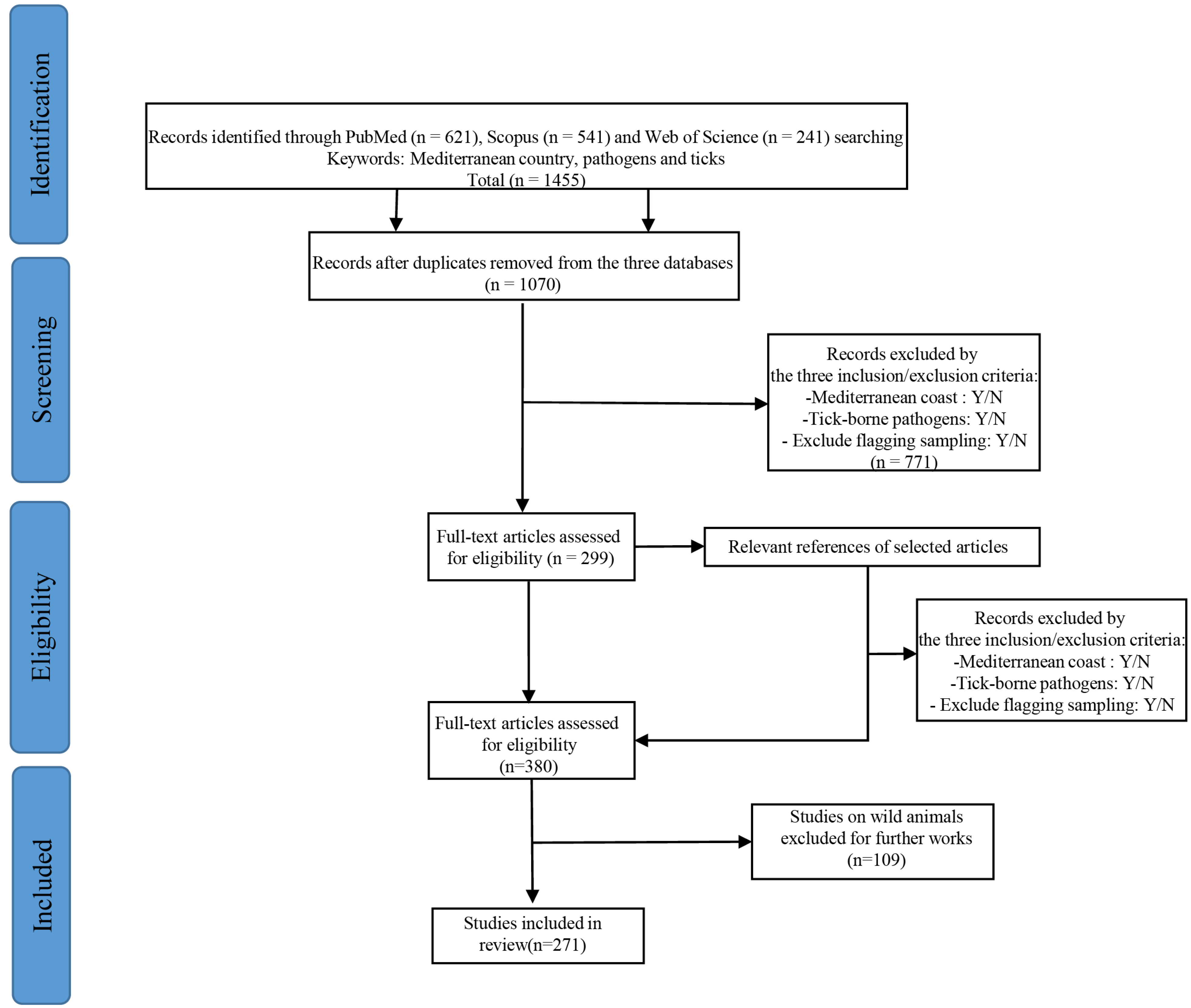
2. Materials and Methods
- Did the study include a country with a Mediterranean coast: Yes/No
- Did the study include tick-borne pathogens: Yes/No
- Did the study exclude ticks collected in vegetation: Yes/No
- Main characteristics of the studies: article ID, years, authors, analytical and statistical methodology
- Pathogen-related information: pathogens screened and detected, species, number of species, zoonotic status, host
- Tick-related information: species, type, number, stage
- Host-related information: groups, sedentary or migratory
- Area of interest: country, type of area, number of sampling sites
3. Results
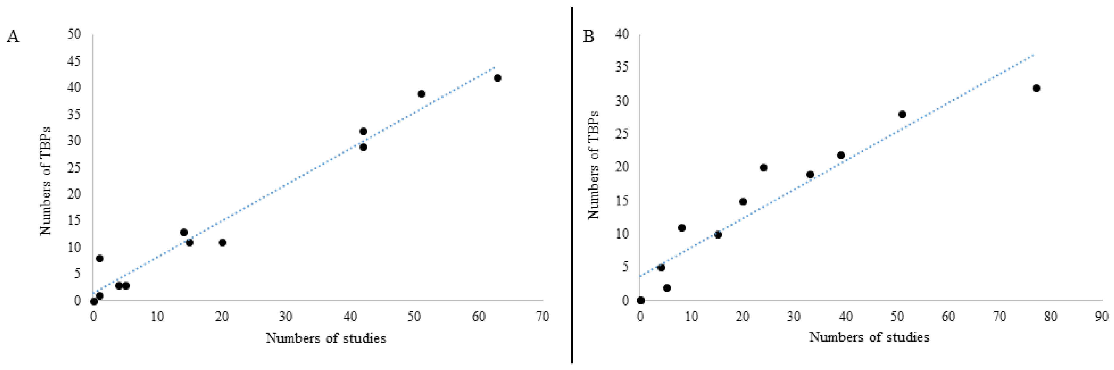
3.1. Bibliographic Analysis
3.2. Tick-Borne Pathogens in Countries of the Mediterranean Basin
3.2.1. Parasites
Nematoda
Apicomplexa
3.2.2. Bacteria
Anaplasma
Bartonella
Borrelia
Chlamydia/Chlamydophila
Coxiella
Ehrlichia
Neoehrlichia
Francisella
Leptospira
Mycoplasma
Rickettsia
3.2.3. Viruses
Capripoxvirus
Flavivirus
Orthonairovirus
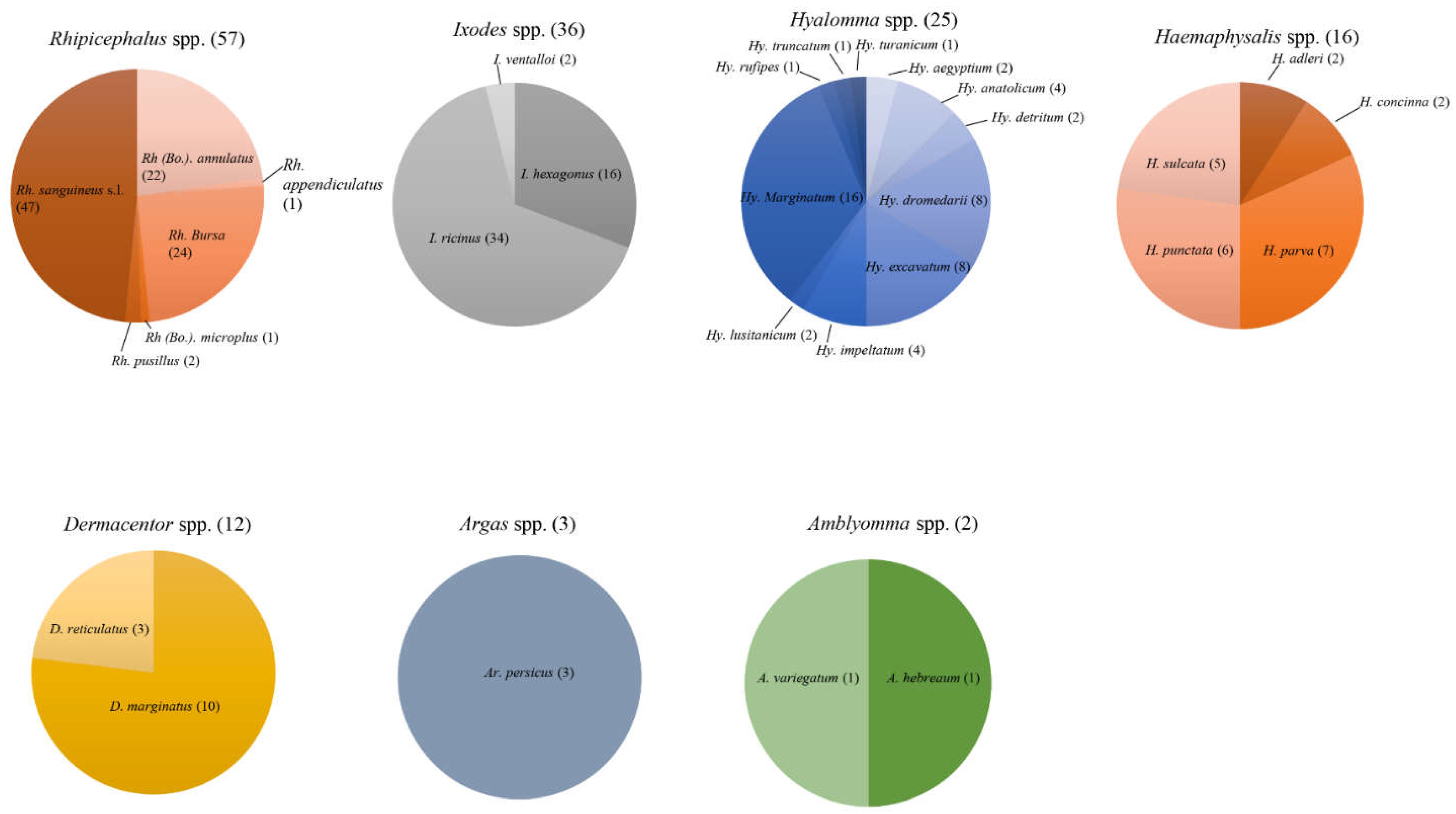
3.3. Ticks Positive for Tick-Borne Pathogens from Domestic Animals in the Mediterranean Basin
3.3.1. Ixodidae
Genus Rhipicephalus
Genus Ixodes
Genus Hyalomma
Genus Haemaphysalis
Genus Dermacentor
Genus Amblyomma
3.3.2. Argasidae
3.4. Domestic Animal Hosts of Both Positive Ticks and TBPs in the Countries of the Mediterranean Basin
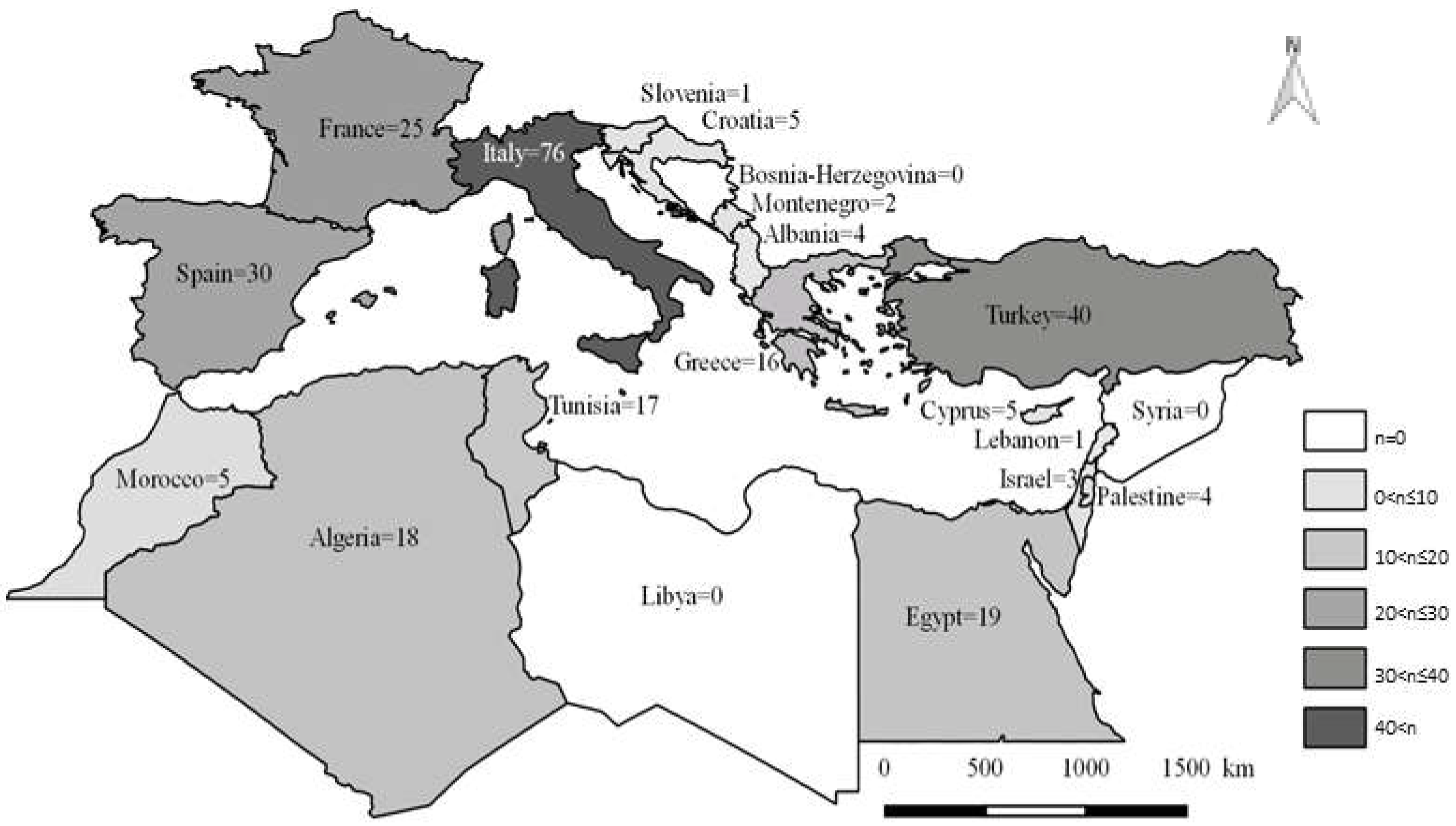
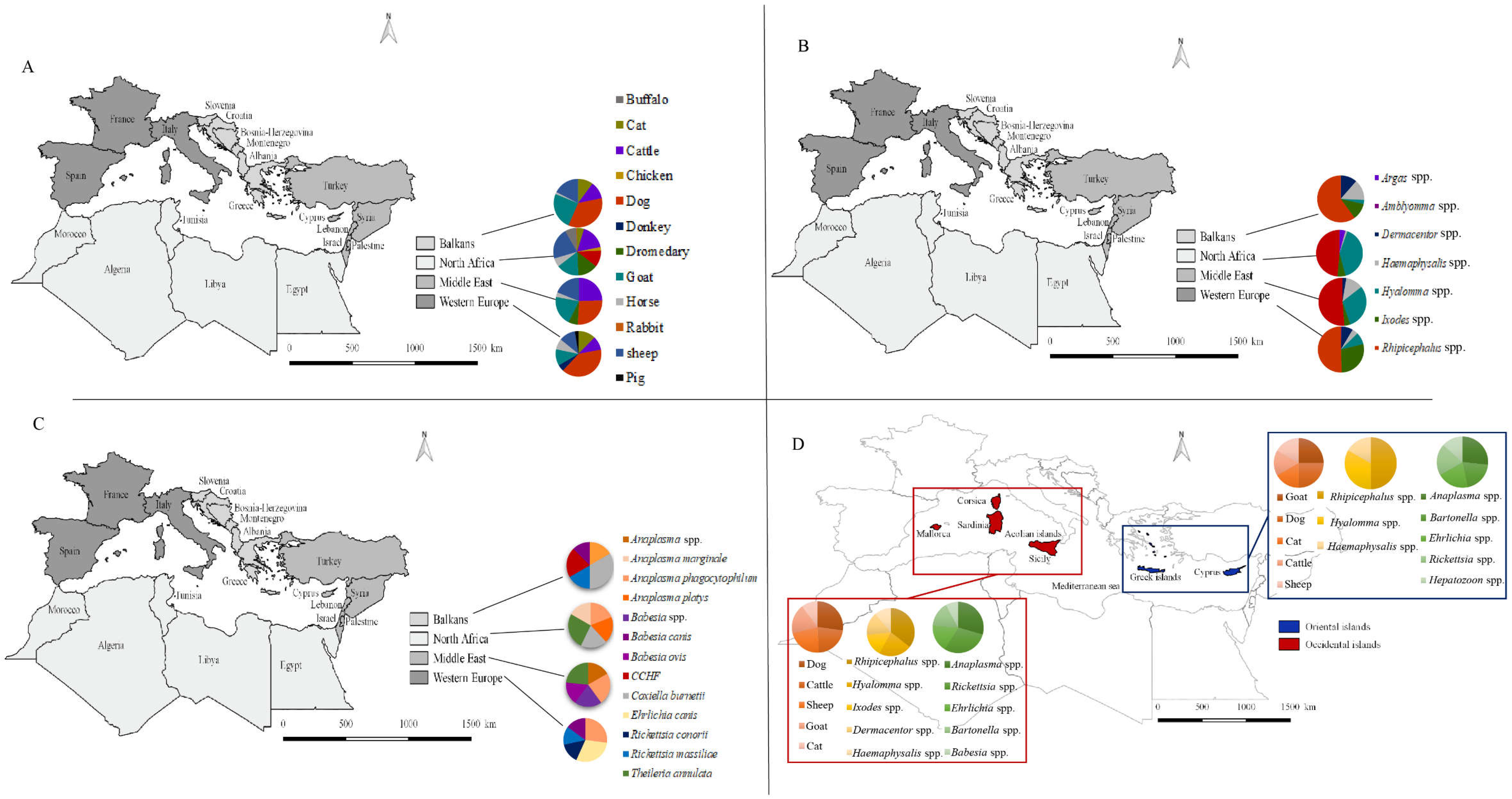
3.5. Biogeography, Diversity and Distribution in the Mediterranean Basin
3.5.1. Overall Analysis of the Four Main Regions in the Mediterranean Basin
Western Europe
North Africa
The Middle East
The Balkans
3.5.2. Focus on Insular Tick-Borne Pathogens in Domestic Animals and Their Ticks
4. Conclusions
Supplementary Materials
Author Contributions
Funding
Institutional Review Board Statement
Informed Consent Statement
Data Availability Statement
Conflicts of Interest
References
- Nicholson, W.L.; Sonenshine, D.E.; Noden, B.H.; Brown, R.N. Chapter 27—Ticks (Ixodida). In Medical and Veterinary Entomology, 3rd ed.; Mullen, G.R., Durden, L.A., Eds.; Academic Press: Cambridge, MA, USA, 2019; pp. 603–672. [Google Scholar] [CrossRef]
- Anderson, J.F.; Magnarelli, L.A. Biology of Ticks. Infect. Dis. Clin. N. Am. 2008, 22, 195–215. [Google Scholar] [CrossRef] [PubMed]
- Bellekom, B.; Hackett, T.D.; Lewis, O.T. A Network Perspective on the Vectoring of Human Disease. Trends Parasitol. 2021, 37, 391–400. [Google Scholar] [CrossRef] [PubMed]
- CDC. Zoonotic Diseases|One Health|CDC. Published 19 February 2020. Available online: https://www.cdc.gov/onehealth/basics/zoonotic-diseases.html (accessed on 11 February 2021).
- Rosenberg, R.; Ben Beard, C. Vector-borne Infections. Emerg. Infect. Dis. 2011, 17, 769–770. [Google Scholar] [CrossRef] [PubMed]
- Chomel, B. Lyme disease. Rev. Sci. Tech. OIE 2015, 34, 569–576. [Google Scholar] [CrossRef]
- WHO. Vector-Borne Diseases. Published 2 March 2020. Available online: https://www.who.int/news-room/fact-sheets/detail/vector-borne-diseases (accessed on 10 February 2020).
- Ahantarig, A.; Trinachartvanit, W.; Baimai, V.; Grubhoffer, L. Hard ticks and their bacterial endosymbionts (or would be pathogens). Folia Microbiol. 2013, 58, 419–428. [Google Scholar] [CrossRef]
- Parola, P.; Raoult, D. Ticks and tickborne bacterial diseases in humans: An emerging infectious threat. Clin. Infect. Dis. 2001, 32, 897–928. [Google Scholar] [CrossRef]
- Beati, L.; Klompen, H. Phylogeography of Ticks (Acari: Ixodida). Annu. Rev. Entomol. 2019, 64, 379–397. [Google Scholar] [CrossRef]
- Eisen, L. Vector competence studies with hard ticks and Borrelia burgdorferi sensu lato spirochetes: A review. Ticks Tick-Borne Dis. 2020, 11, 101359. [Google Scholar] [CrossRef]
- WHO. Crimean-Congo Haemorrhagic Fever. Published 31 January 2013. Available online: https://www.who.int/news-room/fact-sheets/detail/crimean-congo-haemorrhagic-fever (accessed on 11 February 2020).
- Perveen, N.; Muzaffar, S.; Al-Deeb, M. Ticks and Tick-Borne Diseases of Livestock in the Middle East and North Africa: A Review. Insects 2021, 12, 83. [Google Scholar] [CrossRef]
- Hildebrandt, A.; Zintl, A.; Montero, E.; Hunfeld, K.P.; Gray, J. Human Babesiosis in Europe. Pathogens 2021, 10, 1165. [Google Scholar] [CrossRef]
- Giannakopoulos, C.; Bindi, M.; Moriondo, M.; LeSager, P.; Tin, T. Climate Change Impacts in the Mediterranean Resulting from a 2 °C Global Temperature Rise; WWF: Gland, Switzerland, 2005; Volume 1. [Google Scholar]
- Ochoa-Hueso, R.; Munzi, S.; Alonso, R.; Arróniz-Crespo, M.; Avila, A.; Beremjo, V.; Bobbink, R.; Branquinho, C.; Concostrina-Zubiri, L.; Cruz, C.; et al. Ecological impacts of atmospheric pollution and interactions with climate change in terrestrial ecosystems of the Mediterranean Basin: Current research and future directions. Environ. Pollut. 2017, 227, 194–206. [Google Scholar] [CrossRef] [PubMed] [Green Version]
- Al-Abri, S.S.; Abaidani, I.A.; Fazlalipour, M.; Mostafavi, E.; Leblebicioglu, H.; Pshenichnayah, N.; Memish, Z.A.; Hewson, R.; Petersen, E.; Mala, P.; et al. Current status of Crimean-Congo haemorrhagic fever in the World Health Organization Eastern Mediterranean Region: Issues, challenges, and future directions. Int. J. Infect. Dis. 2017, 58, 82–89. [Google Scholar] [CrossRef] [PubMed] [Green Version]
- Hasle, G.; Bjune, G.; Edvardsen, E.; Jakobsen, C.; Linnehol, B.; Røer, J.; Mehl, R.; Røed, K.; Perdersen, J. Transport of Ticks by Migratory Passerine Birds to Norway. J. Parasitol. 2009, 95, 1342–1351. [Google Scholar] [CrossRef] [PubMed]
- Estrada-Peña, A.; De La Fuente, J.; Latapia, T.; Ortega, C. The Impact of Climate Trends on a Tick Affecting Public Health: A Retrospective Modeling Approach for Hyalomma marginatum (Ixodidae). PLoS ONE 2015, 10, e0125760. [Google Scholar] [CrossRef]
- Gray, J.; Dantas-Torres, F.; Estrada-Peña, A.; Levin, M. Systematics and ecology of the brown dog tick, Rhipicephalus sanguineus. Ticks Tick-Borne Dis. 2013, 4, 171–180. [Google Scholar] [CrossRef]
- Moher, D.; Shamseer, L.; Clarke, M.; Ghersi, D.; Liberati, A.; Petticrew, M.; Shekelle, P.; Stewart, L.A.; Prisma-P Group. Preferred reporting items for systematic review and meta-analysis protocols (PRISMA-P) 2015 statement. Syst. Rev. 2015, 4, 1. [Google Scholar] [CrossRef] [Green Version]
- Latrofa, M.S.; Angelou, A.; Giannelli, A.; Annoscia, G.; Ravagnan, S.; Dantas-Torres, F.; Capelli, G.; Halos, L.; Beugnet, F.; Papadopoulos, E.; et al. Ticks and associated pathogens in dogs from Greece. Parasites Vectors 2017, 10, 301. [Google Scholar] [CrossRef] [Green Version]
- Ramos, R.A.N.; Giannelli, A.; Lia, R.P.; Branti, E.; Tarallo, V.D.; Breitshwerdt, E.B.; Dantas-Torres, F.; Stanneck, D.; Otranto, D. Incidence of Cercopithifilaria bainae in dogs and probability of co-infection with other tick-borne pathogens. PLoS ONE 2014, 9, e88198. [Google Scholar] [CrossRef] [Green Version]
- Aktas, M.; Altay, K.; Ozubek, S.; Dumanli, N. A survey of ixodid ticks feeding on cattle and prevalence of tick-borne pathogens in the Black Sea region of Turkey. Vet. Parasitol. 2012, 187, 567–571. [Google Scholar] [CrossRef]
- Aktas, M.; Altay, K.; Dumanli, N. Determination of prevalence and risk factors for infection with Babesia ovis in small ruminants from Turkey by polymerase chain reaction. Parasitol. Res. 2007, 100, 797–802. [Google Scholar] [CrossRef]
- Aktas, M.; Ozubek, S. A molecular survey of hemoplasmas in domestic dogs from Turkey. Vet. Microbiol. 2018, 221, 94–97. [Google Scholar] [CrossRef] [PubMed]
- Aktas, M.; Ozubek, S. A survey of canine haemoprotozoan parasites from Turkey, including molecular evidence of an unnamed Babesia. Comp. Immunol. Microbiol. Infect. Dis. 2017, 52, 36–42. [Google Scholar] [CrossRef] [PubMed]
- Al-Hosary, A.; Ahmed, L.; Ahmed, J.; Nijhof, A.; Clausen, P.H. Epidemiological study on tropical theileriosis (Theileria annulata infection) in the Egyptian Oases with special reference to the molecular characterization of Theileria spp. Ticks Tick Borne Dis. 2018, 9, 1489–1493. [Google Scholar] [CrossRef] [PubMed]
- Cassini, R.; Marcer, F.; di Regalbono, A.F.; Cancrini, G.; Gabrielli, S.; Moretti, A.; Galuppi, R.; Tampieri, M.P.; Pietrobelli, M. New insights into the epidemiology of bovine piroplasmoses in Italy. Vet. Parasitol. 2012, 184, 77–82. [Google Scholar] [CrossRef] [PubMed]
- Ceci, L.; Iarussi, F.; Greco, B.; Lacinio, R.; Fornelli, S.; Carelli, G. Retrospective study of hemoparasites in cattle in southern Italy by reverse line blot hybridization. J. Vet. Med. Sci. 2014, 76, 869–875. [Google Scholar] [CrossRef] [PubMed] [Green Version]
- Guven, E.; Avcioglu, H.; Cengiz, S.; Hayirli, A. Vector-Borne Pathogens in Stray Dogs in Northeastern Turkey. Vector-Borne Zoonotic Dis. 2017, 17, 610–617. [Google Scholar] [CrossRef]
- Torina, A.; Vicente, J.; Alongi, A.; Scimence, S.; Turlá, R.; Nicosia, S.; Marco, V.D.; Caracappa, S.; De la Fuente, J. Observed Prevalence of Tick-borne Pathogens in Domestic Animals in Sicily, Italy during 2003–2005. Zoonoses Public Health. 2007, 54, 8–15. [Google Scholar] [CrossRef]
- Ziam, H.; Kernif, T.; Saidani, K.; Kelanemer, R.; Hammaz, Z.; Geysen, D. Bovine piroplasmosis-anaplasmosis and clinical signs of tropical theileriosis in the plains of Djurdjura (north Algeria). Vet. Med. Sci. 2020, 6, 720–729. [Google Scholar] [CrossRef]
- Açici, M.; Bölükbaş, C.S.; Pekmezcï, G.Z.; Gürler, A.T.; Umur, Ş.; Karaer, K.Z.; Çakmam, A.; Nalbantoğlu, A.S.; Nisbet, C. Seroepidemiological survey of bovine tick-borne infections in the Black Sea Region of Turkey. Turk. J. Vet. Anim. Sci. 2016, 40, 170–174. [Google Scholar] [CrossRef]
- Al-Hosary, A.; Răileanu, C.; Tauchmann, O.; Fischer, S.; Nijhof, A.M.; Silaghi, C. Epidemiology and genotyping of Anaplasma marginale and co-infection with piroplasms and other Anaplasmataceae in cattle and buffaloes from Egypt. Parasites Vectors 2020, 13, 495. [Google Scholar] [CrossRef]
- Altay, K.; Aktas, M.; Dumanli, N. Detection of Babesia ovis by PCR in Rhipicephalus bursa collected from naturally infested sheep and goats. Res. Vet. Sci. 2008, 85, 116–119. [Google Scholar] [CrossRef]
- Aydin, M.F.; Aktas, M.; Dumanli, N. Molecular identification of Theileria and Babesia in ticks collected from sheep and goats in the Black Sea region of Turkey. Parasitol. Res. 2015, 114, 65–69. [Google Scholar] [CrossRef] [PubMed]
- Chisu, V.; Alberti, A.; Zobba, R.; Foxi, C.; Masala, G. Molecular characterization and phylogenetic analysis of Babesia and Theileria spp. in ticks from domestic and wild hosts in Sardinia. Acta Trop. 2019, 196, 60–65. [Google Scholar] [CrossRef] [PubMed]
- El-Ashker, M.; Hotzel, H.; Gwida, M.; El-Beskawy, M.; Silaghi, C.; Tomaso, H. Molecular biological identification of Babesia, Theileria, and Anaplasma species in cattle in Egypt using PCR assays, gene sequence analysis and a novel DNA microarray. Vet. Parasitol. 2015, 207, 329–334. [Google Scholar] [CrossRef] [PubMed] [Green Version]
- Elsify, A.; Sivakumar, T.; Nayel, M.; Salama, A.; Elkhtam, A.; Rizk, M.; Mosaab, O.; Sultan, K.; Elsayed, S.; Igarashi, I.; et al. An epidemiological survey of bovine Babesia and Theileria parasites in cattle, buffaloes, and sheep in Egypt. Parasitol. Int. 2015, 64, 79–85. [Google Scholar] [CrossRef] [Green Version]
- Iori, A.; Gabrielli, S.; Calderini, P.; Moretti, A.; Pietrobelli, M.; Tampieri, M.P.; Galuppi, R.; Cancrini, G. Tick reservoirs for piroplasms in central and northern Italy. Vet. Parasitol. 2010, 170, 291–296. [Google Scholar] [CrossRef]
- Toma, L.; Di Luca, M.; Mancini, F.; Severini, F.; Mariano, C.; Nicolai, G.; Laghezza Masci, V.; Ciervo, A.; Fausto, A.M.; Cacciò, S.M. Molecular characterization of Babesia and Theileria species in ticks collected in the outskirt of Monte Romano, Lazio Region, Central Italy. Ann Ist. Super Sanita 2017, 53, 30–34. [Google Scholar] [CrossRef]
- Cringoli, G.; Otranto, D.; Testini, G.; Buono, V.; Di Giulio, G.; Traversa, D.; Lia, R.; Rinaldi, L.; Veneziano, V.; Puccini, V. Epidemiology of bovine tick-borne diseases in southern Italy. Vet. Res. 2002, 33, 421–428. [Google Scholar] [CrossRef] [Green Version]
- Sadeddine, R.; Diarra, A.Z.; Laroche, M.; Mediannikov, O.; Righi, S.; Benakhla, A.; Dahmana, H.; Raoult, D.; Parola, P. Molecular identification of protozoal and bacterial organisms in domestic animals and their infesting ticks from north-eastern Algeria. Ticks Tick-Borne Dis. 2020, 11, 101330. [Google Scholar] [CrossRef]
- Beck, R.; Vojta, L.; Mrljak, V.; Marinculić, A.; Beck, A.; Živičnjak, T.; Cacciò, S.M. Diversity of Babesia and Theileria species in symptomatic and asymptomatic dogs in Croatia. Int. J. Parasitol. 2009, 39, 843–848. [Google Scholar] [CrossRef]
- Leblond, A.; Pradier, S.; Pitel, P.H.; Fortier, G.; Chadoeuf, J.; Savatier, P. An epidemiological survey of equine anaplasmosis (Anaplasma phagocytophilum) in Southern France. Rev. Sci. Tech. OIE 2006, 24, 899–908. [Google Scholar] [CrossRef]
- Moretti, A.; Mangili, V.; Salvatori, R.; Maresca, C.; Scoccia, E.; Torina, A.; Moretta, I.; Gabrielli, S.; Tampieri, M.P.; Pietrobelli, M. Prevalence and diagnosis of Babesia and Theileria infections in horses in Italy: A preliminary study. Vet. J. 2010, 184, 346–350. [Google Scholar] [CrossRef] [PubMed]
- Camacho, A.T.; Pallas, E.; Gestal, J.J.; Guitiàn, F.J.; Olmeda, A.S.; Telford, S.R.; Speilman, A. Ixodes hexagonus is the main candidate as vector of Theileria annae in northwest Spain. Vet. Parasitol. 2003, 112, 157–163. [Google Scholar] [CrossRef]
- Cassini, R.; Zanutto, S.; di Regalbono, A.F.; Gabrielli, S.; Calderini, P.; Moretti, A.; Tampieri, M.P.; Pietrobelli, M. Canine piroplasmosis in Italy: Epidemiological aspects in vertebrate and invertebrate hosts. Vet. Parasitol. 2009, 165, 30–35. [Google Scholar] [CrossRef] [PubMed]
- Criado-Fornelio, A.; Buling, A.; Pingret, J.L.; Boucraut-Baralon, C.; Alongi, A.; Agnone, A.; Torina, A. Hemoprotozoa of domestic animals in France: Prevalence and molecular characterization. Vet. Parasitol. 2009, 159, 73–76. [Google Scholar] [CrossRef] [PubMed] [Green Version]
- Estrada-Peña, A.; Roura, X.; Sainz, A.; Miró, G.; Solano-Gallego, L. Species of ticks and carried pathogens in owned dogs in Spain: Results of a one-year national survey. Ticks Tick-Borne Dis. 2017, 8, 443–452. [Google Scholar] [CrossRef] [PubMed]
- Guo, H.; Sevinc, F.; Ceylan, O.; Sevinc, M.; Ince, E.; Gao, Y.; Moumouni, P.F.; Liu, M.; Efstratiou, A.; Wang, G.; et al. A PCR survey of vector-borne pathogens in different dog populations from Turkey. Acta Parasitol. 2017, 62, 533–540. [Google Scholar] [CrossRef]
- Mrljak, V.; Kuleš, J.; Mihaljević, Ž.; Torti, M.; Gotić, J.; Crnogaj, M.; Živičnjak, T.; Mayer, I.; Šmit, I.; Bhide, M.; et al. Prevalence and Geographic Distribution of Vector-Borne Pathogens in Apparently Healthy Dogs in Croatia. Vector-Borne Zoonotic Dis. 2017, 17, 398–408. [Google Scholar] [CrossRef]
- Olivieri, E.; Zanzani, S.A.; Latrofa, M.S.; Lia, R.P.; Dantas-Torres, F.; Otranto, D.; Manfredi, M.T. The southernmost foci of Dermacentor reticulatus in Italy and associated Babesia canis infection in dogs. Parasites Vectors 2016, 9, 213. [Google Scholar] [CrossRef] [Green Version]
- Pennisi, M.G.; Caprì, A.; Solano-Gallego, L.; Lombardo, G.; Torina, A.; Masucci, M. Prevalence of antibodies against Rickettsia conorii, Babesia canis, Ehrlichia canis, and Anaplasma phagocytophilum antigens in dogs from the Stretto di Messina area (Italy). Ticks Tick-Borne Dis. 2012, 3, 315–318. [Google Scholar] [CrossRef]
- René-Martellet, M.; Moro, C.V.; Chêne, J.; Bourdoiseau, G.; Chabanne, L.; Mavingui, P. Update on epidemiology of canine babesiosis in Southern France. BMC Vet. Res. 2015, 11, 223. [Google Scholar] [CrossRef] [PubMed]
- Solano-Gallego, L.; Trotta, M.; Carli, E.; Carcy, B.; Caldin, M.; Furlanello, T. Babesia canis canis and Babesia canis vogeli clinicopathological findings and DNA detection by means of PCR-RFLP in blood from Italian dogs suspected of tick-borne disease. Vet. Parasitol. 2008, 157, 211–221. [Google Scholar] [CrossRef] [PubMed]
- Trotta, M.; Nicetto, M.; Fogliazza, A.; Montarsi, F.; Caldin, M.; Furlanello, T.; Solano-Gallego, L. Detection of Leishmania infantum, Babesia canis, and rickettsiae in ticks removed from dogs living in Italy. Ticks Tick-Borne Dis. 2012, 3, 294–297. [Google Scholar] [CrossRef]
- Veneziano, V.; Piantedosi, D.; Ferrari, N.; Neola, B.; Santoro, M.; Pacifico, L.; Sgroi, G.; D’Alessio, N.; Panico, T.; Leutenegger, C.M.; et al. Distribution and risk factors associated with Babesia spp. infection in hunting dogs from Southern Italy. Ticks Tick-Borne Dis. 2018, 9, 1459–1463. [Google Scholar] [CrossRef] [PubMed]
- Zanet, S.; Battisti, E.; Pepe, P.; Cuica, L.; Colombo, L.; Trisciuoglio, A.; Ferroglio, E.; Cringoli, G.; Rinaldi, L.; Maurelli, M.P. Tick-borne pathogens in Ixodidae ticks collected from privately-owned dogs in Italy: A country-wide molecular survey. BMC Vet. Res. 2020, 16, 46. [Google Scholar] [CrossRef] [Green Version]
- Agoulon, A.; Malandrin, L.; Lepigeon, F.; Vénisse, M.; Bonnet, S.; Becker, C.A.M.; Hoch, T.; Bastian, S.; Plantard, O.; Beaudeau, F. A Vegetation Index qualifying pasture edges is related to Ixodes ricinus density and to Babesia divergens seroprevalence in dairy cattle herds. Vet. Parasitol. 2012, 185, 101–109. [Google Scholar] [CrossRef]
- Devos, J.; Geysen, D. Epidemiological study of the prevalence of Babesia divergens in a veterinary practice in the mid-east of France. Vet. Parasitol. 2004, 125, 237–249. [Google Scholar] [CrossRef] [PubMed]
- Passamonti, F.; Fabrizia, V.; Katia, C.; Giacomo, C.; Luisa, M.M.; Daniela, P.F.; Andrea, V.S.; Mauro, C. Anaplasma phagocytophilum in horses and ticks: A preliminary survey of Central Italy. Comp. Immunol. Microbiol. Infect. Dis. 2010, 33, 73–83. [Google Scholar] [CrossRef]
- Spada, E.; Proverbio, D.; Galluzzo, P.; Perego, R.; De Giorgi, G.B.; Roggero, N.; Caracappa, S. Frequency of Piroplasms Babesia microti and Cytauxzoon felis in Stray Cats from Northern Italy. BioMed Res. Int. 2014, 2014, 943754. [Google Scholar] [CrossRef] [Green Version]
- Nagore, D.; García-Sanmartín, J.; García-Pérez, A.L.; Juste, R.A.; Hurtado, A. Identification, genetic diversity and prevalence of Theileria and Babesia species in a sheep population from Northern Spain. Int. J. Parasitol. 2004, 34, 1059–1067. [Google Scholar] [CrossRef]
- Aouadi, A.; Leulmi, H.; Boucheikhchoukh, M.; Benakhla, A.; Raoult, D.; Parola, P. Molecular evidence of tick-borne hemoprotozoan-parasites (Theileria ovis and Babesia ovis) and bacteria in ticks and blood from small ruminants in Northern Algeria. Comp. Immunol. Microbiol. Infect. Dis. 2017, 50, 34–39. [Google Scholar] [CrossRef] [PubMed]
- Azmi, K.; Ereqat, S.; Nasereddin, A.; Al-Jawabreh, A.; Baneth, G.; Abdeen, Z. Molecular detection of Theileria, Babesia, and Hepatozoon spp. in ixodid ticks from Palestine. Ticks Tick-Borne Dis. 2016, 7, 734–741. [Google Scholar] [CrossRef] [PubMed]
- Bilgic, H.B.; Bakırcı, S.; Kose, O.; Unlu, A.H.; Hacilarlioglu, S.; Eren, H.; Weir, W.; Karagenc, T. Prevalence of tick-borne haemoparasites in small ruminants in Turkey and diagnostic sensitivity of single-PCR and RLB. Parasites Vectors 2017, 10, 211. [Google Scholar] [CrossRef] [PubMed]
- Attipa, C.; Papasouliotis, K.; Solano-Gallego, L.; Baneth, G.; Nachum-Biala, Y.; Sarvani, E.; Knowles, T.G.; Mengi, S.; Morris, D.; Helps, C.; et al. Prevalence study and risk factor analysis of selected bacterial, protozoal and viral, including vector-borne, pathogens in cats from Cyprus. Parasites Vectors 2017, 10, 130. [Google Scholar] [CrossRef] [Green Version]
- Bilgic, H.B.; Pekel, G.K.; Hosgor, M.; Karagenc, T. A Retrospective Epidemiological Study: The Prevalence of Ehrlichia canis and Babesia vogeli in Dogs in the Aegean Region of Turkey. Acta Vet. 2019, 69, 164–176. [Google Scholar] [CrossRef] [Green Version]
- Dyachenko, V.; Pantchev, N.; Balzer, H.J.; Meyersen, A.; Straubinger, R.K. First case of Anaplasma platys infection in a dog from Croatia. Parasites Vectors 2012, 5, 49. [Google Scholar] [CrossRef] [Green Version]
- Otranto, D.; Testini, G.; Dantas-Torres, F.; Latrofa, M.S.; Dniz, P.P.V.; De Caprariis, D.; Lia, R.P.; Mencke, N.; Stanneck, D.; Capelli, G.; et al. Diagnosis of canine vector-borne diseases in young dogs: A longitudinal study. J. Clin. Microbiol. 2010, 48, 3316–3324. [Google Scholar] [CrossRef] [Green Version]
- Pennisi, M.G.; Persichetti, M.F.; Serrano, L.; Altet, L.; Reale, S.; Gulotta, L.; Solano-Gallego, L. Ticks and associated pathogens collected from cats in Sicily and Calabria (Italy). Parasites Vectors 2015, 8, 512. [Google Scholar] [CrossRef] [Green Version]
- Ebani, V.V.; Bertelloni, F.; Turchi, B.; Filogari, D.; Cerri, D. Molecular survey of tick-borne pathogens in Ixodid ticks collected from hunted wild animals in Tuscany, Italy. Asian Pac. J. Trop. Med. 2015, 8, 714–717. [Google Scholar] [CrossRef] [Green Version]
- Giannelli, A.; Latrofa, M.S.; Nachum-Biala, Y.; Hodžić, A.; Greco, G.; Attanasi, A.; Annoscia, G.; Otranto, D.; Baneth, G. Three different Hepatozoon species in domestic cats from southern Italy. Ticks Tick-borne Dis. 2017, 8, 721–724. [Google Scholar] [CrossRef]
- Ortuño, A.; Castellà, J.; Criado-Fornelio, A.; Buling, A.; Barba-Carretero, J. Molecular detection of a Hepatozoon species in stray cats from a feline colony in North-eastern Spain. Vet. J. 2008, 177, 134–135. [Google Scholar] [CrossRef] [PubMed]
- Pacifico, L.; Braff, J.; Buono, F.; Beall, M.; Neaola, B.; Buch, J.; Sgroi, G.; Piantedosi, D.; Santoror, M.; Tyrell, P.; et al. Hepatozoon canis in hunting dogs from Southern Italy: Distribution and risk factors. Parasitol. Res. 2020, 119, 3023–3031. [Google Scholar] [CrossRef] [PubMed]
- Ebani, V.V.; Nardoni, S.; Fognani, G.; Bertelloni, F.; Vasta, V.; Papini, R.A.; Verin, R.; Poli, A.; Mancianti, F. Molecular detection of vector-borne bacteria and protozoa in healthy hunting dogs from Central Italy. Asian Pac. J. Trop. Biomed. 2015, 5, 108–112. [Google Scholar] [CrossRef] [Green Version]
- Vojta, L.; Mrljak, V.; Ćurković, S.; Živičnjak, T.; Marinculić, A.; Beck, R. Molecular epizootiology of canine hepatozoonosis in Croatia. Int. J. Parasitol. 2009, 39, 1129–1136. [Google Scholar] [CrossRef] [PubMed]
- Aktas, M.; Özübek, S.; Ipek, D.N.S. Molecular investigations of Hepatozoon species in dogs and developmental stages of Rhipicephalus sanguineus. Parasitol. Res. 2013, 112, 2381–2385. [Google Scholar] [CrossRef] [PubMed]
- Aktas, M.; Altay, K.; Dumanli, N. PCR-based detection of Theileria ovis in Rhipicephalus bursa adult ticks. Vet. Parasitol. 2006, 140, 259–263. [Google Scholar] [CrossRef]
- Aktas, M.; Dumanli, N.; Angin, M. Cattle infestation by Hyalomma ticks and prevalence of Theileria in Hyalomma species in the east of Turkey. Vet. Parasitol. 2004, 119, 1–8. [Google Scholar] [CrossRef]
- Hassan, M.; Gabr, H.; Abdel-Shafy, S.; Hammad, K.; Mokhtar, M. Prevalence of tick-vectors of Theileria annulata infesting the one humped camels in Giza, Egypt. J. Egypt. Soc. Parasitol. 2017, 47, 425–432. [Google Scholar] [CrossRef]
- Ziam, H.; Kelanamer, R.; Aissi, M.; Ababou, A.; Berkvens, D.; Geysen, D. Prevalence of bovine theileriosis in North Central region of Algeria by real-time polymerase chain reaction with a note on its distribution. Trop. Anim. Heal. Prod. 2015, 47, 787–796. [Google Scholar] [CrossRef]
- Tabar, M.D.; Altet, L.; Francino, O.; Sánchez, A.; Ferrer, L.; Roura, X. Vector-borne infections in cats: Molecular study in Barcelona area (Spain). Vet. Parasitol. 2008, 151, 332–336. [Google Scholar] [CrossRef]
- Giménez Pardo, C.; Casado, N.; Criado-Fornelio, A.; de Miguel, F.Á.; Dominguez-Peñafiel, G. A molecular survey of Piroplasmida and Hepatozoon isolated from domestic and wild animals in Burgos (Northern Spain). Vet. Parasitol. 2009, 162, 147–150. [Google Scholar] [CrossRef] [PubMed]
- Aktas, M.; Dumanli, N.; Çetinkaya, B.; Çakmak, A. Field evaluation of PCR in detecting Theileria annulata infection in cattle in eastern Turkey. Vet. Rec. 2002, 150, 548–549. [Google Scholar] [CrossRef] [PubMed]
- Al-Hosary, A.A.T.; Ahmed, L.; Seitzer, U. First Report of Molecular Identification and Characterization of Theileria spp. from Water Buffaloes (Bubalus bubalis) in Egypt. Adv. Anim. Vet. Sci. 2015, 3, 629–633. [Google Scholar] [CrossRef]
- AL-Hosary, A.; Abdel-Rady, A.; Ahmed, L.; Al-Hosary, A. Using Polymerase chain reaction (PCR) for Diagnosis of Bovine Theileriosis in Upper Egypt. IJAVMS 2010, 4, 67–74. [Google Scholar] [CrossRef]
- Dumanli, N.; Aktas, M.; Cetinkaya, B.; Cakmak, A.; Koroglu, E.; Saki, C.E.; Erdogmus, Z.; Nalbantoglu, S.; Ongor, H.; Simşek, S.; et al. Prevalence and distribution of tropical theileriosis in eastern Turkey. Vet. Parasitol. 2005, 127, 9–15. [Google Scholar] [CrossRef]
- Gargano, V.; Blanda, V.; Gambino, D.; La Russa, F.; Di Cataldo, S.; Gentile, A.; Schirò, G.; Torina, A.; Millàn, J.; Vicari, D. Serological Survey and Molecular Characterization of Theileria annulata in Sicilian Cattle. Pathogens 2021, 10, 101. [Google Scholar] [CrossRef]
- Inci, A.; Ica, A.; Yildirim, A.; Vatansever, Z.; Cakmak, A.; Albasan, H.; Çam, Y.; Atasever, A.; Duzlu, O. Epidemiology of tropical theileriosis in the Cappadocia region. Turk J. Vet. Anim. Sci. 2008, 32, 57–64. [Google Scholar]
- Sallemi, S.; Rjeibi, M.R.; Rouatbi, M.; Amairia, S.; Ben Said, M.; Khamassi Khbou, M.; Gharbi, M. Molecular prevalence and phylogenetic analysis of Theileria annulata and Trypanosoma evansi in cattle in Northern Tunisia. Vet. Med. Sci. 2017, 4, 17–25. [Google Scholar] [CrossRef]
- Sayin, F.; Dinçer, S.; Karaer, Z.; Cakmak, A.; INci, A.; Yukari, B.A.; Eren, H.; Vatansever, Z.; Nalbantoğlu, S. Studies on the Epidemiology of Tropical Theileriosis (Theileria annulata Infection) in Cattle in Central Anatolia, Turkey. Trop. Anim. Health Prod. 2003, 35, 521–539. [Google Scholar] [CrossRef]
- Ziam, H.; Benaouf, H. Prevalence of blood parasites in cattle from Wilayates of Annaba and El Tarf East Algeria. Archs Inst. Pasteur. Tunis 2004, 81, 1–4. [Google Scholar]
- Laus, F.; Veronesi, F.; Passamonti, F.; Paggi, E.; Cerquetella, M.; Hyatt, D.; Tesei, B.; Fioretti, D.P. Prevalence of Tick Borne Pathogens in Horses from Italy. J. Vet. Med. Sci. 2013, 75, 715–720. [Google Scholar] [CrossRef] [PubMed] [Green Version]
- Altay, K.; Dumanli, N.; Aktas, M. A study on ovine tick-borne hemoprotozoan parasites (Theileria and Babesia) in the East Black Sea Region of Turkey. Parasitol. Res. 2012, 111, 149–153. [Google Scholar] [CrossRef] [PubMed]
- Aydın, M.F.; Özübek, S.; Aktaş, M. Molecular survey of Anaplasma and Ehrlichia species in cattle from Karaman of Turkey, including a novel tandem report of Anaplasma marginale msp1a gene. Ankara. Üniv. Vet. Fak. Derg. 2019, 66, 255–260. [Google Scholar] [CrossRef]
- Ayllón, T.; Diniz, P.P.V.P.; Breitschwerdt, E.B.; Villaescusa, A.; Rodríguez-Franco, F.; Sainz, A. Vector-Borne Diseases in Client-Owned and Stray Cats from Madrid, Spain. Vector-Borne Zoonotic Dis. 2011, 12, 143–150. [Google Scholar] [CrossRef] [PubMed]
- Belkahia, H.; Ben Said, M.; Ghribi, R.; Selmi, R.; Ben Asker, A.; Yahiaoui, M.; Bousrih, M.; Daaloul-Jedidi, M.; Messadi, L. Molecular detection, genotyping and phylogeny of Anaplasma spp. in Rhipicephalus ticks from Tunisia. Acta Trop. 2018, 191, 38–49. [Google Scholar] [CrossRef]
- Belkahia, H.; Said, M.B.; Sayahi, L.; Alberti, A.; Messadi, L. Detection of novel strains genetically related to Anaplasma platys in Tunisian one-humped camels (Camelus dromedarius). J. Infect. Dev. Ctries. 2015, 9, 1117–1125. [Google Scholar] [CrossRef] [Green Version]
- Boucheikhchoukh, M.; Laroche, M.; Aouadi, A.; Dib, L.; Benakhla, A.; Raoult, D.; Parola, P. MALDI-TOF MS identification of ticks of domestic and wild animals in Algeria and molecular detection of associated microorganisms. Comp. Immunol. Microbiol. Infect. Dis. 2018, 57, 39–49. [Google Scholar] [CrossRef]
- Dahmani, M.; Davoust, B.; Tahir, D.; Raoult, D.; Fenollar, F.; Mediannikov, O. Molecular investigation and phylogeny of Anaplasmataceae species infecting domestic animals and ticks in Corsica, France. Parasites Vectors 2017, 10, 302. [Google Scholar] [CrossRef] [Green Version]
- Dahmani, M.; Davoust, B.; Benterki, M.; Fenollar, F.; Raoult, D.; Mediannikov, O. Development of a new PCR-based assay to detect Anaplasmataceae and the first report of Anaplasma phagocytophilum and Anaplasma platys in cattle from Algeria. Comp. Immunol. Microbiol. Infect. Dis. 2015, 39, 39–45. [Google Scholar] [CrossRef]
- de la Fuente, J.; Torina, A.; Caracappa, S.; Tumino, G.; Furlà, R.; Almazàn, C.; Kocan, K.M. Serologic and molecular characterization of Anaplasma species infection in farm animals and ticks from Sicily. Vet. Parasitol. 2005, 133, 357–362. [Google Scholar] [CrossRef]
- Diakou, A.; Di Cesare, A.; Morelli, S.; Colombo, M.; Halos, L.; Simonato, G.; Tamvakis, A.; Beugnet, F.; Paoletti, B.; Traversa, D. Endoparasites and vector-borne pathogens in dogs from Greek islands: Pathogen distribution and zoonotic implications. PLOS Neglected Trop. Dis. 2019, 13, e0007003. [Google Scholar] [CrossRef] [PubMed] [Green Version]
- Díaz-Regañón, D.; Roura, X.; Suárez, M.L.; León, M.; Sainz, Á. Serological evaluation of selected vector-borne pathogens in owned dogs from northern Spain based on a multicenter study using a commercial test. Parasites Vectors 2020, 13, 301. [Google Scholar] [CrossRef] [PubMed]
- Elhamiani Khatat, S.; Daminet, S.; Kachani, M.; Leutenegger, C.M.; Duchateau, L.; El Amri, H.; Hing, M.; Azrib, R.; Sahibi, H. Anaplasma spp. in dogs and owners in north-western Morocco. Parasites Vectors 2017, 10, 202. [Google Scholar] [CrossRef] [PubMed] [Green Version]
- Lbacha, H.A.; Alali, S.; Zouagui, Z.; Mamoun, L.E.I.; Rhalem, A.; Petit, E.; Haddad, N.; Gandoin, C.; Gandoin, C.; Boulouis, H.-J.; et al. High Prevalence of Anaplasma spp. in Small Ruminants in Morocco. Transbound. Emerg. Dis. 2017, 64, 250–263. [Google Scholar] [CrossRef]
- Petruccelli, A.; Ferrara, G.; Iovane, G.; Schettini, R.; Ciarcia, R.; Caputo, V.; Pompamea, M.; Pagnini, U.; Montagnaro, S. Seroprevalence of Ehrlichia spp., Anaplasma spp., Borrelia burgdorferi sensu lato, and Dirofilaria immitis in Stray Dogs, from 2016 to 2019, in Southern Italy. Animals 2020, 11, 9. [Google Scholar] [CrossRef]
- Zaid, T.; Ereqat, S.; Nasereddin, A.; Al-Jawabreh, A.; Abdelkader, A.; Abdeen, Z. Molecular characterization of Anaplasma and Ehrlichia in ixodid ticks and reservoir hosts from Palestine: A pilot survey. Vet. Med. Sci. 2019, 5, 230–242. [Google Scholar] [CrossRef] [Green Version]
- Aktas, M.; Altay, K.; Dumanli, N. Molecular detection and identification of Anaplasma and Ehrlichia species in cattle from Turkey. Ticks Tick-Borne Dis. 2011, 2, 62–65. [Google Scholar] [CrossRef]
- Seng, P.; Sarih, M.; Socolovschi, C.; Boudebouch, N.; Hassar, M.; Paraola, P.; Raoult, D.; Brouqui, P. Detection of Anaplasmataceae in ticks collected in Morocco. Clin. Microbiol. Infect. 2009, 15, 86–87. [Google Scholar] [CrossRef] [Green Version]
- Elhariri, M.D.; Elhelw, R.A.; Hamza, D.A.; Soliman, D.E. Molecular detection of Anaplasma marginale in the egyptian water buffaloes (Bubalus bubalis) based on major surface protein 1. J. Egypt Soc. Parasitol. 2017, 47, 247–252. [Google Scholar] [CrossRef]
- Torina, A.; Alongi, A.; Naranjo, V.; Estrada-Peña, A.; Vincente, J.; Scimeca, S.; Marino, A.M.F.; Salina, F.; Caracappa, S.; de la Fuente, J. Prevalence and Genotypes of Anaplasma Species and Habitat Suitability for Ticks in a Mediterranean Ecosystem. Appl. Environ. Microbiol. 2008, 74, 7578–7584. [Google Scholar] [CrossRef] [Green Version]
- Aktas, M.; Altay, K.; Dumanli, N.; Kalkan, A. Molecular detection and identification of Ehrlichia and Anaplasma species in ixodid ticks. Parasitol Res. 2009, 104, 1243–1248. [Google Scholar] [CrossRef] [PubMed]
- Altay, K.; Dumanlı, N.; Aktas, M.; Ozubek, S. Survey of anaplasma infections in small ruminants from east part of Turkey. Kafkas Univ. Vet. Fak. Derg. 2014, 20, 1–4. [Google Scholar] [CrossRef]
- Cabezas-Cruz, A.; Gallois, M.; Fontugne, M.; Allain, E.; Denoual, M.; Moutailler, S.; Devillers, E.; Zientara, S.; Memmi, M.; Chauvin, A.; et al. Epidemiology and genetic diversity of Anaplasma ovis in goats in Corsica, France. Parasites Vectors 2019, 12, 3. [Google Scholar] [CrossRef] [PubMed] [Green Version]
- Chisu, V.; Foxi, C.; Mannu, R.; Satta, G.; Masala, G. A five-year survey of tick species and identification of tick-borne bacteria in Sardinia, Italy. Ticks Tick-Borne Dis. 2018, 9, 678–681. [Google Scholar] [CrossRef]
- Oter, K.; Cetinkaya, H.; Vurusaner, C.; Toparlak, M.; Ergunay, K. Molecular Detection and Typing Of Anaplasma Species In Small Ruminants In Thrace Region Of Turkey. Kafkas Univ. Vet. Fak. Derg. 2016, 22, 133–138. [Google Scholar] [CrossRef]
- Selmi, R.; Ben Said, M.; Dhibi, M.; Ben Yahia, H.; Abdelaali, H.; Messadi, L. Genetic diversity of groEL and msp4 sequences of Anaplasma ovis infecting camels from Tunisia. Parasitol. Int. 2020, 74, 101980. [Google Scholar] [CrossRef]
- Alberti, A.; Addis, M.F.; Sparagano, O.; Zobba, R.; Chessa, B.; Cubeddu, T.; Parpaglia, M.L.P.; Ardu, M.; Pittau, M. Anaplasma phagocytophilum, Sardinia, Italy. Emerg. Infect. Dis. 2005, 11, 1322–1324. [Google Scholar] [CrossRef]
- Çetinkaya, H.; Matur, E.; Akyazi, İ.; Ekiz, E.E.; Aydin, L.; Toparlak, M. Serological and molecular investigation of Ehrlichia spp. and Anaplasma spp. in ticks and blood of dogs, in the Thrace Region of Turkey. Ticks Tick-Borne Dis. 2016, 7, 706–714. [Google Scholar] [CrossRef]
- Chastagner, A.; Dugat, T.; Vourc’h, G.; Verheyden, H.; Legrand, L.; Bachy, V.; Chabanne, L.; Joncour, G.; Maillard, R.; Boulouis, H.-J.; et al. Multilocus sequence analysis of Anaplasma phagocytophilum reveals three distinct lineages with different host ranges in clinically ill French cattle. Vet. Res. 2014, 45, 114. [Google Scholar] [CrossRef]
- Ebani, V.; Cerri, D.; Fratini, F.; Ampola, M.; Andreani, E. Seroprevalence of Anaplasma phagocytophilum in domestic and wild animals from central Italy. New Microbiol. 2008, 31, 371–375. [Google Scholar]
- Ebani, V.V.; Guardone, L.; Marra, F.; Altomonte, I.; Nardoni, S.; Mancianti, F. Arthropod-Borne Pathogens in Stray Cats from Northern Italy: A Serological and Molecular Survey. Animals 2020, 10, 2334. [Google Scholar] [CrossRef] [PubMed]
- Ebani, V.V.; Bertelloni, F.; Torracca, B.; Cerri, D. Serological survey of Borrelia burgdorferi sensu lato, Anaplasma phagocytophilum, and Ehrlichia canis infections in rural and urban dogs in Central Italy. Ann. Agric. Environ. Med. 2014, 21, 671–675. [Google Scholar] [CrossRef] [PubMed] [Green Version]
- Ebani, V.V.; Bertelloni, F.; Turchi, B.; Cerri, D. Serological and molecular survey of Anaplasma phagocytophilum in Italian hunting dogs. Ann. Agric. Environ. Med. 2013, 20, 289–292. [Google Scholar] [PubMed]
- Ebani, V.V. Serological Survey of Ehrlichia canis and Anaplasma phagocytophilum in Dogs from Central Italy: An Update (2013–2017). Pathogens 2019, 8, 3. [Google Scholar] [CrossRef] [Green Version]
- Ghafar, M.W.; Amer, S.A. Prevalence and first molecular characterization of Anaplasma phagocytophilum, the agent of human granulocytic anaplasmosis, in Rhipicephalus sanguineus ticks attached to dogs from Egypt. J. Adv. Res. 2012, 3, 189–194. [Google Scholar] [CrossRef] [Green Version]
- Giudice, E.; Giannetto, C.; Furco, V.; Alongi, A.; Torina, A. Anaplasma phagocytophilum seroprevalence in equids: A survey in Sicily (Italy). Parasitol. Res. 2012, 111, 951–955. [Google Scholar] [CrossRef]
- Gokce, I.; Genç, O.; Akca, A.; Vatansever, Z.; Unver, A.; Erdogan, H. Molecular and serological evidence of Anaplasma phagocytophilum infection of farm animals in the Black Sea Region of Turkey. Acta. Vet. Hung. 2008, 56, 281–292. [Google Scholar] [CrossRef]
- Huber, D.; Reil, I.; Duvnjak, S.; Jurković, D.; Lukačević, D.; Pilat, M.; Beck, A.; Mihaljević, Ž.; Vojta, L.; Polkinghorne, A. Molecular detection of Anaplasma platys, Anaplasma phagocytophilum and Wolbachia sp. but not Ehrlichia canis in Croatian dogs. Parasitol. Res. 2017, 116, 3019–3026. [Google Scholar] [CrossRef]
- Laamari, A.; Azzag, N.; Tennah, S.; Derdour, S.Y.; China, B.; Bouadballah, R.; Ghalmi, F. Seroprevalence of Antibodies Against Anaplasma Phagocytophilum and Borrelia Burgdorferi in Horses (Equus Caballus) from Northern Algeria. J. Vet. Res. 2020, 64, 413–419. [Google Scholar] [CrossRef]
- M’ghirbi, Y.; Bèji, M.; Oporto, B.; Khrouf, F.; Hurtado, A.; Bouattour, A. Anaplasma marginale and A. phagocytophilum in cattle in Tunisia. Parasites Vectors 2016, 9, 556. [Google Scholar] [CrossRef] [Green Version]
- Palomar, A.; García-Álvarez, L.; Santibáñez, S.; Portillo, A.; Oteo, J.A. Detection of tick-borne ‘Candidatus Neoehrlichia mikurensis’ and Anaplasma phagocytophilum in Spain in 2013. Parasites Vectors 2014, 7, 57. [Google Scholar] [CrossRef] [PubMed] [Green Version]
- Papa, A.; Tsioka, K.; Kontana, A.; Papadopoulos, C.; Giadinis, N. Bacterial pathogens and endosymbionts in ticks. Ticks Tick Borne Dis. 2017, 8, 31–35. [Google Scholar] [CrossRef] [PubMed]
- Persichetti, M.F.; Solano-Gallego, L.; Serrano, L.; Altet, L.; Reale, S.; Masucci, M.; Pennisi, M.-G. Detection of vector-borne pathogens in cats and their ectoparasites in southern Italy. Parasites Vectors 2016, 9, 247. [Google Scholar] [CrossRef] [PubMed] [Green Version]
- Satta, G.; Chisu, V.; Cabras, P.; Fois, F.; Masala, G. Pathogens and symbionts in ticks: A survey on tick species distribution and presence of tick-transmitted micro-organisms in Sardinia, Italy. J. Med. Microbiol. 2011, 60 Pt 1, 63–68. [Google Scholar] [CrossRef] [Green Version]
- Solano-Gallego, L.; Hegarty, B.; Espada, Y.; Llull, J.; Breitschwerdt, E. Serological and molecular evidence of exposure to arthropod-borne organisms in cats from northeastern Spain. Vet. Microbiol. 2006, 118, 274–277. [Google Scholar] [CrossRef]
- Vascellari, M.; Ravagnan, S.; Carminato, A.; Cazzin, S.; Carli, E.; Da Rold, G.; Lucchese, L.; Natale, A.; Otranto, D.; Capelli, G. Exposure to vector-borne pathogens in candidate blood donor and free-roaming dogs of northeast Italy. Parasites Vectors 2016, 9, 369. [Google Scholar] [CrossRef] [Green Version]
- Aktas, M.; Özübek, S.; Altay, K.; Ipek, N.D.S.; Balkaya, I.; Utuk, A.E.; Kirbas, A.; Şimsek, S.; Dumanli, N. Molecular detection of tick-borne rickettsial and protozoan pathogens in domestic dogs from Turkey. Parasites Vectors 2015, 8, 157. [Google Scholar] [CrossRef] [Green Version]
- Attipa, C.; Solano-Gallego, L.; Leutenegger, C.M.; Papasouliotis, K.; Soutter, F.; Balzer, J.; Carver, S.; Buch, J.S.; Tasker, S. Associations between clinical canine leishmaniosis and multiple vector-borne co-infections: A case-control serological study. BMC Vet. Res. 2019, 15, 331. [Google Scholar] [CrossRef]
- Azzag, N.; Petit, E.; Gandoin, C.; Bouillin, C.; Ghalmi, F.; Haddad, N.; Boulouis, H.-J. Prevalence of select vector-borne pathogens in stray and client-owned dogs from Algiers. Comp. Immunol. Microbiol. Infect. Dis. 2015, 38, 1–7. [Google Scholar] [CrossRef]
- De La Fuente, J.; Torina, A.; Naranjo, V.; Nicosia, S.; Alongi, A.; La Mantia, F.; Kocan, K.M. Molecular characterization of Anaplasma platys strains from dogs in Sicily, Italy. BMC Vet. Res. 2006, 2, 24. [Google Scholar] [CrossRef] [Green Version]
- Hofmann-Lehmann, R.; Wagmann, N.; Meli, M.L.; Riond, B.; NOvacco, M.; JOeket, D.; Centilini, F.; Marsilio, F.; Pennisi, M.G.; Lloret, A.; et al. Detection of “Candidatus Neoehrlichia mikurensis” and other Anaplasmataceae and Rickettsiaceae in Canidae in Switzerland and Mediterranean countries. Schweiz. Arch. Tierh. 2016, 158, 691–700. [Google Scholar] [CrossRef] [PubMed] [Green Version]
- Selmi, R.; Dhibi, M.; Ben Said, M.; Ben Yahia, H.; Abdelaali, H.; Ameur, H.; Baccouche, S.; Gritli, A.; Mhabdi, M. Evidence of natural infections with Trypanosoma, Anaplasma and Babesia spp. in military livestock from Tunisia. Trop Biomed. 2019, 36, 742–757. [Google Scholar] [PubMed]
- Sparagano, O.A.E.; de Vos, A.P.; Paoletti, B.; Cammà, C.; de Santis, P.; Otranto, D.; Giangaspero, A. Molecular Detection of Anaplasma Platys in Dogs Using Polymerase Chain Reaction and Reverse Line Blot Hybridization. J. VET Diagn. Investig. 2003, 15, 527–534. [Google Scholar] [CrossRef] [PubMed] [Green Version]
- Mancini, F.; Vescio, M.F.; Toma, L.; Di Luca, M.; Severini, F.; Cacciò, S.M.; Mariano, C.; Nicolai, G.; Laghezza Masci, V.; Fausto, A.M.; et al. Detection of tick-borne pathogens in ticks collected in the suburban area of Monte Romano, Lazio Region, Central Italy. Ann. Ist Super Sanita 2019, 55, 143–150. [Google Scholar] [CrossRef] [PubMed]
- Otranto, D.; Napoli, E.; Latrofa, M.S.; Annoscia, G.; Tarallo, V.D.; Greco, G.; Lorusso, E.; Gulotta, L.; Falsone, L.; Basano, F.S.; et al. Feline and canine leishmaniosis and other vector-borne diseases in the Aeolian Islands: Pathogen and vector circulation in a confined environment. Vet. Parasitol. 2017, 236, 144–151. [Google Scholar] [CrossRef] [PubMed]
- Pennisi, M.G.; La Camera, E.; Giacobbe, L.; Orlandella, B.M.; Lentini, V.; Zummo, S.; Fera, M.T. Molecular detection of Bartonella henselae and Bartonella clarridgeiae in clinical samples of pet cats from Southern Italy. Res. Vet. Sci. 2009, 88, 379–384. [Google Scholar] [CrossRef] [PubMed]
- Azzag, N.; Haddad, N.; Durand, B.; Petit, E.; Ammouche, A.; Chomel, B.; Boulouis, H.-J. Population Structure of Bartonella henselae in Algerian Urban Stray Cats. PLoS ONE 2012, 7, e43621. [Google Scholar] [CrossRef]
- Bessas, A.; Leulmi, H.; Bitam, I.; Zaidi, S.; Ait-Oudhia, K.; Raoult, D.; Parola, P. Molecular evidence of vector-borne pathogens in dogs and cats and their ectoparasites in Algiers, Algeria. Comp. Immunol. Microbiol. Infect. Dis. 2016, 45, 23–28. [Google Scholar] [CrossRef]
- Chisu, V.; Foxi, C.; Masu, G.; D’Amaddio, B.; Masala, G. Detection of potentially pathogenic bacteria from Ixodes ricinus carried by pets in Tuscany, Italy. Vet. Rec. Open 2020, 7, e000395. [Google Scholar] [CrossRef]
- Diakou, A.; Di Cesare, A.; Accettura, P.M.; Barros, L.; Iorio, R.; Paoletti, B.; Frangipane di Regalbono, A.; Halos, L.; Beugnet, F.; Traverse, D. Intestinal parasites and vector-borne pathogens in stray and free-roaming cats living in continental and insular Greece. PLoS Negl. Trop. Dis. 2017, 11, e0005335. [Google Scholar] [CrossRef] [Green Version]
- Pinna Parpaglia, M.L.; Masu, G.; Masala, G.; Porcu, R.; Zobba, R.; Pintori, G.; Cocco, R. Seroprevalence of Bartonella henselae in Dogs and Cats in Sassari. Vet. Res. Commun. 2007, 31, 317. [Google Scholar] [CrossRef] [PubMed]
- Chochlakis, D.; Cutler, S.; Giadinis, N.D.; Psaroulaki, A. Bartonella vinsonii subsp. arupensis infection in animals of veterinary importance, ticks and biopsy samples. New Microbes New Infect. 2020, 34, 100652. [Google Scholar] [CrossRef] [PubMed]
- Henn, J.B.; Vanhorn, B.A.; Kasten, R.W.; Kachani, M.; Chomel, B.B. Antibodies to Bartonella vinsonii subsp. berkhoffii in Moroccan dogs. Am. J. Trop. Med. Hyg. 2006, 74, 222–223. [Google Scholar] [CrossRef]
- Brinkmann, A.; Hekimoğlu, O.; Dinçer, E.; Hagedorn, P.; Nitsche, A.; Ergünay, K. A cross-sectional screening by next-generation sequencing reveals Rickettsia, Coxiella, Francisella, Borrelia, Babesia, Theileria and Hemolivia species in ticks from Anatolia. Parasites Vectors 2019, 12, 26. [Google Scholar] [CrossRef] [Green Version]
- Chisu, V.; Foxi, C.; Tanda, A.; Masala, G. Molecular evidence of Chlamydiales in ticks from wild and domestic hosts in Sardinia, Italy. Parasitol. Res. 2018, 117, 981–987. [Google Scholar] [CrossRef] [PubMed]
- Abdullah, H.H.A.M.; El-Shanawany, E.E.; Abdel-Shafy, S.; Abou-Zeina, H.A.A.; Abdel-Rahman, E.H. Molecular and immunological characterization of Hyalomma dromedarii and Hyalomma excavatum (Acari: Ixodidae) vectors of Q fever in camels. Vet. World 2018, 11, 1109–1119. [Google Scholar] [CrossRef] [PubMed] [Green Version]
- Barkallah, M.; Gharbi, Y.; Hmani, M.; Mallek, Z.; Gautier, M.; Gdoura, R.; Fendri, I. Serological and molecular evidence of coxiellosis and risk factors in sheep flocks in central-eastern Tunisia. Comp. Immunol. Microbiol. Infect. Dis. 2018, 57, 15–21. [Google Scholar] [CrossRef]
- Benaissa, M.H.; Ansel, S.; Mohamed-Cherif, A.; Benfodil, K.; Khelef, D.; Youngs, C.R.; Kaidi, R.; Ait-Oudhia, K. Seroprevalence and risk factors for Coxiella burnetii, the causative agent of Q fever in the dromedary camel (Camelus dromedarius) population in Algeria. Onderstepoort J. Vet. Res. 2017, 84, 1461. [Google Scholar] [CrossRef] [Green Version]
- Chochlakis, D.; Santos, A.S.; Giadinis, N.D.; Papadopoulos, D.; Boubaris, L.; Kalaitzakis, E.; Psaroulaki, A.; Kritas, S.K.; Petridou, E.I. Genotyping of Coxiella burnetii in sheep and goat abortion samples. BMC Microbiol. 2018, 18, 204. [Google Scholar] [CrossRef] [Green Version]
- Di Domenico, M.; Curini, V.; De Massis, F.; Di Provvido, A.; Scacchia, M.; Cammà, C. Coxiella burnetii in Central Italy: Novel Genotypes Are Circulating in Cattle and Goats. Vector-Borne Zoonotic Dis. 2014, 14, 710–715. [Google Scholar] [CrossRef] [Green Version]
- Ebani, V.V. Retrospective Study on the Occurrence of Antibodies against Coxiella burnetii in Dogs from Central Italy. Pathogens 2020, 9, 1068. [Google Scholar] [CrossRef]
- Filioussis, G.; Theodoridis, A.; Papadopoulos, D.; Gelasakis, A.I.; Vouraki, S.; Bramis, G.; Arsenos, G. Serological prevalence of Coxiella burnetii in dairy goats and ewes diagnosed with adverse pregnancy outcomes in Greece. Ann. Agric. Environ. Med. 2017, 24, 702–705. [Google Scholar] [CrossRef] [PubMed]
- Knap, N.; Žele, D.; Glinšek Biškup, U.; Avšič-Županc, T.; Vengušt, G. The prevalence of Coxiella burnetii in ticks and animals in Slovenia. BMC Vet. Res. 2019, 15, 368. [Google Scholar] [CrossRef] [PubMed] [Green Version]
- Lausevic, D. Prevalence of Coxiella burnetii antibodies in sheep in the territory of Montenegro. Acta Vet. Beo. 2001, 51, 149–156. [Google Scholar]
- Loftis, A.; Reeves, W.; Szumlas, D.; Abbassy, M.; Helmy, I.; Moriarity, J.; Dash, G. Rickettsial agents in Egyptian ticks collected from domestic animals. Exp. Appl. Acarol. 2006, 40, 67–81. [Google Scholar] [CrossRef] [PubMed]
- Masala, G.; Porcu, R.; Sanna, G.; Chessa, G.; Cillara, G.; Chisu, V.; Tola, S. Occurrence, distribution, and role in abortion of Coxiella burnetii in sheep and goats in Sardinia, Italy. Vet. Microbiol. 2004, 99, 301–305. [Google Scholar] [CrossRef]
- Pape, M.; Bouzalas, E.G.; Koptopoulos, G.S.; Mandraveli, K.; Arvanitidou-Vagiona, M.; Nikolaidis, P.; Alexou-Daniel, S. The serological prevalence of Coxiella burnetii antibodies in sheep and goats in northern Greece. Clin. Microbiol. Infect. 2009, 15, 146–147. [Google Scholar] [CrossRef] [Green Version]
- Psaroulaki, A.; Ragiadakou, D.; Kouris, G.; Papadopoulos, B.; Chaniotis, B.; Tselentis, Y. Ticks, Tick-Borne Rickettsiae, and Coxiella burnetii in the Greek Island of Cephalonia. Ann. N. Y. Acad. Sci. 2006, 1078, 389–399. [Google Scholar] [CrossRef]
- Ruiz-Fons, F.; Rodríguez, Ó.; Torina, A.; Naranjo, V.; Gortázar, C.; de la Fuente, J. Prevalence of Coxiella burnetti infection in wild and farmed ungulates. Vet. Microbiol. 2008, 126, 282–286. [Google Scholar] [CrossRef]
- Selmi, R.; Ben Said, M.; Mamlouk, A.; Ben Yahia, H.; Messadi, L. Molecular detection and genetic characterization of the potentially pathogenic Coxiella burnetii and the endosymbiotic Candidatus Midichloria mitochondrii in ticks infesting camels (Camelus dromedarius) from Tunisia. Microb. Pathog. 2019, 136, 103655. [Google Scholar] [CrossRef]
- Selmi, R.; Mamlouk, A.; Ben Yahia, H.; Abdelaali, H.; Ben Said, M.; Sellami, K.; Daaloul-Jedidi, M.; Jemli, M.H.; Messadi, L. Coxiella burnetii in Tunisian dromedary camels (Camelus dromedarius): Seroprevalence, associated risk factors and seasonal dynamics. Acta Trop. 2018, 188, 234–239. [Google Scholar] [CrossRef] [PubMed]
- Spyridaki, I.; Psaroulaki, A.; Loukaides, F.; Antoniou, M.; Hadjichristodolou, C.; Tselentis, Y. Isolation of Coxiella burnetii by a centrifugation shell-vial assay from ticks collected in Cyprus: Detection by nested polymerase chain reaction (PCR) and by PCR-restriction fragment length polymorphism analyses. Am. J. Trop. Med. Hyg. 2002, 66, 86–90. [Google Scholar] [CrossRef] [PubMed] [Green Version]
- Attipa, C.; Hicks, C.A.E.; Barker, E.N.; Christodoulou, V.; Neofytou, K.; Mylonakis, M.E.; Siarkou, V.I.; Vingopoulou, E.I.; Soutter, F.; Chochlakis, D. Canine tick-borne pathogens in Cyprus and a unique canine case of multiple co-infections. Ticks Tick Borne Dis. 2017, 8, 341–346. [Google Scholar] [CrossRef] [Green Version]
- Migliore, S.; Gargano, V.; De Maria, C.; Gambino, D.; Gentile, A.; Vitale Badaco, V.; Shirò, G.; Mira, F.; Galluzzo, P.; Vicari, D. A Cross Sectional Study on Serological Prevalence of Ehrlichia canis and Rickettsia conorii in Different Canine Population of Sicily (South-Italy) during 2017–2019. Animals 2020, 10, 2444. [Google Scholar] [CrossRef] [PubMed]
- Miro, G.; Montoya, A.; Roura, X.; Galvez, R.; Sainz, A. Seropositivity rates for agents of canine vector-borne diseases in Spain: A multicentre study. Parasites Vectors 2013, 6, 117. [Google Scholar] [CrossRef] [PubMed] [Green Version]
- Piantedosi, D.; Neola, B.; D’Alessio, N.; Di Prisco, F.; Santoro, M.; Pacifico, L.; Sgroi, G.; Auletta, L.; Buch, J.; Chandrashekar, R. Seroprevalence and risk factors associated with Ehrlichia canis, Anaplasma spp., Borrelia burgdorferi sensu lato, and D. immitis in hunting dogs from southern Italy. Parasitol. Res. 2017, 116, 2651–2660. [Google Scholar] [CrossRef] [PubMed]
- Solano-Gallego, L.; Llull, J.; Osso, M.; Hegarty, B.; Breitschwerdt, E. A serological study of exposure to arthropod-borne pathogens in dogs from northeastern Spain. Vet. Res. 2006, 37, 231–244. [Google Scholar] [CrossRef] [Green Version]
- Cicculli, V.; Capai, L.; Quilichini, Y.; Masse, S.; Fernández-Alvarez, A.; Minodier, L.; Bompard, P.; Charrel, R.; Falchi, A. Molecular investigation of tick-borne pathogens in ixodid ticks infesting domestic animals (cattle and sheep) and small rodents (black rats) of Corsica, France. Ticks Tick-Borne Dis. 2019, 10, 606–613. [Google Scholar] [CrossRef]
- Horton, K.; Wasfy, M.; Samaha, H.; Abdel-Rahman, B.; Safwat, S.; Fadeel, M.; Mohareb, E.; Dueger, E. Serosurvey for Zoonotic Viral and Bacterial Pathogens Among Slaughtered Livestock in Egypt. Vector-Borne Zoonotic Dis. 2014, 14, 633–639. [Google Scholar] [CrossRef] [Green Version]
- Aktas, M.; Ozubek, S. Molecular survey of haemoplasmas in shelter dogs and associations with Rhipicephalus sanguineus sensu lato. Med. Vet. Entomol. 2017, 31, 457–461. [Google Scholar] [CrossRef]
- Tennant, K.V.; Barker, E.N.; Polizopoulou, Z.; Helps, C.R.; Tasker, S. Real-time quantitative polymerase chain reaction detection of haemoplasmas in healthy and unhealthy dogs from Central Macedonia, Greece. J. Small Anim. Pract. 2011, 52, 645–649. [Google Scholar] [CrossRef] [PubMed]
- Abdullah, H.H.A.M.; El-Molla, A.; Salib, F.A.; Allam, N.A.T.; Ghazy, A.A.; Abdel-Shafy, S. Morphological and molecular identification of the brown dog tick Rhipicephalus sanguineus and the camel tick Hyalomma dromedarii (Acari: Ixodidae) vectors of Rickettsioses in Egypt. Vet. World 2016, 9, 1087–1101. [Google Scholar] [CrossRef] [PubMed] [Green Version]
- Azagi, T.; Klement, E.; Perlman, G.; Lustig, Y.; Mumcuoglu, K.Y.; Apanaskevich, D.A.; Gottlieb, Y. Francisella-Like Endosymbionts and Rickettsia Species in Local and Imported Hyalomma Ticks. Appl. Environ. Microbiol. 2017, 83, e01302-17. [Google Scholar] [CrossRef] [PubMed] [Green Version]
- Beninati, T.; Genchi, C.; Torina, A.; Caracappa, S.; Bandi, C.; Lo, N. Rickettsiae in Ixodid Ticks, Sicily. Emerg. Infect. Dis. 2005, 11, 509–511. [Google Scholar] [CrossRef]
- Ereqat, S.; Nasereddin, A.; Al-Jawabreh, A.; Azmi, K.; Harrus, S.; Mumcuoglo, K.; Apanaskevich, D.; Abdeen, Z. Molecular Detection and Identification of Spotted Fever Group Rickettsiae in Ticks Collected from the West Bank, Palestinian Territories. PLoS Negl. Trop. Dis. 2016, 10, e0004348. [Google Scholar] [CrossRef]
- Khrouf, F.; M’Ghirbi, Y.; Znazen, A.; Ben Jemaa, M.; Hammami, A.; Bouattour, A. Detection of Rickettsia in Rhipicephalus sanguineus Ticks and Ctenocephalides felis Fleas from Southeastern Tunisia by Reverse Line Blot Assay. J. Clin. Microbiol. 2014, 52, 268–274. [Google Scholar] [CrossRef] [Green Version]
- Márquez, F.J. Spotted fever group Rickettsia in ticks from southeastern Spain natural parks. Exp. Appl. Acarol. 2008, 45, 185–194. [Google Scholar] [CrossRef]
- Oteo, J.A.; Portillo, A.; Santibáñez, S.; Pérez-Martínez, L.; Blanco, J.R.; Jiménez, S.; Ibarra, V.; Pérez-Palacios, A.; Sanz, M. Prevalence of spotted fever group Rickettsia species detected in ticks in La Rioja, Spain. Ann. N. Y. Acad. Sci. 2006, 1078, 320–323. [Google Scholar] [CrossRef]
- Abdel-Shafy, S.; Allam, N.; Mediannikov, O.; Parola, P.; Raoult, D. Molecular Detection of Spotted Fever Group Rickettsiae Associated with Ixodid Ticks in Egypt. Vector-Borne Zoonotic Dis. 2012, 12, 346–359. [Google Scholar] [CrossRef]
- Cicculli, V.; Oscar, M.; Casabianca, F.; Villechenaud, N.; Charrel, R.; de Lamballerie, X.; Falchi, A. Molecular Detection of Spotted-Fever Group Rickettsiae in Ticks Collected from Domestic and Wild Animals in Corsica, France. Pathogens 2019, 8, 138. [Google Scholar] [CrossRef] [Green Version]
- Demoncheaux, J.P.; Socolovschi, C.; Davoust, B.; Haddad, S.; Raoult, D.; Parola, P. First detection of Rickettsia aeschlimannii in Hyalomma dromedarii ticks from Tunisia. Ticks Tick-Borne Dis. 2012, 3, 398–402. [Google Scholar] [CrossRef] [PubMed]
- Kleinerman, G.; Baneth, G.; Mumcuoglu, K.; Van Straten, M.; Berlin, D.; Apanaskevich, D.; Abdeen, Z.; Nasereddin, A.; Harrus, S. Molecular Detection of Rickettsia africae, Rickettsia aeschlimannii, and Rickettsia sibirica mongolitimonae in Camels and Hyalomma spp. Ticks from Israel. Vector Borne Zoonotic Dis. 2013, 13, 851–856. [Google Scholar] [CrossRef] [PubMed]
- Matsumoto, K.; Parola, P.; Brouqui, P.; Raoult, D. Rickettsia aeschlimannii in Hyalomma ticks from Corsica. Eur. J. Clin. Microbiol. Infect. Dis. 2004, 23, 732–734. [Google Scholar] [CrossRef] [PubMed]
- Cicculli, V.; de Lamballerie, X.; Charrel, R.; Falchi, A. First molecular detection of Rickettsia africae in a tropical bont tick, Amblyomma variegatum, collected in Corsica, France. Exp. Appl. Acarol. 2019, 77, 207–214. [Google Scholar] [CrossRef] [Green Version]
- Orkun, Ö.; Karaer, Z.; Çakmak, A.; Nalbantoğlu, S. Spotted fever group rickettsiae in ticks in Turkey. Ticks Tick-Borne Dis. 2014, 5, 213–218. [Google Scholar] [CrossRef]
- Selmi, R.; Ben Said, M.; Ben Yahia, H.; Abdelaali, H.; Messadi, L. Molecular epidemiology and phylogeny of spotted fever group Rickettsia in camels (Camelus dromedarius) and their infesting ticks from Tunisia. Transbound. Emerg. Dis. 2020, 67, 733–744. [Google Scholar] [CrossRef]
- Espejo, E.; Andrés, M.; Pérez, J.; Prat, J.; Guerrero, C.; Muñoz, M.T.; Alegre, M.D.; Lite, J.; Bella, F. Prevalence of antibodies to Rickettsia conorii in human beings and dogs from Catalonia: A 20-year perspective. Epidemiol. Infect. 2016, 144, 1889–1894. [Google Scholar] [CrossRef] [Green Version]
- Morganti, G.; Veronesi, F.; Stefanetti, V.; Di Murcio, T.; Fiorentino, E.; Diaferia, M.; Santoro, A.; Passamonti, F.; Gramiccia, M. Emerging feline vector-borne pathogens in Italy. Parasites Vectors 2019, 12, 193. [Google Scholar] [CrossRef]
- Ortuño, A.; Pons, I.; Nogueras, M.M.; Castellà, J.; Segura, F. The dog as an epidemiological marker of Rickettsia conorii infection. Clin. Microbiol. Infect. 2009, 15, 241–242. [Google Scholar] [CrossRef] [Green Version]
- Psaroulaki, A.; Spyridaki, I.; Ioannidis, A.; Babalis, T.; Gikas, A.; Tselentis, Y. First isolation and identification of Rickettsia conorii from ticks collected in the region of Fokida in Central Greece. J. Clin. Microbiol. 2003, 41, 3317–3319. [Google Scholar] [CrossRef] [Green Version]
- Solano-Gallego, L.; Caprì, A.; Pennisi, M.G.; Caldin, M.; Furlanello, T.; Trotta, M. Acute febrile illness is associated with Rickettsia spp infection in dogs. Parasites Vectors 2015, 8, 216. [Google Scholar] [CrossRef] [PubMed] [Green Version]
- Chisu, V.; Masala, G.; Foxi, C.; Socolovschi, C.; Raoult, D.; Parola, P. Rickettsia conorii israelensis in Rhipicephalus sanguineus ticks, Sardinia, Italy. Ticks Tick-Borne Dis. 2014, 5, 446–448. [Google Scholar] [CrossRef] [PubMed]
- Raele, D.A.; Galante, D.; Pugliese, N.; Salandra, G.L.; Cafiero, M.A. Spotted fever group rickettsiae associated with ixodid ticks in wild environment in Southern Italy. Microbiologyopen 2018, 7, e00527. [Google Scholar] [CrossRef] [PubMed] [Green Version]
- Toledo, A.; Olmeda, A.S.; Escudero, R.; Jado, I.; Valcárcel, F.; Casado-Nistal, M.A.; Rodríguez-Vargas, M.; Gil, H.; Anda, P. Tick-borne zoonotic bacteria in ticks collected from central Spain. Am. J. Trop. Med. Hyg. 2009, 81, 67–74. [Google Scholar] [CrossRef]
- Dib, L.; Lafri, I.; Boucheikhchoukh, M.; Dendani, Z.; Bitam, I.; Benakhla, A. Seasonal distribution of Rickettsia spp. in ticks in northeast Algeria. New Microbes New Infect 2018, 27, 48–52. [Google Scholar] [CrossRef]
- Bitam, I.; Parola, P.; Matsumoto, K.; Rolain, J.M.; Baziz, B.; Boudidi, S.C.; Harrat, Z.; Belkaid, M.; Raoult, D. First Molecular Detection of R. conorii, R. aeschlimannii, and R. massiliae in Ticks from Algeria. Ann. N. Y. Acad. Sci. 2006, 1078, 368–372. [Google Scholar] [CrossRef]
- Chochlakis, D.; Ioannou, I.; Sandalakis, V.; Dimitriou, T.; Kassinis, N.; Papadopoulos, B.; Tselentis, Y.; Psaroulaki, A. Spotted Fever Group Rickettsiae in Ticks in Cyprus. Microb. Ecol. 2012, 63, 314–323. [Google Scholar] [CrossRef]
- Leulmi, H.; Aouadi, A.; Bitam, I.; Bessas, A.; Benakhla, A.; Raoult, D.; Parola, P. Detection of Bartonella tamiae, Coxiella burnetii and rickettsiae in arthropods and tissues from wild and domestic animals in northeastern Algeria. Parasites Vectors 2016, 9, 27. [Google Scholar] [CrossRef]
- Márquez, F.J.; Millán, J. Rickettsiae in ticks from wild and domestic carnivores of Doñana National Park (Spain) and surrounding area. Clin Microbiol Infect. 2009, 15 (Suppl. 2), 224–226. [Google Scholar] [CrossRef] [Green Version]
- Mura, A.; Masala, G.; Tola, S.; Satta, G.; Fois, F.; Piras, P.; Rolain, J.-M.; Raoult, D.; Parola, P. First direct detection of rickettsial pathogens and a new rickettsia, “Candidatus Rickettsia barbariae”, in ticks from Sardinia, Italy. Clin. Microbiol. Infect. 2008, 14, 1028–1033. [Google Scholar] [CrossRef] [Green Version]
- Scarpulla, M.; Barlozzari, G.; Marcario, A.; Salvato, L.; Blanda, V.; De Liberato, C.; D’Agostini, C.; Torina, A.; Macri, G. Molecular detection and characterization of spotted fever group rickettsiae in ticks from Central Italy. Ticks Tick-Borne Dis. 2016, 7, 1052–1056. [Google Scholar] [CrossRef] [PubMed] [Green Version]
- Waner, T.; Keysary, A.; Eremeeva, M.E.; Mumcuoglu, K.Y.; Atiya-Nasagi, Y. Rickettsia africae and Candidatus Rickettsia barbariae in Ticks in Israel. Am. J. Trop. Med. Hyg. 2014, 90, 920–922. [Google Scholar] [CrossRef] [Green Version]
- Dib, L.; Bitam, I.; Bensouilah, M.; Parola, P.; Raoult, D. First description of Rickettsia monacensis in Ixodes ricinus in Algeria. Clin. Microbiol. Infect. 2009, 15, 261–262. [Google Scholar] [CrossRef] [PubMed] [Green Version]
- Pascucci, I.; Di Domenico, M.; Curini, V.; Cocco, A.; Averaimo, D.; D’Alterio, N.; Cammà, C. Diversity of Rickettsia in Ticks Collected in Abruzzi and Molise Regions (Central Italy). Microorganisms 2019, 7, 696. [Google Scholar] [CrossRef] [PubMed] [Green Version]
- Vila, A.; Estrada-Peña, A.; Altet, L.; Cusco, A.; Dandreano, S.; Francino, O.; Halos, L.; Roura, X. Endosymbionts carried by ticks feeding on dogs in Spain. Ticks Tick Borne Dis. 2019, 10, 848–852. [Google Scholar] [CrossRef] [Green Version]
- Chisu, V.; Leulmi, H.; Masala, G.; Piredda, M.; Foxi, C.; Parola, P. Detection of Rickettsia hoogstraalii, Rickettsia helvetica, Rickettsia massiliae, Rickettsia slovaca and Rickettsia aeschlimannii in ticks from Sardinia, Italy. Ticks Tick-Borne Dis. 2017, 8, 347–352. [Google Scholar] [CrossRef]
- Ortuño, A.; Pons, I.; Quesada, M.; Lario, S.; Anton, E.; Gil, A.; Castellà, J.; Segura, F. Evaluation of the Presence of Rickettsia slovaca Infection in Domestic Ruminants in Catalonia, Northeastern Spain. Vector-Borne Zoonotic Dis. 2012, 12, 1019–1022. [Google Scholar] [CrossRef] [Green Version]
- Tuppurainen, E.S.M.; Venter, E.H.; Coetzer, J.A.W.; Bell-Sakyi, L. Lumpy skin disease: Attempted propagation in tick cell lines and presence of viral DNA in field ticks collected from naturally-infected cattle. Ticks Tick Borne Dis. 2015, 6, 134–140. [Google Scholar] [CrossRef] [Green Version]
- Alfano, N.; Tagliapietra, V.; Rosso, F.; Ziegler, U.; Arnoldi, D.; Rizzoli, A. Tick-borne encephalitis foci in northeast Italy revealed by combined virus detection in ticks, serosurvey on goats and human cases. Emerg. Microbes Infect. 2020, 9, 474–484. [Google Scholar] [CrossRef]
- Papa, A.; Pavlidou, V.; Antoniadis, A. Greek Goat Encephalitis Virus Strain Isolated from Ixodes ricinus, Greece. Emerg. Infect. Dis. 2008, 14, 330–332. [Google Scholar] [CrossRef]
- Albayrak, H.; Ozan, E.; Kurt, M. Molecular Detection of Crimean-Congo Haemorrhagic Fever Virus (CCHFV) but not West Nile Virus (WNV) in Hard Ticks from Provinces in Northern Turkey. Zoonoses Public Health 2010, 57, e156–e160. [Google Scholar] [CrossRef] [PubMed]
- Chisholm, K.; Dueger, E.; Fahmy, N.T.; Samaha, H.A.T.; Zayed, A.; Abdel-Dayem, M.; Villinski, J.T. Crimean-Congo Hemorrhagic Fever Virus in Ticks from Imported Livestock, Egypt. Emerg. Infect. Dis. 2012, 18, 181–182. [Google Scholar] [CrossRef] [PubMed]
- Dinçer, E.; Brinkmann, A.; Hekimoğlu, O.; Hacioğlu, S.; Földes, K.; Karapinar, Z.; Polat, P.F.; Oğuz, B.; Orunç Kilinç, Ö.; Hagedorn, P.; et al. Generic amplification and next generation sequencing reveal Crimean-Congo hemorrhagic fever virus AP92-like strain and distinct tick phleboviruses in Anatolia, Turkey. Parasites Vectors 2017, 10, 335. [Google Scholar] [CrossRef] [PubMed]
- Lugaj, A.; Koni, M.; Mertens, M.; Groschup, M.H.; Bërxholi, K. Serological survey of Crimean-Congo hemorrhagic fever virus in cattle in Berat and Kolonje, Albania. Alb. J. Agric. 2014, 13, 325–328. [Google Scholar]
- Lugaj, A.; Mynyr, K.; Isolde, S.; Kristaq, B. A Seroepidemiological Survey of Crimean Congo Hemorrhagic Fever among Goats and Sheep in Lezhe Torovica Province, Albania. Available online: https://agris.fao.org/agris-search/search.do?recordID=AL2015102705 (accessed on 12 March 2021).
- Lugaj, A.; Mertens, M.; Groschup, M.; Berxholi, K. Serological survey of CCHFV in cattle in 10 regions of Albania. Int. J. Res. Appl. Nat. Soc. Sci. 2014, 2, 55–60. [Google Scholar]
- Mohamed, M.; Said, A.-R.; Murad, A.; Graham, R. A serological survey of Crimean-Congo haemorrhagic fever in animals in the Sharkia Governorate of Egypt. Vet. Ital. 2010, 44, 513–517. [Google Scholar]
- Negredo, A.; Habela, M.Á.; Ramírez de Arellano, E.; Diez, F.; Lasala, F.; López, P.; Sarriá, A.; Labiod, N.; Calero-Bernal, R.; Arenas, M.; et al. Survey of Crimean-Congo Hemorrhagic Fever Enzootic Focus, Spain, 2011–2015. Emerg. Infect. Dis. 2019, 25, 1177–1184. [Google Scholar] [CrossRef]
- Schuster, I.; Mertens, M.; Mrenoshki, S.; Staubach, C.; Mertens, C.; Brüning, F.; Wernike, K.; Hechinger, S.; Berxholi, K.; Mitrov, D.; et al. Sheep and goats as indicator animals for the circulation of CCHFV in the environment. Exp. Appl. Acarol. 2016, 68, 337–346. [Google Scholar] [CrossRef] [Green Version]
- Tonbak, S.; Aktas, M.; Altay, K.; Azkur, A.K.; Kalkan, A.; Bolat, Y.; Dumanli, N.; Ozdarendeli, A. Crimean-Congo hemorrhagic fever virus: Genetic analysis and tick survey in Turkey. J. Clin. Microbiol. 2006, 44, 4120–4124. [Google Scholar] [CrossRef] [Green Version]
- Tahir, D.; Davoust, B.; Parola, P. Vector-borne nematode diseases in pets and humans in the Mediterranean Basin: An update. Vet. World 2019, 12, 1630–1643. [Google Scholar] [CrossRef] [Green Version]
- Ewing, S.A.; Panciera, R.J. American canine hepatozoonosis. Clin. Microbiol. Rev. 2003, 16, 688–697. [Google Scholar] [CrossRef] [PubMed] [Green Version]
- Aktas, M.; Özübek, S.; Altay, K.; Balkaya, I.; Utuk, A.E.; Kirbas, A.; Şimsek, S.; Dumanli, N. A molecular and parasitological survey of Hepatozoon canis in domestic dogs in Turkey. Vet. Parasitol. 2015, 209, 264–267. [Google Scholar] [CrossRef] [PubMed]
- Mans, B.J.; Pienaar, R.; Latif, A.A. A review of Theileria diagnostics and epidemiology. Int. J. Parasitol. Parasites Wildl. 2015, 4, 104–118. [Google Scholar] [CrossRef] [PubMed] [Green Version]
- Altay, K.; Aydin, M.F.; Dumanli, N.; Aktas, M. Molecular detection of Theileria and Babesia infections in cattle. Vet. Parasitol. 2008, 158, 295–301. [Google Scholar] [CrossRef]
- Ben Said, M.; Belkahia, H.; Messadi, L. Anaplasma spp. in North Africa: A review on molecular epidemiology, associated risk factors and genetic characteristics. Ticks Tick Borne Dis. 2018, 9, 543–555. [Google Scholar] [CrossRef]
- Kocan, K.M.; De La Fuente, J.; Cabezas-Cruz, A. The genus Anaplasma: New challenges after reclassification. Rev. Sci. Tech. OIE 2015, 34, 577. [Google Scholar] [CrossRef]
- Houpikian, P.; Raoult, D. Molecular phylogeny of the genus Bartonella: What is the current knowledge? FEMS Microbiol. Lett. 2001, 200, 1–7. [Google Scholar] [CrossRef]
- Álvarez-Fernández, A.; Breitschwerdt, E.B.; Solano-Gallego, L. Bartonella infections in cats and dogs including zoonotic aspects. Parasites Vectors 2018, 11, 624. [Google Scholar] [CrossRef]
- Margos, G.; Gofton, A.; Wibberg, D.; Dangel, A.; Marosevic, D.; Loh, L.-M.; Oskam, C.; Fingerle, V. The genus Borrelia reloaded. PLoS ONE 2018, 13, e0208432. [Google Scholar] [CrossRef]
- Kernif, T.; Leulmi, H.; Raoult, D.; Parola, P. Emerging Tick-Borne Bacterial Pathogens. Microbiol. Spectr. 2016, 4, 295–310. [Google Scholar] [CrossRef]
- Angelakis, E.; Raoult, D. Q fever. Vet. Microbiol. 2010, 140, 297–309. [Google Scholar] [CrossRef] [PubMed] [Green Version]
- Ismail, N.; Bloch, K.C.; McBride, J.W. Human Ehrlichiosis and Anaplasmosis. Clin. Lab. Med. 2010, 30, 261–292. [Google Scholar] [CrossRef] [PubMed] [Green Version]
- Portillo, A.; Santibáñez, P.; Palomar, A.M.; Santibáñez, S.; Oteo, J.A. ‘Candidatus Neoehrlichia mikurensis’ in Europe. New Microbes New Infect. 2018, 22, 30–36. [Google Scholar] [CrossRef] [PubMed]
- Rohani, M.; Shahraki, A.H.; Ghasemi, A.; Esmaeili, S.; Karadenizli, A.; Mostafavi, E. The prevalence of Francisella spp. in different natural surface water samples collected from northwest of Iran. Iran. J. Microbiol. 2019, 11, 19–24. [Google Scholar] [CrossRef] [PubMed] [Green Version]
- Levett, P.N. Leptospirosis. Clin. Microbiol. Rev. 2001, 14, 296–326. [Google Scholar] [CrossRef] [Green Version]
- Wójcik-Fatla, A.; Zając, V.; Cisak, E.; Sroka, J.; Sawczyn, A.; Dutkiewicz, J. Leptospirosis as a tick-borne disease? Detection of Leptospira spp. in Ixodes ricinus ticks in eastern Poland. Ann. Agric. Environ. Med. 2012, 19, 656–659. [Google Scholar]
- Jambhekar, A.; Robin, E.; Le Boedec, K. A systematic review and meta-analyses of the association between 4 mycoplasma species and lower respiratory tract disease in dogs. J. Vet. Intern. Med. 2019, 33, 1880–1891. [Google Scholar] [CrossRef]
- Mascarelli, P.E.; Keel, M.K.; Yabsley, M.; Last, L.A.; Breitschwerdt, E.B.; Maggi, R.G. Hemotropic mycoplasmas in little brown bats (Myotis lucifugus). Parasites Vectors 2015, 7, 117. [Google Scholar] [CrossRef] [Green Version]
- Parker, A.M.; Sheehy, P.A.; Hazelton, M.S.; Bosward, K.L.; House, J.K. A review of mycoplasma diagnostics in cattle. J. Vet. Intern. Med. 2018, 32, 1241–1252. [Google Scholar] [CrossRef]
- Abdad, M.Y.; Abou Abdallah, R.; Fournier, P.E.; Stenos, J.; Vasoo, S. A Concise Review of the Epidemiology and Diagnostics of Rickettsioses: Rickettsia and Orientia spp. J. Clin. Microbiol. 2018, 56, e01728-17. [Google Scholar] [CrossRef] [Green Version]
- Tomassone, L.; Berriatua, E.; De Sousa, R.; Duscher, G.G.; Mihalca, A.D.; Silaghi, C.; Sprong, H.; Zintl, A. Neglected vector-borne zoonoses in Europe: Into the wild. Vet. Parasitol. 2018, 251, 17–26. [Google Scholar] [CrossRef] [PubMed] [Green Version]
- Tuppurainen, E.S.M.; Venter, E.H.; Shisler, J.L.; Gari, G.; Mekonnen, G.A.; Juleff, N.; Lyons, N.A.; De Clercq, K.; Upton, C.; Bowden, T.R.; et al. Review: Capripoxvirus Diseases: Current Status and Opportunities for Control. Transbound. Emerg. Dis. 2017, 64, 729–745. [Google Scholar] [CrossRef] [PubMed]
- Holbrook, M.R. Historical Perspectives on Flavivirus Research. Viruses 2017, 9, 97. [Google Scholar] [CrossRef] [PubMed] [Green Version]
- Portillo, A.; Palomar, A.; Santibáñez, P.; Oteo, J. Epidemiological Aspects of Crimean-Congo Hemorrhagic Fever in Western Europe: What about the Future? Microorganisms 2021, 9, 649. [Google Scholar] [CrossRef] [PubMed]
- Estrada-Peña, A. Ticks of Domestic Animals in the Mediterranean Region: A Guide to Identification of Species; University of Zaragoza: Zaragoza, Spain, 2004. [Google Scholar]
- Vayssier-Taussat, M.; Cosson, J.F.; Degeilh, B.; Eloit, M.; Fontanet, A.; Moutailler, S.; Raoult, D.; Sellal, E.; Ungehuer, M.-N.; Zylbermann, P. How a multidisciplinary ‘One Health’ approach can combat the tick-borne pathogen threat in Europe. Futur. Microbiol. 2015, 10, 809–818. [Google Scholar] [CrossRef]
- Pérez-Eid, C. Les Tiques : Identification, Biologie, Importance Médicale et Vétérinaire; Lavoisier: Cachan, France, 2007. [Google Scholar]
- Vatansever, Z. Haemaphysalis parva (Neumann, 1897) (Figs. 100–102). In Ticks of Europe and North Africa: A Guide to Species Identification; Estrada-Peña, A., Mihalca, A.D., Petney, T.N., Eds.; Springer International Publishing: Berlin/Heidelberg, Germany, 2017; pp. 259–263. [Google Scholar] [CrossRef]
- Yonow, T. The life-cycle of Amblyomma variegatum (Acari: Ixodidae): A literature synthesis with a view to modelling. Int. J. Parasitol. 1995, 25, 1023–1060. [Google Scholar] [CrossRef]
- Hornok, S.; Kontschán, J.; Takács, N.; Chaber, A.-L.; Halajian, A.; Abichu, G.; Kamani, J.; Szekeres, S.; Plantard, O. Molecular phylogeny of Amblyomma exornatum and Amblyomma transversale, with reinstatement of the genus Africaniella (Acari: Ixodidae) for the latter. Ticks Tick-Borne Dis. 2020, 11, 101494. [Google Scholar] [CrossRef]
- Hasan, M. Detection of ectoparasites in different birds. Iraqi J. Vet. Sci. 2019, 33, 37–41. [Google Scholar] [CrossRef]
- de Rancourt, M.; Mottet, A. Mediterranean animal production: Development or decline? Options Mediterr. 2006, 78, 13–22. [Google Scholar]
- Capek, M.; Literak, I.; Kocianova, E.; Sychra, O.; Najer, T.; Trnka, A.; Kverek, P. Ticks of the Hyalomma marginatum complex transported by migratory birds into Central Europe. Ticks Tick-Borne Dis. 2014, 5, 489–493. [Google Scholar] [CrossRef]
- De Liberato, C.; Frontoso, R.; Magliano, A.; Montemaggiori, A.; Antorino, G.L.; Sala, M.; Bosworth, A.; Scicluna, M.T. Monitoring for the possible introduction of Crimean-Congo haemorrhagic fever virus in Italy based on tick sampling on migratory birds and serological survey of sheep flocks. Prev. Vet. Med. 2018, 149, 47–52. [Google Scholar] [CrossRef] [PubMed]
- Bruderer, B.; Liechti, F. Bird migration across the Mediterranean. Proc. Int. Ornithol. Congr. 1999, 22, 1983–1999. [Google Scholar]
- Grech-Angelini, S.; Stachurski, F.; Vayssier-Taussat, M.; Devillers, E.; Casabianca, F.; Lancelot, R.; Uilenberg, G.; Moutailler, S. Tick-borne pathogens in ticks (Acari: Ixodidae) collected from various domestic and wild hosts in Corsica (France), a Mediterranean island environment. Transbound. Emerg. Dis. 2020, 67, 745–757. [Google Scholar] [CrossRef] [PubMed]
- Galluzzo, P.; Grippi, F.; Di Bella, S.; Santangelo, F.; Sciortino, S.; Castiglia, A.; Sciacca, C.; Arnone, M.; Alduina, R.; Chiarenza, G. Seroprevalence of Borrelia burgdorferi in Stray Dogs from Southern Italy. Microorganisms 2020, 8, 1688. [Google Scholar] [CrossRef]
- Otranto, D.; Brianti, E.; Latrofa, M.S.; Annoscia, M.S.; Weigl, S.; Lia, R.P.; Gaglio, G.; Napoli, E.; Giannetto, S.; Papadopoulos, E.; et al. On a Cercopithifilaria sp. transmitted by Rhipicephalus sanguineus: A neglected, but widespread filarioid of dogs. Parasites Vectors 2012, 5, 1. [Google Scholar] [CrossRef] [Green Version]


| Pathogen | Zoonotic (Yes/No) | Engorged Positive Ticks Collected from Hosts | Positive Tick Hosts | Positive Pathogen Hosts | Countries | References |
|---|---|---|---|---|---|---|
| Parasites: Nematoda | ||||||
| Cercopithifilaria | ||||||
| Cercopithifilaria bainae | No | Rhipicephalus sanguineus s.l. | Dog | Dog | Greece, Italy | [22,23] |
| Parasites: Apicomplexa | ||||||
| Babesia | ||||||
| Babesia spp. | Hyalomma excavatum, Hyalomma marginatum, Rhipicephalus sanguineus s.l. | Cattle | Cattle, Dog, Donkey, Goats, Pigs, Sheep | Egypt, Italy, Morocco, Turkey | [24,25,26,27,28,29,30,31,32,33] | |
| Babesia bigemina | No | Hyalomma marginatum, Rhipicephalus (B) annulatus, Rhipicephalus bursa, Rhipicephalus sanguineus s.l. | Cattle, Dog, Sheep | Cattle, Pig, Sheep | Algeria, Egypt, Italy, Turkey | [29,32,33,34,35,36,37,38,39,40,41,42] |
| Babesia bovis | Yes | Dermacentor marginatus, Hyalomma marginatum, Ixodes ricinus, Rhipicephalus sanguineus s.l. | Cattle, Goat, Sheep | Buffalo, Cattle | Algeria, Egypt, Italy, Turkey | [29,30,32,34,35,39,40,41,43,44] |
| Babesia caballi | No | Rhipicephalus (B) annulatus | Cattle | Dog, Donkey, Horse | Croatia, France, Italy, | [32,42,45,46,47] |
| Babesia canis | No | Dermacentor reticulatus, Ixodes hexagonus, Rhipicephalus sanguineus s.l. | Dog | Dog | Croatia, France, Italy, Spain, Turkey | [27,32,37,41,45,48,49,50,51,52,53,54,55,56,57,58,59,60] |
| Babesia capreoli | No | Ixodes hexagonus, Ixodes ricinus, Rhipicephalus sanguineus s.l. | Dog | data not found | Italy | [60] |
| Babesia divergens | Yes | Ixodes ricinus, Rhipicephalus sanguineus s.l. | Dog | Cattle | France, Italy | [60,61,62] |
| Babesia equi | No | data not found | data not found | Horse | Italy | [63] |
| Babesia gibsoni | No | Dermacentor reticulatus, Ixodes hexagonus, Ixodes ricinus, Rhipicephalus sanguineus s.l. | Dog | Dog | Croatia, Italy, Spain | [45,48,51,59] |
| Babesia major | No | data not found | data not found | Cattle | France, Turkey | [36,50] |
| Babesia microti | Yes | Dermacentor marginatus, Ixodes hexagonus, Ixodes ricinus, Rhipicephalus sanguineus s.l. | Dog, Goat, Sheep | Cat, Dog | Italy, Turkey | [32,37,41,60,64] |
| Babesia motasi | No | data not found | data not found | Sheep | Spain | [65] |
| Babesia occultans | No | data not found | data not found | Cattle | Egypt | [35] |
| Babesia ovis | No | Dermacentor marginatus, Haemaphysalis concinna, Haemaphysalis parva, Ixodes ricinus, Rhipicephalus bursa, Rhipicephalus sanguineus s.l. | Dog, Goat, Sheep | Cattle, Goat, Sheep | Algeria, Italy, Palestine, Spain, Turkey | [25,33,36,37,41,65,66,67,68] |
| Babesia venatorum | Yes | Ixodes hexagonus, Ixodes ricinus, Rhipicephalus sanguineus s.l. | Dog | data not found | Italy | [60] |
| Babesia vogeli | No | Dermacentor marginatus, Ixodes ricinus, Rhipicephalus sanguineus s.l. | Cat, Dog, Goat, Sheep | Dog | Croatia, Cyprus, France, Italy, Palestine, Spain, Turkey | [27,41,45,49,51,56,57,59,67,69,70,71,72,73] |
| Hepatozoon | ||||||
| Hepatozoon spp. | Rhipicephalus sanguineus s.l. | Dog | Cat, Dog | Croatia, Cyprus, Italy, Spain, Turkey | [22,23,27,51,52,67,69,72,74,75,76,77] | |
| Hepatozoon canis | No | Haemaphysalis parva, Ixodes hexagonus, Ixodes ricinus, Rhipicephalus sanguineus s.l. | Dog | Cat, Dog | Croatia, Cyprus, France, Greece, Italy, Palestine, Spain, Turkey | [22,23,27,50,51,52,67,69,72,75,77,78,79] |
| Hepatozoon felis | No | Haemaphysalis concinna, Rhipicephalus sanguineus s.l. | Dog | Cat | Greece, Italy, Turkey | [22,75,80] |
| Theileria | ||||||
| Theileria spp. | Hyalomma anatolicum, Hyalomma dromedarii, Rhipicephalus sanguineus s.l. | Cattle, Dromedary | Cat, Cattle, Dogs, Donkey, Goat, Pigs, Sheep | Algeria, Egypt, Italy, Spain, Turkey | [26,30,32,51,65,68,81,82,83,84,85] | |
| Theileria annae | No | Dermacentor reticulatus, Ixodes hexagonus, Ixodes ricinus, Rhipicephalus sanguineus s.l. | Dog | Cattle, Dog, Donkey, Horse | Croatia, France, Italy, Spain | [29,45,47,48,56,86] |
| Theileria annulata | No | Hyalomma anatolicum, Hyalomma detritum, Hyalomma dromedarii, Hyalomma excavatum, Rhipicephalus (B) annulatus | Cattle, Dromedary, Sheep | Buffalo, Cattle, Dromedary, Donkey, Goat, Pig, Sheep | Algeria, Egypt, Italy, Spain, Tunisia, Turkey | [28,32,33,34,35,36,37,40,44,83,84,86,87,88,89,90,91,92,93,94,95] |
| Theileria buffeli | No | Dermacentor marginatus, Haemaphysalis punctata, Hyalomma marginatum, Ixodes hexagonus, Ixodes ricinus, Rhipicephalus (B) annulatus, Rhipicephalus bursa, Rhipicephalus sanguineus s.l. | Cat, Cattle, Dog, Goat | Cattle, Horse | Algeria, France, Italy, Spain, Turkey | [36,38,42,44,50,60,86] |
| Theileria cervi | No | Ixodes ricinus, Rhipicephalus sanguineus s.l. | Dog | Data not found | Italy | [60] |
| Theileria equi | No | Hyalomma marginatum, Ixodes ricinus, Rhipicephalus (B) annulatus, Rhipicephalus sanguineus s.l. | Cattle, Dog, Horse | Cattle, Dog, Donkey, Horse | Algeria, Croatia, Italy, Spain | [32,38,41,42,44,45,47,51,63,78,96] |
| Theileria lestoquardi | No | data not found | data not found | Buffalo, Sheep | Egypt, Turkey | [82,89] |
| Theileria luwenshuni | No | data not found | data not found | Goat, Sheep | Turkey | [68] |
| Theileria orientalis | No | Ixodes hexagonus, Ixodes ricinus, Rhipicephalus (B) annulatus, Rhipicephalus sanguineus s.l. | Cattle, Dog | Buffalo, Cattle | Algeria, Egypt, Italy | [40,42,60,95] |
| Theileria ovis | No | Ixodes ricinus, Rhipicephalus bursa, Rhipicephalus sanguineus s.l. | Goat, Sheep | Buffalo, Goat, Sheep | Algeria, France, Greece, Palestine, Spain, Turkey | [22,37,44,50,65,66,67,68,81,97] |
| Theileria uilenbergi | No | data not found | data not found | Buffalo, Goat, Sheep | Egypt, Turkey | [68,89] |
| Theileria sergenti | No | Ixodes hexagonus, Ixodes ricinus, Rhipicephalus (B) annulatus, Rhipicephalus sanguineus s.l. | Cattle, Dog | Cattle, Horse | Italy, Spain | [42,47,60,86] |
| Bacteria | ||||||
| Anaplasma | ||||||
| Anaplasma spp. | Argas persicus, Hyalomma excavatum, Hyalomma marginatum, Rhipicephalus spp., Rhipicephalus (B) annulatus, Rhipicephalus bursa, Rhipicephalus sanguineus s.l. | Cattle, Chicken, Dogs, Dromedary, Goats, Horse, Sheep | Cat, Cattle, Dog, Donkey, Dromedary, Goat, Horse, Pig, Sheep | Algeria, France, Greece, Italy, Morocco, Spain, Tunisia, Turkey, Palestine | [24,26,68,98,99,100,101,102,103,104,105,106,107,108,109,110,111] | |
| Anaplasma bovis | No | data not found | data not found | Cattle, Goat, Sheep | Italy, Tunisia | [30,100] |
| Anaplasma centrale | No | Rhipicephalus bursa, Rhipicephalus sanguineus s.l. | Cattle, Dog | Cattle, Goat | Italy, Morocco, Turkey | [24,30,98,100,112,113] |
| Anaplasma marginale | No | Haemaphysalis punctata, Rhipicephalus bursa, Rhipicephalus sanguineus s.l. | Cattle, Dog | Buffalo, Cat, Cattle, Dog, Donkey, Goat, Horse, Pig, Sheep | Algeria, Egypt, France, Italy, Morocco, Tunisia, Turkey | [30,32,33,34,35,39,43,44,98,100,103,105,112,113,114,115] |
| Anaplasma ovis | No | Haemaphysalis punctata, Rhipicephalus bursa, Rhipicephalus sanguineus s.l. | Cattle, Dog, Horse, Goat, Sheep | Cattle, Dog, Goat, Horse, Sheep | Algeria, France, Italy, Morocco, Tunisia, Turkey | [44,60,66,68,100,101,105,113,115,116,117,118,119,120,121] |
| Anaplasma phagocytophilum | Yes | Haemaphysalis sulcata, Hyalomma marginatum, Ixodes hexagonus, Ixodes ricinus, Rhipicephalus bursa, Rhipicephalus sanguineus s.l. | Cat, Dog, Goat, Horse, Sheep | Cat, Cattle, Dog, Donkey, Goat, Horse, Sheep | Algeria, Croatia, Egypt, France, Greece, Italy, Morocco, Spain, Tunisia, Turkey | [24,32,46,51,53,55,63,68,78,96,100,104,105,112,115,117,120,122,123,124,125,126,127,128,129,130,131,132,133,134,135,136,137,138,139,140,141] |
| Anaplasma platys | Yes | Hyalomma spp., Ixodes hexagonus, Ixodes ricinus, Rhipicephalus (B) annulatus, Rhipicephalus sanguineus s.l. | Cattle, Dog, Dromedary, Goat, Horse, Sheep | Buffalo, Cattle, Dog, Dromedary | Algeria, Croatia, Cyprus, Egypt, Greece, Italy, Morocco, Palestine, Spain, Tunisia, Turkey | [22,23,35,44,51,60,69,71,72,101,104,108,111,113,116,123,133,140,142,143,144,145,146,147,148] |
| Bartonella | ||||||
| Bartonella spp. | Rhipicephalus (B) annulatus, Rhipicephalus bursa, Rhipicephalus sanguineus s.l. | Cattle, Dog, Goat | Cat, Dog | Italy, Spain | [85,119,139,149,150] | |
| Bartonella clarridgeiae | Yes | data not found | data not found | Cat | Italy | [151] |
| Bartonella henselae | Yes | Ixodes ricinus | Cat, Dog | Cat, Dog | Algeria, Cyprus, Greece, Italy, Spain | [69,99,126,138,140,144,150,151,152,153,154,155,156] |
| Bartonella vinsonii | Yes | data not found | data not found | Cattle, Dog, Goat | Greece, Morocco | [157,158] |
| Bartonella vinsonii berkhoffi | Yes | Rhipicephalus sanguineus s.l. | Dog | Dog | Italy, Spain | [72,78,140] |
| Borrelia | ||||||
| Borrelia spp. | Argas persicus, Hyalomma aegyptium, Rhipicephalus (B) annulatus | Cattle, chicken, Dog, Goat, Horses, Sheep | Cattle | Algeria, Turkey | [44,102,159] | |
| Borrelia afzelii | Yes | Ixodes hexagonus, Ixodes ricinus | Dog | data not found | Italy, Spain | [51,60] |
| Borrelia burgdoferi sensus lato | Yes | Hyalomma marginatum, Ixodes hexagonus, Ixodes ricinus, Rhipicephalus (B) annulatus, Rhipicephalus bursa | Cattle, Dog | Dog, Horse | Croatia, Italy | [53,60,110,128,149] |
| Borrelia garinii | Yes | Ixodes ricinus | Dog | data not found | Spain | [51] |
| Borrelia theileri | No | data not found | data not found | Goat, Sheep | Algeria | [66] |
| Borrelia valaisiana | Yes | Ixodes ricinus | Dog | data not found | Spain | [51] |
| Chlamydia | ||||||
| Chlamydia abortus | Yes | Ixodes ricinus, Rhipicephalus (B) annulatus, Rhipicephalus bursa, Rhipicephalus sanguineus s.l. | Cat, Cattle, Dog, Goat, Sheep | data not found | Italy | [154,160] |
| Chlamydophila | ||||||
| Chlamydophila psittaci | Yes | Ixodes ricinus, Rhipicephalus sanguineus s.l. | Cat, Cattle, Dog, Goat, Sheep | data not found | Italy | [154,160] |
| Coxiella | ||||||
| Coxiella burnetii | Yes | Argas persicus, Dermacentor marginatus, Haemaphysalis punctata, Haemaphysalis sulcata, Hyalomma spp., Hyalomma dromedarii, Hyalomma marginatum, Ixodes ricinus, Rhipicephalus bursa, Rhipicephalus sanguineus s.l. | Cattle, Chicken, Dog, Dromedary, Goat, Rabbit, Sheep | Buffalo, Cat, Cattle, Dromedary, Goat, Horse, Sheep | Algeria, Cyprus, Egypt, Greece, Italy, Montenegro, Slovenia, Spain, Tunisia | [32,66,74,78,119,139,153,161,162,163,164,165,166,167,168,169,170,171,172,173,174,175,176,177] |
| Ehrlichia | ||||||
| Ehrlichia spp. | Haemaphysalis parva, Hyalomma spp., Hyalomma excavatum, Hyalomma marginatum, Ixodes hexagonus, Ixodes ricinus, Rhipicephalus (B) annulatus, Rhipicephalus bursa, Rhipicephalus sanguineus s.l. | Buffalo, Cattle, Dog, Sheep | Cattle, Dog, Goat, Sheep | Egypt, France, Italy, Palestine, Spain, Turkey | [26,60,68,85,99,110,111,112,149,170] | |
| Ehrlichia canis | Yes | Dermacentor marginatus, Haemaphysalis punctata, Haemaphysalis sulcata Hyalomma spp., Hyalomma excavatum, Ixodes hexagonus, Ixodes ricinus, Ixodes ventalloi, Rhipicephalus (B) annulatus, Rhipicephalus bursa, Rhipicephalus sanguineus s.l. | Buffalo, Cat, Cattle, Dog, Goat, Sheep | Cat, Dog | Algeria, Croatia, Cyprus, Egypt, France, Greece, Italy, Palestine, Spain, Turkey | [32,53,55,60,72,73,74,78,103,106,107,111,119,123,126,127,129,138,139,143,144,146,153,170,178,179,180,181,182] |
| Ehrlichia equi | Yes | data not found | data not found | Horse | Italy | [32] |
| Ehrlichia ewingii | Yes | data not found | data not found | Dog | Cyprus | [143] |
| Ehrlichia minancensis | No | Hyalomma marginatum, Rhipicephalus bursa | Cattle, Goat | data not found | France | [183] |
| Candidatus | ||||||
| Candidatus Ehrlichia urmitei | No | Rhipicephalus (B) annulatus | Cattle, Goat, Horse, Sheep | data not found | Algeria | [44] |
| Noehrlichia | ||||||
| Noehrlichia mikurensis | Yes | Ixodes ricinus | Cattle | data not found | Spain | [137] |
| Francisella | ||||||
| Francisella spp. | Yes | Hyalomma marginatum, Rhipicephalus (B) annulatus, Rhipicephalus bursa | Cattle | data not found | Italy | [149] |
| Leptopsira | ||||||
| Leptopsira spp. | Yes | data not found | data not found | Buffalo, Cattle, Dromedary, Sheep | Egypt | [184] |
| Mycoplasma | ||||||
| Mycoplasma spp. | data not found | data not found | Goat, Sheep | Morocco | [109] | |
| Mycoplasma haemocanis | No | data not found | data not found | Dog | Greece, Turkey | [26,52,185,186] |
| Mycoplasma haemofelis | No | data not found | data not found | Cat | Cyprus | [178] |
| Candidatus | ||||||
| Candidatus Mycoplasma haemonutum | No | data not found | data not found | Cat | Cyprus | [178] |
| Candidatus Mycoplasma haematoparvum | No | data not found | data not found | Dog | Greece, Turkey | [26,52,185,186] |
| Candidatus Mycoplasma turicensis | No | data not found | data not found | Cat | Cyprus | [178] |
| Rickettsia | ||||||
| Rickettsia spp. | Dermacentor marginatus, Haemaphysalis sulcata, Hyalomma spp., Hyalomma aegyptium, Hyalomma detritum, Hyalomma dromaderii, Hyalomma impeltatum, Hyalomma marginatum, Ixodes ricinus, Rhipicephalus spp. Rhipicephalus (B) annulatus, Rhipicephalus bursa, Rhipicephalus pusillus, Rhipicephalus sanguineus s.l. | Cat, Cattle, Chicken, Dog, Donkey, Dromedary, Goat, Horse, Sheep | Cat, Dog, Horse | Algeria, Egypt, Israel, Italy, Palestine, Spain, Tunisia, Turkey | [85,99,102,138,139,149,159,170,187,188,189,190,191,192,193] | |
| Rickettsia aeschlimannii | Yes | Hyalomma dromedarii, Hyalomma impeltatum, Hyalomma marginatum, Hyalomma rufipes, Ixodes ricinus | Cattle, Dromedary, Goat | Dromedary, Horse | Egypt, France, Israel, Spain, Tunisia | [170,175,193,194,195,196,197,198] |
| Rickettsia africae | Yes | Amblyomma variegatum, Hyalomma spp., Hyalomma anatolicum, Hyalomma dromedarii, Hyalomma excavatum, Hyalomma impeltatum, Hyalomma marginatum, Hyalomma turanicum | Cattle, Donkey, Dromedary, Goat, Sheep | Dromedary | Egypt, France, Israel, Italy, Lebanon, Palestine, Tunisia, Turkey | [189,190,194,197,199,200,201] |
| Rickettsia conorii | Yes | Hyalomma marginatum, Rhipicephalus bursa, Rhipicephalus sanguineus s.l. | Cattle, Dog, Donkey, Goat, Sheep | Cat, Dog | Greece, Italy, Spain, Tunisia | [106,119,138,140,141,173,179,182,189,191,202,203,204,205,206] |
| Rickettsia conorii israelensis | Yes | Rhipicephalus sanguineus s.l. | Dog, Goat | data not found | Italy | [119,207] |
| Rickettsia felis | Yes | Rhipicephalus sanguineus s.l. | Cat, Dog, Sheep | data not found | Italy, Spain | [208,209] |
| Rickettsia helvetica | Yes | Hyalomma impeltatum, Ixodes ricinus, Ixodes ventalloi | Cat, Cattle, Dromedary | Dromedary | Algeria, Italy, Tunisia | [73,154,201,210] |
| Rickettsia hoogstraalii | Unknown | Haemaphysalis parva, Haemaphysalis sulcata | Dog, Sheep | data not found | Greece, Italy | [22,119] |
| Rickettsia massiliae | Yes | Haemaphysalis adleri, Haemaphysalis parva, Rhipicephalus (B) annulatus, Rhipicephalus bursa Rhipicephalus pusillus, Rhipicephalus sanguineus s.l. | Cattle, Dog, Goat, Horse, Sheep | Cat, Dog, Dromedary | Algeria, Cyprus, France, Greece, Israel, Italy, Lebanon, Palestine, Spain, Tunisia | [22,44,51,58,137,153,173,190,191,192,193,195,201,205,207,209,210,211,212,213,214,215,216,217] |
| Rickettsia monacensis | Yes | Hyalomma impeltatum, Ixodes ricinus, Rhipicephalus sanguineus s.l. | Cat, Cattle, Dog, Dromedary, Goat | Dromedary | Algeria, France, Greece, Italy, Spain, Tunisia | [51,137,195,201,210,216,218,219] |
| Rickettsia raoultii | Yes | Dermacentor reticulatus, Ixodes ricinus, Rhipicephalus sanguineus s.l. | Cattle, Dog | data not found | Algeria, Spain, Turkey | [200,210,220] |
| Rickettsia rickettsii | Yes | data not found | data not found | Dog | Italy | [141] |
| Rickettsia rhipicephali | Unkown | Rhipicephalus bursa, Rhipicephalus sanguineus s.l. | Cattle, Dog, Goat | data not found | Greece | [173,205] |
| Rickettsia sibirica mongolotimonae | Yes | Hyalomma excavatum | Goat, Sheep | data not found | Cyprus | [212] |
| Rickettsia slovaca | Yes | Dermacento marginatus, Haemaphysalis punctata | Cattle, Dog, Donkey, Sheep, Pig | Cattle, Goat, Sheep | France, Italy, Spain, Turkey | [189,192,193,195,200,209,221,222] |
| Rickettsia typhi | Yes | data not found | data not found | Cat | Greece | [155] |
| Candidatus | ||||||
| Candidatus Rickettsia goldwasserii | Unknown | Haemaphysalis adleri, Haemaphysalis parva, Rhipicephalus sanguineus s.l. | Dog, Goat, Horse, Sheep | data not found | Palestine | [190] |
| Candidatus Rickettsia barbariae | Unknown | Hyalomma dromedarii, Rhipicephalus (B) annulatus, Rhipicephalus bursa, Rhipicephalus sanguineus s.l. | Cattle, Dog, Goat, Horse, Sheep | data not found | Cyprus, France, Italy, Lebanon, Palestine | [119,190,195,212] |
| Viruses | ||||||
| Capripoxvirus | ||||||
| Lumpy skin disease virus | Yes | Amblyomma spp., Amblyomma hebreaum, Hyalomma truncatum, Rhipicephalus (B) annulatus, Rhipicephalus appendiculatus, Rhipicephalus (B) microplus | Cattle | data not found | Egypt | [223] |
| Flavivirus | ||||||
| Tick-borne encephalitis | Yes | Ixodes ricinus | Goat | Goat | Greece, Italy | [224,225] |
| Orthonairovirus | ||||||
| Crimean-Congo Hemorrhagic Fever | Yes | Dermacentor marginatus, Haemaphysalis parva, Hyalomma anatolicum, Hyalomma dromedarii, Hyalomma excavatum, Hyalomma lusitanicum, Hyalomma marginatum, Ixodes ricinus, Rhipicephalus spp., Rhipicephalus bursa s.l., Rhipicephalus sanguineus s.l. | Buffalo, Cattle, Dromedary, Dog, Goat, Sheep | Buffalo, Cattle, Goat, Sheep | Albania, Greece, Egypt, Spain, Turkey | [184,226,227,228,229,230,231,232,233,234,235] |
| Categories of Domestic Animals | Livestock | Pets | ||||||||||
|---|---|---|---|---|---|---|---|---|---|---|---|---|
| Animal names (number of countries with data available) | Cattle (13) (Bos taurus)  | Goat (13) (Capra hircus)  | Sheep (15) (Ovis aries)  | Dromedary (6) (Camelus dromedarius)  | Horse (7) (Equs caballus)  | Buffalo (1) (Bubalus spp.)  | Chicken (2) (Gallus gallus)  | Donkey (1) (Equs asinus)  | Pig (1) (Sus domesticus)  | Dog (14) (Canis lupus familiaris)  | Cat (5) (Felis silvestris)  | Rabbit (1) (Oryctolagus sp.)  |
| Pathogens found in ticks | ||||||||||||
| Number of studies | 51 | 42 | 42 | 20 | 14 | 5 | 4 | 1 | 0 | 63 | 15 | 1 |
| Number of TBPs found in ticks | Bacteria: 29 Parasite: 10 | Bacteria: 25 Parasite: 5 Virus: 2 | Bacteria: 21 Parasite: 7 Virus: 1 | Bacteria: 8 Parasite: 2 Virus: 1 | Bacteria: 12 Parasite: 1 | Bacteria: 2 Virus: 1 | Bacteria: 3 | Bacteria: 4 Parasite: 4 | 0 | Bacteria: 26 Parasite: 17 Virus: 1 | Bacteria: 9 Parasite: 2 | Bacteria: 1 |
| Percentage of TBPs in ticks | 59.1% | 48.5% | 43.9% | 16.7% | 19.7% | 4.5% | 4.5% | 12.1% | 0% | 66.7% | 16.7% | 1.5% |
| Pathogens found in animals | ||||||||||||
| Number of studies | 51 | 33 | 39 | 15 | 20 | 8 | 0 | 4 | 5 | 77 | 24 | 0 |
| Number of TBPs found in animals | Bacteria: 13 Parasites: 13 Viruses: 2 | Bacteria: 12 Parasites: 5 Viruses: 2 | Bacteria: 11 Parasites: 10 Viruses: 1 | Bacteria: 9 Parasites: 1 | Bacteria: 9 Parasites: 6 | Bacteria: 4 Parasites: 6 Viruses: 1 | 0 | Bacteria: 3 Parasites: 2 | Parasites: 2 | Bacteria: 19 Parasites: 13 | Bacteria: 15 Parasites: 5 | 0 |
| Percentage of TBPs in animals | 40.6% | 27.5% | 21.7% | 14.5% | 21.7% | 15.9% | 0% | 7.2% | 2.9% | 46.4% | 29% | 0% |
| Country | Island | Surface Area | Western Basin/Eastern Basin | Pathogen (Found in A for Positive Animal and/or T for Positive Tick) | Positive Ticks | Positive Tick Hosts | Positive Pathogen Hosts | References |
|---|---|---|---|---|---|---|---|---|
| Cyprus | Cyprus | 9251 km2 | Eastern | A. platysA, B. vogeliA, Ba. henselaeA, C. burnetiiT, E. canisA, E. ewingiiA, H. canis, H. felisAM. haemofelisA, Candidatus M. haemotumum A, Candidatus M. turicensis A, R. aeschlimanii T, R. hoostraalii T, R. sibirica mongolotimonae T and Candidatus R. barbariae T | H. punctata, H sulcata, Hyalomma spp., Hy. excavatum, Hy. marginatum, Hy. rufipes, I. gibbosus, Rh. bursa and Rh. sanguineus s.l. | Cattle, Goat and Sheep | Cat and Dog | [69,143,177,178,212] |
| France | Corsica | 8722 km2 | Western | Anaplasma spp. A,T, A. ovis A, A. marginale A,T, A. phagocytophilum T, B. bigemina T, B. canis A,T, B. vogeli A,T, Ba. henselae T, Bo. afzelii T, Bo. burgdoferi s.l. T, Bo. miyamotoi T, E. canis T, E. minancensis, R. aeschlimanii T, R. africae T, R. helvetica T, R. massiliae T, R. monacensis T, R. slovaca T, Candidatus R. barbariae T, T. annae A and T. equi T | A. variegatum, D. marginatus, D. reticulatus, I. ricinus, Hy. aegyptium, Hy. marginatum, Hy. rufipes, Rh. bursa and Rh. sanguineus s.l. | Cat, Cattle, Dog and Sheep | Cattle, dog, Horse, goat and sheep | [56,103,183,195,198,199,272] |
| Greece | Crete | 8450 km2 | Eastern | Ba. henselaeA, Ba. vinsoniiT, R. felisAand R. TyphiA | Rh. bursa and Rh. sanguineus s.l. | Goat, Cattle | Cat | [155,157] |
| Ios | 109 km2 | Eastern | Anaplasma spp. A, B. canis A, Bo. burgdoferi A, E. canis A and R. conorii A | data not found | data not found | Dog | [106] | |
| Mykonos | 85.5 km2 | Eastern | Ba. henselae A and Rickettsia spp. A | data not found | data not found | Cat | [155] | |
| Santorini | 76.19 km2 | Eastern | Anaplasma spp. A, B. canis A, Bo. burgdoferi A, E. canis A and R. conorii A | data not found | data not found | Dog | [106] | |
| Skiathos | 49.9 km2 | Eastern | Anaplasma spp.A, B. canis A, Bo. burgdoferi A, E. canis A and R. conorii A | data not found | data not found | Dog | [106] | |
| Skopelos | 96.3 km2 | Eastern | Ba. henselaeAand Rickettsia spp. A | data not found | data not found | Cat | [155] | |
| Tinos | 197 km2 | Eastern | Anaplasma spp. A, B. canis A, Bo. burgdoferi A, E. canis A and R. conorii A | data not found | data not found | Dog | [106] | |
| Italy | Aeolian Island | 114.7 km2 | Western | Anaplasma spp. A, B. vogeli A, Bartonella spp. A, Ba. clarridgeiae A, Ba. henselae A, Ehrlichia canis A, H. canis A, H. felis A, R. helvetica A and R. monacensis A | I. ricinus, I. ventalloi. Rh. pusillus and Rh. sanguineus s.l. | Cat | Cat and Dog | [73,150] |
| Sardinia | 24090 km2 | Western | A. phagocytophilumA, A. ovisA, B. bigeminaTBartonella spp. T, Ba. henselae A,T, Chlamydia abortus T, Chlamydophila psittaci T, C. burnetii A,T, E. canis T, Rickettsia spp. T, R. aeschlimannii T, R. conorii israelensis T, R. helvetica T, R. hoogstralii T, R. massiliae T, R. slovaca T, Candidatus R. barbariae T, T. buffeli T, T. equi T, T. orientalis Tand T. sergenti T | D. marginatus, H. punctata, H. sulcata, Hy. marginatum, I. festai. Rhipicephalus spp., Rh (B). annulatus, Rh. bursa, Rh. sanguineus s.l. | Cat, Cattle, Dog, Goat, Horse, Pig and Sheep | Cat, Dog, Goat, Horse and Sheep | [38,119,122,139,156,160,171,207,215,221] | |
| Sicily | 25711 km2 | Western | Anaplasma spp. A, A. marginale A, A. phagocytophilum A, A. platys A, A. ovis A, Babesia spp. A, B. bigemina A, B. bovis A, B. caballi A, B. canis A, B. microti A, B. vogeli T, Ba. claridgeiae A,T, Ba. henselae A, Bo. burgdoferi s.l A, Cercopithifilaria spp. A,T, C. burnetii A, Ehrlichia spp. A, E. canis A, E. equi A, Hepatozoon spp. A,T, Rickettsia spp. A,T, R. aeschlimannii T, R. africae T, R. conorii A,T, R. felis A, R. helvetica T, R. monacensis T, R. rickettsii A, R. slovaca T, Theileria spp. A, T. annulata A and T. equi A | D. reticulatus, D. marginatus, H. punctata, Hy. lusitacum, Hy. marginatum, Ixodes spp., I. ricinus, I. ventalloi, Rh. bursa, Rh. pusillus, Rh. sanguineus s.l. | Cat, Cattle, Dog, Donkey and Sheep | Cat, Cattle, Dog, Donkey, Goat, Horse, Pig and Sheep | [32,91,105,115,131,138,145,146,151,179,189,273,274] | |
| Spain | Mallorca | 3640 km2 | Western | A. phagocytophilumA, Ba. henselaeA, Ba. vinsonii berkhoffiA, E. canisAand R. conoriiA | data not found | data not found | Cat and Dog | [140,182] |
Publisher’s Note: MDPI stays neutral with regard to jurisdictional claims in published maps and institutional affiliations. |
© 2022 by the authors. Licensee MDPI, Basel, Switzerland. This article is an open access article distributed under the terms and conditions of the Creative Commons Attribution (CC BY) license (https://creativecommons.org/licenses/by/4.0/).
Share and Cite
Defaye, B.; Moutailler, S.; Pasqualini, V.; Quilichini, Y. Distribution of Tick-Borne Pathogens in Domestic Animals and Their Ticks in the Countries of the Mediterranean Basin between 2000 and 2021: A Systematic Review. Microorganisms 2022, 10, 1236. https://doi.org/10.3390/microorganisms10061236
Defaye B, Moutailler S, Pasqualini V, Quilichini Y. Distribution of Tick-Borne Pathogens in Domestic Animals and Their Ticks in the Countries of the Mediterranean Basin between 2000 and 2021: A Systematic Review. Microorganisms. 2022; 10(6):1236. https://doi.org/10.3390/microorganisms10061236
Chicago/Turabian StyleDefaye, Baptiste, Sara Moutailler, Vanina Pasqualini, and Yann Quilichini. 2022. "Distribution of Tick-Borne Pathogens in Domestic Animals and Their Ticks in the Countries of the Mediterranean Basin between 2000 and 2021: A Systematic Review" Microorganisms 10, no. 6: 1236. https://doi.org/10.3390/microorganisms10061236