Review

Domestic Environment and Gut Microbiota: Lessons from Pet Dogs

1
Microbiota Interaction with Human and Animal Team (MIHA), Micalis Institute, AgroParisTech, Université Paris-Saclay, Institut National de Recherche Pour l’Agriculture, l’Alimentation et l’Environnement, 78 350 Jouy-en-Josas, France
2
Oniris, Department of Clinical Sciences, Nantes-Atlantic College of Veterinary Medicine and Food Sciences, 44 300 Nantes, France
3
Royal Canin Research & Development Center, 30 470 Aimargues, France
4
Laboratory of Biotechnology and Biomonitoring of the Environment and Oasis Systems, University of Gafsa, Gafsa 2112, Tunisia
*
Author to whom correspondence should be addressed.
Microorganisms 2022, 10(5), 949; https://doi.org/10.3390/microorganisms10050949
Submission received: 25 February 2022 / Revised: 25 April 2022 / Accepted: 26 April 2022 / Published: 30 April 2022

Abstract

:
Accumulating data show the involvement of intestinal microbiota in the development and maintenance of numerous diseases. Many environmental factors influence the composition and function of the gut microbiota. An animal model subjected to the same environmental constraints that will allow better characterization of the microbiota–host dialogue is awaited. The domestic dog has physiological, dietary and pathological characteristics similar to those of humans and shares the domestic environment and lifestyle of its owner. This review exposes how the domestication of dogs has brought them closer to humans based on their intrinsic and extrinsic similarities which were discerned through examining and comparing the current knowledge and data on the intestinal microbiota of humans and canines in the context of several spontaneous pathologies, including inflammatory bowel disease, obesity and diabetes mellitus.

1. Introduction

It is estimated that there are 100 trillion microorganisms in the human body containing more than 11 million genes [1]. Most of these microbial cells reside within the gut and have a profound influence on physiology [2]. Causal links between several noncommunicable chronic diseases and gut microbiota have now been established using animal models [3,4]. Mice have been chosen as a model to study nutritional impacts, the development of illnesses, and the effects of antimicrobials. Aiming to extrapolate such an understanding from mice to humans, the commonalities and disparities between their gut microbiota were reviewed [5,6]. Many differences between the physiologies of the mouse and human intestinal and immune systems have now been identified [7]. Comparative genomic studies show that the immune system and its regulatory pathways underwent major changes throughout evolution, demonstrating the host-specific adaptation of the gastrointestinal immune system [8]. Furthermore, wild mice are vegetarian omnivores, while humans are historically omnivorous, and only 4% of the microbial genes of the mouse microbiota catalog share at least 95% sequence identity with the human microbiota catalog [6,9]. The two datasets exhibit partial functional overlap. It is therefore essential to expand such studies to other animal models before extrapolating the findings to humans.
The domestic dog, Canis lupus familiaris, is regarded as the first domesticated animal [10]. The dog provides a large animal model that is more comparable to humans than mice from physical and clinical perspectives (Table 1). Indeed, the domestic dog has an omnivorous metabolism and can digest, absorb and metabolize dietary carbohydrates [11]. More importantly, pet dogs also share their owners’ environments and are hence affected by their “lifestyles” in addition to their own genetic traits. Many naturally occurring canine diseases have similar human counterparts, notably noncommunicable chronic disorders such as chronic inflammatory diseases, diabetes mellitus and obesity [12] (Figure 1). Additionally, a comparison of the canine and human microbiota reveals both commonalities and discrepancies [13]. When comparing the human gut microbiota gene catalog to the catalogs of swine, mice and dogs built with datasets obtained via similar technology, the canine gastrointestinal microbiota has the highest taxonomic and functional overlap with the human intestinal microbiota [6,7,14,15]. In this review, we provide a concise analysis of the domestic environment shared by humans and pet dogs, and we focus on comparing the composition and function of the gut microbiota of both species in addition to the relevance of the canine model for studying interactions between the gut microbiota and its host.

2. Pet Dogs Share Domestic Environments with Their Owners

2.1. Domestic Dogs’ Lifestyles

The domestic dog and humans share a long common history that has intensified over time to the point of the dog becoming “man’s best friend”. Today, we can distinguish, on the one hand, the pet dog of industrialized countries, which lives in one’s home and occupies the full place of a family member, and on the other hand, the stray dog of developing countries, which can be stray or semi-stray. The former is in daily contact with its owner and shares resting and cooking areas, which promotes the transmission of commensals and pathogens in both directions [16]. Its food is mainly industrially composed of sources of protein, lipids and cooked starch. Its health is closely monitored, which exposes it to the same constraints of hygiene and medicalization (vaccination, antiparasitic drugs, etc.) as the humans around it. In 2018, 34% of American households owned a pet dog [17].

2.2. Domestic Environmental Exposure

The pet dog shares the domestic environment of its owners and is therefore exposed to the same environmental factors. Some studies tend to show that the owners of dogs affected by noncommunicable diseases are more prone to said diseases than those with disease-free dogs. Delicano et al. reported an increased risk of type 2 diabetes in the owners of diabetic dogs compared to those of nondiabetic dogs, supporting their role as sentinels of shared diabetogenic health behaviors or environmental exposures [18]. Previously, Glickman et al. reported a higher risk of mesothelioma in dogs whose owners are exposed to asbestos, confirming the sentinel role of the pet dog in identifying environmental health hazards for humans [19]. This sentinel role has also been demonstrated for exposure to various environmental toxins, such as diethylhexyl phthalate, polychlorinated biphenyl 153 and lead [20,21].
In addition, several diseases are common to pet dogs and humans, including metabolic disorders (diabetes mellitus, obesity, etc.), cardiovascular diseases (systemic arterial hypertension, etc.), chronic inflammatory diseases (inflammatory bowel disease, chronic bronchitis, etc.), neuropsychiatric diseases, and neoplastic diseases [13]. Recently, Yaglom et al. detected cross-species SARS-CoV-2 transmission between humans and dogs [22]. Veterinary therapies for dogs are similar to those for humans, so pet dogs are subjected to the same pharmacopeia used in humans [12]. Some of these diseases are favored by factors clearly identified in humans that are shared by their pets, such as eating, physical activity habits and exposure to xenobiotics.

2.3. Pet Dogs’ Diets

Like humans, pet dogs are considered omnivores [23]. After domestication, the dog made a transition from a carnivorous diet, facilitated by hunting, to an industrialized omnivorous diet higher in fiber and starch [11]. Recent studies have shown that three genes (AMY2B, MGAM and SGLT1) involved in the digestion of starch and in the uptake of glucose have been positively selected during dog domestication and are considered to represent evolutionary adaptation to their modern starch-rich diet. As is the case in humans, dogs digest starch very well, with an apparent ileal digestibility greater than 90% and a proportion of resistant starch available for the colonic microbiota [24,25,26]. Other recent studies showed that certain metabolic characteristics of the dog, such as the ability to synthesize enough niacin, taurine, and arginine, make it more similar to omnivores such as humans [27].
The pet dog provides valuable insights, as it shares the home environment, diet and eating habits, as well as spontaneous diseases and therapies of its owner. We can therefore anticipate that the environmental factors that interact with the human gut microbiota could have similar effects on the gut microbiota of pet dogs.

3. Pet and Human Gut Microbiota in Health Conditions

3.1. From Birth to Adulthood

It has been widely documented, both in pets and humans, that the composition of the gut microbiota changes over time.
In dogs, the bacterial composition of the fecal microbiota shows significant interindividual variability. At the age of 2 days, it was mainly represented by 29–95% Firmicutes, followed by Proteobacteria and Fusobacteria [28]. At the age of 2 months, the fecal microbiota presented a higher diversity at the phylum level, with a predominance of Bacteroidetes, followed by Firmicutes, Fusobacteria and Proteobacteria [28]. At this age, the fecal microbiota profile was markedly different from that of bitches, predominantly represented by Firmicutes, Fusobacteria and Bacteroidetes [28]. A significant depletion in the representation of the Bifidobacterium genus was identified in adult and senior dogs compared to puppies [2].
Early studies of human neonatal development showed that the gut microbiota starts to exhibit adult-like characteristics by the age of three years, but recent studies have suggested that its complete development may take longer [29,30]. Many factors not yet studied in pet dogs influence the composition of the human gut microbiota of a newborn and its evolution, such as the mode of feeding (breastfeeding or formula feeding) and delivery (natural delivery or C-section) [31]. The gut microbiota of 0–1- and 1–6-month-old groups are characterized by low biodiversity and are mostly represented by two main phyla, Actinobacteria (Bifidobacterium genus) and Proteobacteria. Firmicutes are poorly represented at this age, in contrast to pet dogs. However, the humans of 6–36 months are characterized by the presence of the Bacteroides, Faecalibacterium, Blautia and Ruminococcus genera, which are typical of adult microbiota [31]. The bacterial species found in adult human microbiota mostly belong to Proteobacteria, Firmicutes, Actinobacteria and Bacteroidetes.
These observations give rise to the hypothesis that the bifidobacterial community shows comparable trends in the canine and human gut microbiota [2,30].

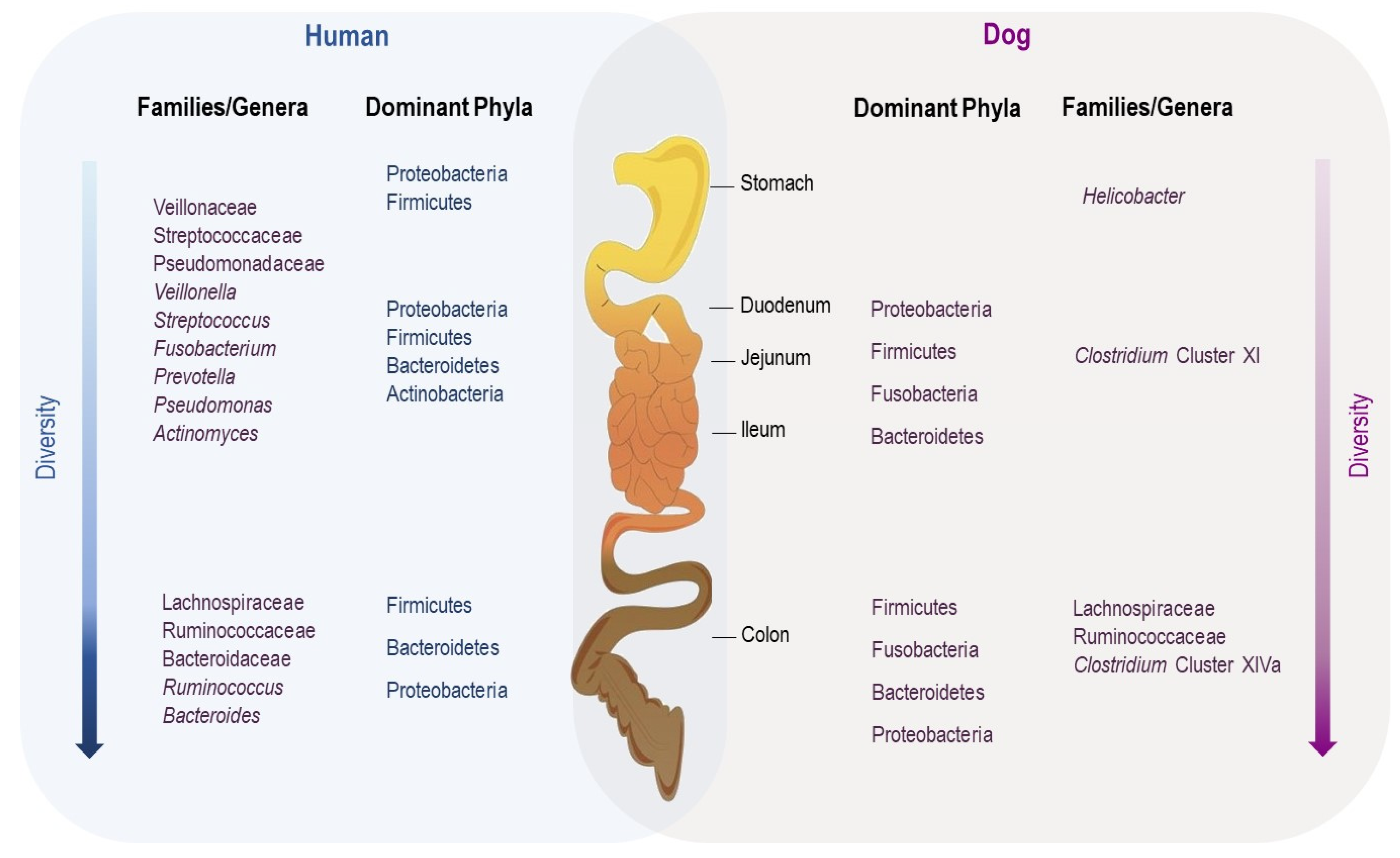
3.2. Gut Microbiota along the Gastrointestinal Tract

It is widely known that the gut microbiota varies along the gastrointestinal tract [32]. The particularity of each region of the GI tract, such as the acidic nature of the stomach, the profile of bile acids and enzyme richness in the small intestine and the low oxygen availability in the colon, have a great impact on which microbiota species colonize each segment [32,33]. In dogs, most reports focus on the analysis of fecal microbiota due to the practical difficulties and ethical constraints related to sample collection from each intestinal compartment in privately owned animals [28,29]. Suchodolski et al. evaluated the microbial communities of the duodenal, jejunal, ileal and colonic digesta of healthy dogs through 16S rRNA gene analysis [33]. Firmicutes, Fusobacteria, Bacteroidetes and Proteobacteria were the predominant phyla from the four sites, and there was a gradual increase in bacterial diversity along the gastrointestinal tract from the duodenum to the colon [30]. Similarly, Honneffer et al. analyzed the contents of the duodenum, ileum, colon and rectum from six healthy dogs via Illumina sequencing of 16S rRNA genes [34]. In addition to the four previously mentioned phyla, Actinobacteria was identified but contributed minimally in each segment [32].
For humans, there are only a few studies describing the bacterial biogeography of the entire GI tract. Vuik et al. characterized the mucosal microbiota along the entire GI tract using mucosal biopsies taken from nine different regions from the distal esophagus to rectum in 14 individuals [35]. The upper GI tract was dominated by Proteobacteria and Firmicutes, followed by Bacteroidetes and Actinobacteria at low concentrations. However, in the lower GI tract, the representation of Proteobacteria consistently decreased, while that of Firmicutes increased, and they dominated the large intestine in the distal colon, followed by Bacteroidetes, which had become a dominant phylum [32,33,34,35].
Although it is difficult to compare human and canine data due to methodological differences, the examination of the horizontal distribution of the gut microbiota reveals similarities and differences between the two species (Figure 2). Further studies using a standardized methodology are needed in the future.

3.3. Effects of Diet

In both humans and their pet dogs, diet is considered a key factor that influences the gut microbiota structure and host metabolic functions [36,37]. Evidence shows that the gut microbiota evolves to adapt to high intakes of fiber, carbohydrate and proteins. Several studies have compared the effects on the gut microbiota of feeding dogs with bones and raw food (BARF) diets (uncooked meat and bones) vs. commercial food (nutritionally balanced for omnivores, with a high abundance of fibers and carbohydrates). Bermingham et al. and Schmidt et al. demonstrated an overall decrease in the relative abundance of Firmicutes and Bacteroidetes involved in the digestion of dietary fibers in dogs fed with BARF diets [38,39]. They also identified a higher abundance of Proteobacteria, Fusobacterium, Lactobacillus and Clostridium. Most of the bacteria that decreased in abundance are associated with the production of short-chain fatty acids (SCFAs) from dietary carbohydrates, indicating a decrease in the fermentation of carbohydrates due to a decrease in carbohydrate intake [40]. Alessandri et al. showed a gradual rise in the relative abundance of bacterial taxa such as Prevotella and Sutterella, which break down carbohydrates, and a notable decline in the relative abundance of Parabacteroides and Ruminococcaceae in dogs fed with commercial food compared to dogs fed with a BARF diet [2].
These data mirror those for humans, especially when we compare Mediterranean-style with Western-style diets. While the Mediterranean diet (MD) is characterized by a high amount of dietary fiber, the Western diet (WD) is rich in animal protein and saturated fat [41,42]. In the majority of published studies, the gut microbiota composition differs between those consuming MD- and WD-style diets [41]. For example, high levels of Bacteroides sp. are found in WD-style diets, while Prevotella sp. have been observed to increase under an MD diet [41,43]. Shankar et al. demonstrated that the differences in gut metabolites and microbial composition and functions between Egyptian and US children are consistent with their diets [43]. While the gut microbiota of Egyptian children was found to be characterized by higher levels of SCFAs and increases in several polysaccharide-degrading microbes and end-products of polysaccharide fermentation, the gut microbiota of US children was found to have increased proteolytic microbes and end-products of protein and fat metabolism [43].

3.4. Effects of Xenobiotics

Pet dogs benefit from advanced veterinary care and are treated with the same classes of drugs as humans (antibiotics, anticancer chemotherapeutic drugs, proton-pump inhibitors, anti-inflammatory agents, vaccines, etc.). Therefore, pet dogs can contribute to studies of the impact of exposure to these substances on gut microbiota and health throughout life.
In recent years, several studies have evaluated the effects of certain xenobiotics on the gut microbiota of humans and pet dogs. The administration of omeprazole (a proton-pump inhibitor (PPI)) to healthy dogs results in higher proportions of Firmicutes and Fusobacteria, a decrease in gastric Helicobacter and an increase in total bacteria in the duodenum [44]. In humans, PPI administration also alters the gut microbiota composition but results in a different profile [45,46]. Jackson et al. reported a lower abundance of Firmicutes and higher abundance of Bacteroidales in PPI users [46].
Other changes in the composition of the gut microbiota community following exposure to xenobiotics were reviewed by Lu et al. [47].
In addition to the administration of xenobiotics for medical reasons, humans and pet dogs are exposed to environmental chemicals that affect the host and resident gut microbiota on a daily basis. In this context, the study of Koestel et al. evidenced an effect of bisphenol A, an endocrine-disrupting chemical widely present in food-can linings, on the bacterial gut composition in pet dogs [48]. The authors identified disturbances in many bacterial genera and species, including Bacteroides spp., Clostridium hiranonis, Bacteroides uniformis, Ruminococcus spp., Roseburia spp., Megamonas spp., Fusobacterium spp., Ruminococcus spp., Pectinatus spp., Catenibacterium spp. and Faecalibacterium prausnitzii. According to the authors, the effects of bisphenol A could be extrapolated to humans, and therefore, dogs are considered to be bio-sentinels for human health concerns.

4. Pet Dog and Human Gut Microbiota in Disease Conditions

The gut microbiota is essential in the gastrointestinal homeostasis of animals and humans. Numerous studies have reported an association between the imbalances in the intestinal microbial ecosystem of pets and humans, also known as dysbiosis, with the presence of several inflammatory and endocrine diseases. The gut microbiota is also involved in extraintestinal diseases such as obesity, atopic dermatitis and diabetes mellitus [49].

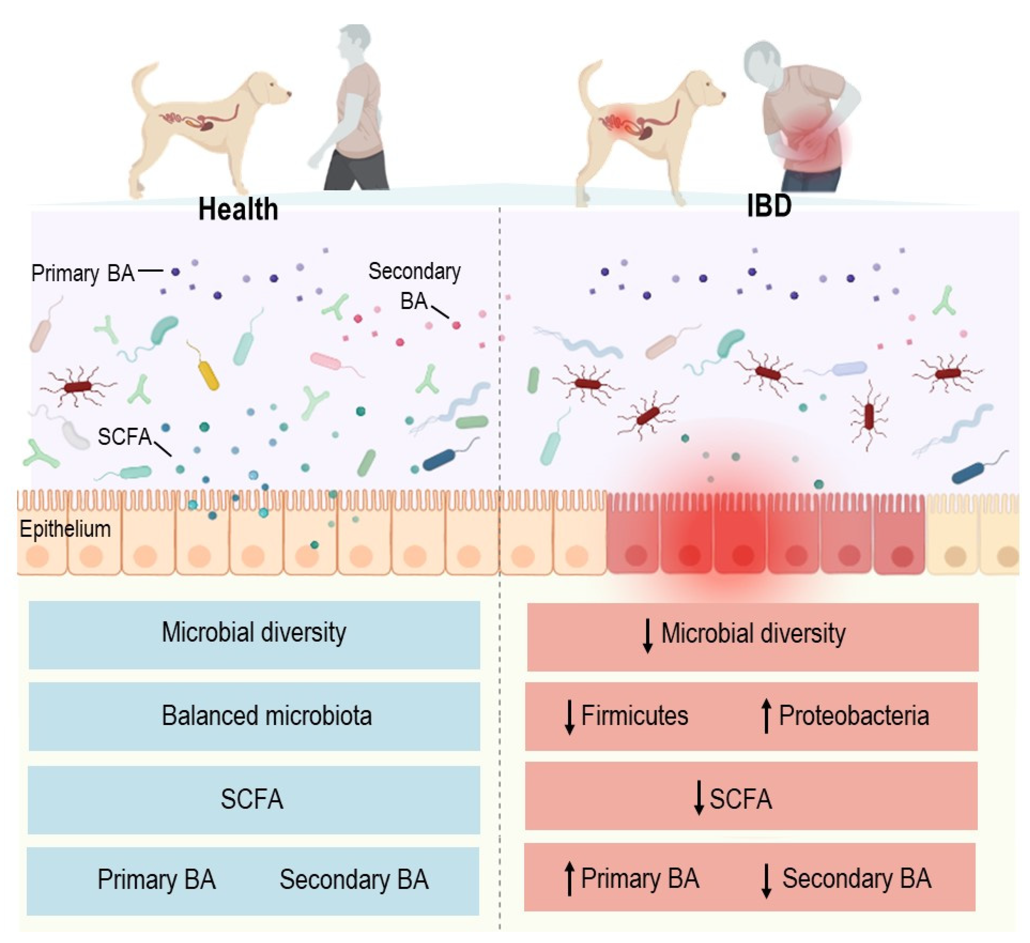
4.1. Inflammatory Bowel Diseases

Inflammatory bowel diseases (IBDs) have emerged as a global health problem [49,50,51]. Similarly to humans, dogs also develop IBD as a result of genetic and environmental factors, aberrant immune responses, and the gut microbiota [14,52]. Unlike in humans, little is known about canine gut microbiota in IBD, and most observations originate from the analysis of fecal samples using 16S rDNA gene sequencing on limited numbers of dogs. The comparison of microbial populations from the studies available in dogs show noticeable variability among studies, and this may be related to numerous factors such as differences in the methodology (e.g., total headcounts, DNA extraction method and primer bias), age, breed, diet, geographical origin and housing environment of the studies together with the disease stage and medications.
Alterations in the gut microbiota composition and function are also associated with canine IBD [53] (Figure 3). Similarly to human patients, dogs with IBD have a decreased microbial richness and diversity compared to healthy subjects [54]. Some deviations in the gut microbiota are common between humans and dogs with IBD, whereas others are host-specific [13]. Common modifications are characterized by both a superabundance of Enterobacteriaceae and Proteobacteria and a decline in Bacteroidetes and Firmicutes in the majority of subjects [55,56,57,58]. A decline in Faecalibacterium prausnitzii is reported in both humans and dogs with IBD in comparison to healthy individuals [13,59,60,61]. More specific changes include the overrepresentation of species of the Paraprevotellaceae family and Porphyromonas genus in dogs with IBD. Adherent-invasive E. coli (AIEC) is found in Crohn’s disease (CD) and ulcerative colitis (UC) patients [62,63,64] and in dogs with granulomatous colitis [65] (Figure 4). This breed-specific canine disorder has been described in Boxer and French Bulldogs and is sporadically reported in other breeds. The clinical and histopathological features are similar to those of CD and parallel to those of Whipple’s disease in humans [66,67]. Membranous colitis induced by Clostridioides difficile infection has been extensively studied in humans [67]. In dogs, the clinical involvement of C. difficile has not been established, given there is a prevalence of between 30 and 36% in the stools of asymptomatic dogs [68,69].
Dysbiosis induces metabolic alterations, including changes in SCFA and tryptophan metabolite production, which may affect gut homeostasis and immunological tolerance [70,71]. Increasing evidence demonstrates that the abundance of SCFA-producing bacteria dramatically decreases in fecal samples from human IBD patients, leading to reduced levels of SCFAs in the gut and the exacerbation of intestinal inflammation [68]. In dogs with chronic enteropathy, the fecal concentrations of acetate and propionate are also lower than those in healthy dogs [70]. As previously mentioned, the representation of the SFCA-producing bacterial phylum Bacteroidetes is decreased in human and canine IBD patients [70,71,72,73]. Other SFCA-producing bacteria, including some strains of Faecalibacterium spp., Roseburia, Eubacterium and Ruminococcus, are also reduced in human and canine IBD patients [70,71,72,73,74,75]. Butyrate is known to inhibit neutrophil recruitment, restore intestinal barrier function and alleviate the clinical and pathological features of Clostridioides difficile-induced colitis in mice [76,77].
The biotransformation of bile acids (BAs) by colonic microbiota is also involved in the pathogenesis of IBD in humans and pet dogs. The secondary bile acids (deoxycholic acid (DCA), lithocholic acid (LCA) and ursodeoxycholic acid (UDCA)) exhibit intestinal anti-inflammatory properties as demonstrated in vitro and in vivo in rodent models [78,79]. However, the formation of secondary BAs involves a deconjugation step mediated by the microbial bile salt hydrolase (BSH), followed by 7-alpha-dehydroxylation and epimerization supported by the colonic microbiota [80]. BSH has been identified in many bacterial genera, including Lactobacillus, Bifidobacterium, Clostridium, Bacteroides, Faecalibacterium and Enterococcus [78,81]. The conversion to secondary BAs is attributed to a smaller number of bacteria with bile-acid-inducible enzymes, including Clostridium cluster XIVa and Eubacterium among the genera of the phylum Firmicutes, whose populations are reduced during the course of human and canine IBD [81,82]. A preliminary study using untargeted metabolomics on fecal specimens from dogs with IBD confirmed increased primary BAs and decreased in secondary BAs in comparison to healthy dogs [61]. This pattern for secondary BAs in dogs with IBD was confirmed by two other studies [83,84], and the findings were similar to those of numerous reports on CD and UC [69,85].
Altogether, the data published for humans and dogs show a reduction in the diversity and richness of the intestinal microbiota with the progression of IBD. Functionally, the conversion of primary bile acids to secondary bile acids and SCFA synthesis by the gut microbiota appear to be similarly impaired in both species. Further studies with a standardized methodology and sufficient sample sizes are needed to compare the characteristics of the gut microbiota in humans and dogs with IBD.

4.2. Diabetes Mellitus

In recent years, changes in gut microbiota composition were suggested to be a potential contributor to type 2 diabetes mellitus (DM) [86]. While much of this understanding comes from studies in mice, which have highlighted the influence of the gut microbiota on glucose homeostasis, alterations in gut microbiota composition have also been noted in pet dogs, with DM. Jergens et al. observed intestinal dysbiosis and altered fecal bile acid (BA) levels in dogs with insulin-dependent DM, found to be similar to humans with T2DM [87]. Bacteria from the Enterobacteriaceae family were more abundant in diabetic dogs, whereas those from the Erysipelotrichia class and from Mogibacteriaceae and Anaeroplasmataceae families were overrepresented in healthy controls. At the species level, the proportion of an unclassified bacterial species from the Enterobacteriaceae family was most significantly correlated with DM, whereas the abundances of Bacteroides plebeius and Lactobacillus reuteri was associated with healthy individuals. Dogs suffering from DM had higher levels of total primary fecal unconjugated BAs in comparison to healthy dogs. The level of cholic acid was increased in the feces of diabetic dogs relative to controls. The link between BAs and host–microbiota interactions appears to be complex and bidirectional. Several pathways are involved in the microbial metabolism of BAs, including BA deconjugation by bacterial species possessing bile salt hydrolase activity and the generation of iso-BA by bacteria-producing hydroxysteroid dehydrogenases [88].
In humans, the relative abundance of Firmicutes, for instance, and Clostridia was significantly reduced in the diabetic group, compared to in healthy controls, in a study by Larsen et al. [89]. The ratios of Bacteroidetes/Firmicutes and the Bacteroides–Prevotella group to the Clostridium coccoides–Eubacterium rectal group were positively correlated with plasma glucose concentrations [89]. Other differences included decreased abundances of Roseburia species and F. prausnitzii, which are known to be producers of SCFAs [86,89,90,91]. Butyrate provides energy to colonic epithelial cells and has the potential to increase insulin sensitivity and energy expenditure [92].

4.3. Obesity

According to the World Health Organization (WHO), approximately 39% of human adults are considered obese and overweight [93,94]. The etiology is related to various factors, and the gut microbiota continues to draw attention as an element that affects disease status.
In pets, overweight and obesity are also frequent conditions that decrease life expectancy and trigger several comorbidities such as insulin resistance, systemic arterial hypertension and osteoarthritis [95]. Several authors have reported decreased bacterial diversity in the fecal microbiota of overweight (OW) and obese (OB) dogs when compared to normal weight (NW) dogs [96,97]. Handl et al. identified a greater abundance of the phylum Actinobacteria and the genus Roseburia in OB dogs [98]. When comparing 17 NW, 27 OW and 22 OB dogs, Forster et al. showed that the Erysipelotrichi class was more abundant in OW compared to OB dogs, and this was essentially led by differences in the Eubacterium genus [99]. The Actinobacteria class was determined to be present at higher levels in OB dogs relative to NW dogs. At the order level, Bifidobacteriales were significantly less abundant and Aeromonadales showed a tendency to be more abundant in OW relative to OB dogs. Comparatively to OB dogs, NW individuals exhibited higher levels of Erysipelotrichaceae, Erysipelotrichales and Erysipelotrichi and also had a lower abundance of the order Bifidobacteriales. When studied as operational taxonomic units (OTUs), the genus Blautia was more represented in NW and OW dogs than in OB dogs, as was the Lachnospiraceae family and the Eubacterium biforme species. The Ruminococcus family was more relatively abundant in NW than OB dogs. OTUs within the Prevotella copri species and the Clostridium genus were more abundant in OW than OB dogs. The order Clostridiales was also shown to increase in research dogs subjected to long-term ad libitum feeding when compared with NW control dogs [98]. Other authors reported a higher abundance of Fusobacteria, and more specifically, of the species Fusobacteria perfoetens in OW dogs in comparison to LN dogs [97]. Another study identified a predominance of the phylum Proteobacteria in OB dogs [96]. From a functional perspective, they speculated that an enrichment of Gram-negative bacteria may be implicated in chronic low-grade inflammation in OB dogs via increased levels of intestinal LPSs [100]. Gram-positive bacteria could modulate inflammation. In fact, Kainulainen et al. reported a beneficial role for the canine indigenous strain Lactobacillus acidophilus LAB20 and its ability to attenuate LPS-induced IL-8 production in HT-29 cells [101].
The impacts of dietary intervention and exercise on the canine gut microbiota have been investigated in several clinical trials [102,103]. Kieler et al. did not show any effects of exercise on the gut microbiota composition during a weight-loss program based on a commercial low-fat, high-protein and high-fiber, dry diet [103]. A negative correlation between the abundance of Megamonas and weight-loss rate was identified. The relative abundance of Ruminococcaceae was significantly lower at the end of the trial and the mid-term fecal concentrations of acetic and propionic acid were lower in dogs with rapid weight loss compared to dogs with slow weight loss. These data suggest that obese dogs exhibiting fecal bacteria that are able to produce acetic and propionic acids may be less amenable to weight loss due to an increased ability to extract energy from the diet through the production of SCFAs.
In humans, it was demonstrated that obese people present a lower diversity and richness in gut microbiota composition [104,105]. Indeed, several studies highlight an increase in the Firmicutes/Bacteroidetes ratio in obese people compared to healthy individuals [97,99,100]. Recently, a study by Palmas et al. showed an altered abundance of several taxa belonging to Bacteroidetes (Bacteroides, Rikenella and Parabacteroides), Firmicutes (Eubacterium, Ruminococcus and Streptococcus) and Proteobacteria (Escherichia, Enterobacter and Klebsiella) [106]. To better characterize the extent of its contribution to the disease, some mechanisms were proposed. Metabolites, especially SFCAs, produced by gut microbiota can regulate host energy metabolism, thereby increasing de novo lipogenesis in the liver and lipid accumulation in host adipocytes [107,108,109]. Studies of obese people have demonstrated a positive correlation between fecal SCFA concentrations and obesity [110,111]; however, others have reported a negative relationship between SCFA levels and the obese phenotype [112]. Moreover, Morrison and Preston postulated that SCFAs constitute signaling molecules mediating crosstalk between the host and its corresponding gut microbiota [108]. Hence, changes in SCFAs are representative of major carbon fluxes from the diet through the gut microbiota to the host, which serves as evident of their regulatory role in the overall metabolism [113]. Another proposed mechanism includes an increase in lipopolysaccharides (LPSs) produced by gut microbiota [93]. Indeed, LPSs can affect intestinal permeability, leading to an increase in their plasmatic concentration, which is correlated with the chronic low-grade inflammation characteristic of obese human patients. Additionally, it is well known that LPSs are able to bind to Toll-like receptor-4 (TLR-4), which upregulates the production of inflammatory cytokines and chemokines [114].

4.4. Kidney and Urinary Tract Diseases

The link between the gut microbiota and chronic kidney disease (CKD) has been investigated in large human cohorts showing a significant reduction in microbial diversity compared to healthy controls. The bacterial communities are distinct with an enrichment of the genera Akkermansia, Klebsiella and Enterobacteriaceae and a depletion of the genera Blautia and Roseburia in patients with non-hemodialyzed CKD. Some functions are predicted upward such as the metabolism of tryptophan and phenylalanine and others are predicted downward such as the metabolism of arginine and proline during CKD [115,116]. Patients with end-stage kidney disease on peritoneal dialysis were less likely to have Bifidobacterium catenulatum, Bifidobacterium longum, Bifidobacterium bifidum, Lactobacillus plantarum, Lactobacillus paracasei and Klebsiella pneumoniae than healthy controls [117]. These populational and functional changes are suspected to be due to the exposure of intestinal bacteria to urea crossing the gut barrier leading to a selection of bacterial families containing urease, uricase or indole- and p-cresyl enzymes [118]. Unfortunately, there is no comparative data available for dogs.
Few data on the link between gut microbiota and urolithiasis in humans and dogs are available. In dogs, Oxalobacter formigenes, a bacterium that degrades oxalates, was shown to exhibit significantly contrasting prevalence between dogs with oxalate calcium uroliths (25%), healthy dogs belonging to a breed predisposed to oxalate calcium uroliths (50%), and healthy non-predisposed dogs (75%) [119]. The significant underabundance of O. formigenes in the human gut has also been reported in patients with calcium oxalate urolithiasis since the early 2000s [120].
Very recently published data for humans make it possible to establish a correlation between a decrease in the abundance of SCFA-producing bacteria and the formation of calcium oxalate nephrolithiasis [121]. Liu et al. showed that SCFAs have the ability to reduce the formation of stones via regulating the expression of the intestinal transporter SLC26A6 involved in oxalate excretion [121]. This communication route between gut microbiota and the host has not been studied in the context of lithiasic disease in dogs.

4.5. Neuropsychiatric Diseases

Understanding the microbiota–gut–brain axis, with the goal of identifying innovative therapeutic approaches for mental disorders in humans and dogs is a current challenge [116]. Laboratory animals, and especially germ-free and gnotobiotic mice, have been invaluable tools for proof-of-principle studies demonstrating the impact of the gut microbiota in the healthy development and homeostasis of the nervous system and in psychiatric diseases, autism spectrum disorder, schizophrenia, Alzheimer’s disease, Parkinson’s disease and stroke [117]. Communication between the gut microbiota and the brain takes place via endocrine, immune, humoral and nervous channels, with particular attention given to the vagus nerve [118]. Data published by Cummings et al. show that 96.4% of new drugs developed for treating Alzheimer’s disease as a result of basic research using mouse models fail in human clinical trials [122]. There is an urgent need for novel models that will allow scientists to better translate the findings to humans.
Dogs exhibit behavioral problems of interest for exploring the links between gut microbiota and host behavior. They could serve as a model of spontaneous pathologies for investigating new therapeutic pathways. In particular, Kirchoff et al. studied a cohort of 21 conspecifically aggressive dogs and showed that the composition of the gut microbiota differed between aggressive and non-aggressive dogs [123]. More precisely, Proteobacteria and Fusobacteria displayed greater relative abundances in samples from non-aggressive animals, whereas Firmicutes were more abundant in samples from aggressive dogs. The Fusobacteriaceae family and more specifically, the Fusobacteria genus, were more abundant in specimens from non-aggressive dogs, whereas the Lactobacillaceae family and more specifically, the Lactobacillus genus, were more abundant in specimens from aggressive dogs. Mondo et al. compared the gut microbiota of dogs with behavioral disorders (aggressive and phobic conditions) with that of healthy controls [124]. The gut microbiota structure exhibited a robust dissociation of the aggressive population that seemed to be driven by a greater abundance of classically subdominant taxa, such as Blautia, Catenibacterium, Collinsella, Ruminococcus Dorea, Megamonas and Slackia. In contrast, the phobic population showed an enrichment of the Lactobacillus genus which comprises well-documented GABA producers. The major determinants of the segregation of the normal-behavior population were Bacteroides, Faecalibacterium, Fusobacterium, Phascolarctobacterium and Prevotella, evidencing the preponderance of bacterial genera typically contributing to the gut microbiota of healthy dogs. Mondo et al. applied a machine-learning technique (random forest) to their genus-level dataset and established Catenibacterium and Megamonas as bacterial determinant factors of aggressiveness [125]. Regarding idiopathic epilepsy, a pilot study using 16S rRNA gene amplicon sequencing failed to identify differences in overall fecal bacterial patterns and did not show quantitative variations in Lactobacillus species between untreated epileptic dogs and paired healthy dogs from the same household [125].

5. Conclusions

As shown in this review, the physiologies of the domestic dog and human are more similar than those of the human and mouse. Thus, the dog has the potential to be a useful animal model. The pet dog may be utilized in the field of translational sciences to identify novel therapies and maximize clinical benefits for both species. To the best of our knowledge, there are few (or no) studies in the field of gut microbiota that have used dogs as a larger animal model for the assessment of treatments to improve human wellbeing or health.
The vast majority of canine studies to date have focused on the phylogenetic structure of the microbiota and, in most cases, the bacterial fraction of the canine microbiota. Although this will increase the complexity of microbiota analyses, the characterization of the whole microbe community residing in the canine gut—i.e., the bacteria and archaea, fungi and yeasts, and protozoans and viruses—along with their interplay and their dynamics over time is likely to be key for understanding the ecology and physiology of the microbiota. The microbiota’s phylogenetic structure is undoubtedly an important parameter, but it is becoming clearer that functional information has to be associated with the microbiome structure in order to decipher the basis of the symbiotic crosstalk between the host and its microbiota to identify active compounds or species in addition to obtaining hints regarding the mode of action that determines the success of interventions.
Another challenge is to define what a healthy (or unhealthy) microbiota is and when appropriate, to evaluate whether microbiota or microbiota-produced compound(s) could help to better characterize a population of patients in order to develop efficient diagnostic tools using a combination of microbiota-based marker(s). It is likely that the meta-analyses of datasets, provided that the procedures are described and correspond to standards and best practices, would allow overcoming the limited number of dog samples included in most of the published studies. This raises the question of how the scientific community can develop and regularly update the required/useful best practices, standards and reference materials and define the type of information that has to be disclosed with the datasets. On these latter aspects, several international efforts are in progress, as are more specific initiatives. This question of larger numbers of samples and diversity of dog environments also highlights the potential of large international collaborative projects that would allow overcoming some of the putative biases associated with microbiome/population studies and determining whether local or worldwide scales will have to be considered for further developments in research and innovation.

Author Contributions

J.H., S.R., A.K., V.M., H.B., A.D., A.J., H.M., A.S., M.A.B., V.B., E.M. and M.R. conceived the scientific ideas. J.H., S.R., A.K., V.M., A.D., A.S., V.B., M.A.B., E.M. and M.R. performed and discussed the work and edited the manuscript. All the authors reviewed the manuscript and provided critical feedback. All authors have read and agreed to the published version of the manuscript.

Funding

This work received funding from Royal Canin (PS-Com-Pro Project No. 13001516-300), CMCU-PHC Utique (No. 19G0819)—Campus France (No. 41786NC) and Twinning European project MICAfrica (No. 952583).

Conflicts of Interest

The authors declare no conflict of interest.

References

  1. Xie, H.; Guo, R.; Zhong, H.; Feng, Q.; Lan, Z.; Qin, B.; Ward, K.J.; Jackson, M.A.; Xia, Y.; Chen, X.; et al. Shotgun Metagenomics of 250 Adult Twins Reveals Genetic and Environmental Impacts on the Gut Microbiome. Cell Syst. 2016, 3, 572–584.e3. [Google Scholar] [CrossRef] [PubMed] [Green Version]
  2. Alessandri, G.; Milani, C.; Mancabelli, L.; Mangifesta, M.; Lugli, G.A.; Viappiani, A.; Duranti, S.; Turroni, F.; Ossiprandi, M.C.; van Sinderen, D.; et al. Metagenomic Dissection of the Canine Gut Microbiota: Insights into Taxonomic, Metabolic and Nutritional Features. Environ. Microbiol. 2019, 21, 1331–1343. [Google Scholar] [CrossRef] [PubMed]
  3. West, C.E.; Renz, H.; Jenmalm, M.C.; Kozyrskyj, A.L.; Allen, K.J.; Vuillermin, P.; Prescott, S.L.; in-FLAME Microbiome Interest Group. The Gut Microbiota and Inflammatory Noncommunicable Diseases: Associations and Potentials for Gut Microbiota Therapies. J. Allergy Clin. Immunol. 2015, 135, 3–13. [Google Scholar] [CrossRef] [PubMed] [Green Version]
  4. Huang, R.; Ju, Z.; Zhou, P.-K. A Gut Dysbiotic Microbiota-Based Hypothesis of Human-to-Human Transmission of Non-Communicable Diseases. Sci. Total Environ. 2020, 745, 141030. [Google Scholar] [CrossRef]
  5. Hugenholtz, F.; de Vos, W.M. Mouse Models for Human Intestinal Microbiota Research: A Critical Evaluation. Cell. Mol. Life Sci. 2018, 75, 149–160. [Google Scholar] [CrossRef] [PubMed] [Green Version]
  6. Xiao, L.; Feng, Q.; Liang, S.; Sonne, S.B.; Xia, Z.; Qiu, X.; Li, X.; Long, H.; Zhang, J.; Zhang, D.; et al. A Catalog of the Mouse Gut Metagenome. Nat. Biotechnol. 2015, 33, 1103–1108. [Google Scholar] [CrossRef] [PubMed]
  7. Xiao, L.; Estellé, J.; Kiilerich, P.; Ramayo-Caldas, Y.; Xia, Z.; Feng, Q.; Liang, S.; Pedersen, A.Ø.; Kjeldsen, N.J.; Liu, C.; et al. A Reference Gene Catalogue of the Pig Gut Microbiome. Nat. Microbiol. 2016, 1, 16161. [Google Scholar] [CrossRef] [PubMed]
  8. Yue, F.; The Mouse ENCODE Consortium; Cheng, Y.; Breschi, A.; Vierstra, J.; Wu, W.; Ryba, T.; Sandstrom, R.; Samantha, K.; Davis, C.; et al. A Comparative Encyclopedia of DNA Elements in the Mouse Genome. Nature 2014, 515, 355–364. [Google Scholar] [CrossRef] [Green Version]
  9. Seok, J.; Warren, H.S.; Cuenca, A.G.; Mindrinos, M.N.; Baker, H.V.; Xu, W.; Richards, D.R.; McDonald-Smith, G.P.; Gao, H.; Hennessy, L.; et al. Genomic Responses in Mouse Models Poorly Mimic Human Inflammatory Diseases. Proc. Natl. Acad. Sci. USA 2013, 110, 3507–3512. [Google Scholar] [CrossRef] [Green Version]
  10. MetaHIT Consortium; Li, J.; Jia, H.; Cai, X.; Zhong, H.; Feng, Q.; Sunagawa, S.; Arumugam, M.; Kultima, J.R.; Prifti, E.; et al. An Integrated Catalog of Reference Genes in the Human Gut Microbiome. Nat. Biotechnol. 2014, 32, 834–841. [Google Scholar] [CrossRef]
  11. Botigué, L.R.; Song, S.; Scheu, A.; Gopalan, S.; Pendleton, A.L.; Oetjens, M.; Taravella, A.M.; Seregély, T.; Zeeb-Lanz, A.; Arbogast, R.-M.; et al. Ancient European Dog Genomes Reveal Continuity since the Early Neolithic. Nat. Commun. 2017, 8, 16082. [Google Scholar] [CrossRef] [PubMed]
  12. Axelsson, E.; Ratnakumar, A.; Arendt, M.-L.; Maqbool, K.; Webster, M.T.; Perloski, M.; Liberg, O.; Arnemo, J.M.; Hedhammar, Å.; Lindblad-Toh, K. The Genomic Signature of Dog Domestication Reveals Adaptation to a Starch-Rich Diet. Nature 2013, 495, 360–364. [Google Scholar] [CrossRef] [PubMed]
  13. Ettinger, S.J.; Feldman, E.C.; Côté, E. (Eds.) Textbook of Veterinary Internal Medicine: Diseases of the Dog and the Cat, 11th ed.; Elsevier: St. Louis, MO, USA, 2017; ISBN 978-0-323-31211-0. [Google Scholar]
  14. Vázquez-Baeza, Y.; Hyde, E.R.; Suchodolski, J.S.; Knight, R. Dog and Human Inflammatory Bowel Disease Rely on Overlapping yet Distinct Dysbiosis Networks. Nat. Microbiol. 2016, 1, 16177. [Google Scholar] [CrossRef]
  15. Gómez-Gallego, C.; Forsgren, M.; Selma-Royo, M.; Nermes, M.; Collado, M.C.; Salminen, S.; Beasley, S.; Isolauri, E. The Composition and Diversity of the Gut Microbiota in Children Is Modifiable by the Household Dogs: Impact of a Canine-Specific Probiotic. Microorganisms 2021, 9, 557. [Google Scholar] [CrossRef] [PubMed]
  16. American Veterinary Medical Association. AVMA Pet Ownership and Demographics Sourcebook: 2017–2018 Edition; American Veterinary Medical Association: Schaumburg, IL, USA, 2018; ISBN 978-1-882691-53-1. [Google Scholar]
  17. Delicano, R.A.; Hammar, U.; Egenvall, A.; Westgarth, C.; Mubanga, M.; Byberg, L.; Fall, T.; Kennedy, B. The Shared Risk of Diabetes between Dog and Cat Owners and Their Pets: Register Based Cohort Study. BMJ 2020, 371, m4337. [Google Scholar] [CrossRef] [PubMed]
  18. Glickman, L.T.; Domanski, L.M.; Maguire, T.G.; Dubielzig, R.R.; Churg, A. Mesothelioma in Pet Dogs Associated with Exposure of Their Owners to Asbestos. Environ. Res. 1983, 32, 305–313. [Google Scholar] [CrossRef]
  19. Sumner, R.N.; Tomlinson, M.; Craigon, J.; England, G.C.W.; Lea, R.G. Independent and Combined Effects of Diethylhexyl Phthalate and Polychlorinated Biphenyl 153 on Sperm Quality in the Human and Dog. Sci. Rep. 2019, 9, 3409. [Google Scholar] [CrossRef]
  20. Bischoff, K.; Priest, H.; Mount-Long, A. Animals as Sentinels for Human Lead Exposure: A Case Report. J. Med. Toxicol. 2010, 6, 185–189. [Google Scholar] [CrossRef] [Green Version]
  21. Yaglom, H.D.; Hecht, G.; Goedderz, A.; Jasso-Selles, D.; Ely, J.L.; Ruberto, I.; Bowers, J.R.; Engelthaler, D.M.; Venkat, H. Genomic Investigation of a Household SARS-CoV-2 Disease Cluster in Arizona Involving a Cat, Dog, and Pet Owner. One Health 2021, 13, 100333. [Google Scholar] [CrossRef]
  22. National Research Council (NRC). Energy. In Nutrient Requirements of Dogs and Cats; The National Academies Press: Washington, DC, USA, 2006; pp. 28–48. [Google Scholar]
  23. Walker, J.A.; Harmon, D.L.; Gross, K.L.; Collings, G.F. Evaluation of Nutrient Utilization in the Canine Using the Ileal Cannulation Technique. J. Nutr. 1994, 124, 2672S–2676S. [Google Scholar] [CrossRef] [Green Version]
  24. Bello-Perez, L.A.; Flores-Silva, P.C.; Agama-Acevedo, E.; Tovar, J. Starch Digestibility: Past, Present, and Future. J. Sci. Food Agric. 2020, 100, 5009–5016. [Google Scholar] [CrossRef] [PubMed]
  25. Corsato Alvarenga, I.; Aldrich, C.G. Starch Characterization of Commercial Extruded Dry Pet Foods. Transl. Anim. Sci. 2020, 4, txaa018. [Google Scholar] [CrossRef] [PubMed]
  26. Harmon, D.L. 0437 Idiosyncrasies of Amino Acid Metabolism in Dogs and Cats. J. Anim. Sci. 2016, 94, 210–211. [Google Scholar] [CrossRef] [Green Version]
  27. Guard, B.C.; Mila, H.; Steiner, J.M.; Mariani, C.; Suchodolski, J.S.; Chastant-Maillard, S. Characterization of the Fecal Microbiome during Neonatal and Early Pediatric Development in Puppies. PLoS ONE 2017, 12, e0175718. [Google Scholar] [CrossRef] [PubMed] [Green Version]
  28. Roswall, J.; Olsson, L.M.; Kovatcheva-Datchary, P.; Nilsson, S.; Tremaroli, V.; Simon, M.-C.; Kiilerich, P.; Akrami, R.; Krämer, M.; Uhlén, M.; et al. Developmental Trajectory of the Healthy Human Gut Microbiota during the First 5 Years of Life. Cell Host Microbe 2021, 29, 765–776.e3. [Google Scholar] [CrossRef]
  29. Derrien, M.; Alvarez, A.-S.; de Vos, W.M. The Gut Microbiota in the First Decade of Life. Trends Microbiol. 2019, 27, 997–1010. [Google Scholar] [CrossRef] [Green Version]
  30. Mancabelli, L.; Tarracchini, C.; Milani, C.; Lugli, G.A.; Fontana, F.; Turroni, F.; van Sinderen, D.; Ventura, M. Multi-Population Cohort Meta-Analysis of Human Intestinal Microbiota in Early Life Reveals the Existence of Infant Community State Types (ICSTs). Comput. Struct. Biotechnol. J. 2020, 18, 2480–2493. [Google Scholar] [CrossRef]
  31. Vasapolli, R.; Schütte, K.; Schulz, C.; Vital, M.; Schomburg, D.; Pieper, D.H.; Vilchez-Vargas, R.; Malfertheiner, P. Analysis of Transcriptionally Active Bacteria Throughout the Gastrointestinal Tract of Healthy Individuals. Gastroenterology 2019, 157, 1081–1092.e3. [Google Scholar] [CrossRef] [Green Version]
  32. Suchodolski, J.S.; Camacho, J.; Steiner, J.M. Analysis of Bacterial Diversity in the Canine Duodenum, Jejunum, Ileum, and Colon by Comparative 16S RRNA Gene Analysis. FEMS Microbiol. Ecol. 2008, 66, 567–578. [Google Scholar] [CrossRef]
  33. Honneffer, J.B.; Steiner, J.M.; Lidbury, J.A.; Suchodolski, J.S. Variation of the Microbiota and Metabolome along the Canine Gastrointestinal Tract. Metabolomics 2017, 13, 26. [Google Scholar] [CrossRef]
  34. Vuik, F.; Dicksved, J.; Lam, S.Y.; Fuhler, G.M.; van der Laan, L.; van de Winkel, A.; Konstantinov, S.R.; Spaander, M.; Peppelenbosch, M.P.; Engstrand, L.; et al. Composition of the Mucosa-Associated Microbiota along the Entire Gastrointestinal Tract of Human Individuals. United Eur. Gastroenterol. J. 2019, 7, 897–907. [Google Scholar] [CrossRef] [PubMed] [Green Version]
  35. Goldsmith, J.R.; Sartor, R.B. The Role of Diet on Intestinal Microbiota Metabolism: Downstream Impacts on Host Immune Function and Health, and Therapeutic Implications. J. Gastroenterol. 2014, 49, 785–798. [Google Scholar] [CrossRef] [PubMed] [Green Version]
  36. Wernimont, S.M.; Radosevich, J.; Jackson, M.I.; Ephraim, E.; Badri, D.V.; MacLeay, J.M.; Jewell, D.E.; Suchodolski, J.S. The Effects of Nutrition on the Gastrointestinal Microbiome of Cats and Dogs: Impact on Health and Disease. Front. Microbiol. 2020, 11, 1266. [Google Scholar] [CrossRef] [PubMed]
  37. Bermingham, E.N.; Maclean, P.; Thomas, D.G.; Cave, N.J.; Young, W. Key Bacterial Families (Clostridiaceae, Erysipelotrichaceae and Bacteroidaceae) Are Related to the Digestion of Protein and Energy in Dogs. PeerJ 2017, 5, e3019. [Google Scholar] [CrossRef]
  38. Schmidt, M.; Unterer, S.; Suchodolski, J.S.; Honneffer, J.B.; Guard, B.C.; Lidbury, J.A.; Steiner, J.M.; Fritz, J.; Kölle, P. The Fecal Microbiome and Metabolome Differs between Dogs Fed Bones and Raw Food (BARF) Diets and Dogs Fed Commercial Diets. PLoS ONE 2018, 13, e0201279. [Google Scholar] [CrossRef] [Green Version]
  39. Marchesi, J.R.; Holmes, E.; Khan, F.; Kochhar, S.; Scanlan, P.; Shanahan, F.; Wilson, I.D.; Wang, Y. Rapid and Noninvasive Metabonomic Characterization of Inflammatory Bowel Disease. J. Proteome Res. 2007, 6, 546–551. [Google Scholar] [CrossRef]
  40. Merra, G.; Noce, A.; Marrone, G.; Cintoni, M.; Tarsitano, M.G.; Capacci, A.; De Lorenzo, A. Influence of Mediterranean Diet on Human Gut Microbiota. Nutrients 2020, 13, 7. [Google Scholar] [CrossRef]
  41. Jin, Q.; Black, A.; Kales, S.N.; Vattem, D.; Ruiz-Canela, M.; Sotos-Prieto, M. Metabolomics and Microbiomes as Potential Tools to Evaluate the Effects of the Mediterranean Diet. Nutrients 2019, 11, 207. [Google Scholar] [CrossRef] [Green Version]
  42. Shankar, V.; Gouda, M.; Moncivaiz, J.; Gordon, A.; Reo, N.V.; Hussein, L.; Paliy, O. Differences in Gut Metabolites and Microbial Composition and Functions between Egyptian and U.S. Children Are Consistent with Their Diets. mSystems 2017, 2, e00169-16. [Google Scholar] [CrossRef] [Green Version]
  43. Garcia-Mazcorro, J.F.; Suchodolski, J.S.; Jones, K.R.; Clark-Price, S.C.; Dowd, S.E.; Minamoto, Y.; Markel, M.; Steiner, J.M.; Dossin, O. Effect of the Proton Pump Inhibitor Omeprazole on the Gastrointestinal Bacterial Microbiota of Healthy Dogs. FEMS Microbiol. Ecol. 2012, 80, 624–636. [Google Scholar] [CrossRef]
  44. Freedberg, D.E.; Toussaint, N.C.; Chen, S.P.; Ratner, A.J.; Whittier, S.; Wang, T.C.; Wang, H.H.; Abrams, J.A. Proton Pump Inhibitors Alter Specific Taxa in the Human Gastrointestinal Microbiome: A Crossover Trial. Gastroenterology 2015, 149, 883–885.e9. [Google Scholar] [CrossRef] [PubMed] [Green Version]
  45. Jackson, M.A.; Goodrich, J.K.; Maxan, M.-E.; Freedberg, D.E.; Abrams, J.A.; Poole, A.C.; Sutter, J.L.; Welter, D.; Ley, R.E.; Bell, J.T.; et al. Proton Pump Inhibitors Alter the Composition of the Gut Microbiota. Gut 2016, 65, 749–756. [Google Scholar] [CrossRef] [PubMed] [Green Version]
  46. Lu, K.; Mahbub, R.; Fox, J.G. Xenobiotics: Interaction with the Intestinal Microflora. ILAR J. 2015, 56, 218–227. [Google Scholar] [CrossRef] [PubMed]
  47. Koestel, Z.L.; Backus, R.C.; Tsuruta, K.; Spollen, W.G.; Johnson, S.A.; Javurek, A.B.; Ellersieck, M.R.; Wiedmeyer, C.E.; Kannan, K.; Xue, J.; et al. Bisphenol A (BPA) in the Serum of Pet Dogs Following Short-Term Consumption of Canned Dog Food and Potential Health Consequences of Exposure to BPA. Sci. Total Environ. 2017, 579, 1804–1814. [Google Scholar] [CrossRef]
  48. Blake, A.B.; Suchodolski, J.S. Importance of Gut Microbiota for the Health and Disease of Dogs and Cats. Anim. Front. 2016, 6, 37–42. [Google Scholar] [CrossRef]
  49. Kaplan, G.G. The Global Burden of IBD: From 2015 to 2025. Nat. Rev. Gastroenterol. Hepatol. 2015, 12, 720–727. [Google Scholar] [CrossRef]
  50. Molodecky, N.A.; Soon, I.S.; Rabi, D.M.; Ghali, W.A.; Ferris, M.; Chernoff, G.; Benchimol, E.I.; Panaccione, R.; Ghosh, S.; Barkema, H.W.; et al. Increasing Incidence and Prevalence of the Inflammatory Bowel Diseases with Time, Based on Systematic Review. Gastroenterology 2012, 142, 46–54.e42. [Google Scholar] [CrossRef] [Green Version]
  51. Ng, S.C.; Shi, H.Y.; Hamidi, N.; Underwood, F.E.; Tang, W.; Benchimol, E.I.; Panaccione, R.; Ghosh, S.; Wu, J.C.Y.; Chan, F.K.L.; et al. Worldwide Incidence and Prevalence of Inflammatory Bowel Disease in the 21st Century: A Systematic Review of Population-Based Studies. Lancet 2017, 390, 2769–2778. [Google Scholar] [CrossRef]
  52. Kathrani, A.; Lee, H.; White, C.; Catchpole, B.; Murphy, A.; German, A.; Werling, D.; Allenspach, K. Association between Nucleotide Oligomerisation Domain Two (Nod2) Gene Polymorphisms and Canine Inflammatory Bowel Disease. Vet. Immunol. Immunopathol. 2014, 161, 32–41. [Google Scholar] [CrossRef]
  53. Deng, P.; Swanson, K.S. Gut Microbiota of Humans, Dogs and Cats: Current Knowledge and Future Opportunities and Challenges. Br. J. Nutr. 2015, 113, S6–S17. [Google Scholar] [CrossRef]
  54. Alam, M.T.; Amos, G.C.A.; Murphy, A.R.J.; Murch, S.; Wellington, E.M.H.; Arasaradnam, R.P. Microbial Imbalance in Inflammatory Bowel Disease Patients at Different Taxonomic Levels. Gut Pathog. 2020, 12, 1–8. [Google Scholar] [CrossRef] [PubMed]
  55. Walker, A.W.; Sanderson, J.D.; Churcher, C.; Parkes, G.C.; Hudspith, B.N.; Rayment, N.; Brostoff, J.; Parkhill, J.; Dougan, G.; Petrovska, L. High-Throughput Clone Library Analysis of the Mucosa-Associated Microbiota Reveals Dysbiosis and Differences between Inflamed and Non-Inflamed Regions of the Intestine in Inflammatory Bowel Disease. BMC Microbiol. 2011, 11, 7. [Google Scholar] [CrossRef] [PubMed] [Green Version]
  56. Sokol, H.; Seksik, P.; Furet, J.P.; Firmesse, O.; Nion-Larmurier, I.; Beaugerie, L.; Cosnes, J.; Corthier, G.; Marteau, P.; Doré, J. Low Counts of Faecalibacterium Prausnitzii in Colitis Microbiota. Inflamm. Bowel Dis. 2009, 15, 1183–1189. [Google Scholar] [CrossRef]
  57. Frank, D.N.; St Amand, A.L.; Feldman, R.A.; Boedeker, E.C.; Harpaz, N.; Pace, N.R. Molecular-Phylogenetic Characterization of Microbial Community Imbalances in Human Inflammatory Bowel Diseases. Proc. Natl. Acad. Sci. USA 2007, 104, 13780–13785. [Google Scholar] [CrossRef] [Green Version]
  58. Xenoulis, P.G.; Palculict, B.; Allenspach, K.; Steiner, J.M.; Van House, A.M.; Suchodolski, J.S. Molecular-Phylogenetic Characterization of Microbial Communities Imbalances in the Small Intestine of Dogs with Inflammatory Bowel Disease: Small Intestinal Microbial Communities in Canine IBD. FEMS Microbiol. Ecol. 2008, 66, 579–589. [Google Scholar] [CrossRef] [PubMed] [Green Version]
  59. Suchodolski, J.S.; Xenoulis, P.G.; Paddock, C.G.; Steiner, J.M.; Jergens, A.E. Molecular Analysis of the Bacterial Microbiota in Duodenal Biopsies from Dogs with Idiopathic Inflammatory Bowel Disease. Vet. Microbiol. 2010, 142, 394–400. [Google Scholar] [CrossRef]
  60. Minamoto, Y.; Otoni, C.C.; Steelman, S.M.; Büyükleblebici, O.; Steiner, J.M.; Jergens, A.E.; Suchodolski, J.S. Alteration of the Fecal Microbiota and Serum Metabolite Profiles in Dogs with Idiopathic Inflammatory Bowel Disease. Gut Microbes 2015, 6, 33–47. [Google Scholar] [CrossRef] [Green Version]
  61. Darfeuille-Michaud, A.; Boudeau, J.; Bulois, P.; Neut, C.; Glasser, A.-L.; Barnich, N.; Bringer, M.-A.; Swidsinski, A.; Beaugerie, L.; Colombel, J.-F. High Prevalence of Adherent-Invasive Escherichia Coli Associated with Ileal Mucosa in Crohn’s Disease. Gastroenterology 2004, 127, 412–421. [Google Scholar] [CrossRef]
  62. Sokol, H.; Lepage, P.; Seksik, P.; Doré, J.; Marteau, P. Temperature Gradient Gel Electrophoresis of Fecal 16S RRNA Reveals Active Escherichia Coli in the Microbiota of Patients with Ulcerative Colitis. J. Clin. Microbiol. 2006, 44, 3172–3177. [Google Scholar] [CrossRef] [Green Version]
  63. Palmela, C.; Chevarin, C.; Xu, Z.; Torres, J.; Sevrin, G.; Hirten, R.; Barnich, N.; Ng, S.C.; Colombel, J.-F. Adherent-Invasive Escherichia Coli in Inflammatory Bowel Disease. Gut 2018, 67, 574–587. [Google Scholar] [CrossRef]
  64. Simpson, K.W.; Dogan, B.; Rishniw, M.; Goldstein, R.E.; Klaessig, S.; McDonough, P.L.; German, A.J.; Yates, R.M.; Russell, D.G.; Johnson, S.E.; et al. Adherent and Invasive Escherichia Coli Is Associated with Granulomatous Colitis in Boxer Dogs. Infect. Immun. 2006, 74, 4778–4792. [Google Scholar] [CrossRef] [PubMed] [Green Version]
  65. Baumgart, M.; Dogan, B.; Rishniw, M.; Weitzman, G.; Bosworth, B.; Yantiss, R.; Orsi, R.H.; Wiedmann, M.; McDonough, P.; Kim, S.G.; et al. Culture Independent Analysis of Ileal Mucosa Reveals a Selective Increase in Invasive Escherichia Coli of Novel Phylogeny Relative to Depletion of Clostridiales in Crohn’s Disease Involving the Ileum. ISME J. 2007, 1, 403–418. [Google Scholar] [CrossRef] [PubMed] [Green Version]
  66. Van Kruiningen, H.J.; Montali, R.J.; Strandberg, J.D.; Kirk, R.W. A Granulomatous Colitis of Dogs with Histologic Resemblance to Whipple’s Disease. Pathol. Vet. 1965, 2, 521–544. [Google Scholar] [CrossRef] [PubMed]
  67. Khoruts, A.; Staley, C.; Sadowsky, M.J. Faecal Microbiota Transplantation for Clostridioides Difficile: Mechanisms and Pharmacology. Nat. Rev. Gastroenterol. Hepatol. 2021, 18, 67–80. [Google Scholar] [CrossRef] [PubMed]
  68. Berry, A.S.F.; Kelly, B.J.; Barnhart, D.; Kelly, D.J.; Beiting, D.P.; Baldassano, R.N.; Redding, L.E. Gut Microbiota Features Associated with Clostridioides Difficile Colonization in Puppies. PLoS ONE 2019, 14, e0215497. [Google Scholar] [CrossRef] [PubMed] [Green Version]
  69. Thanissery, R.; McLaren, M.R.; Rivera, A.; Reed, A.D.; Betrapally, N.S.; Burdette, T.; Winston, J.A.; Jacob, M.; Callahan, B.J.; Theriot, C.M. Clostridioides Difficile Carriage in Animals and the Associated Changes in the Host Fecal Microbiota. Anaerobe 2020, 66, 102279. [Google Scholar] [CrossRef]
  70. Lavelle, A.; Sokol, H. Gut Microbiota-Derived Metabolites as Key Actors in Inflammatory Bowel Disease. Nat. Rev. Gastroenterol. Hepatol. 2020, 17, 223–237. [Google Scholar] [CrossRef]
  71. Franzosa, E.A.; Sirota-Madi, A.; Avila-Pacheco, J.; Fornelos, N.; Haiser, H.J.; Reinker, S.; Vatanen, T.; Hall, A.B.; Mallick, H.; McIver, L.J.; et al. Gut Microbiome Structure and Metabolic Activity in Inflammatory Bowel Disease. Nat. Microbiol. 2019, 4, 293–305. [Google Scholar] [CrossRef]
  72. Minamoto, Y.; Minamoto, T.; Isaiah, A.; Sattasathuchana, P.; Buono, A.; Rangachari, V.R.; McNeely, I.H.; Lidbury, J.; Steiner, J.M.; Suchodolski, J.S. Fecal Short-Chain Fatty Acid Concentrations and Dysbiosis in Dogs with Chronic Enteropathy. J. Vet. Intern. Med. 2019, 33, 1608–1618. [Google Scholar] [CrossRef] [Green Version]
  73. Vich Vila, A.; Imhann, F.; Collij, V.; Jankipersadsing, S.A.; Gurry, T.; Mujagic, Z.; Kurilshikov, A.; Bonder, M.J.; Jiang, X.; Tigchelaar, E.F.; et al. Gut Microbiota Composition and Functional Changes in Inflammatory Bowel Disease and Irritable Bowel Syndrome. Sci. Transl. Med. 2018, 10, eaap8914. [Google Scholar] [CrossRef] [Green Version]
  74. Machiels, K.; Joossens, M.; Sabino, J.; De Preter, V.; Arijs, I.; Eeckhaut, V.; Ballet, V.; Claes, K.; Van Immerseel, F.; Verbeke, K.; et al. A Decrease of the Butyrate-Producing Species Roseburia Hominis and Faecalibacterium Prausnitzii Defines Dysbiosis in Patients with Ulcerative Colitis. Gut 2014, 63, 1275–1283. [Google Scholar] [CrossRef] [PubMed]
  75. Sokol, H.; Pigneur, B.; Watterlot, L.; Lakhdari, O.; Bermudez-Humaran, L.G.; Gratadoux, J.-J.; Blugeon, S.; Bridonneau, C.; Furet, J.-P.; Corthier, G.; et al. Faecalibacterium Prausnitzii Is an Anti-Inflammatory Commensal Bacterium Identified by Gut Microbiota Analysis of Crohn Disease Patients. Proc. Natl. Acad. Sci. USA 2008, 105, 16731–16736. [Google Scholar] [CrossRef] [PubMed] [Green Version]
  76. Simeoli, R.; Mattace Raso, G.; Pirozzi, C.; Lama, A.; Santoro, A.; Russo, R.; Montero-Melendez, T.; Berni Canani, R.; Calignano, A.; Perretti, M.; et al. An Orally Administered Butyrate-Releasing Derivative Reduces Neutrophil Recruitment and Inflammation in Dextran Sulphate Sodium-Induced Murine Colitis. Br. J. Pharmacol. 2017, 174, 1484–1496. [Google Scholar] [CrossRef] [PubMed] [Green Version]
  77. Fachi, J.L.; Felipe, J.D.S.; Pral, L.P.; Da Silva, B.K.; Corrêa, R.O.; De Andrade, M.C.P.; da Fonseca, D.M.; Basso, P.J.; Câmara, N.O.S.; Éricka Lorennade, D.S.E.S.; et al. Butyrate Protects Mice from Clostridium Difficile-Induced Colitis through an HIF-1-Dependent Mechanism. Cell Rep. 2019, 27, 750–761.e7. [Google Scholar] [CrossRef] [PubMed] [Green Version]
  78. Duboc, H.; Rajca, S.; Rainteau, D.; Benarous, D.; Maubert, M.-A.; Quervain, E.; Thomas, G.; Barbu, V.; Humbert, L.; Despras, G.; et al. Connecting Dysbiosis, Bile-Acid Dysmetabolism and Gut Inflammation in Inflammatory Bowel Diseases. Gut 2013, 62, 531–539. [Google Scholar] [CrossRef]
  79. Sinha, S.R.; Haileselassie, Y.; Nguyen, L.P.; Tropini, C.; Wang, M.; Becker, L.S.; Sim, D.; Jarr, K.; Spear, E.T.; Singh, G.; et al. Dysbiosis-Induced Secondary Bile Acid Deficiency Promotes Intestinal Inflammation. Cell Host Microbe 2020, 27, 659–670.e5. [Google Scholar] [CrossRef]
  80. Bourgin, M.; Kriaa, A.; Mkaouar, H.; Mariaule, V.; Jablaoui, A.; Maguin, E.; Rhimi, M. Bile Salt Hydrolases: At the Crossroads of Microbiota and Human Health. Microorganisms 2021, 9, 1122. [Google Scholar] [CrossRef]
  81. Long, S.L.; Gahan, C.G.M.; Joyce, S.A. Interactions between Gut Bacteria and Bile in Health and Disease. Molecular Aspects of Medicine 2017, 56, 54–65. [Google Scholar] [CrossRef]
  82. Honneffer, J.B.; Minamoto, Y.; Suchodolski, J.S. Microbiota Alterations in Acute and Chronic Gastrointestinal Inflammation of Cats and Dogs. World J. Gastroenterol. 2014, 20, 16489–16497. [Google Scholar] [CrossRef]
  83. Blake, A.B.; Guard, B.C.; Honneffer, J.B.; Lidbury, J.A.; Steiner, J.M.; Suchodolski, J.S. Altered Microbiota, Fecal Lactate, and Fecal Bile Acids in Dogs with Gastrointestinal Disease. PLoS ONE 2019, 14, e0224454. [Google Scholar] [CrossRef] [Green Version]
  84. Guard, B.C.; Honneffer, J.B.; Jergens, A.E.; Jonika, M.M.; Toresson, L.; Lawrence, Y.A.; Webb, C.B.; Hill, S.; Lidbury, J.A.; Steiner, J.M.; et al. Longitudinal Assessment of Microbial Dysbiosis, Fecal Unconjugated Bile Acid Concentrations, and Disease Activity in Dogs with Steroid-Responsive Chronic Inflammatory Enteropathy. J. Vet. Intern. Med. 2019, 33, 1295–1305. [Google Scholar] [CrossRef] [PubMed] [Green Version]
  85. Yang, Z.-H.; Liu, F.; Zhu, X.-R.; Suo, F.-Y.; Jia, Z.; Yao, S.-K. Altered Profiles of Fecal Bile Acids Correlate with Gut Microbiota and Inflammatory Responses in Patients with Ulcerative Colitis. WJG 2021, 27, 3609–3629. [Google Scholar] [CrossRef] [PubMed]
  86. Qin, J.; Li, Y.; Cai, Z.; Li, S.; Zhu, J.; Zhang, F.; Liang, S.; Zhang, W.; Guan, Y.; Shen, D.; et al. A Metagenome-Wide Association Study of Gut Microbiota in Type 2 Diabetes. Nature 2012, 490, 55–60. [Google Scholar] [CrossRef] [PubMed]
  87. Jergens, A.E.; Guard, B.C.; Redfern, A.; Rossi, G.; Mochel, J.P.; Pilla, R.; Chandra, L.; Seo, Y.-J.; Steiner, J.M.; Lidbury, J.; et al. Microbiota-Related Changes in Unconjugated Fecal Bile Acids Are Associated with Naturally Occurring, Insulin-Dependent Diabetes Mellitus in Dogs. Front. Vet. Sci. 2019, 6, 199. [Google Scholar] [CrossRef]
  88. Ridlon, J.M.; Kang, D.J.; Hylemon, P.B.; Bajaj, J.S. Bile Acids and the Gut Microbiome. Curr. Opin. Gastroenterol. 2014, 30, 332–338. [Google Scholar] [CrossRef] [Green Version]
  89. Larsen, N.; Vogensen, F.K.; van den Berg, F.W.J.; Nielsen, D.S.; Andreasen, A.S.; Pedersen, B.K.; Al-Soud, W.A.; Sørensen, S.J.; Hansen, L.H.; Jakobsen, M. Gut Microbiota in Human Adults with Type 2 Diabetes Differs from Non-Diabetic Adults. PLoS ONE 2010, 5, e9085. [Google Scholar] [CrossRef]
  90. Karlsson, F.H.; Tremaroli, V.; Nookaew, I.; Bergström, G.; Behre, C.J.; Fagerberg, B.; Nielsen, J.; Bäckhed, F. Gut Metagenome in European Women with Normal, Impaired and Diabetic Glucose Control. Nature 2013, 498, 99–103. [Google Scholar] [CrossRef]
  91. Forslund, K.; Hildebrand, F.; Nielsen, T.; Falony, G.; Le Chatelier, E.; Sunagawa, S.; Prifti, E.; Vieira-Silva, S.; Gudmundsdottir, V.; Pedersen, H.K.; et al. Disentangling Type 2 Diabetes and Metformin Treatment Signatures in the Human Gut Microbiota. Nature 2015, 528, 262–266. [Google Scholar] [CrossRef]
  92. Gao, Z.; Yin, J.; Zhang, J.; Ward, R.E.; Martin, R.J.; Lefevre, M.; Cefalu, W.T.; Ye, J. Butyrate Improves Insulin Sensitivity and Increases Energy Expenditure in Mice. Diabetes 2009, 58, 1509–1517. [Google Scholar] [CrossRef] [Green Version]
  93. Castaner, O.; Goday, A.; Park, Y.-M.; Lee, S.-H.; Magkos, F.; Shiow, S.-A.T.E.; Schröder, H. The Gut Microbiome Profile in Obesity: A Systematic Review. Int. J. Endocrinol. 2018, 2018, 4095789. [Google Scholar] [CrossRef]
  94. Obesity and Overweight. Available online: https://www.who.int/news-room/fact-sheets/detail/obesity-and-overweight (accessed on 7 August 2021).
  95. Blees, N.R.; Wolfswinkel, J.; Kooistra, H.S.; Corbee, R.J. Influence of Macronutrient Composition of Commercial Diets on Circulating Leptin and Adiponectin Concentrations in Overweight Dogs. J. Anim. Physiol. Anim. Nutr. 2020, 104, 698–706. [Google Scholar] [CrossRef] [PubMed]
  96. Park, H.-J.; Lee, S.-E.; Kim, H.-B.; Isaacson, R.E.; Seo, K.-W.; Song, K.-H. Association of Obesity with Serum Leptin, Adiponectin, and Serotonin and Gut Microflora in Beagle Dogs. J. Vet. Intern. Med. 2015, 29, 43–50. [Google Scholar] [CrossRef] [PubMed]
  97. Chun, J.L.; Ji, S.Y.; Lee, S.D.; Lee, Y.K.; Kim, B.; Kim, K.H. Difference of Gut Microbiota Composition Based on the Body Condition Scores in Dogs. J. Anim. Sci. Technol. 2020, 62, 239–246. [Google Scholar] [CrossRef] [PubMed] [Green Version]
  98. Handl, S.; German, A.J.; Holden, S.L.; Dowd, S.E.; Steiner, J.M.; Heilmann, R.M.; Grant, R.W.; Swanson, K.S.; Suchodolski, J.S. Faecal Microbiota in Lean and Obese Dogs. FEMS Microbiol. Ecol. 2013, 84, 332–343. [Google Scholar] [CrossRef] [PubMed] [Green Version]
  99. Forster, G.M.; Stockman, J.; Noyes, N.; Heuberger, A.L.; Broeckling, C.D.; Bantle, C.M.; Ryan, E.P. A Comparative Study of Serum Biochemistry, Metabolome and Microbiome Parameters of Clinically Healthy, Normal Weight, Overweight, and Obese Companion Dogs. Top. Companion Anim. Med. 2018, 33, 126–135. [Google Scholar] [CrossRef]
  100. Moinard, A.; Payen, C.; Ouguerram, K.; André, A.; Hernandez, J.; Drut, A.; Biourge, V.C.; Suchodolski, J.S.; Flanagan, J.; Nguyen, P.; et al. Effects of High-Fat Diet at Two Energetic Levels on Fecal Microbiota, Colonic Barrier, and Metabolic Parameters in Dogs. Front. Vet. Sci. 2020, 7, 566282. [Google Scholar] [CrossRef]
  101. Kainulainen, V.; Tang, Y.; Spillmann, T.; Kilpinen, S.; Reunanen, J.; Saris, P.; Satokari, R. The Canine Isolate Lactobacillus Acidophilus LAB20 Adheres to Intestinal Epithelium and Attenuates LPS-Induced IL-8 Secretion of Enterocytes in Vitro. BMC Microbiol. 2015, 15, 4. [Google Scholar] [CrossRef] [Green Version]
  102. Coelho, L.P.; Kultima, J.R.; Costea, P.I.; Fournier, C.; Pan, Y.; Czarnecki-Maulden, G.; Hayward, M.R.; Forslund, S.K.; Schmidt, T.S.B.; Descombes, P.; et al. Similarity of the Dog and Human Gut Microbiomes in Gene Content and Response to Diet. Microbiome 2018, 6, 72. [Google Scholar] [CrossRef]
  103. Kieler, I.N.; Shamzir Kamal, S.; Vitger, A.D.; Nielsen, D.S.; Lauridsen, C.; Bjornvad, C.R. Gut Microbiota Composition May Relate to Weight Loss Rate in Obese Pet Dogs. Vet. Med. Sci. 2017, 3, 252–262. [Google Scholar] [CrossRef]
  104. Ley, R.E.; Turnbaugh, P.J.; Klein, S.; Gordon, J.I. Microbial Ecology: Human Gut Microbes Associated with Obesity. Nature 2006, 444, 1022–1023. [Google Scholar] [CrossRef]
  105. Le Chatelier, E.; Nielsen, T.; Qin, J.; Prifti, E.; Hildebrand, F.; Falony, G.; Almeida, M.; Arumugam, M.; Batto, J.-M.; Kennedy, S.; et al. Richness of Human Gut Microbiome Correlates with Metabolic Markers. Nature 2013, 500, 541–546. [Google Scholar] [CrossRef] [PubMed]
  106. Palmas, V.; Pisanu, S.; Madau, V.; Casula, E.; Deledda, A.; Cusano, R.; Uva, P.; Vascellari, S.; Loviselli, A.; Manzin, A.; et al. Gut Microbiota Markers Associated with Obesity and Overweight in Italian Adults. Sci. Rep. 2021, 11, 5532. [Google Scholar] [CrossRef] [PubMed]
  107. Liou, A.P.; Paziuk, M.; Luevano, J.-M.; Machineni, S.; Turnbaugh, P.J.; Kaplan, L.M. Conserved Shifts in the Gut Microbiota Due to Gastric Bypass Reduce Host Weight and Adiposity. Sci. Transl. Med. 2013, 5, 178ra41. [Google Scholar] [CrossRef] [PubMed] [Green Version]
  108. Perry, R.J.; Peng, L.; Barry, N.A.; Cline, G.W.; Zhang, D.; Cardone, R.L.; Petersen, K.F.; Kibbey, R.G.; Goodman, A.L.; Shulman, G.I. Acetate Mediates a Microbiome-Brain-β-Cell Axis to Promote Metabolic Syndrome. Nature 2016, 534, 213–217. [Google Scholar] [CrossRef] [Green Version]
  109. Khan, M.J.; Gerasimidis, K.; Edwards, C.A.; Shaikh, M.G. Role of Gut Microbiota in the Aetiology of Obesity: Proposed Mechanisms and Review of the Literature. J. Obes. 2016, 2016, 7353642. [Google Scholar] [CrossRef] [Green Version]
  110. Fernandes, J.; Su, W.; Rahat-Rozenbloom, S.; Wolever, T.M.S.; Comelli, E.M. Adiposity, Gut Microbiota and Faecal Short Chain Fatty Acids Are Linked in Adult Humans. Nutr. Diabetes 2014, 4, e121. [Google Scholar] [CrossRef]
  111. Riva, A.; Borgo, F.; Lassandro, C.; Verduci, E.; Morace, G.; Borghi, E.; Berry, D. Pediatric Obesity Is Associated with an Altered Gut Microbiota and Discordant Shifts in Firmicutes Populations. Environ. Microbiol. 2017, 19, 95–105. [Google Scholar] [CrossRef]
  112. Barczyńska, R.; Litwin, M.; Sliżewska, K.; Szalecki, M.; Berdowska, A.; Bandurska, K.; Libudzisz, Z.; Kapuśniak, J. Bacterial Microbiota and Fatty Acids in the Faeces of Overweight and Obese Children. Pol. J. Microbiol. 2018, 67, 339–345. [Google Scholar] [CrossRef] [Green Version]
  113. Morrison, D.J.; Preston, T. Formation of Short Chain Fatty Acids by the Gut Microbiota and Their Impact on Human Metabolism. Gut Microbes 2016, 7, 189–200. [Google Scholar] [CrossRef] [Green Version]
  114. Cani, P.D.; Amar, J.; Iglesias, M.A.; Poggi, M.; Knauf, C.; Bastelica, D.; Neyrinck, A.M.; Fava, F.; Tuohy, K.M.; Chabo, C.; et al. Metabolic Endotoxemia Initiates Obesity and Insulin Resistance. Diabetes 2007, 56, 1761–1772. [Google Scholar] [CrossRef] [Green Version]
  115. Liu, Y.; Jin, X.; Ma, Y.; Jian, Z.; Wei, Z.; Xiang, L.; Sun, Q.; Qi, S.; Wang, K.; Li, H. Short-Chain Fatty Acids Reduced Renal Calcium Oxalate Stones by Regulating the Expression of Intestinal Oxalate Transporter SLC26A6. mSystems 2021, 6, e01045-21. [Google Scholar] [CrossRef] [PubMed]
  116. Grochowska, M.; Wojnar, M.; Radkowski, M. The Gut Microbiota in Neuropsychiatric Disorders. Acta Neurobiol. Exp. (Wars) 2018, 78, 69–81. [Google Scholar] [CrossRef] [PubMed] [Green Version]
  117. Schächtle, M.A.; Rosshart, S.P. The Microbiota-Gut-Brain Axis in Health and Disease and Its Implications for Translational Research. Front. Cell. Neurosci. 2021, 15, 698172. [Google Scholar] [CrossRef] [PubMed]
  118. Breit, S.; Kupferberg, A.; Rogler, G.; Hasler, G. Vagus Nerve as Modulator of the Brain–Gut Ax-is in Psychiatric and Inflammatory Disorders. Front. Psychiatry 2018, 9, 44. [Google Scholar] [CrossRef] [PubMed] [Green Version]
  119. Gnanandarajah, J.S.; Abrahante, J.E.; Lulich, J.P.; Murtaugh, M.P. Presence of Oxalobacter Formigenes in the Intestinal Tract Is Associated with the Absence of Calcium Oxalate Urolith Formation in Dogs. Urol. Res. 2012, 40, 467–473. [Google Scholar] [CrossRef]
  120. Kumar, R.; Mukherjee, M.; Bhandari, M.; Kumar, A.; Sidhu, H.; Mittal, R.D. Role of Oxalobacter Formigenes in Calcium Oxalate Stone Disease: A Study from North India. Eur. Urol. 2002, 41, 318–322. [Google Scholar] [CrossRef]
  121. Liu, Y.; Jin, X.; Hong, H.G.; Xiang, L.; Jiang, Q.; Ma, Y.; Chen, Z.; Cheng, L.; Jian, Z.; Wei, Z.; et al. The Relationship between Gut Microbiota and Short Chain Fatty Acids in the Renal Calcium Oxalate Stones Disease. FASEB J. 2020, 34, 11200–11214. [Google Scholar] [CrossRef]
  122. Frank, L.; Mann, S.; Levine, C.B.; Cummings, B.P.; Wakshlag, J.J. Increasing Body Condition Score Is Positively Associated Interleukin-6 and Monocyte Chemoattractant Protein-1 in Labrador Retrievers. Vet. Immunol. Immunopathol. 2015, 167, 104–109. [Google Scholar] [CrossRef]
  123. Kirchoff, N.S.; Udell, M.A.R.; Sharpton, T.J. The Gut Microbiome Correlates with Conspecific Aggression in a Small Population of Rescued Dogs (Canis familiaris). PeerJ 2019, 7, e6103. [Google Scholar] [CrossRef] [Green Version]
  124. Mondo, E.; Barone, M.; Soverini, M.; D’Amico, F.; Cocchi, M.; Petrulli, C.; Mattioli, M.; Marliani, G.; Candela, M.; Accorsi, P.A. Gut Microbiome Structure and Adrenocortical Activity in Dogs with Aggressive and Phobic Behavioral Disorders. Heliyon 2020, 6, e03311. [Google Scholar] [CrossRef]
  125. Muñana, K.R.; Jacob, M.E.; Callahan, B.J. Evaluation of Fecal Lactobacillus Populations in Dogs with Idiopathic Epilepsy: A Pilot Study. Anim. Microbiome 2020, 2, 19. [Google Scholar] [CrossRef] [PubMed]
Figure 1. Overview of the impact of the domestic environment on the gut microbiota and health of humans and pet dogs.
Microorganisms 10 00949 g001
Figure 2. Comparative biogeography of the gut microbiota.
Microorganisms 10 00949 g002
Figure 3. Humans and pet dogs show similar gut microbiota disturbances during IBD, characterized by a reduction in microbial diversity, a reduction in Firmicutes and an increase in Proteobacteria; a reduction in short-chain fatty acids (SCFAs); an increase in primary bile acids (BAs); and a reduction in secondary BAs.
Microorganisms 10 00949 g003
Figure 4. Bacterial features associated with human and canine IBD.
Microorganisms 10 00949 g004
Table 1. Comparative physiology of humans, dogs, mice and pigs.
HumanPet DogMousePig
LifestyleSedentary, active, athleticFrequently similar to its ownerStandard laboratory accommodationStandard laboratory accommodation
Environmental exposureDomestic environmentDomestic environmentLaboratory environmentLaboratory or farm environment
DietOmnivorousOmnivorousVegetarian omnivoresOmnivorous
DiseasesSpontaneous diseases (IBD, obesity, diabetes mellitus, etc.)Spontaneous diseases similar to those in humans (IBD, obesity, diabetes mellitus, etc.)Induced models of IBD, obesity, diabetes mellitus, etc.Induced models of IBD, obesity, diabetes mellitus, etc.
Publisher’s Note: MDPI stays neutral with regard to jurisdictional claims in published maps and institutional affiliations.

