Article

Deciphering the Microbiota and Volatile Profiles of Algerian Smen, a Traditional Fermented Butter

1
Laboratoire de Biotechnologie et Qualité des Aliments (BIOQUAL), Institut de la Nutrition, de l’Alimentation et des Technologies Agro-Alimentaires (INATAA), Université Frères Mentouri Constantine 1, Route de Ain-El-Bey, Constantine 25000, Algeria
2
Laboratoire Universitaire de Biodiversité et Ecologie Microbienne, INRAE, Univ Brest, F-29280 Plouzané, France
3
STLO, Institut Agro, INRAE, F-35000 Rennes, France
*
Author to whom correspondence should be addressed.
Microorganisms 2022, 10(4), 736; https://doi.org/10.3390/microorganisms10040736
Submission received: 8 March 2022 / Revised: 25 March 2022 / Accepted: 26 March 2022 / Published: 29 March 2022
(This article belongs to the Section Food Microbiology)

Abstract

:
In Algeria, Smen is a fermented butter produced in households using empirical methods. Smen fermentation is driven by autochthonous microorganisms; it improves butter shelf-life and yields highly fragrant products used as ingredients in traditional dishes as well as in traditional medicine. The present study is aimed at investigating microbial diversity and dynamics during Algerian Smen fermentation using both culture-dependent and culture-independent approaches, as well as by monitoring volatile organic compound production. To reach this goal, fifteen Smen samples (final products) produced in households from different regions in Algeria were collected and analyzed. In addition, microbial and volatile compound dynamics at the different stages of Smen manufacturing were investigated for one Smen preparation. The results showed that Smen is a microbiologically safe product, as all hygiene and safety criteria were respected. The dominant microorganisms identified by both techniques were LAB and yeasts. Lactococcus spp. and Streptococcus thermophilus were the main bacterial species involved in spontaneous raw milk fermentation preceding butter-making, while lactobacilli and enterococci were the only bacteria found to be viable during Smen maturation. Regarding fungal diversity, yeast species were only recovered from two mature Smen samples by culturing, while different species (e.g., Geotrichum candidum, Moniliella sp.) were identified in all samples by the culture-independent approach. Using microbial analysis of a single batch, many of these were found viable during manufacturing. Concerning the volatile profiles, they were highly diverse and characterized by a high prevalence of short chain fatty acids, methylketones, and esters. Correlation analysis between microbial diversity and volatile profiles showed that several yeast (Moniliella sp., K. marxianus) and LAB (e.g., Lactococcus spp., S. thermophilus) species were strongly correlated with one or more volatile organic compound families, including several ethyl esters and methyl ketones that can be linked to pleasant, sweetly floral, fruity, buttery, and creamy odors. This study clearly identified key microorganisms involved in Smen fermentation and maturation that could be used in the future for better fermentation control and improvement of quality attributes.

1. Introduction

Fermented foods constitute one of the most important diet components in many communities around the world, and represent about one-third of diets worldwide [1,2]. Traditional fermentation was empirically developed centuries ago by ancient human communities, and was used together with smoking, drying, and salting processes to preserve foods for human consumption as well as to provide new organoleptic or nutritional qualities to the final products [3,4]. This constituted a crucial step in human food culture history [1]. Fermented food products harbour complex microbial communities composed of bacteria, yeast, and/or filamentous fungi. These may be naturally present or deliberately added as starter or secondary cultures. This microbiota, with their continuously evolving and successive microbial community structures, play a central role in product-manufacturing [5]. The subtleties of the character of fermented foods as well as product shelf-life and safety are largely determined by microbiota composition, evolution, and functions [6].
Many fermented foods and beverages are produced worldwide; however, many of them have not yet been subjected to scientific study, and the biological and microbiological bases of their fermentation processes are poorly understood [7]. Understanding their microbial diversity could lead to the identification of quality biomarkers, and this knowledge could be exploited to design specific starter cultures to produce safer and higher quality foods [8].
Africa is a very large reservoir of fermented foods derived from a wide variety of raw materials including crops, cereals, oil seeds, roots, and milk [8,9]. These fermentation methods are often carried out at small scales and in households without the use of starters, and are therefore naturally driven. Among African countries, North African countries share many traditional foods which have been passed from one generation to another [10], such as Smen. Smen is a traditional fermented butter made from raw whole milk (cow, goat, or ewe’s milk either alone or as a mixture) by traditional methods [11]. Produced in Mahgreb countries, it is defined as a rancid butter obtained from raw salted butter (8% to 10%) made from spontaneously acidified milk (Raib) and matured in the dark under anaerobic cool conditions (13 to 15 °C) for a period of 1 to 12 months, and sometimes for even longer time periods [10,11]. This fermented butter is highly aromatic, and is especially used to improve the taste of many traditional dishes, in particular meat and pastries [11,12]. Products that are matured for long time-periods are used in traditional medicine for treating ailments such as coughs [11]. It is worth noting that the name (Smen or Dhan in Algeria, Smen in Morocco, Semna in Egypt, and Samin in Sudan) and preparation method can differ from one country to another.
In Algeria, Smen preparation methods vary from one region to another, and two main preparation modes are used (Figure 1). The first method consists of butter salting with a salt concentration between 50 and 80 g/kg of butter, and the second method consists of heat treatment of the butter followed by addition of one or more ingredients, including salt, coarse semolina, and herbs. After packing in a traditional ceramic container or other containers, a maturation step is applied, with a duration varying from one month to several years [11].
This fermented butter has been the subject of many scientific studies [12,13,14,15,16,17]. However, no detailed studies have ever characterized its microbial diversity, including both bacteria and fungi, using culture-dependent and culture-independent methods. Indeed, in the studies cited above, description of microbial diversity was achieved using culture-dependent methods and focused mainly on lactic acid bacteria and in a few cases yeast diversity. It is well established that culture-dependent methods are time-consuming, although they can reveal a large number of species in many ecosystems, including food-associated ones. In addition, only the cultivable microbiota are obtained with the selected media; it should be noted that certain microorganisms may be in a viable but non-cultivable state, and thus go undetected [18]. On the other hand, culture-independent methods are readily used to monitor evolving microbial populations over space or time and to understand microbial biodiversity in an ecosystem without using culture media [5]. It is worth mentioning that culture-dependent methods remain crucial for understanding the molecular adaptations of microbial guilds, especially those with potential biotechnological applications [19]. Moreover, use of combined approaches is of clear interest to fully understand and preserve microbiological diversity within a given ecosystem. In this context, the aim of the present study was to investigate for the first time the microbial diversity and dynamics involved in Algerian Smen fermentation by applying both culture-dependent and culture-independent approaches, as well as to identify the volatile organic compounds produced. To reach this goal, fifteen Smen samples (final product) produced in households were collected from different Algerian regions, while Smen microbial and volatile compound dynamics were investigated for one household preparation.

2. Materials and Methods

2.1. Sampling

2.1.1. Collected Smen Samples

A total of fifteen samples were collected from households in different regions of Algeria (Batna, Biskra, El Oued, Jijel, Khenchela, Setif and Ouargla). The collected samples were prepared according to traditional methods, as previously described [11]; all information gathered on these products, including maturation age and milk type, is provided in Table 1. Samples were transported and stored at 4 °C for culture-dependent microbial analyses, and portions of each sample were stored at −20 °C for further analysis, including culture-independent microbial analysis.

2.1.2. Dynamic Follow-Up of a Smen Preparation

In order to follow the microbial diversity and dynamics at different Smen preparation stages, Smen was prepared in a household in the region of Setif using locally produced butter from fresh raw milk in a small dairy. To prepare butter, 30 L of raw cow’s milk was spontaneously fermented at ambient temperature for 48 h. After milk coagulation, the coagulum (called Raib) was churned with a traditional churn for 55 min. During churning, warm milk was added to the mixture in order to favor agglomeration of butter grains. At the end of churning, butter was recovered using a perforated ladle; the butter yield was about 750 g. Then, at the household, butter was washed several times to remove buttermilk and impurity traces. The washed butter was salted with two tablespoons (approximately 6%) of salt, well homogenized, and covered for one week to remove water, then packaged in a glass recipient and left at room temperature for a further maturation period of six months.
Different samples were collected during manufacture, i.e., raw milk, Raib, added milk during churning, butter, added salt, and Smen after one, two, three, and six months of maturation. In addition, churn and container surfaces were sampled using a wet swabbing technique. To do this, a sterile cotton swab was moistened by immersion in 10 mL of saline solution (0.9% NaCl) in a 20 mL sterile container and the swab was rubbed on the sampling site. All samples were transported at 4 °C and analyzed directly in the laboratory using culture-dependent microbiological analyses. Milk, Raib and swab suspensions were centrifugated at 4000 g for 10 min and the cell pellet was stored at −20 °C for further analysis. For the other samples, an aliquot was directly stored at −20 °C for further analysis.

2.2. Microbiological Analyses

2.2.1. Safety and Hygienic Quality

Microbiological analyses of butter and Smen samples were performed on the aqueous phase as previously described [20], with minor modifications. Briefly, each sample was fragmented using a sterile knife and 2.5 g was placed in a tube with 2.1 mL of 1/4 Ringer’s solution (2.25 g/L NaCl, 0.105 g/L KCl, 0.12 g/L CaCl2, 0.2 g/L NaHCO3). After vigorous homogenization, the mixture was incubated at 40 °C until fusion, and separation of the two phases was then carried out by centrifugation for 10 min at 3500 rpm. The aqueous phase was recovered with a pipette and used for serial dilutions. Milk and Raib samples were serially diluted in 1/4 Ringer’s solution, and inoculated on appropriate media.
Concerning hygienic quality assessment, total aerobic mesophilic bacteria were enumerated on plate count agar (Condalab, Madrid, Spain) after incubation at 30 °C for 72 h [21], while total and thermotolerant coliforms were enumerated using violet red bile lactose agar (Condalab, Spain) after incubation at 37 and 44 °C for 48 h, respectively [22]. Sulfite reducing anaerobic bacteria were enumerated on meat liver agar (Condalab, Madrid, Spain) after incubation at 37 °C for 48 h [23].
Coagulase positive staphylococci were enumerated on Baird Parker agar (Condalab, Madrid, Spain) after incubation at 37 °C for 24 h followed by confirmation of typical colonies of Staphylococcus aureus after microscopic characterization, Gram staining, catalase, and DNase production tests [24]. Salmonella detection was performed as follows: briefly, 25 g of each sample was homogenized with 225 mL of buffered peptone water (Condalab, Madrid, Spain) and incubated at 37 °C for 18 h followed by an enrichment in Müller–Kauffmann tetrathionate and selenite cystine broths at 37 °C and 45 °C for 24 h. Enrichment cultures were then plated on Hektoen agar and brilliant green bile lactose agar (Condalab, Madrid, Spain) and incubated at 37 °C for 24 h [25].

2.2.2. Enumeration and Isolation of Lactic Acid Bacteria and Yeasts

Lactic acid bacteria (LAB) were enumerated on MRS agar (Condalab, Madrid, Spain) and incubated anaerobically at 30 °C for 72 h, while yeast and molds were enumerated on Sabouraud chloramphenicol agar (Condalab, Madrid, Spain) incubated at 25 °C for 3 to 7 days. LAB and yeast colonies were randomly picked from agar plates containing between 30 and 300 colonies, then purified by successively restreaking them twice using the same medium. In addition, presumptive LAB isolates were subjected to microscopic observation, Gram staining, and catalase tests. Overall, a total of 121 lactic acid bacteria and 102 yeast isolates were obtained from the studied samples. Isolates were stored at −20 °C in MRS for lactic acid bacteria and BHI broth containing 30% (v/v) glycerol for yeast prior to molecular identifications.

2.2.3. Identification of Lactic Acid Bacteria and Yeast Isolates

M13 RAPD-PCR fingerprinting was first used for isolate dereplication based on their respective band patterns. Freshly grown colonies scraped from MRS or Sabouraud dextrose agar and incubated as described above, were suspended in 200 μL sterile water and stored at −20 °C. Total DNA was extracted using the FastDNA Spin kit following manufacturer’s instructions (MP Biomedicals, Illkirch-Graffenstaden, France). RAPD-PCR fingerprinting was performed using M13 primer (5′-GAGGGTGGCGGCTCT-3′) [26]. DNA amplification was carried out in an Analytik Jena thermal cycler using the following conditions: 95 °C for 5 min; 45 cycle;: 95 °C for 1 min, 36 °C for 1 min, and a final extension at 72 °C for 4 min. PCR sample aliquots (10 μL) were analyzed using 1.2% (w/v) agarose gels (Promega, Charbonnières-les-Bains, France) in 1X TAE buffer at 120 V for 1.33 h and then visualized with Midori Green staining (Nippon Genetics, Düren, Germany). RAPD band patterns were analyzed using the BioNumerics software (version 6.6; Applied Maths) and 1–3 representative isolates (82 LAB and 48 yeast isolates) of each band pattern were selected for sequencing.
Selected LAB isolates were identified by 16S rRNA gene sequencing [27] using the primers 16S-F (5′-CCGAATTCGTCGACAACAGAGTTTGATCCTGGCTCAG-3′) and 16S-R (5′-CCCGGGATCCAAGCTTACGGCTACCTTGTTACGACTT-3′) while yeast identification was performed by sequencing of the D1/D2 region of the 26S rRNA gene [28] with NL1 (5′-GCATATCAATAAGCGGAGGAAAAG-3′) and NL4 (5′-GGTCCGTGTTTCAAGACGG-3′) primers. PCR products were verified by agarose gel electrophoresis (0.8% m/v in 1X TAE buffer), and were sequenced by Eurofins sequencing platform (Eurofins Genomics, Ebersberg, Germany) using the same primers. The sequences were assembled into contigs using DNA baser software and the sequences were compared with the GenBank database using the “Basic Local Alignment Search Tool” (BLAST) (https://www.ncbi.nlm.nih.gov/BLAST, accessed on 1 December 2021) for assignment to the closest known relatives.

2.3. Metataxonomic Analysis of Bacterial and Fungal Communities

2.3.1. Sample Preparation and DNA Extraction

Prior to DNA extraction from Smen or butter samples, a 20-g aliquot was solubilized with 90 mL of 2% trisodium citrate previously heated to 42 °C, then the mixture was homogenized for 3 min in a Stomacher Bag® (Interscience, Saint Nom la Bretêche, France). Aliquots of 50 mL were centrifuged at 8420× g for 15 min at 4 °C and the supernatant and fat layer were removed. For milk and Raib samples, pellets stored at −20 °C and obtained as described above were solubilized in 10 mL of 2% trisodium citrate previously heated at 42 °C and vortexed vigorously, and the mixture was then centrifuged at 8420× g for 15 min at 4 °C. Pellets from swab samples stored at −20 °C were processed directly.
Total DNA extraction was performed using the DNeasy® Blood and Tissue Kit (Qiagen, Hilden, Germany) with an additional enzymatic lysis step as described by Penland et al. [29]. Briefly, after thawing pellets at room temperature, they were resuspended in 400 µL of lysis buffer (Tris-HCl 20 mM at pH 8.0, EDTA 2 mM, 1% Triton X-100 supplemented with 20 mg/mL lysozyme and 50 U mutanolysin). Rnase A (10 µL from a 1 mg/mL stock solution) and lyticase (10 µL at 200 U) were added separately to each sample. Samples were then incubated at 37 °C for 2 h and the mixture was gently shaken at regular time-intervals. After enzymatic treatment, a mechanical lysis step was performed after addition of 300 mg of zirconium beads using a bead mill (MM400, Retsch GmbH, Haan, Germany) set to a speed of 30 Hz for 1 min. A final treatment with proteinase K (20 mg/mL) was applied for 1 h at 54 °C. Remaining extraction and purification steps were performed according to the manufacturer’s instructions. DNA quantification was performed with Nanodrop spectrophotometer and DNA was stored at −20 °C.

2.3.2. PCR and Metagenetic Sequencing Conditions

PCR was carried out using the primers S-D-bact-0341-b-S-17 (5′-CCTACGGGNGGCWGCAG-3′) and S-D-BAct-0785-a-A-21 (5′-GACTACHVGGGTATCTAATCC-3′) to amplify the V3–V4 regions of the bacterial 16S RNA [30]. For fungi, ITS3f (5′-GCATCGATGAAGAACGCAGC-3′) and ITS4_Kyo1 (5′-TCCTCCGCTTWTTGWTWTGC-3′) primers were used to amplify the ITS2 region [31].
Amplifications and sequencing steps were successively performed in the same run at the Genome Quebec sequencing platform (McGill University, Montreal, QC, Canada) using Illumina Miseq PE300 230 technology to generate 2 × 300 bp reads. The sequences obtained in this study were submitted to DDBJ\EMBL\GenBank under the accession numbers provided in Tables S6 and S7.

2.3.3. Bioinformatic and Phylogenetic Analysis

Sequence data were analyzed using the FROGS pipelines (Find Rapidly OTU with Galaxy Solution) with the Galaxy platform (https://galaxy.migale.inra.fr/, accessed on 26 March 2020). Paired-end reads were first merged using Flash, then reads were clustered into OTUs using the SWARM algorithm, with an aggregation distance of 3. Chimera removal was performed using the UCHIME method on the VSEARCH tool. Prior to taxonomic affiliation, an abundance filter was applied to screen sequence clusters; sequences with relative abundance below 5 × 10⁻⁵ were excluded. Finally, assignment was performed using the 16S SILVA Pintail100–138 and ITS UNITE Fungi 8.2 databases for 16S and ITS data, respectively [32]. Multi-affiliations were corrected using the NCBI website. OTU affiliated with chloroplast and mitochondrial sequences were removed from the 16S dataset.

2.3.4. Biodiversity and Statistical Analysis

Alpha and beta diversity analyses were performed using the QIIME 2 pipeline [33] and the Calypso software tool v8.84 [34] after normalization to the lowest sequence number in a sample and total sum normalization of count data combined with square root transformation (Hellinger transformation). Principal Coordinate Analysis (PCoA) based on Bray–Curtis distances was performed to compare biodiversity distribution at the OTU level between different categories (milk origin, geographical region, preparation method). Statistical differences between groups were evaluated using an Adonis test. Furthermore, the linear discriminant analysis (LDA) effect size (LEfSe) algorithm was applied to identify the representative bacterial and fungal taxa of each fermentation stage as well as of each collected sample.

2.4. Physicochemical Analysis

pH was measured using a pH meter. For Smen and butter samples, 10 g was melted and thoroughly mixed, then the electrode was directly placed into the sample. Milk and Raib pH were measured by inserting the electrode into 10 mL samples. These measurements were performed in triplicate. Moisture content was determined by heating 5 g of each Smen sample in a 103 °C oven until a constant weight was reached. For acid index, 2.5 g of Smen sample was dissolved in a mixture of diethyl ether/ethanol (1:1, v/v) previously neutralized with a KOH solution (0.1 N). The mixture was then titrated with KOH solution (0.5 N) in the presence of phenolphthalein as a pH indicator [35].
Determination of salt content was performed as follows: 5 g of each sample was solubilized in 100 mL of boiling water and titrated with silver nitrate solution at 55 °C in the presence of potassium chromate as an indicator [36]. Titratable acidity for milk and Raib was determined using the AOAC method; 25 mL of each sample was titrated with a solution of NaOH (0.11 N) in the presence of phenolphthalein as an indicator [37].
Volatile compounds were analyzed by headspace (HS) gas chromatography-mass spectrometry (GC-MS) using a Turbomatrix HS-40 trap, Clarus 680 gas chromatograph, and Clarus 600 T quadrupole mass spectrometer (PerkinElmer, Courtaboeuf, France). Samples of 2.5 ± 0.05 g were placed in 20 mL vials (Perkin Elmer vial) and stored at −20 °C until analysis. Compounds were eluted on an Elite WAX ETR column (30 m × 0.25 mm × 0.25 µm; Perkin Elmer, Waltham, MA, USA), with helium as the mobile phase, in the following conditions: initial temperature 35 °C maintained for 10 min, then increased at 5 °C/min up to 230 °C. The MS was operated within a mass range of m/z 29–206 and detection was by ionization impact at 70 eV [38].
Volatile compounds were identified by comparison to retention indexes, mass spectral data of standards, and from the NIST 2008 Mass Spectral Library data (Scientific Instrument Services, Ringoes, NJ, USA). GC-MS data were processed as described by Pogačić et al. [38]. Volatile compounds were quantified using the abundance of one selected mass fragment (m/z) in arbitrary units.
Differences between volatile profiles for each sample were investigated by comparing the abundance of identified compounds using principal component analysis under R software. Hierarchical clustering of the identified compounds was performed in order to investigate their abundance and dynamics in each sample using Ward’s minimum variance linkage and the Euclidian distance method with R software.

2.5. Correlation Analyses between Microbial Diversity and Volatile Profiles

In order to define the relationship between microbial diversity and aroma production, a Spearman rank correlation test was performed between the culture-independent and volatile compound data; p values were adjusted using Benjamini–Hochberg correction and significance level was set at 0.05 [39]. Results for species showing at least one significant correlation were visualized on a heat-map with hierarchical clustering using Ward’s minimum variance linkage and the Euclidean distance method. Correlations with p > 0.05 were considered null and set to r = 0. All analyses were performed with R software using the FactoMiner, Factoextra, Hmisc, Psych, and ggplots packages [40,41].

3. Results

3.1. Microbial Diversity and Biochemical Profiles of Smen Samples Produced in Algerian Households

3.1.1. Safety and Hygienic Quality

The fifteen Smen samples satisfied all tested safety and hygienic quality criteria as coagulase-positive staphylococci counts were below 2 log10 CFU/g, Salmonella spp. were absent, and total and thermotolerant coliforms as well as sulfite-reducing anaerobic bacteria were below their respective detection thresholds (Table 2). Total aerobic mesophilic bacteria were only detected in seven samples. All samples except O1 were produced using the salting method; counts varied between 3.2 and 5.4 log10 CFU/g. Lactic acid bacteria were enumerated for the same seven samples, with counts reaching up to 6.7 log10 CFU/g. Among these samples, two were characterized by the presence of yeasts, with counts up to 4.7 log10 CFU/g.

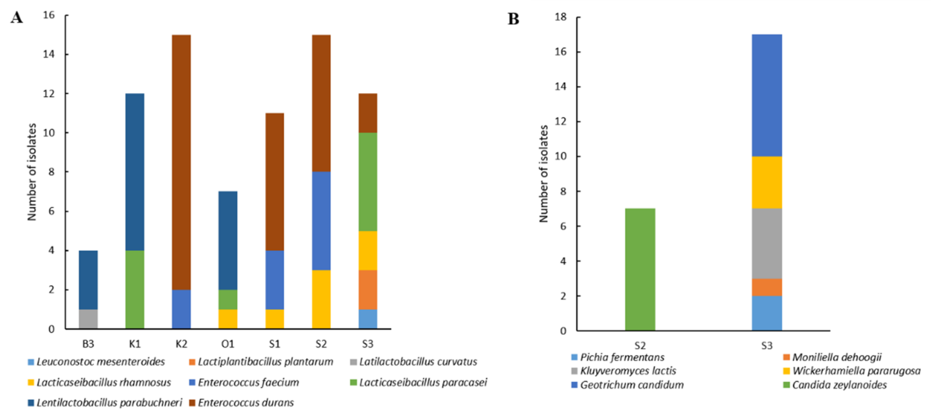
3.1.2. Lactic Acid Bacteria and Yeast Diversity Using Culture-Dependent Analysis

A total of 76 LAB and 25 yeast isolates were obtained from seven and two Smen samples, respectively. Following M13 RAPD-PCR dereplication, 52 LAB and nine yeast isolates representing the different band patterns and considered different strains, were identified to the species level by sequencing either the 16S rRNA or D1-D2 domain of the 26S rRNA genes, respectively.
As shown in Figure 2A, among LAB, enterococci and lactobacilli represented 51.31% and 47.36%, respectively, of the total isolates, and were thus clearly dominant. For enterococci, two species were identified, Enterococcus faecium and Enterococcus durans, which were dominant in three samples, S1, S2, and K2. Lactobacilli diversity was much higher and they dominated in the four remaining samples with, in decreasing order of frequency, Lentilactobacillus parabuchneri, Lacticaseibacillus paracasei, Lacticaseibacillus rhamnosus, Lactiplantibacillus plantarum, and Latilactobacillus curvatus. One Leuconostoc mesenteroides isolate was identified in one sample.
Concerning yeast diversity (Figure 2B), six species were identified. Sample S2 contained only one species, Candida zeylanoides, while five species were identified in the S3 sample. Geotrichum candidum was the most dominant, followed by Wickerhamiella pararugosa, Kluyveromyces lactis, Pichia fermentans, and Moniliella dehoogii.

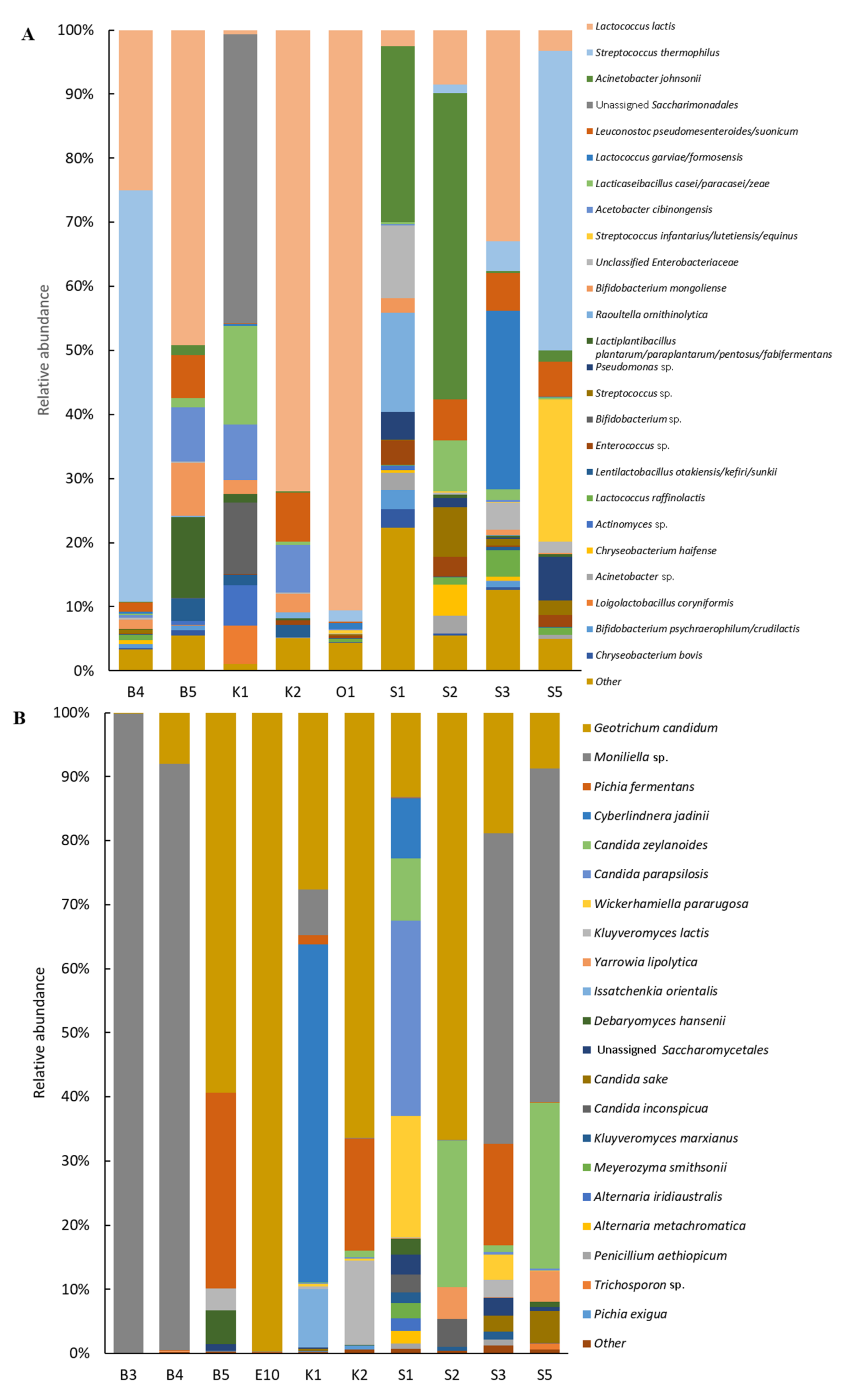
3.1.3. Microbial Diversity Using Metagenetic Analyses

Bacterial Diversity

After total DNA extraction from the studied Smen samples, nine samples (B4, B5, K1, K2, O1, S1, S2, S3, S5) yielded sufficient DNA for metagenetic analysis. With the exception of O1, all samples were prepared using the salting method, and six (K1, K2, O1, S1, S2, S3) had bacterial populations detectable by culturing. From these nine Smen samples, a total of 297,203 quality filtered sequences were generated, which were clustered into 160 OTUs. After normalization to the lowest read number in a sample (17,291), the number of observed species (richness) ranged between 44 and 110, while the Chao1 index (estimated richness) ranged from 45 to 97.5, evenness from 0.15 to 0.69, the Shannon index from 0.56 to 3.14, and Simpson’s index of diversity from 0.18 to 0.90 (Table S1). Significant differences within bacterial communities were found taking into account categorical variables that were correctly represented among the studied samples. Indeed, the Chao1 index was significantly higher in Smen samples made from cow’s milk than in Smen obtained from goat’s milk (p ˂ 0.05). In addition, the Chao1 and Simpson indices were significantly different (p < 0.05) according to the region of origin, with those collected from Setif having the highest values for both indices. It is worth mentioning that all Smen samples from the Setif region were made out of cow’s milk. Finally, sample O1 (prepared using a heat-treatment method) harboured the lowest species richness, Chao1, and Simpson index.
Compositional analysis of Smen samples revealed that OTUs belonged to eight phyla, with Bacillota representing 67.5% of total sequences followed by Pseudomonadota (20.4%), Actinomycetota (5.7%), Candidate Phyla Radiation (CPR)/”Patescibacteria” (5%), and Bacteroidota (1.4%). As shown in Figure 3A, which represents the top 25 species distribution in all samples, Smen samples harboured complex bacterial communities. Overall, Lactococcus lactis, Streptococcus thermophilus, and Acinetobacter johnsonii were the main representative species. L. lactis was dominant in the B4, B5, K2, S3, and O1 samples, with a relative abundance of 25.0%, 49.2%, 72.0%, 33.0%, and 90.6%, respectively, although it was observed in smaller proportions in K1 (0.7%), S2 (8.5%), S1 (2.5%), and S5 (3.3%). Other Lactococcus species included Lactococcus raffinolactis and Lactococcus garvieae/formosensis, the latter having a high relative abundance in the S3 sample. S. thermophilus was present in five samples, B4, S3, S2, O1, and S5, and was dominant in B4 (64.2%) and S5 (46.7%). Another Streptococcus sp. related to Streptococcus infantarius/lutetiensis/equinus was identified in sample S5. A. johnsonii dominated in S1 and S2, with a relative abundance of 27.5 and 47.7%, respectively, while an unidentified member of the Saccharimonadales order belonging to the Candidate Phyla Radiation (CPR)/”Patescibacteria” was dominant in the K1 sample. In contrast to the culture-dependent results, lactobacilli and enterococci were not the most dominant taxa, although reads from several Enterococcus and Lactobacillus related-species were found to be part of the top 25 taxa present in these Smen samples. Other species encountered in the different samples, with mean relative abundances over 1% of total sequences, included Bifidobacterium, Acetobacter, an unassigned member of the Enterobacteriaceae family, Pseudomonas, Raoultella, Enterococcus, and Chryseobacterium spp.
Beta-diversity among Smen bacterial communities was investigated using PCoA based on Bray–Curtis distances; variance analysis of these distances was then performed using a multivariate permutation Adonis test. At the OTU level, milk origin was the only factor which significantly shaped bacterial communities (p < 0.05, R2 = 0.257). It should be noted that the effect of Smen preparation methods on bacterial community structuring could not be evaluated as only one sample prepared according to that method was included in our dataset.
To further identify bacterial taxa which were over- or underrepresented in Smen produced from cow or goat milk, a linear discriminant analysis (LDA) coupled with effect size measurements (LEfSe) was performed at the genus, species, and OTU levels. Six genera, twelve species, and seventeen OTUs exhibited significantly different abundances depending on milk origin. Indeed, the microbiota of goat milk Smen were enriched in OTUs related to Acinetobacter cibinongensis, Lentilactobacillus otakiensis/kefiri/sunkii, and Levilactobacillus brevis, while those from cow’s milk were enriched in OTUs from species of the genera Streptococcus, Chryseobacterium, Pseudomonas, Enterococcus, Serratia, Acinetobacter, Kocuria, and Brachybacterium.

Fungal Diversity

The ITS2 region metabarcoding analysis generated a total of 646,883 quality-filtered sequences from ten samples (B3, B4, B5, E10, K1, K2, S1, S2, S3, S5) clustered into seventy OTUs. After rarefaction to the lowest number of sequences in a sample (50,092), richness ranged from 21 to 53, while the Chao1 index ranged from 19 to 54, evenness from 0.007 to 0.56, the Shannon index from 0.02 to 2.16 and Simpson’s index of diversity from 0.004 to 0.83 (Table S2). Comparison of alpha-diversity indices according to categorical variables showed that the Shannon and Simpson indices were significantly different according to geographical origin, as samples collected from Setif and Khenchela presented the highest values. Sample E10, which was prepared using the heat-treatment method, harboured the lowest alpha-diversity indices.
Fungal communities from Smen samples were dominated by Ascomycota and Basidiomycota members, which represented 69.9 and 30.1% of total sequences, respectively. Figure 3B shows fungal species distribution in the different Smen samples. Overall, Smen mycobiota of each sample was dominated by one to five species. Interestingly, samples such as S1 and S3 were characterized by much higher diversity than the other samples, with respectively twelve and nine species (relative abundances over 1%). G. candidum was the only species found at high relative abundance (ranging from 8% to 99.7%) in all samples with the exception of sample B3, followed by Moniliella sp. (abundances over 1% in five out of ten samples). This species was the most dominant in samples where G. candidum relative abundance was low. Other abundant species included P. fermentans, C. zeylanoides, K. lactis, Cyberlindnera jadinii, and W. pararugosa.
Concerning beta-diversity analysis, as previously observed for bacterial communities, milk origin was the only factor which significantly shaped Smen mycobiota (p < 0.05, R2 = 0.258). LefSe analysis at the OTU level showed that Smen prepared from goat milk were enriched in Pichia exigua and K. lactis OTUs, whereas an OTU related to Y. lipolytica was more prevalent in Smen made from cow’s milk.

3.1.4. Physico-Chemical Analysis and Volatile Profiles

The physico-chemical properties of the studied Smen samples are shown in Table 2. The pH values ranged between 5.2 and 3.1, and were generally below pH 4, higher pH values being found in <3 months old Smen samples. Moisture values did not exceed 19.9% and acid index values ranged between 2.2 and 153.7 mg KOH/g, with increased values in samples with increased maturation times. This was especially true for the B1 sample, which was matured for ten years. Salt content was variable and found between 7.4 and 0.1%, with the lowest values in two samples (i.e., Jn and O1) prepared using the heat-treatment method.
Volatile compound analyses by HS-GC-MS identified 81 compounds belonging to different families, i.e., esters (20), ketones (20), aldehydes (13), fatty acids (12), alcohols (7), furans (3), terpenes (3), styrene (1), and others (2) (Table S3 and Figure S1).
Based on the abundance distribution of volatile compounds for each sample, five different groups could be differentiated (Figure 4). A first group gathered 25 compounds, mainly ketones (twelve compounds, e.g., pentanone, diacetyl, hexanone) and esters (eleven compounds, e.g., ethyl butanoate, ethyl acetate, methyl butanoate). One alcohol (2-pentanol) and one fatty acid (nonanoic acid) was identified in this group. These compounds were abundant in the B3, B4, and S5 samples, while only twelve of them were present at high abundances in the S3 sample. Group II gathered nine compounds. Among them, three compounds were present at high abundances in sample B3, i.e., methylbutyl-2-methylpropanoate, styrene, and heptyl acetate. The remaining compounds corresponded to alcohols (n = 3) and esters (n = 3) and were abundant in both B3 and S3 samples. Group III gathered thirteen compounds present at high abundances in the Bi3 sample. These compounds included nine aldehydes (e.g., heptadienal and 2-nonenal) and four ketones (e.g., hydroxypropanone, and 3,5-octadien-2-one). Group IV gathered eighteen compounds which were much more abundant in the B1 sample. These mainly corresponded to fatty acids (n = 7, e.g., acetic acid, hexanoic acid, octanoic acid) and aldehydes (n = 4; e.g., benzaldehyde and nonanal). The same fatty acids, except for pentanoic acid, were abundant in the B3 sample, whereas ethylfuran, nonanal, formic acid, and pentanol were found at high abundances in both the B1 and B3 samples. Group V gathered sixteen compounds belonging to different families, i.e., ketones, esters, alcohols, fatty acids, terpenes, and others. In this group, the K1 sample was associated with methylbutyl acetate, phenylethanol, 3-methylbutanol, 3-methylbutanoic acid, propanoic acid, methylpropanoic acid, phenylethyl acetate, dimethylsulfone, and undecanone. Acetoin was related to the K2, B4 and O3 samples. In addition, the O3 sample was associated with dimethyl sulfone, phenylethyl acetate, undecanone, and tetrahydro-6-methyl-2H-pyran-2-one, while the O1 sample was associated with dimethyl sulfone and phenylethyl acetate and Jn was associated with two ketones, undecanone and tetrahydro-6-methyl-2H-pyran-2-one.
Terpenes were found at high abundances in the B5 sample, i.e., β-phellandrene, α-pinene, limonene. Limonene was found at high abundance in the O3 sample and at lower levels in E10 and S2.

3.2. Microbial Dynamics and Diversity and Biochemical Profiles throughout Different Preparation Stages of Smen

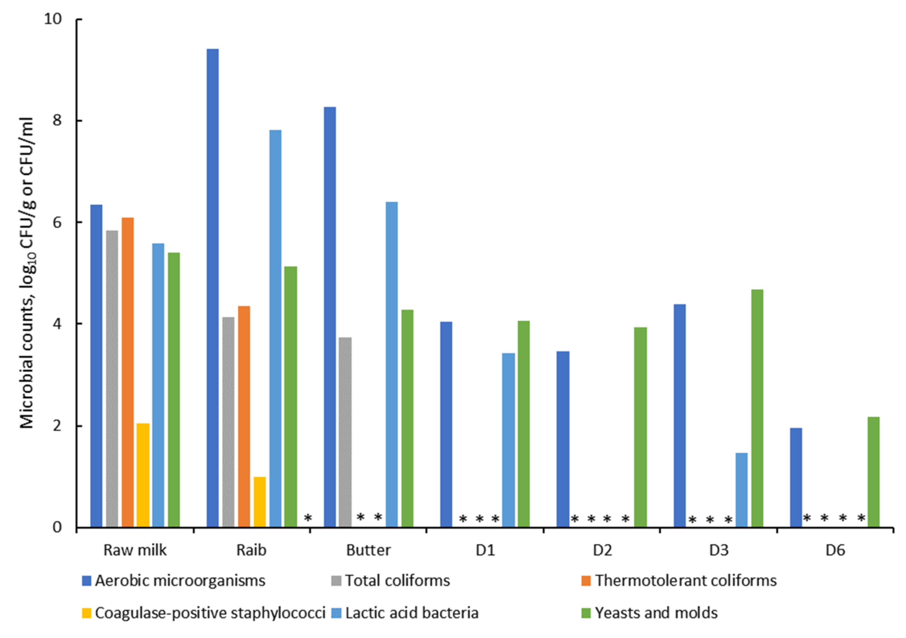
3.2.1. Safety and Hygienic Quality

In the second part of this study, the microbial diversity and dynamics during Smen manufacture were investigated using culture-dependent and culture-independent approaches. As shown in Figure 5, there were important variations in microbial counts at the different Smen production stages. Concerning total aerobic microorganisms and LAB, their counts increased during raw milk fermentation to Raib, with counts reaching 9.42 and 7.81 log10 CFU/g, respectively. A slight decrease in the latter populations was observed in butter, followed by a gradual decrease during Smen maturation. It should be underlined that LAB were found at very low populations, i.e., 1.41 log10 CFU/g, in Smen matured for three months (D3), and were below the detection limit in Smen matured for two (D2) and six (D6) months. In contrast, yeast and mold counts remained stable during Smen manufacture up to three months of maturation, with counts ranging from 4 to 5 log10 CFU/g. For total and thermotolerant coliforms, counts were high in raw milk (~6 log10 CFU/mL), decreased in Raib, and were below detection thresholds in Smen matured for one month (D1) and butter for total coliforms and thermotolerant coliforms, respectively. Coagulase-positive staphylococci followed the same trend, with an initial 2 log10 CFU/mL population in raw milk, and were not detected after butter making. Noteworthily, Salmonella spp. and sulfite-reducing anaerobic bacteria were systematically absent during all Smen manufacturing steps.

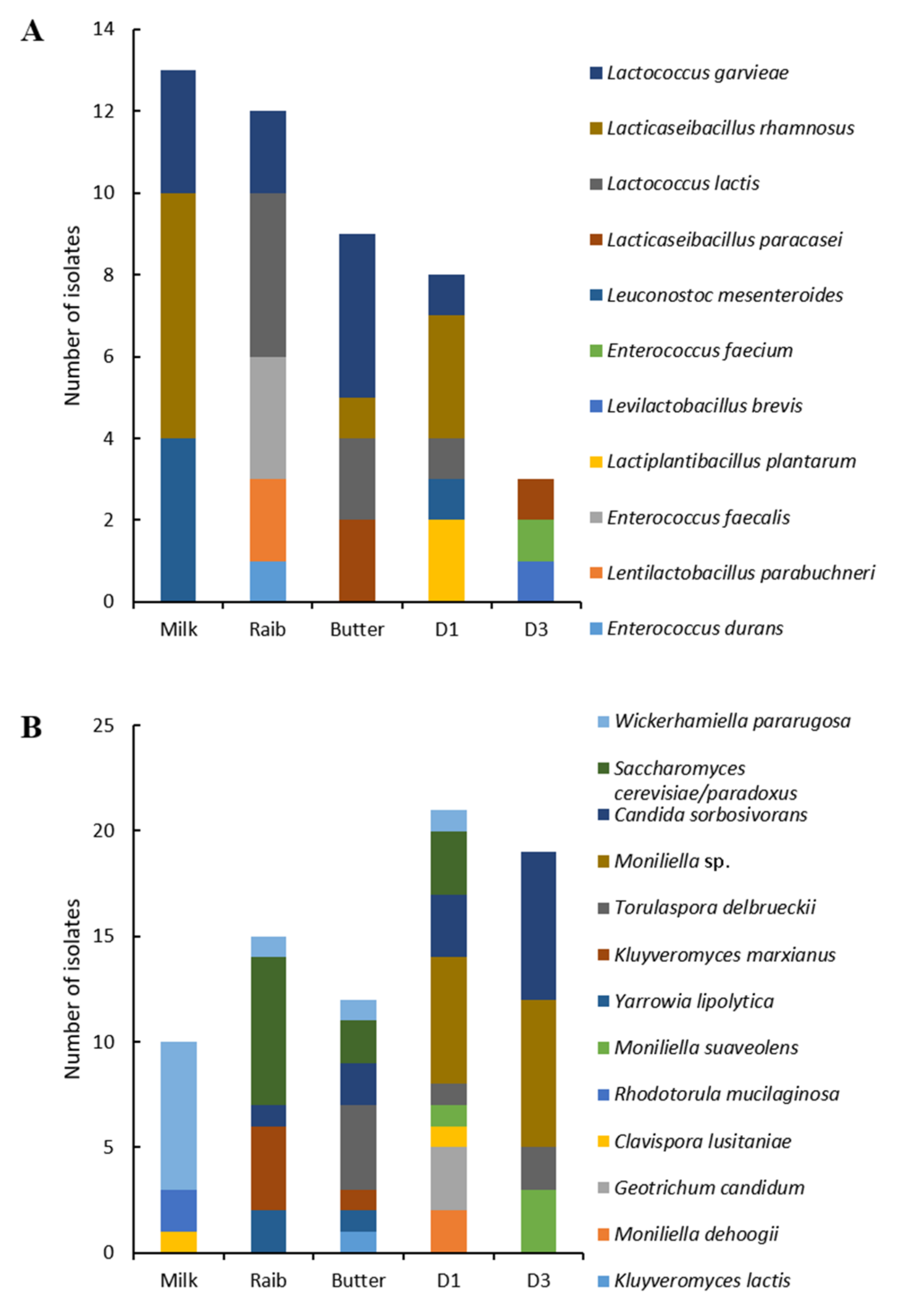
3.2.2. Lactic Acid Bacteria and Yeast Diversity Using Culture-Dependent Analysis

A total of 45 LAB and 77 yeast isolates were obtained from different preparation stages (raw milk, Raib, butter and Smen/Dhan with different maturation times, i.e., one and three months) of which 30 LAB and 39 yeast isolates representative of the different RAPD patterns were identified by sequencing.
Concerning LAB diversity, eleven species including enterococci, lactobacilli, leuconostoc, and lactococci were identified, i.e., E. faecium, E. durans, Enterococcus faecalis, L. plantarum, L. rhamnosus, Levilactobacillus brevis, L. paracasei, L. parabuchneri, L. mesenteroides, L. lactis, and L. garvieae. Shifts in LAB communities were observed throughout the different Smen preparation stages (Figure 6A). Indeed, while L. rhamnosus, L. mesenteroides, and L. garvieae dominated in raw milk, L. lactis, L. garvieae, L. parabuchneri, E. faecalis, and E. durans were dominant in Raib and L. paracasei, L. lactis, L. rhamnosus and L. garvieae were dominant in butter. The latter three species were found in one-month matured Smen along with L. mesenteroides, previously isolated from raw milk. Only three isolates were identified in three-month matured Smen, and belonged to the L. brevis, E. faecium, and L. paracasei species.
As shown in Figure 6B, yeast isolates collected throughout Smen manufacturing and maturation were assigned to thirteen different species, Clavispora lusitaniae, Rhodotorula mucilaginosa, W. pararugosa, Kluyveromyces marxianus, Saccharomyces cerevisiae/paradoxus, Y. lipolytica, Candida sorbosivorans, Torulaspora delbrueckii, K. lactis, G. candidum, M. dehoogii, Moniliella suaveolens, and an unidentified Moniliella species. Noteworthily, the latter isolates which were dominant in one-month and three-month Smen samples likely correspond to a novel species, as their D1-D2 domain 26S rRNA gene sequences shared less than 95% similarity with M. dehoogii, the closest phylogenetic relative (data not shown). As previously observed for LAB, there were shifts in fungal communities during Smen manufacture and maturation. In raw milk, three dominant species were identified, W. pararugosa, R. mucilaginosa, C. lusitaniae, while in Raib, R. mucilaginosa and C. lusitaniae were no longer detected and instead the dominants yeasts were S. cerevisiae followed by K. marxianus, Y. lipolytica, C. sorbosivorans, and W. pararugosa. The butter mycobiota were quite similar to that of Raib except that T. delbrueckii was detected in addition to the previously mentioned species. After one month of maturation, Smen harboured species previously found in raw milk, Raib and butter as well as other species that were not previously detected, i.e., G. candidum and Moniliella spp. Finally, Smen samples matured for three months were characterized by a high dominance of Moniliella spp. and C. sorbosivorans, while other species, e.g., S. cerevisiae and G. candidum, were no longer detected.

3.2.3. Microbial Diversity Using Culture-Independent Analysis

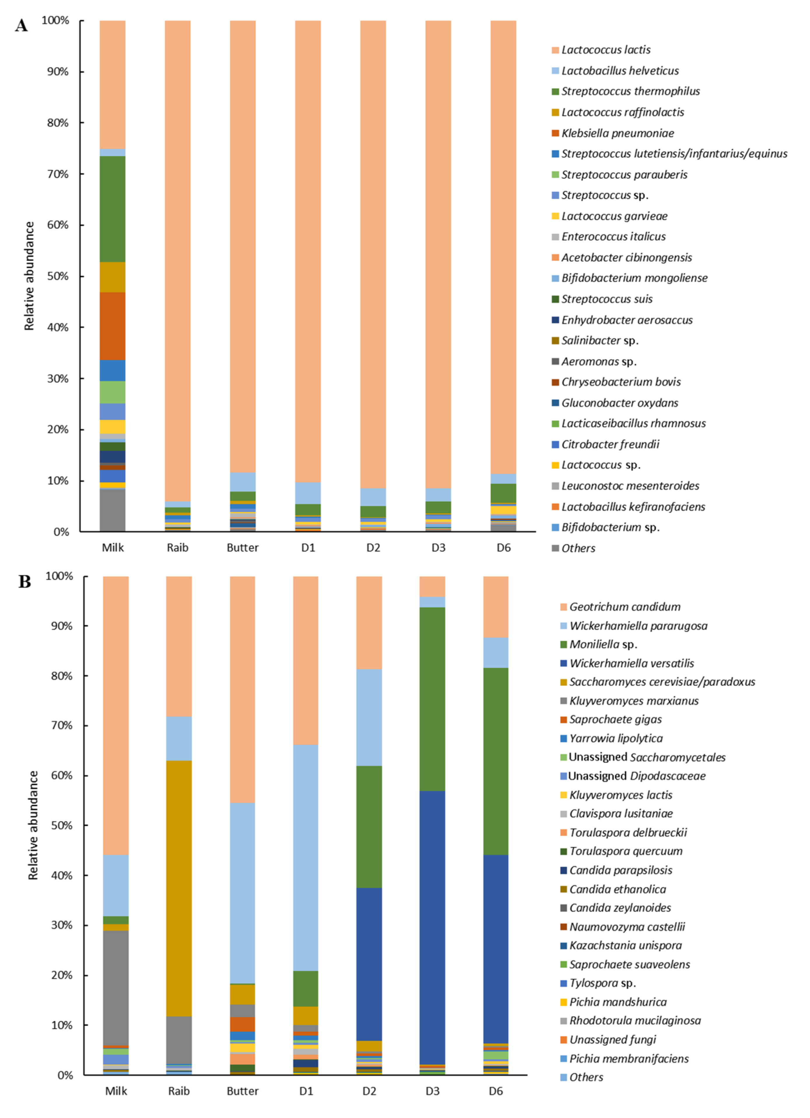
Bacterial Diversity Using V3–V4 Metabarcoding Analysis

Metabarcoding analysis was performed at the different stages of Smen preparation. In addition, this analysis was applied on potent microbial reservoirs including added milk during churning, traditional churn surface, added salt and, Smen containers. The total number of filtered high-quality reads generated from all samples was 368,039; these were clustered into 104 OTUs. After rarefaction to the lowest sequence number in a sample (22,483), richness ranged between 53 to 75, while the Chao1 index ranged from 54 to 79, evenness from 0.12 to 0.64, the Shannon index from 0.67 to 3.95, and Simpson’s index of diversity from 0.14 to 0.88 (Table S4). Considering only alpha-diversity indices of samples collected throughout Smen production and maturation, the raw milk bacterial microbiota were characterized by the highest species richness (75), Simpson index (0.88), evenness (0.64), and Shannon index (3.95). While species richness remained quite high after milk fermentation, with values between 53 (Raib) and 72 (butter), the Simpson, evenness and Shannon indexes of samples collected after milk fermentation, namely, Raib, butter and Smen after one, three, and six months maturation, decreased to values ranging from 0.14 to 0.24, 0.12 to 0.18, 0.67 and 1.1, respectively. This observation indicates that following raw milk fermentation, the bacterial microbiota of the latter samples were unequally distributed and less diverse, i.e., dominated by a relatively limited number of species.
This result was confirmed by the compositional analysis of samples throughout Smen manufacturing (Figure 7A). Indeed, raw milk harboured a complex bacterial community, with fourteen species present at a relative abundance >1%. These species corresponded on the one hand to members of the Bacillota phylum, including lactococci (L. lactis, L. raffinolactis, L. garvieae), streptococci (S. thermophilus, Streptococcus lutetiensis/infantarius/equinus, Streptococcus parauberis, Streptococcus suis), and to a lesser extent to enterococci (Enterococcus italicus) and lactobacilli (Lactobacillus helveticus). On the other hand, raw milk was characterized by the presence of members of the Pseudomonadota phylum either belonging to the Enterobacterales (i.e., Klebsiella pneumoniae, Citrobacter freundii) and Pseudomonadales order (i.e., Enhydrobacter aerosaccus). In contrast, the bacterial diversity of Raib, butter, and Smen samples during maturation was dominated by few species, with, in decreasing order of abundance, L. lactis (with relative abundances ranging from 88% to 91%), L. helveticus, and S. thermophilus. Other minor species (relative abundance < 1%) included those observed in raw milk, e.g., enterococci, other lactococci (i.e., L. garvieae and L. raffinolactis), lactobacilli species (i.e., Lactobacillus kefiranofaciens) and Leuconostoc spp. (i.e., L. mesenteroides) as well as members of the Pseudomonadota phylum. Moreover, while bacterial species distribution in Smen containers was, as expected, quite similar to that of butter, Smen species distribution in milk added during churning, at the traditional churn surface, and in the added salt were quite different (Figure S2). In particular, the milk added during churning and the surface of the traditional churn were characterized by high relative abundances of L. helveticus and L. raffinolactis OTUs, respectively, whereas the salt bacterial community was dominated by OTUs related to Haloparvum and Salinibacter sp.

Fungal Diversity Using ITS2 Metabarcoding Analysis

After filtering out low-quality and chimeric sequences, a total of 368,039 high-quality sequences were obtained and clustered into 78 OTUs. After rarefaction to the lowest number of sequences in a sample (39,443), the number of observed species (richness) ranged from 47 to 60, while the Chao1 index ranged from 49 to 90, evenness from 0.34 to 0.53, the Shannon index from 1.9 to 3.39, and Simpson’s index of diversity from 0.64 to 0.82 (Table S5). Overall, comparison of alpha-indices from the different studied samples did not show any important variations, the only exception being the salt used for butter salting, which was characterized by a high Chao1 index (i.e., 90) compared to the other samples.
Regarding fungal diversity during Smen manufacturing steps, our results highlighted major shifts in the dominant fungi (Figure 7B). Indeed, while the raw milk mycobiota were dominated by G. candidum, K. marxianus and W. pararugosa, Raib was dominated by the latter three species as well as S. cerevisiae/paradoxus, which was present in raw milk at low relative abundance. In butter, both G. candidum and W. pararugosa remained dominant while other species, including the other previously mentioned species as well as C. lusitaniae, Saprochaete gigas, K. lactis, T. delbrueckii, Torulaspora quercuum, and Y. lipolytica, were found at relative abundances over 1%. After one month maturation, the Smen mycobiota were quite similar to the one observed for butter, except that a Moniliella sp. was found at 7.2% relative abundance. After two, three, and six months maturation, the latter species became progressively dominant together with Wickerhamiella versatilis, while the relative abundance of G. candidum and W. pararugosa decreased. LefSe analysis at the OTU level confirmed that Smen samples were enriched with Moniliella sp. and W. versatilis-related OTUs. Interestingly, regarding potent fungal reservoirs encountered during Smen manufacturing (Figure S2), most harbored a high relative abundance of G. candidum (with the exception of the Smen containers) and W. pararugosa. In addition, the churning equipment and added milk during churning harboured high relative abundances of S. gigas and K. marxianus, while the salt and Smen containers appeared to be important sources of Moniliella sp. and to a lesser extent of W. versatilis.

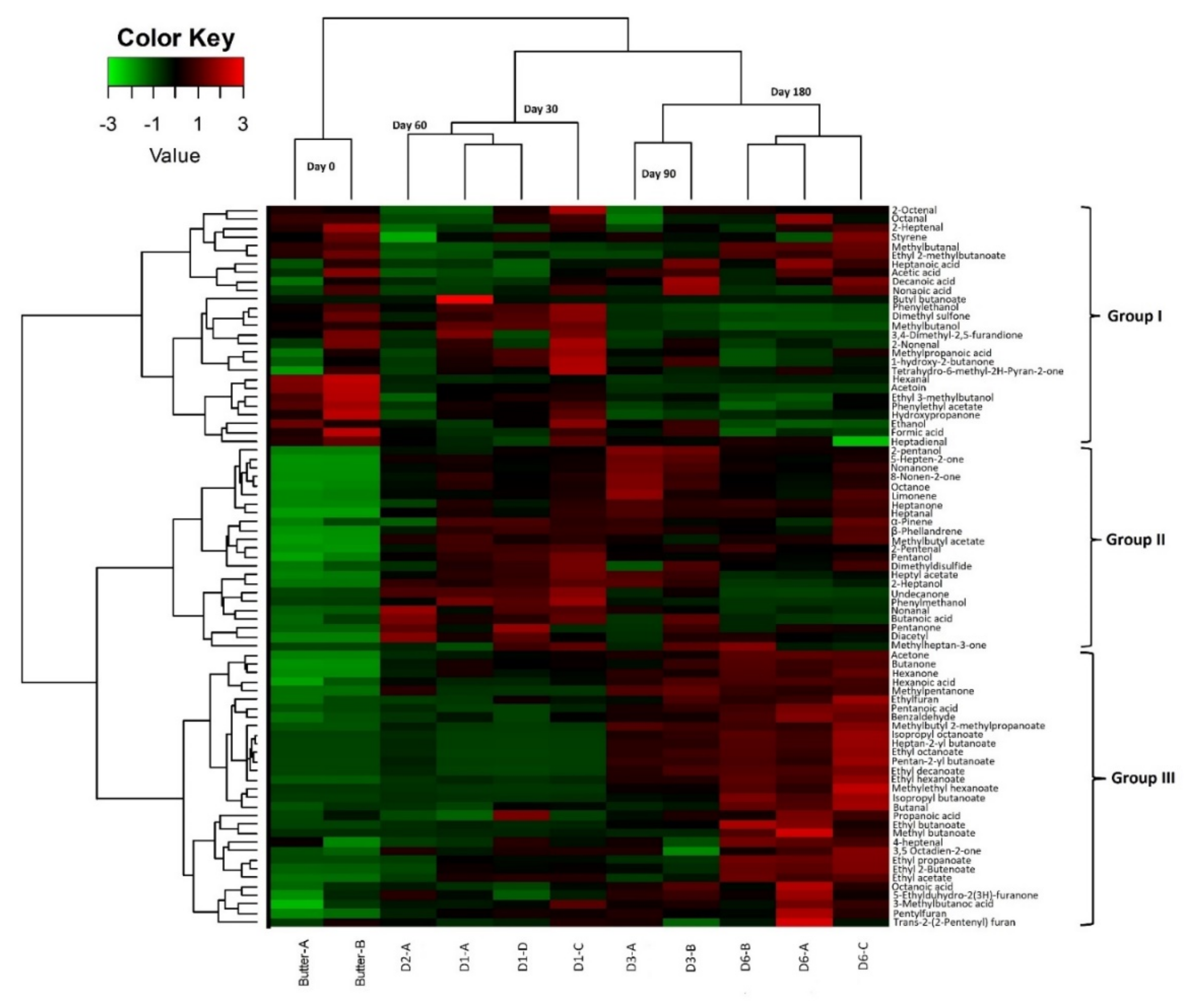
3.2.4. Physico-Chemical Analysis and Volatile Profiles

Physico-chemical changes during Smen preparation are shown in Table 3. The pH value of raw milk was 6.7 and, after 48 h fermentation, Raib pH was 4.2 and associated with an increase in Dornic acidity from 16.3 to 47. After churning, butter pH slightly increased to 4.5, then, after Smen preparation and one month maturation, decreased by ~0.5 units to reach 4 and remained constant after six months maturation. The acid index value for butter was 6.7 mg KOH/g. It increased during Smen maturation and ranged between 10.1 and 21.1 mg KOH/g after one and six months, respectively. Moisture values slightly decreased before and after Smen preparation. They varied between 22.6% for butter and between 15.9% and 14.8% after one and six months’ maturation, respectively. Prior to salting, butter contained 0.2% while after salting and one month maturation, it reached 1% before slowly increasing to 1.3% at the end of maturation.
During Smen maturation, a large number of volatile compounds were generated (Figure S3). A total of 81 compounds were identified, as described above in Section 3.1.4. Sample projection using PCA (Figure S4) showed that the first two dimensions, which accounted for 44.10% and 21.02%, respectively, separated samples according to their maturation time (0 maturation time (butter) on the left to 180 days (D6) maturation on the right). As shown in Figure S4, the matured samples (D3, D6) were found in the right quadrant, while the other samples with 30 (D1) and 60 (D2) maturation days were identified in the upper quadrant. Butter samples formed a separate group in the lower left quadrant. The D3 and D6 samples were associated with many compounds, mainly esters (n = 16, e.g., methylbutyl acetate, ethyl acetate, methyl butanoate), ketones (n = 15, e.g., diacetyl, octanone, acetone) as well as other compounds such as alcohols (n = 2, e.g., 2 pentanol, pentanol), aldehydes (n = 5, e.g., butanal, benzaldehyde, heptanal), fatty acids (n = 6, e.g., heptanoic, hexanoic, decanoic acids), furans (n = 2, i.e., ethylfuran, pentylfuran), and terpenes (n = 3, i.e., α-pinene, β-phellandrene, limonene), while the D1 and D2 samples were specifically associated with certain alcohols (methylbutanol, phenylethanol, ethanol), aldehydes (heptadienal, 2 nonenal), esters (phenylethyl acetate, ethyl-3-methyl butanoate), fatty acids (formic acid), and ketones (dimethyl-2,5- furandione, hydroxypropanone, acetoin, dimethyl sulfone). On the other hand, the butter sample was linked to heptanal, methylbutanal, ethyl 2-methylbutanoate, and acetic acid.
In order to investigate volatile compound dynamics during maturation, a heatmap representation was performed presenting the 81 volatile compounds by hierarchical clustering analysis (Figure 8). We were able to define three different clusters. The first group included 27 compounds: seven aldehydes (e.g., 2 octenal, 2 heptenal, hexanal), six fatty acids (e.g., heptanoic acid, decanoic acid, acetic acid), and five ketones (e.g., hydroxypropanone, acetoin, 1- hydroxy-2-butanone) together with four esters, three alcohols, one sulfone, and styrene. Certain aldehydes (2 octenal, octanal, 2 heptenal, hexanal, heptadienal) were found to be more abundant in raw butter, and their abundance gradually decreased between the first and third month of maturation. However, their abundance increased (except for hexanal) at six months maturation along with an increase in branched esters, free fatty acids, and styrene (methylbutanal, ethyl 2- methylbutanoate, butyl butanoate, heptanoic acid, acetic acid, nonanoic acid,). A slight decrease in acetoin, hydroxypropanone, phenylethyl acetate, ethanol, and formic acid was noted at the end of maturation compared to raw butter. The first month of maturation (D1) was characterized by the abundance of several compounds, mainly ketones (n = 3), esters (n = 2), aldehydes (n = 3), and alcohols (n = 3). Group II gathered 23 compounds, mainly ketones, i.e., 5-hepten-2-one, nonanone, 8-nonen-2-one, and octanone, the abundance of which increased from the first month of Smen maturation and continued to slightly increase to the end of maturation. The same trend was observed for 2-pentanol, limonene, heptanal, α-pinene, β-phellandrene, and methylbutyl acetate. At the mid-maturation stage (30 and 60 days), several compounds, i.e., pentanone, diacetyl, methylheptan-3-one, pentanol, 2-heptanol, nonanal, 2-heptanal, butanoic acid, heptyl acetate, phenylmethanol, undecanone, and dimethyl disulfide, presented their highest abundance before slightly decreasing at the end of maturation (90 days and 180 days). Finally, group III gathered 31 compounds including fourteen esters (e.g., isopropyl octanoate, ethyl octanoate, methylethyl hexanoate), six ketones (e.g., acetone, butanone, hexanone), five fatty acids (e.g., hexanoic acid, pentanoic acid, propanoic acid), three aldehydes (benzaldehyde, butanal, 4- heptenal), and three furans (ethylfuran, pentylfuran, trans-2,2-pentyl furan). The abundance of these compounds was the highest at the end of maturation.

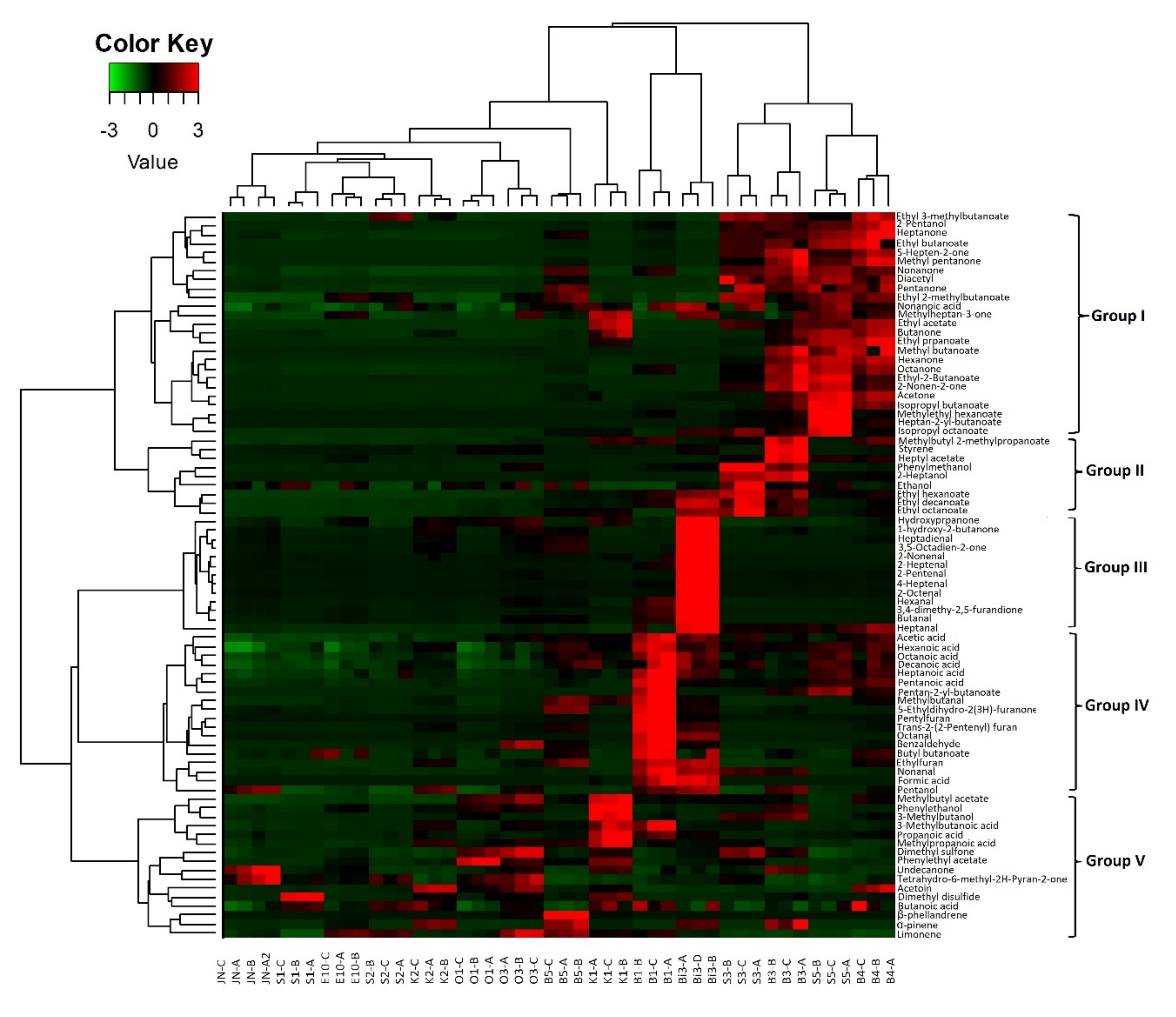
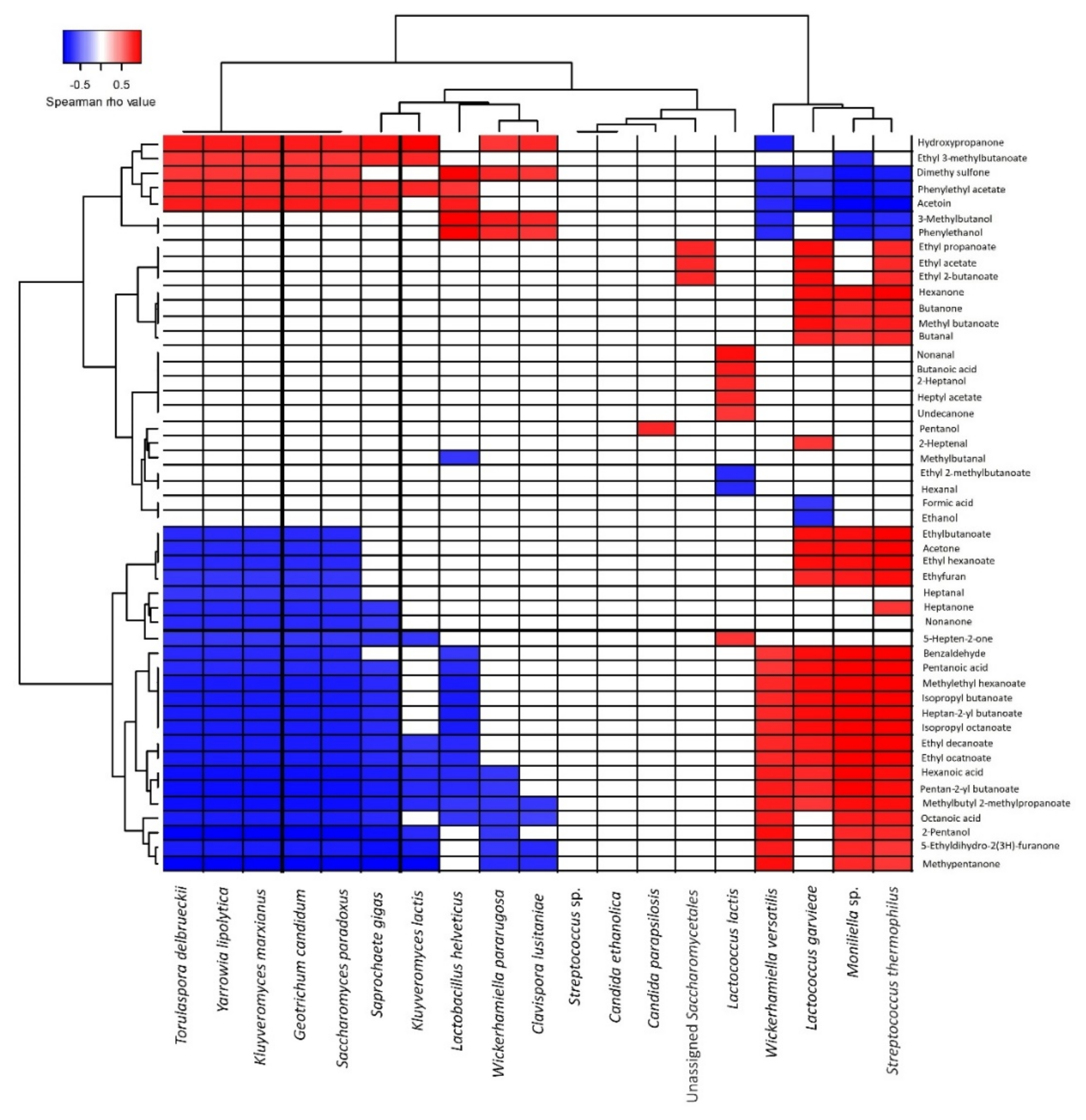
3.2.5. Correlation between Volatile Profiles and Microbiota during Maturation

To better understand the nature of the correlations between volatile compounds and microbial species, Spearman’s correlations were calculated between the two parameters, i.e., volatile profiles and independent microbial data for which a relative abundance cut-off was set to ˃0.5%. Different profiles could be differentiated (Figure 9). Nine species presented strong negative correlations (r ˃ 0.7) with esters i.e., L. helveticus (n = 9), G. candidum (n = 12), S. cerevisiae/paradoxus (n = 12), K. marxianus (n = 10), S. gigas (n = 9), Y. lipolytica (n = 11), C. lusitaniae (n = 4), K. lactis (n = 4), and T. delbrueckii (n = 10), while opposite correlations were observed for L. garvieae (n = 14), S. thermophilus (n = 11), W. versatilis (n = 8), and Moniliella sp. (n = 12). On the other hand, Y. lipolytica and T. delbrueckii showed a high positive correlation with phenylethyl acetate (r ˃ 0.8). By comparison, three species showed the highest correlations with different esters (r ˃ 0.9): Moniliella sp. with ten esters and L. garvieae and S. thermophilus with eight different esters.
Moniliella sp. and W. versatilis were the only species that showed strong positive correlations (r > 0.8) with fatty acids, i.e., octanoic acid, pentanoic acid, and hexanoic acid, while other species showed strong negative correlations with the same fatty acids. Regarding acetoin production, it was related to L. helveticus, G. candidum, K. marxianus, S. gigas, Y. lipolytica and T. delbrueckii. Several species showed strong positive correlation with certain ketones, i.e., Moniliella sp. and K. marxianus. The production of 2-pentanol was related to S. thermophilus (r = 0.96), Moniliella sp. (r = 0.8), and W. versatilis (r = 0.88). On the other hand, for dimethyl sulfone, positive correlations were noted for L. helveticus, K. marxianus, Y. lipolytica, C. lusitaniae, and T. delbrueckii.

4. Discussion

In the present study, the microbial diversity and dynamics as well as physico-chemical and volatile profiles of Algerian Smen, a spontaneously fermented butter produced using empirical methods, were investigated using several microbial, molecular and biochemical methods. To the best of our knowledge, this is the first time such an extensive study has been carried out on this fermented product.
Regarding Smen safety and hygiene, the present study confirmed, in agreement with a previous study [13], that Smen is a safe product, as all tested hygiene and safety criteria were satisfactory. This result is not surprising, as several hurdles during the Smen manufacturing process are encountered and contribute to its safety and salubrity. Indeed, spontaneous fermentation of raw milk to Raib by LAB leads to competition-exclusion phenomena as well as production of antimicrobial compounds (e.g., lactic acid) and a pH decrease. D Products are also rich in free fatty acids, the latter showing an increase during the maturation process. In the present study, the impact of these hurdles on microbial counts, and in particular on coagulase-positive staphylococci and total and thermotolerant coliforms, was clearly noticeable throughout the different Smen preparation stages. Indeed, coagulase-positive staphylococci, thermotolerant coliforms, and total coliforms were all below the detection threshold in butter and one-month matured Smen, respectively. It is worth noting that, among the fifteen products collected from Algerian households, all were satisfactory regarding hygiene and safety criteria and only two samples harboured viable yeasts with counts of ~4 log10 CFU/g, while seven samples harboured viable bacteria with counts quite similar to LAB, mainly identified as enterococci and lactobacilli. While lactobacilli are generally recognized as safe, enterococci, especially E. faecium, have a paradoxical status, as they are responsible for nosocomial infections and can transfer antibiotic resistance genes and can play a positive role in many fermented foods as well, including dairy products [42]. In this regard, it would be of interest to investigate Smen enterococci isolates for virulence genes and their acquired antibiotic resistance properties, as well as to go further in the taxonomical identification of E. faecium isolates, in order to determine whether they belong to clade A (hospital associated) or B (community associated), the latter clade being recently reassigned to Enteroccocus lactis [43].
Regarding Smen microbial diversity, two separate complementary studies were undertaken using both culture-dependent and metagenetic approaches. First, the microbial diversity and volatile profiles of Smen samples collected from different households were investigated. As expected, there were important differences in the results obtained by each microbiological approach, as culture-dependent approaches revealed certain lactobacilli and enterococci as well as yeast species, in agreement with previous studies [44,45,46,47,48] in samples matured for up to one year, with counts up to ~6 and ~4 log10 CFU/g, respectively. In contrast, metagenetic analysis, because it is based on total DNA either derived from live and dead cells, provided a general overview of microbial species that left their DNA signature during Smen manufacture and maturation. In Smen samples (those where sufficient DNA quantity was extracted for downstream metagenetic analysis), bacterial communities were found to be quite diverse, with L. lactis, S. thermophilus, and/or A. johnsonii, being the main representative species. L. lactis and S. thermophilus were previously identified as the main species involved in the spontaneous fermentation of raw milk to Raib, called Rayeb in Morocco, which can be consumed as is, processed further to a traditional white cheese called Jben, or churned to obtain buttermilk (Moroccan and Algerian lben, Tunisian leben, Egyptian, Iraqi and Lebanese laban) and raw butter (Zebda), the latter being used for Smen production [16,17,49,50,51,52]. Concerning A. johnsonii, identified at a high relative abundance in two samples (S1 and S2), together with other Gram-negative species belonging to the Pseudomonadota phylum and enterococci, their higher relative abundances, when compared to LAB species, is likely the result of either a fermentation deviation during spontaneous milk fermentation or the use of spoiled raw milk, and/or of poor hygiene quality, as this species is considered a major milk spoilage species [53].
For the first time, the present study provided an overview of Smen mycobiota, which have been systematically overlooked to date. While most identified species (e.g., G. candidum, C. zeylanoides, K. lactis) are regularly encountered in raw milk and dairy products [54,55,56,57], an interesting finding was the high prevalence of Moniliella spp., a black yeast from the Basidiomycota phylum. Members of this genus are generally considered as food spoilage agents in acidic and/or low aw foods, including fat-rich dairy products [58,59]. Its presence in Smen is not surprising, as this genus comprises species which are considered acid-resistant, xerophilic, and fermentative; as demonstrated in the second part of this study, Moniliella sp. can grow during Smen maturation. Interestingly, the type species of Moniliella spathulata (CBS 241.79) was originally isolated from Indian Ghee made from buffalo milk [60].
The investigation of the volatile profiles of Smen collected from households led to the identification of 81 compounds belonging to different families, mainly derived from fat metabolism; most of them were similar to those previously detected in Moroccan Smen [12,61]. Indeed, as previously reported [12], in addition to short chain (C4 to C10) fatty acids, which are important contributors to Smen aromatic profiles, key compounds included methylketones and esters, particularly butanoic and hexanoic ethyl esters and 2-pentanone. Enterococci (e.g., E. faecalis and E. durans) [62], lactobacilli (e.g., L. paracasei, L. plantarum, L. rhamnosus) [63,64], and yeast (e.g., C. zeylanoides, G. candidum, M. dehoogii, W. pararugosa) [47,65,66] species isolated in the present study have been previously shown to possess lipase and esterase activities. Thus, through lipolysis and conversion of free fatty acids to other compounds (e.g., short-chain fatty acids, methylketones, and ethyl esters), they likely contribute to Smen maturation during storage, as further discussed below. Moreover, as for Moroccan Smen [12], these products displayed diverse volatile profiles with no clear clustering according to maturation time, milk origin, or preparation method. This result is not surprising given the fact that Smen manufacturing processes can vary from one household to another and these processes rely on the activity of complex microbial communities composed of autochthonous microorganisms.
Several hypotheses driven by the aforementioned results were confirmed in the second part of this study by following microbial community and volatile profile dynamics during different stages of Smen manufacture and maturation. It was found that L. lactis dominated immediately after raw milk fermentation, as confirmed by both the culture-dependent and culture-independent analyses. It then became progressively subdominant after churning and subsequent maturation, as shown by the culture-dependent approach, while cultivable bacteria in matured Smen belonged to lactobacilli and enterococci. This result clearly highlighted that the strong dominance of L. lactis, revealed using a metagenetic approach, in Smen collected from households could be the result of a bias due to the presence of VBNC, or more likely, dead cells, as L. lactis can be isolated on MRS medium; a similar observation concerning discrepancies between culture-dependent and metagenetic results regarding L. lactis prevalence in dairy products was reported for Pélardon cheese [29]. To overcome this potential bias, the use of a DNA stain (e.g., propidium monoazide) which can bind to DNA from dead cells, thus rendering it inaccessible for PCR amplification, could have been useful. There was another discrepancy between the results of the culture-dependent and culture-independent approaches regarding G. candidum prevalence; while this species was found to dominate in Raib by metagenetic analysis, it was not isolated. We have no clear explanation for this result.
The results of this part of this study defined the possible technological/functional roles played by bacterial and fungal species encountered during Smen manufacturing and maturation. In the studied batch, L. lactis and to a lesser extent S. thermophilus and L. helveticus were the main bacterial species involved in raw milk spontaneous fermentation to Raib. During this process, lactose is fermented into lactate, while citrate can be metabolized by wild L. lactis to CO2, acetic acid, and α-acetolactate. The latter can be enzymatically transformed into acetoin or chemically degraded to diacetyl, which has buttery and creamy notes [61]. In raw butter, acetate and acetoin were found at high abundances, while diacetyl was present at higher abundances in Smen matured for one month. During maturation, Smen was progressively enriched in short-chain fatty acids, including even-numbered (e.g., butanoic, hexanoic, octanoic and decanoic acids) and odd-numbered (e.g., pentanoic, heptanoic, nonanoic) fatty acids. These compounds are characterized by strong odors described as fatty, rancid buttery, ripened cheese, whey-flowery, cheesy-musty, rancid buttery, and soapy [61]. The increase in fatty acid abundance can be attributed to residual milk lipase activity as well as to the lipase and esterase activities of LAB and more importantly yeasts. In this regard, strong positive correlations were found between fatty acids and W. versatilis and Moniliella sp. In addition to short-chain fatty acids, ethyl esters, i.e., ethyl acetate, ethyl butanoate, and ethyl hexanoate, were the most dominant identified compounds in Smen, especially at the end of maturation. Ethyl esters, which have pleasant, sweetly floral, and fruity odors [61], can be formed via two different reactions catalyzed by microbial esterases/lipases, namely, esterification and alcoholysis. This explains the decrease in abundances of alcohols such as ethanol, methylbutanol, and phenylethanol at the early stages of maturation and the increase in ester abundances at the end. Strong positive correlations were noted between ethyl esters and L. helveticus, L. garvieae, S. thermophilus, Moniliella sp. and W. versatilis, all of which are already known for their esterase activities [67,68,69,70]. The second-most abundant compounds were ketones. Their abundance increased after the first month of maturation. Ketones are produced through oxidation of fatty acids to β-ketoacids and decarboxylation [71]. These compounds, which have fruity, floral, and musty notes, were strongly correlated with K. marxianus and Moniliella sp. Nevertheless, to the best of our knowledge, K. marxianus is not a ketone producer [72]. Moniliella spp. are able to produce γ-decalactone and other γ-lactones and δ-lactones [73], however, nothing is known regarding their ability to produce methyl ketones. In contrast, G. candidum, which was identified at high relative abundance using metagenetics, is a very well-known ketone producer [65].

5. Conclusions

In conclusion, the present study provided new knowledge about the microbial diversity, dynamics, and volatile profiles of Algerian Smen and identified the key microorganisms during Smen manufacturing and maturation as well as their relation with volatile compound production. The diversity and abundances of the volatile compounds identified here were highly related to maturation time and lipolysis degree as well as to their autochthonous microbiota. This knowledge and the collection of microorganisms built through this study could be useful to select LAB and yeast strains to design future specific starter and non-starter cultures for Raib and Smen production at a larger scale, e.g., cooperatives of local producers to better control these fermentation processes and improve Smen quality attributes [11].

Supplementary Materials

The following supporting information can be downloaded at: https://www.mdpi.com/article/10.3390/microorganisms10040736/s1, Table S1: Alpha-diversity indices of bacterial communities from Smen samples collected in different households, Table S2: Alpha-diversity indices of fungal communities from Smen samples collected in different households, Table S3: Volatile compounds identified in different Smen samples using HS-GC-MS, Table S4: Alpha-diversity indices of bacterial communities from Smen samples throughout the different preparation stages of Smen, Table S5: Alpha-diversity indices of fungal communities from Smen samples throughout the different preparation stages of Smen, Table S6: Accession numbers of metabarcoding sequences (16S), Table S7. Accession numbers of metabarcoding sequences (ITS), Figure S1: Examples of chromatograms obtained using headspace (HS) gas chromatography-mass spectrometry (GC-MS) of Smen samples collected in different households, Figure S2: Bacterial (A) and fungal (B) diversity in potent microbial reservoirs encountered during Smen manufacturing revealed using a metagenetic approach, Figure S3: Examples of chromatograms obtained using headspace (HS) gas chromatography-mass spectrometry (GC-MS) of Smen during maturation, Figure S4: PCA plots of volatile compounds from the 81 volatile compounds detected throughout Smen maturation. PCA plot of individuals (A) and variables (B) showing principal component 1 (Dim1, 44.10%) versus 2 (Dim2, 21.02%).

Author Contributions

F.B. and J.M. were responsible for conceptualization, study design, and funding application; R.B. collected samples and performed all experimental work except volatilome analysis under F.B., J.M., M.C., S.D., A.P. and M.B. supervision; A.T. and M.-B.M. analyzed Smen volatilome; R.B. performed metagenetic reads processing under H.F. supervision; R.B., J.M., M.C., A.T., M.B. performed data statistical analysis; R.B., J.M. and F.B. drafted the manuscript. All authors contributed to the final revision and approved the submission. All authors have read and agreed to the published version of the manuscript.

Funding

This work, which was conducted in the framework of the ProMed Foods project ‘Promotion of local Mediterranean fermented foods through a better knowledge and management of microbial resources’, was funded through the ARIMNet2 2016 Call by the following funding agencies: ANR (France), MERS (Algeria), ELGO-DEMETER (Greece), MIPAF (Italy), INIA (Spain), and MHESR (Tunisia). ARIMNet2 (ERA-NET) has received funding from the European Union’s Seventh Framework Programme for research, technological development, and demonstration under grant agreement no. 618127.

Institutional Review Board Statement

Not applicable.

Informed Consent Statement

Not applicable.

Data Availability Statement

Original Research sequence data have been deposited in the Sequence Read Archive (SRA) database at the NCBI under accession PRJNA775076.

Acknowledgments

The authors wish to address their special gratitude to the Algerian Ministry of Higher Education and Scientific Research for the attribution of a scholarship within the Algerian–French cooperation program PROFAS B+. We are also grateful to the INRAE MIGALE bioinformatics facility (MIGALE, INRAE, 2020. Migale bioinformatics Facility, doi: 10.15454/1.5572390655343293E12) for providing computing and storage resources. Finally, we are grateful to the participating households for providing the samples and to the milkman for welcoming us to his dairy and for participating in the elaboration of the butter sample.

Conflicts of Interest

The authors declare no conflict of interest.

References

  1. Tamang, J.P.; Kailasapathy, K. Fermented Foods and Beverages of the World; CRC Press, Taylor and Francis Group: New York, NY, USA, 2010. [Google Scholar]
  2. El Sheikha, A.F.; Hu, D.-M. Molecular techniques reveal more secrets of fermented foods. Crit. Rev. Food Sci. Nutr. 2018, 60, 11–32. [Google Scholar] [CrossRef] [PubMed]
  3. Blandino, A.; Al-Aseeri, M.E.; Pandiella, S.S.; Cantero, D.; Webb, C. Cereal-based fermented foods and beverages. Food Res. Int. 2003, 36, 527–543. [Google Scholar] [CrossRef]
  4. Schoustra, S.E.; Kasase, C.; Toarta, C.; Kassen, R.; Poulain, A.J. Microbial Community Structure of Three Traditional Zambian Fermented Products: Mabisi, Chibwantu and Munkoyo. PLoS ONE 2013, 8, e63948. [Google Scholar] [CrossRef] [PubMed] [Green Version]
  5. Giraffa, G. Studying the dynamics of microbial populations during food fermentation. FEMS Microbiol. Rev. 2004, 28, 251–260. [Google Scholar] [CrossRef] [PubMed] [Green Version]
  6. Cocolin, L.; Ercolini, D. Zooming into Food-Associated Microbial Consortia: A ‘Cultural’ Evolution. Curr. Opin. Food Sci. 2015, 2, 43–50. [Google Scholar] [CrossRef]
  7. Tamang, J.P.; Thapa, N.; Tamang, B.; Rai, A.; Chettri, R. Microorganisms in Fermented Foods and Beverages. In Health Benefits of Fermented Foods and Beverages; Tamang, J.P., Ed.; CRC Press, Taylor and Francis Group: New York, NY, USA, 2015; pp. 2–87. [Google Scholar]
  8. Diaz, M.; Kellingray, L.; Akinyemi, N.; Adefiranye, O.O.; Olaonipekun, A.B.; Bayili, G.R.; Ibezim, J.; Du Plessis, A.S.; Houngbédji, M.; Kamya, D.; et al. Comparison of the microbial composition of African fermented foods using amplicon sequencing. Sci. Rep. 2019, 9, 13863. [Google Scholar] [CrossRef] [PubMed] [Green Version]
  9. Wafula, E.N.; Franz, C.M.; Rohn, S.; Huch, M.; Mathara, J.M.; Trierweiler, B. Fermentation of African indigenous leafy vegetables to lower post-harvest losses, maintain quality and increase product safety. Afr. J. Hortic. Sci. 2016, 9, 1–13. [Google Scholar]
  10. Benkerroum, N. Traditional Fermented Foods of North African Countries: Technology and Food Safety Challenges with Regard to Microbiological Risks. Compr. Rev. Food Sci. Food Saf. 2013, 12, 54–89. [Google Scholar] [CrossRef]
  11. Boussekine, R.; Merabti, R.; Barkat, M.; Becila, F.-Z.; Belhoula, N.; Mounier, J.; Bekhouche, F. Traditional Fermented Butter Smen/Dhan: Current Knowledge, Production and Consumption in Algeria. J. Food Res. 2020, 9, 71–82. [Google Scholar] [CrossRef]
  12. Iradukunda, C.; Aida, W.M.W.; Ouafi, A.T.; Barkouch, Y.; Boussaid, A. Aroma profile of a traditionally fermented butter (smen). J. Dairy Res. 2018, 85, 114–120. [Google Scholar] [CrossRef]
  13. El Marrakchi, A.; Tantaoui-Elarraki, A.; El Mane, A.; Tifrit, L. La flore microbienne du smen marocain I. Flore naturelle et flore d’intérêt hygiénique. Le Lait 1988, 68, 205–217. [Google Scholar] [CrossRef] [Green Version]
  14. Triqui, R.; Guth, H. Potent odorants in “Smen”, a traditional fermented butter product. Eur. Food Res. Technol. 2001, 212, 292–295. [Google Scholar] [CrossRef]
  15. Bensalah, F.; Labtar, A.; Delorme, C.; Renault, P. Occurrence, isolation and DNA identification of Streptococcus thermophilus involved in algerian traditional butter ‘Smen’. Afr. J. Biotechnol. 2011, 10, 17251–17257. [Google Scholar] [CrossRef]
  16. Bettache, G.; Fatma, A.; Miloud, H.; Mebrouk, K. Isolation and identification of lactic acid bacteria from Dhan, a traditional butter and their major technological traits. World Appl. Sci. J. 2012, 17, 480–488. [Google Scholar]
  17. Labtar, A.; Larouci, S.; Guermouche, A.; Bensalah, F. Study on molecular identification of lactic acid bacteria from fermented milks and" Smen"(a traditional steppe butter) and their enzyme producing attributes. SAJEB 2019, 9, 95–103. [Google Scholar] [CrossRef]
  18. Ongol, M.P.; Asano, K. Main microorganisms involved in the fermentation of Ugandan ghee. Int. J. Food Microbiol. 2009, 133, 286–291. [Google Scholar] [CrossRef] [Green Version]
  19. Bezuidt, O.; Gomri, M.A.; Pierneef, R.; Van Goethem, M.; Kharroub, K.; Cowan, D.; Makhalanyane, T.P. Draft genome sequence of Thermoactinomyces sp. strain AS95 isolated from a Sebkha in Thamelaht, Algeria. Stand. Genom. Sci. 2016, 11, 1–6. [Google Scholar] [CrossRef] [Green Version]
  20. Idoui, T.; Karam, N.E. Lactic acid bacteria from Jijel’s traditional butter: Isolation, identification and major technological traits. Grasas Aceites 2008, 59, 361–367. [Google Scholar] [CrossRef]
  21. ISO 4833:2003; Microbiology of Food and Animal Feeding Stuffs—Horizontal Method for the Enumeration of Microorganisms—Colony-Count Technique at 30 Degrees C. ISO: Geneva, Switzerland. Available online: https://www.iso.org/standard/34524.html (accessed on 21 March 2021).
  22. ISO 4832:2006; Microbiology of Food and Animal Feeding Stuffs—Horizontal Method for the Enumeration of Coliforms—Colony-Count Technique. ISO: Geneva, Switzerland. Available online: https://www.iso.org/standard/38280.html (accessed on 21 March 2021).
  23. ISO 7937:2004; Microbiology of Food and Animal Feeding Stuffs—Horizontal Method for the Enumeration of Clostridium Perfringens—Colony-Count Technique. ISO: Geneva, Switzerland. Available online: https://standards.iteh.ai/catalog/standards/cen/4f03b11b-ede0-4640-ad00-7169594d489b/en-iso-7937-2004 (accessed on 21 March 2021).
  24. ISO 6888-1:1999; Microbiology of Food and Animal Feeding Stuffs Horizontal Method for the Enumeration of Coagulase Positive Staphylococci (Staphylococcus aureus and Other Species)—Part 1: Technique Using Baird-Parker Agar Medium. ISO: Geneva, Switzerland. Available online: https://www.iso.org/standard/23036.html (accessed on 21 March 2021).
  25. Salmonella, A.G. Food Microbiology Protocols; Spencer, J.F.T., Ragout de Spencer, A.L., Eds.; Humana Press Inc.: Totowa, NJ, USA, 2001; pp. 113–118. [Google Scholar]
  26. Guinebretiere, M.H.; Nguyen-The, C. Sources of Bacillus cereus contamination in a pasteurized zucchini puree processing line, differentiated by two PCR-based methods. FEMS Microbiol. Ecol. 2003, 43, 207–215. [Google Scholar] [CrossRef]
  27. Weisburg, W.G.; Barns, S.M.; Pelletier, D.A.; Lane, D.J. 16S ribosomal DNA amplification for phylogenetic study. J. Bacteriol. 1991, 173, 697–703. [Google Scholar] [CrossRef] [Green Version]
  28. Kurtzman, C.P.; Robnett, C.J. Identification of clinically important ascomycetous yeasts based on nucleotide divergence in the 5’ end of the large-subunit (26S) ribosomal DNA gene. J. Clin. Microbiol. 1997, 35, 1216–1223. [Google Scholar] [CrossRef] [Green Version]
  29. Penland, M.; Falentin, H.; Parayre, S.; Pawtowski, A.; Maillard, M.-B.; Thierry, A.; Mounier, J.; Coton, M.; Deutsch, S.-M. Linking Pélardon artisanal goat cheese microbial communities to aroma compounds during cheese-making and ripening. Int. J. Food Microbiol. 2021, 345, 109130. [Google Scholar] [CrossRef]
  30. Klindworth, A.; Pruesse, E.; Schweer, T.; Peplies, J.; Quast, C.; Horn, M.; Glöckner, F.O. Evaluation of general 16S ribosomal RNA gene PCR primers for classical and next-generation sequencing-based diversity studies. Nucleic Acids Res. 2013, 41, e1. [Google Scholar] [CrossRef]
  31. Toju, H.; Tanabe, A.; Yamamoto, S.; Sato, H. High-Coverage ITS Primers for the DNA-Based Identification of Ascomycetes and Basidiomycetes in Environmental Samples. PLoS ONE 2012, 7, e40863. [Google Scholar] [CrossRef] [Green Version]
  32. Escudié, F.; Auer, L.; Bernard, M.; Mariadassou, M.; Cauquil, L.; Vidal, K.; Maman, S.; Hernandez-Raquet, G.; Combes, S.; Pascal, G. FROGS: Find, Rapidly, OTUs with Galaxy Solution. Bioinformatics 2018, 34, 1287–1294. [Google Scholar] [CrossRef]
  33. Caporaso, J.G.; Kuczynski, J.; Stombaugh, J.; Bittinger, K.; Bushman, F.D.; Costello, E.K.; Fierer, N.; Gonzalez Peña, A.; Goodrich, J.K.; Gordon, J.I.; et al. QIIME allows analysis of high-throughput community sequencing data. Nat. Methods 2010, 7, 335–336. [Google Scholar] [CrossRef] [PubMed] [Green Version]
  34. Zakrzewski, M.; Proietti, C.; Ellis, J.; Hasan, S.; Brion, M.-J.; Berger, B.; Krause, L. Calypso: A user-friendly web-server for mining and visualizing microbiome–environment interactions. Bioinformatics 2016, 33, 782–783. [Google Scholar] [CrossRef] [PubMed] [Green Version]
  35. Paquot, C. IUPAC Standard Methods for the Analysis of Oils, Fats and Derivatives, 6th ed.; Pergamon Press: Oxford, UK, 1979. [Google Scholar]
  36. Wolff, J.P. Manuel d’Analyse des Corps Gras; Azoulay: Paris, France, 1968. [Google Scholar]
  37. AOAC. Official Methods of Analysis, 12th ed.; Association of Official Analytical Chemists: Washington, DC, USA, 1975. [Google Scholar]
  38. Pogačić, T.; Maillard, M.-B.; Leclerc, A.; Hervé, C.; Chuat, V.; Yee, A.L.; Valence, F.; Thierry, A. A methodological approach to screen diverse cheese-related bacteria for their ability to produce aroma compounds. Food Microbiol. 2015, 46, 145–153. [Google Scholar] [CrossRef]
  39. Benjamini, Y.; Hochberg, Y. Controlling the False Discovery Rate: A Practical and Powerful Approach to Multiple Testing. J. R. Stat. Soc. Ser. B Methodol. 1995, 57, 289–300. Available online: http://www.jstor.org/stable/2346101 (accessed on 21 March 2022). [CrossRef]
  40. Lê, S.; Josse, J.; Husson, F. “FactoMineR: An R Package for Multivariate Analysis”. J. Stat. Softw. 2008, 25, 1–18. [Google Scholar] [CrossRef] [Green Version]
  41. Wickham, H. ggplot2: Elegant Graphics for Data Analysis, 2nd ed.; Springer International Publishing: Berlin/Heidelberg, Germany, 2016. [Google Scholar]
  42. Ogier, J.C.; Serror, P. Safety assessment of dairy microorganisms: The Enterococcus genus. Int. J. Food Microbiol. 2008, 126, 291–301. [Google Scholar] [CrossRef] [PubMed]
  43. Daza, M.V.B.; Cortimiglia, C.; Bassi, D.; Cocconcelli, P.S. Genome-based studies indicate that the Enterococcus faecium Clade B strains belong to Enterococcus lactis species and lack of the hospital infection associated markers. Int. J. Syst. Evol. Microbiol. 2021, 71, 004948. [Google Scholar] [CrossRef]
  44. Sakili, D.; Isoual, D. Les Bactéries Lactiques dans l’élaboration du Smen Marocain; Académie d’Agriculture de France: Paris, France, 2003. [Google Scholar]
  45. Savadogo, A.; Traore, A. La flore microbienne et les propriétés fonctionnelles des yaourts et laits fermentés. Int. J. Biol. Chem. Sci. 2012, 5, 2057. [Google Scholar] [CrossRef] [Green Version]
  46. Berthoud, H.; Wüthrich, D.; Bruggmann, R.; Wechsler, D.; Fröhlich-Wyder, M.-T.; Irmler, S. Development of new methods for the quantitative detection and typing of Lactobacillus parabuchneri in dairy products. Int. Dairy J. 2017, 70, 65–71. [Google Scholar] [CrossRef] [Green Version]
  47. Da Rosa, P.D.; Mattanna, P.; Carboni, D.; Amorim, L.; Richards, N.; Valente, P. Candida zeylanoides as a new yeast model for lipid metabolism studies: Effect of nitrogen sources on fatty acid accumulation. Folia Microbiol. 2014, 59, 477–484. [Google Scholar] [CrossRef]
  48. Čanak, I.; Berkics, A.; Bajcsi, N.; Kovacs, M.; Belak, A.; Teparić, R.; Maraz, A.; Mrša, V. Purification and Characterization of a Novel Cold-Active Lipase from the Yeast Candida zeylanoides. Microb. Physiol. 2015, 25, 403–411. [Google Scholar] [CrossRef]
  49. Tantaoui-Elaraki, A.; El Marrakchi, A. Study of Moroccan Dairy Products: Lben and Smen. Mircen J. 1987, 3, 211–220. [Google Scholar] [CrossRef]
  50. Abd-El-Malek, Y. Traditional Egyptian dairy fermentations. In Global Impact of Food Microbiology (GIAM). State of the Art: GIAM and Its Relevance to Development in Developing Countries; Stantan, W.R., Da Silva, E.J., Eds.; University of Malaya Press: Kuala Lumpur, Malaysia, 1978; pp. 198–208. [Google Scholar]
  51. Ben Amor, K.; Cornelieus, C.; Mahjoub, A.; Thonart, P. Identification de La Flore Lactique Du Lait Fermenté Traditionnel Tunisien (Lben) et Évaluation Des Composés Aromatisants. Microb. Hyg. Alim. 1998, 10, 31–36. [Google Scholar]
  52. Hamama, A.; Ha, D. Improvements of the manufacture of traditional fermented products in Morocco: Case of jben (moroccan traditional fresh cheese). In Emerging Technology Series, Food Processing Technologies for Africa; UNIDO: Vienna, Austria, 1997; pp. 85–102. [Google Scholar]
  53. Schmidt, V.S.; Kaufmann, V.; Kulozik, U.; Scherer, S.; Wenning, M. Microbial biodiversity, quality and shelf life of microfiltered and pasteurized extended shelf life (ESL) milk from Germany, Austria and Switzerland. Int. J. Food Microbiol. 2012, 154, 1–9. [Google Scholar] [CrossRef]
  54. Delavenne, E.; Mounier, J.; Déniel, F.; Barbier, G.; Le Blay, G. Biodiversity of antifungal lactic acid bacteria isolated from raw milk samples from cow, ewe and goat over one-year period. Int. J. Food Microbiol. 2012, 155, 185–190. [Google Scholar] [CrossRef]
  55. Wouters, J.T.; Ayad, E.H.; Hugenholtz, J.; Smit, G. Microbes from raw milk for fermented dairy products. Int. Dairy J. 2002, 12, 91–109. [Google Scholar] [CrossRef]
  56. Gardini, F.; Tofalo, R.; Belletti, N.; Iucci, L.; Suzzi, G.; Torriani, S.; Guerzoni, M.; Lanciotti, R. Characterization of yeasts involved in the ripening of Pecorino Crotonese cheese. Food Microbiol. 2006, 23, 641–648. [Google Scholar] [CrossRef]
  57. Sessou, P.; Keisam, S.; Tuikhar, N.; Gagara, M.; Farougou, S.; Jeyaram, K. High-Throughput Illumina MiSeq amplicon sequencing of yeast communities associated with indigenous dairy products from republics of benin and niger. Front. Microbiol. 2019, 10, 594. [Google Scholar] [CrossRef]
  58. Rosa, C.A.; Jindamorakot, S.; Limtong, S.; Nakase, T.; Lachance, M.-A.; Fidalgo-Jiménez, A.; Daniel, H.-M.; Pagnocca, F.C.; Inácio, J.; Morais, P.B. Synonymy of the yeast genera Moniliella and Trichosporonoides and proposal of Moniliella fonsecae sp. nov. and five new species combinations. Int. J. Syst. Evol. Microbiol. 2009, 59, 425–429. [Google Scholar] [CrossRef]
  59. Nakayama, M.; Hosoya, K.; Shimizu-Imanishi, Y.; Chibana, H.; Yaguchi, T. Development of Rapid Identification and Risk Analysis of Moniliella spp. in Acidic Processed Foods. Biocontrol Sci. 2016, 21, 73–80. [Google Scholar] [CrossRef] [Green Version]
  60. De Hoog, G.S. Taxonomic review of Moniliella, Trichosporonoides and Hyalodendron. In The Black Yeasts, II: Moniliella and Allied Genera; Institute of the Royal Netherlands: Amsterdam, The Netherlands, 1979; pp. 1–36. [Google Scholar]
  61. Sarhir, S.T.; Amanpour, A.; Bouseta, A.; Selli, S. Fingerprint of aroma-active compounds and odor activity values in a traditional Moroccan fermented butter “Smen” using GC–MS–Olfactometry. J. Food Compost. Anal. 2021, 96, 103761. [Google Scholar] [CrossRef]
  62. Serio, A.; Chaves-López, C.; Paparella, A.; Suzzi, G. Evaluation of metabolic activities of enterococci isolated from Pecorino Abruzzese cheese. Int. Dairy J. 2010, 20, 459–464. [Google Scholar] [CrossRef]
  63. García-Cano, I.; Rocha-Mendoza, D.; Ortega-Anaya, J.; Wang, K.; Kosmerl, E.; Jiménez-Flores, R. Lactic acid bacteria isolated from dairy products as potential producers of lipolytic, proteolytic and antibacterial proteins. Appl. Microbiol. Biotechnol. 2019, 103, 5243–5257. [Google Scholar] [CrossRef] [PubMed] [Green Version]
  64. Oliszewski, R.; Medina, R.B.; Gonzalez, S.N.; Perezchaia, A.; Chaia, A.B.P. Esterase activities of indigenous lactic acid bacteria from Argentinean goats’ milk and cheeses. Food Chem. 2007, 101, 1446–1450. [Google Scholar] [CrossRef]
  65. Boutrou, R.; Guéguen, M. Interests in Geotrichum candidum for cheese technology. Int. J. Food Microbiol. 2005, 102, 1–20. [Google Scholar] [CrossRef] [PubMed]
  66. Sakpuntoon, V.; Angchuan, J.; Boontham, W.; Khunnamwong, P.; Boonmak, C.; Srisuk, N. Grease Waste as a Reservoir of Lipase-Producing Yeast and Description of Limtongella siamensis gen. nov., sp. nov. Microorganisms 2019, 8, 27. [Google Scholar] [CrossRef] [Green Version]
  67. Collins, Y.F.; McSweeney, P.L.H.; Wilkinson, M.G. Lipolysis and free fatty acid catabolism in cheese: A review of current knowledge. Int. Dairy J. 2003, 13, 841–866. [Google Scholar] [CrossRef]
  68. Fernández, E.; Alegría, A.; Delgado, S.; Mayo, B. Phenotypic, genetic and technological characterization of Lactococcus garvieae strains isolated from a raw milk cheese. Int. Dairy J. 2010, 20, 142–148. [Google Scholar] [CrossRef] [Green Version]
  69. Kalantzopoulos, G.; Tsakalidou, E.; Manolopoulou, E. Proteinase, peptidase and esterase activities of cell-free extracts from wild strains of Lactobacillus delbrueckii subsp. bulgaricus and Streptococcus salivarius subsp. thermophilus isolated from traditional Greek yogurt. J. Dairy Res. 1990, 57, 593–601. [Google Scholar] [CrossRef]
  70. Higuchi, T.; Aoki, T.; Uchida, K. Esterase activity in soy sauce Moromi as a factor hydrolyzing flavor esters. J. Ferment. Bioeng. 1991, 71, 163–167. [Google Scholar] [CrossRef]
  71. McSweeney, P.L.; Sousa, M.J. Biochemical pathways for the production of flavour compounds in cheeses during ripening: A review. Le Lait. 2000, 80, 293–324. [Google Scholar] [CrossRef]
  72. Padilla, B.; Belloch, C.; López-Díez, J.J.; Flores, M.; Manzanares, P. Potential impact of dairy yeasts on the typical flavour of traditional ewes’ and goats’ cheeses. Int. Dairy J. 2014, 35, 122–129. [Google Scholar] [CrossRef] [Green Version]
  73. Laufenberg, G.; Rosato, P.; Kunz, B. Adding value to vegetable waste: Oil press cakes as substrates for microbial decalactone production. Eur. J. Lipid Sci. Technol. 2004, 106, 207–217. [Google Scholar] [CrossRef]
Figure 1. Flow diagram showing the traditional Smen production process according to the salting (A) and heat-treatment (B) methods.
Microorganisms 10 00736 g001
Figure 2. Bacterial (A) and fungal (B) diversity in Smen samples collected from Algerian households determined by culture-dependent analyses.
Microorganisms 10 00736 g002
Figure 3. Bacterial (A) and fungal (B) diversity in Smen samples from different Algerian households determined by a metagenetic approach.
Microorganisms 10 00736 g003
Figure 4. Normalized heat-map representation of volatile abundances in fifteen different Smen samples collected from Algerian households. Hierarchical clustering was performed using Ward’s Linkage and Euclidean distance.
Microorganisms 10 00736 g004
Figure 5. Microbial counts during the different preparation stages of Smen (D1, D2, D3, D6: Smen after one, two, three, and six months of maturation, respectively). * indicates counts below the detection threshold.
Microorganisms 10 00736 g005
Figure 6. Bacterial (A) and fungal (B) diversity during the different preparation stages of Smen (D1, D3: Smen after one and three months of maturation, respectively) determined by the culture-dependent approach.
Microorganisms 10 00736 g006
Figure 7. Bacterial (A) and fungal (B) diversity during Smen preparation stages (D1, D2, D3, D6: Smen after one, two, three, and six months of maturation, respectively) determined by a metagenetic approach.
Microorganisms 10 00736 g007
Figure 8. Normalized heat-map representation of volatile abundances during Smen maturation. Hierarchical clustering was performed using Ward’s Linkage and Euclidean distance. (D1, D2, D3, D6: Smen after one, two, three, and six months of maturation, respectively).
Microorganisms 10 00736 g008
Figure 9. Spearman correlation matrices between microbial species composition and volatile compounds during Smen maturation (butter and Smen samples matured for one to six months). Only significant correlations are shown (p < 0.05). Positive correlations are indicated in red while negative ones are indicated in blue.
Microorganisms 10 00736 g009
Table 1. Description of collected Smen samples.
SamplesMilk OriginMaturation TimeRegionPreparation MethodStorage Temperature
B1Cow10 yearsBatnaSaltingAmbient
B3Cow<1 yearBatnaSaltingAmbient
B4Cow<1 yearBatnaSaltingAmbient
B5Goat2 yearsBatnaSaltingAmbient
Bi3Ewe and goat<1 yearBiskraHeat-treatmentRefrigerated
E10Goat1 yearEl OuedHeat-treatmentAmbient
JnCow<3 monthsJijelHeat-treatmentAmbient
K1Goat6 monthsKhenchelaSaltingAmbient
K2Goat10 monthsKhenchelaSaltingAmbient
O1Goat1 yearOuarglaHeat-treatmentRefrigerated
O3Goat1 yearOuarglaHeat-treatmentAmbient
S1Cow6 monthsSetifSaltingAmbient
S2Cow2 monthsSetifSaltingAmbient
S3Cow1 monthsSetifSaltingAmbient
S5Cow2 yearsSetifSaltingAmbient
Table 2. Physicochemical properties and microbial counts (log10 CFU/g) of the fifteen different Smen samples produced in Algerian households.
SamplespHMoisture (%)Acid Index (mg KOH/g)Salt Content (%)Total Aerobic Mesophilic BacteriaTotal
Coliforms
Thermo-Tolerant ColiformsLAB 1Yeast and MoldsCoagulase-Positive StaphylococciSalmonella spp.Sulphite-Reducing
Anaerobic Bacteria
B13.1 ± 0.019.9153.7 ± 1.7ND 1<1<1<1<1<2<2n.d. 3n.d.
B33.5 ± 0.012.235.9 ± 0.0ND4.5<1<12.3<2<2n.d.n.d.
B43.4 ± 0.01240.3 ± 0.1ND<1<1<1<1<2<2n.d.n.d.
B53.6 ± 0.012.631.4 ± 0.0ND<1<1<1<1<2<2n.d.n.d.
Bi33.8 ± 0.00.819.1 ± 1.1ND<1<1<1<1<2<2n.d.n.d.
E10NDNDNDND<1<1<1<1<2<2n.d.n.d.
Jn4.3 ± 0.003.9 ± 0.10.1 ± 0.0<1<1<1<1<2<2n.d.n.d.
K13.3 ± 0.012.76.7 ± 0.03.3 ± 0.05.2<1<15.1<2<2n.d.n.d.
K23.8 ± 0.09.014.6 ± 1.52.2 ± 0.04.1<1<13.1<2<2n.d.n.d.
O15.2 ± 0.002.2 ± 0.00.1 ± 0.03.2<1<13.3<2<2n.d.n.d.
O3ND02.2 ± 0.0ND<1<1<1<1<2<2n.d.n.d.
S13.3 ± 0.012.83.3 ± 0.07.4 ± 0.13.7<1<14.5<2<2n.d.n.d.
S23.6 ± 0.07.33.6 ± 0.03.9 ± 0.95.3<1<15.54.2<2n.d.n.d.
S34.4 ± 0.010.54.4 ± 0.61.4 ± 0.05.4<1<16.74.7<2n.d.n.d.
S5ND2.543.8 ± 1.9ND<1<1<1<1<2<2n.d.n.d.
ND, Not Determined due to limited sample size; 1 LAB, lactic acid bacteria; n.d., not detected.
Table 3. Physicochemical properties of samples collected during Smen manufacture stages.
SamplepHMoisture (%)Dornic Acidity (D°)Salt Content (%)Acid Index
(mg KOH/g)
Milk6.7 ± 0.0ND16.3 ± 0.0NDND
Raib4.2 ± 0.0ND47.0 ± 0.0NDND
Butter4.5 ± 0.122.6ND0.20 ± 0.06.7 ± 0.0
1-month Smen (D1)4.0 ± 0.115.9ND1.10 ± 0.010.1 ± 1.1
2-month Smen (D2)4.3 ± 0.115.1ND1.10 ± 0.018.4 ± 0.0
3-month Smen (D3)4.1 ± 0.015.0ND1.20 ± 0.019.1 ± 1.1
6-month Smen (D6)4.0 ± 0.014.8ND1.30 ± 0.121.1 ± 0.3
ND, Not Determined.
Publisher’s Note: MDPI stays neutral with regard to jurisdictional claims in published maps and institutional affiliations.

