Article

Loop-Mediated Isothermal Amplification as Point-of-Care Testing for EGFR-Mutated Lung Adenocarcinoma

1
Department of Surgery, Teikyo University School of Medicine, 2-11-1 Kaga, Itabashi-ku 173-8605, Tokyo, Japan
2
Department of Thoracic Surgery, Saitama Cardiovascular and Respiratory Center, 1696 Itai, Kumagaya 360-0197, Saitama, Japan
3
Biochemical Research Laboratory II, Eiken Chemical Co., Ltd., 1381-3 Shimoishigami, Otawara-shi 324-0036, Tochigi, Japan
4
Department of Pathology, Saitama Cardiovascular and Respiratory Center, 1696 Itai, Kumagaya 360-0197, Saitama, Japan
*
Author to whom correspondence should be addressed.
Micromachines 2022, 13(6), 897; https://doi.org/10.3390/mi13060897
Submission received: 13 March 2022 / Revised: 21 April 2022 / Accepted: 22 April 2022 / Published: 6 June 2022
(This article belongs to the Special Issue Point-of-Care Diagnostic Devices)

Abstract

:
Liquid biopsy has been adapted as a diagnostic test for EGFR mutations in patients with advanced or metastatic non-small cell lung cancer (NSCLC). Loop-mediated isothermal amplification (LAMP) has been widely used for the rapid detection of pathogens through DNA amplification. This study investigated the efficacy of an EGFR-LAMP assay using plasma samples of patients with resected NSCLC tumors. The EGFR status was investigated using both LAMP and next-generation sequencing (NGS) assays in cases that met the following criteria: (1) pulmonary adenocarcinoma with EGFR mutation detected by the Therascreen EGFR PCR Kit and (2) preoperative plasma samples contained enough DNA for the LAMP and NGS experiments. Among 51 specimens from patients with EGFR-mutated tumors or metastatic lymph nodes, the LAMP assay detected 1 EGFR mutation that was also detected in the NGS assay. However, a plasma sample that demonstrated EGFR wild type in the LAMP assay showed an EGFR mutant status in NGS. The detection rates (1.9% in LAMP and 3.9% in NGS) were very low in both assays, demonstrating a similar performance in detecting EGFR mutations in NSCLC tumors; therefore, it could be a more suitable test for the advanced stage, not the early stage. Notably, the LAMP assay was more time-saving, cost-effective, and straightforward. However, further investigation is required to develop a more sensitive assay.

1. Introduction

Lung cancer remained the leading cause of cancer death and the second most frequently diagnosed cancer in the GLOBOCAN 2020 database [1]. The 5-year survival rate after lung cancer diagnosis was only 10% to 20% in most countries from 2010 to 2014, although these rates were higher in Japan (33%), Israel (27%), and Korea (25%) [2]. In the last two decades, advances in lung cancer therapeutics have led to the adaptation of comprehensive molecular profiling of novel driver mutations, and the epidermal growth factor receptor (EGFR) tyrosine kinase inhibitors (TKIs) have been shown to improve overall response rate and median progression-free survival when compared with platinum-based chemotherapy in patients with advanced or metastatic non-small cell lung cancer (NSCLC) harboring activating EGFR mutations [3,4,5,6,7,8,9,10]. Thus, EGFR TKIs are established effective therapies in patients with activating and sensitizing mutation in exons 18–21 of EGFR [11,12,13,14].
Recently, NCCN guidelines noted that osimertinib, a third-generation TKI, should be considered an adjuvant treatment even after complete resection of NSCLC [14]. Until recently, osimertinib was used only as a second-line treatment for those with T790M mutations [12,13]. However, Wu et al. reported that osimertinib demonstrated statistically longer disease-free survival in patients with stage IB to IIIA EGFR-mutated NSCLC than in those who received a placebo [15]. Based on published evidence regarding osimertinib use in the double-blind, phase III ADAURA clinical trials, routine molecular biomarker testing of EGFR mutation is now recommended not only for patients with advanced or metastatic NSCLC but also for patients with completely resected stage IB-IIIA NSCLC according to the NCCN guidelines [14].
These clinical trials have called for new or revised recommendations in the molecular testing guidelines for patients with NSCLC [16,17,18,19,20,21]. Currently, several assays are accepted as EGFR testing methods based on the technologies of real-time polymerase chain reaction (RT-PCR) or next-generation sequencing (NGS). The cobas® EGFR Mutation Test v2 (Hoffman-La Roche Ltd., Basel, Switzerland) and the Therascreen EGFR Rotor-Gene Q (RGQ) PCR Kit® (Qiagen, K.K., Tokyo, Japan) were approved as companion diagnostics with TKIs by the United States Food and Drug Administration and the Ministry of Health, Labour and Welfare in Japan [18]. Similarly, FoundationOne® CDx (Foundation Medicine Inc., Cambridge, MA, USA) and OncomineTM Dx Target Test (Thermo Fisher Scientific Inc., Waltham, MA, USA) were adapted for EGFR screening for patients with advanced or metastatic NSCLC. However, these methods have been notably complicated, time-consuming, and expensive in clinical practice.
Loop-mediated isothermal amplification (LAMP) is an alternative PCR-based technology that has been widely used globally in bacteriology [22,23,24,25] and virology, including for SARS-CoV-2 [26,27,28,29,30,31]. For instance, TB-LAMP has been used extensively, with multiple meta-analyses reporting sensitivity and specificity in the range of 89–93% and 94–95%, respectively [25,32,33]. In contrast to RT-PCR, LAMP uses 4–6 primers, does not require an expensive thermocycler, and has the potential to decrease testing cost and time to diagnosis in laboratory or community settings. Thus, the LAMP assay has been recommended in guidelines [34,35], and point-of-care diagnostics for tuberculosis have been developed with a turnaround time of about 30 min and a cost of about 7 euros per test [36]. In our previous study, 117 NSCLC tumor tissues were investigated to compare the results of the EGFR-LAMP and Therascreen assays. The receiver operating characteristics curve analysis of the EGFR-LAMP assay for LAMP values demonstrated 0.973 (95% CI: 0.929–1.00) in exon 19 deletion, and 0.952 (95% CI: 0.885–1.00) in L858R [37]. The sensitivity and specificity were 89.3% and 98.9%, respectively. Although the LAMP assay is not yet widely used in clinical oncology, it could provide a more rapid, simple, and inexpensive method for detecting oncogenes than conventional methods.
Recently, many researchers have been investigating liquid biopsy as a promising non-invasive technology for the early detection of cancer as well as continuous monitoring of disease progression and/or treatment efficacy [38,39,40,41]. In this study, we aimed to develop a new liquid biopsy system using EGFR-LAMP primers for NSCLC. We compared findings between the LAMP and NGS assays and evaluated their sensitivity and specificity.

2. Materials and Methods
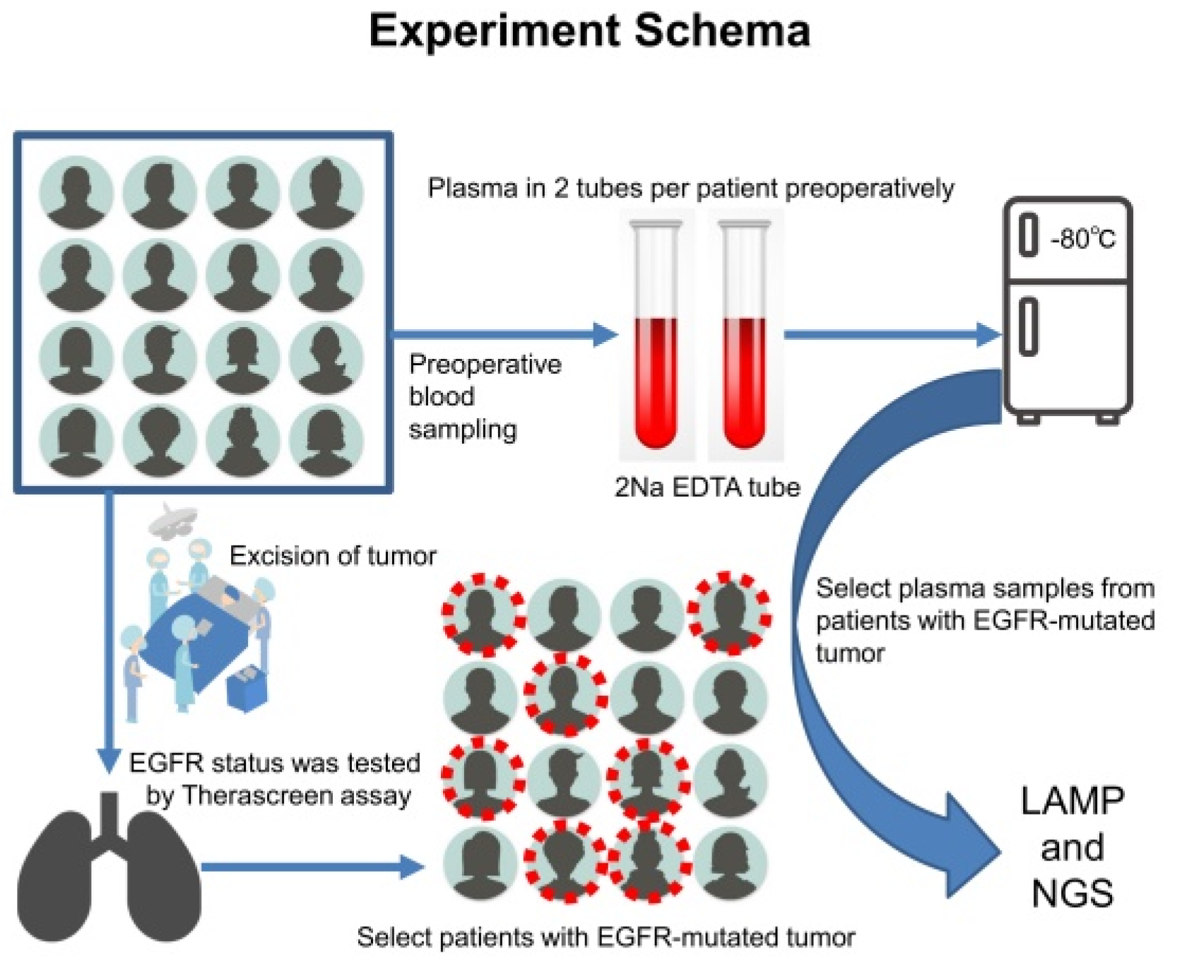
2.1. Study Design

This study was a prospective study without interventions to investigate the sensitivity and specificity of the EGFR-LAMP liquid biopsy compared with those of the NGS assay. The objects of this study were patients who had surgical biopsy or treatment for NSCLC suspected at Saitama Cardiovascular and Respiratory Center (Saitama, Japan) from January 2019 to September 2020. Clinical data and plasma samples were collected from patients preoperatively and consecutively, and tumor tissue samples were obtained through surgery or surgical biopsy (Figure 1). Any other pulmonary diseases without NSCLC were excluded from this study. Only EGFR-mutated pulmonary adenocarcinomas detected using the Therascreen assay were selected for further investigation for liquid biopsy. The present study was approved by The Institutional Review Board of the Saitama Cardiovascular and Respiratory Center (approval no. 2018033). Written informed consent was provided by all patients. The study was conducted in accordance with the Declaration of Helsinki (as revised in 2013). There were 3 primary endpoints for the study: (i) confirmation of the detection of EGFR mutations in NSCLC using the EGFR-LAMP primers, (ii) calculation of sensitivity and specificity, and (iii) comparison with results of the NGS assay. We planned to collect 50 EGFR-mutated tumors to increase the probability of detecting at least one EGFR mutation in the LAMP assay.

2.2. Plasma Samples and DNA Extraction

Blood samples were obtained preoperatively and consecutively from patients with a strong suspicion of primary lung cancer. Through normal centrifugation (4 °C, 3000 rpm, 15 min) within 8 h from blood sampling, plasma separation was performed by sampling from the center of the plasma supernatant. Among these, postoperative pathology demonstrated 57 EGFR-mutated primary adenocarcinomas considered eligible for this study. After excluding 6 patients who did not provide written informed consent, cell-free DNA (cfDNA) from plasma samples was extracted using the cobas® cfDNA Sample Preparation Kit (Roche Diagnostics, Hague Road, IN, USA) according to the manufacturer’s protocol.

2.3. Pathological Diagnosis and Therascreen qPCR Mutation Analysis

All tumor tissues were obtained through surgery or surgical biopsy and fixed with 10% buffer formalin at 20–25 °C (24–36 h) to create formalin-fixed, paraffin-embedded tumor blocks according to the recommendation of The Japanese Society of Pathology [42]. Hematoxylin–eosin (HE) staining was performed by the standard methods using Tissue-Tek Prisma® (Sakura Finetek Japan Co., Ltd., Tokyo, Japan) according to the manufacturer’s protocol. All pathological diagnoses were made based on the WHO classification version 8 by an expert pulmonary pathologist who examined HE-stained slides in low and high magnification using an ECLIPSE Ni-u light microscope (Nikon Co., Ltd., Tokyo, Japan). The presence of EGFR mutations was determined using a Therascreen EGFR PCR kit® (Qiagen, K.K.) [43].

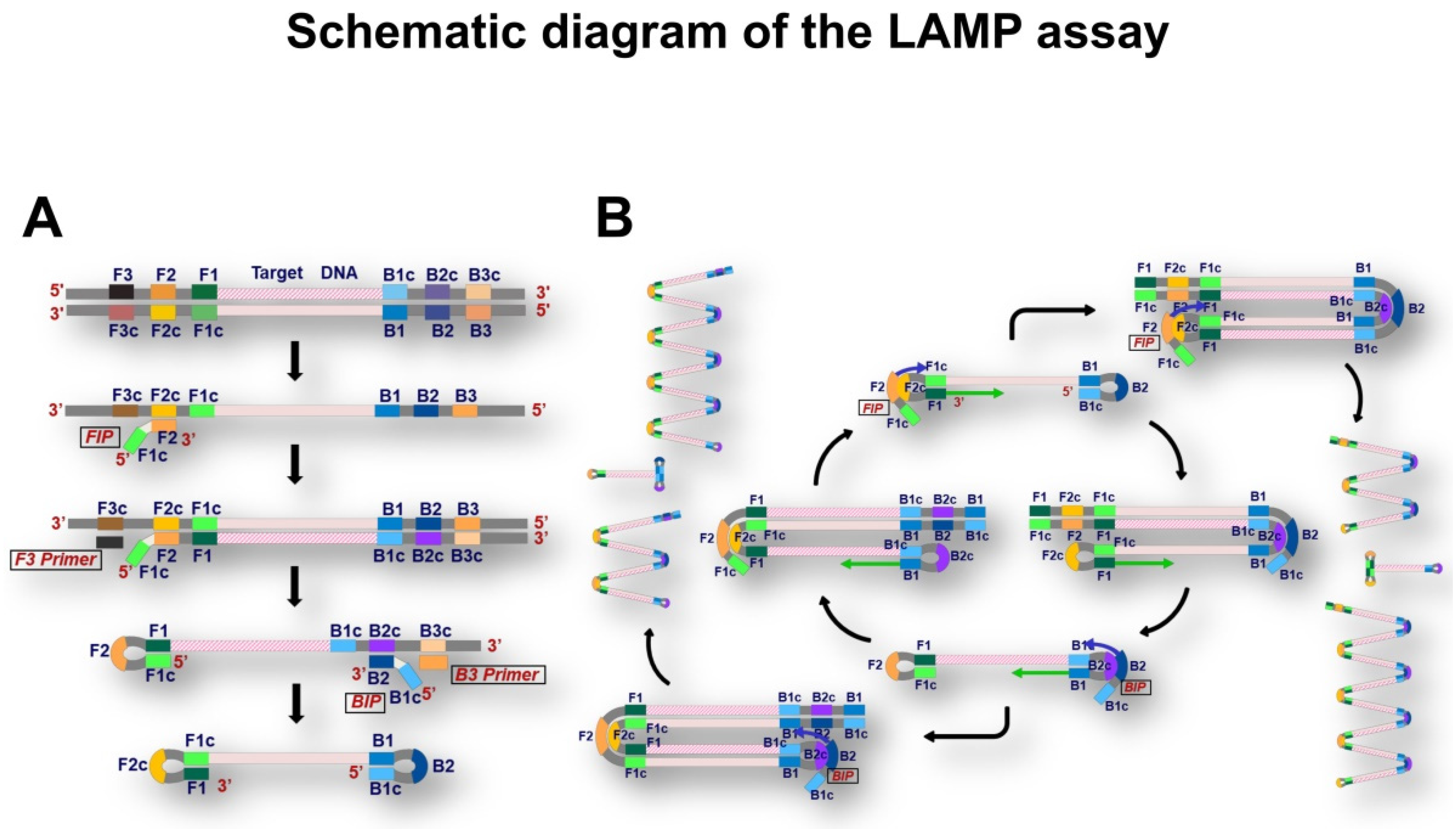
2.4. LAMP Mutation Analysis

The LAMP assay was performed according to the standard protocol described in our previous studies [37,44]. Briefly, the LAMP assay was conducted at 65 °C for 120 min using the LightCycler 480® (Roche Diagnostics KK, Tokyo, Japan) and a primer set that was developed in our laboratory (Figure 2). After denaturing the amplicon at 95 °C for 5 min followed by hybridization at 37 °C for 5 min, the temperature was gradually raised to 80 °C, and the fluorescence intensity was measured 7 times per 1 °C increment., The data were analyzed using the LightCycler 480 software® (version 1.5.1.62; Roche Diagnostics KK) to calculate the melting peak.

2.5. Next-Generation Sequencing

Amplicon-based NGS was performed using the MiSeq system (Illumina KK, Tokyo, Japan), and the primer sets for amplification of EGFR exons 18, 19, 20, and 21 were used as previously reported [37,40]. The resulting FASTQ files were mapped to the GRCh38 human reference sequence using BWA-MEM (http://bio-bwa.sourceforge.net/ (accessed on 5 June 2022)). The variant data was extracted from the mapped data using Samtools ver. 1.9 (http://www.htslib.org/ (accessed on 5 June 2022)) and GATK4 (https://gatk.broadinstitute.org/hc/en-us (accessed on 5 June 2022)).

2.6. Statistical Analysis

Descriptive statistics and categorical variables were calculated using standard formulae with Excel 2019 ver. 16.0.12527.20260® (Microsoft Corp, Tokyo, Japan) and SPSS Statistics® version 28 (IBM Corp., Armonk, NY, USA). The sensitivity and specificity of the LAMP and the NGS assays were obtained, and figures were created using Excel 2019 and the statistical software R version 3.6.3 (R Foundation for Statistical Computing, Vienna, Austria) with the ggplot2 package. Clinical and pathological findings were extracted from the electronic medical records of the patients. p < 0.05 was considered significant.

3. Results

3.1. Examination of the Efficacy of EGFR-LAMP Liquid Biopsy

To investigate the performance of EGFR-LAMP liquid biopsy in detecting EGFR mutations, four kinds of solutions with different concentrations of mutated EGFR copies (0%, 5%, and 100% in 10,000 copies and 5% in 1000 copies) were prepared in the preliminary study (Figure 3). The detection of EGFR mutations was marked by 29 EGFR-LAMP primers in a solution of 5% in 10,000 copies. Detection was achieved in 26 primers but not in three primers. Thus, a concentration of at least 5% in 10,000 copies may be required to detect EGFR mutations using the LAMP assay.

3.2. Characteristics of Patients with EGFR-Mutated Adenocarcinoma

A total of 264 plasma sampling and surgical procedures were performed as a biopsy or treatment for patients with highly suspicious lung cancer. Among them, 57 resected tumors demonstrated primary lung cancer with EGFR mutations, which was confirmed by the Therascreen assay using tumor tissues. After excluding patients without informed consent, 51 patients were enrolled in this study. The mean age of the 51 patients was 68.7 years (49–85 years), and most patients were female (n = 31, 70.8%) (Table 1). There were 22 never-smokers (43.1%), 23 former-smokers (45.1%), and 6 current smokers (11.8%). In terms of histologic subtype of adenocarcinoma, there were 45 papillary predominant (88.0%), 4 lepidic (8.0%), 1 micropapillary (2.0%), and solid predominant (2.0%) adenocarcinomas. Most cases were pathological stage I, consisting of 12 p-IA1 (24.0%), 21 p-IA2 (41.0%), and 6 p-IA3 tumors (12.0%).

3.3. Amount of cfDNA Extracted from Plasma Samples

The cfDNA was obtained from clinical plasma samples using the QIAamp DNA Blood Mini Kit® (Qiagen, K.K.). Figure 4 illustrates the amount of cfDNA extracted from each specimen. In extracted cfDNA, the average value was 50.89 ng (25.76–110.24), and over 0.162 ng/μL cfDNA was available for use in most cases (Supplementary table). Among them, the quantity of cfDNA that we actually used was 1.59 ng (0.81–3.45) on average for the LAMP assay and 5.54 ng (2.83–10.00) for the NGS assay. Therefore, the cfDNA volume was considered enough for both the LAMP and NGS assays.

3.4. Therascreen EGFR PCR Mutation Analysis for Resected or Biopsied Tumors

Among the 51 adenocarcinomas, there were 27 tumors with exon 19 deletion (52.9%), 18 tumors with exon 21 L858R point mutations (35.3%), 2 tumors with exon 20 in-frame insertion (3.9%), 2 tumors with exon 19 deletion/exon 20 in-frame insertion (double mutation) (3.9%), and 2 uncommon EGFR mutations (exon 19 deletion/G719X point mutation and G719X/exon 18 point mutation) (Table 2).

3.5. Comparison of Results of the LAMP and the NGS Assays

Among the 51 tumors, there was one tumor with EGFR mutations in both the LAMP and the NGS assays, another one with EGFR wild type in the LAMP assay but EGFR mutations in the NGS assay, and the others with EGFR wild type in both assays. Both the LAMP and the NGS assays were not able to detect EGFR mutations in 49 plasma samples, even though the mutation was confirmed through the Therascreen assay in the resected or biopsied tumors. Among two cases with EGFR mutations in the NGS assay, one case had p-stage IVB adenocarcinoma with an L858R point mutation in the LAMP assay, while the other had p-stage IIIA adenocarcinoma with an L858R point mutation only in the NGS assay (Table 3). In the supplementary table, the amount of cfDNA was 1.44 ng in the LAMP assay detecting EGFR mutation (average 1.59 ng in all LAMP experiments), and it was 5.04 ng and 6.58 ng in the NGS assay resulting in EGFR mutations (average 5.54 ng in all NGS experiments). A relationship was not found between the amount of cf DNA and the detection of EGFR mutation in both assays. The positivity rate of the EGFR-LAMP liquid biopsy was 2.0%, and that of the NGS assay was 3.9%.

4. Discussion

The identification of EGFR mutations is necessary for decision-making in the treatment of patients with advanced or metastatic NSCLC. Furthermore, the latest NCCN guidelines recommend testing EGFR status even after complete resection for patients with early-stage NSCLC to consider the administration of adjuvant chemotherapy [16,17,18,45]. Due to the expanded indications for EGFR examination, the demand for a rapid and cheap approach to identifying EGFR status has been steadily increasing. However, health insurance only allows testing through conventional PCR-based or NGS-based methods in most countries. Since the latter method is time-consuming, expensive, and highly complex, there is an urgent need to develop a more convenient approach for molecular testing of oncogenes.
Point-of-care testing (POCT) is a concept where testing for healthcare is provided close to or near the patients. Although various definitions have been provided in the medical or scientific field, it was defined as “testing performed near or at the site of a patient with the result leading to a possible change in the care of the patient” [46]. POCT is being increasingly used in general practice inside and outside the hospital, such as in the case of the reverse transcription LAMP (RT-LAMP) for COVID-19 [47]. The standard for COVID-19 diagnostics are PCR-based tests that are highly sensitive, specific, and remarkably reliable, but these tests are limited by the requirement for sophisticated laboratories and skilled personnel, the complex protocol, the long waiting times for results, and an overall high cost per test. The development of RT-LAMP can be a robust solution for the rapid and cost-effective diagnosis of COVID-19 in low-resource laboratory settings. Although basic research into POCT for several diseases has been progressing drastically, there are only a few reports of POCT in oncology for now.
In 2018, Asaka et al. reported a POCT system for detecting EGFR mutations based on a droplet-polymerase chain reaction (d-PCR) assay using cfDNA from patients with lung cancer [48]. The EGFR d-PCR assay was designed to detect three EGFR mutations: L858R in exon 21, E746_A750del in exon 19, and T790M in exon 20. However, it has not been adopted in clinical practice yet. In this manner, the development of POCT is just beginning in oncology, and it is widely expected that POCT will conquer some technical limitations of conventional methods.
In this pioneering study, the EGFR-LAMP assay successfully detected EGFR mutations using 29 EGFR-LAMP primers developed and reported in our previous study [37,44]. However, an EGFR mutation was only detected in one case. The low positivity rate for EGFR mutations is a clinical issue in the development of the EGFR-LAMP liquid biopsy. The reason for this is likely not the limits of abilities of the LAMP assay but the low concentration of circulating tumor DNA (ctDNA) because the NGS assay also demonstrated a low positivity rate. The ctDNA concentration could be below 5% in 1000 copies according to the relationship between the detection rate and DNA concentration (Figure 3). Consequently, using plasma samples from patients with advanced or metastatic tumors instead of early-stage tumors is recommended to confirm the positivity rate of EGFR mutations in EGFR-LAMP liquid biopsy. Another improvement will be the development of a multiplex platform equipped with multiple EGFR-LAMP primers, allowing us to avoid the subdivision of plasma samples for each primer. However, it is necessary to elucidate the optimal combinations of EGFR-LAMP primers to allow accurate gene amplification.
Currently, the cobas EGFR assay® (Roche Molecular Systems Inc., CA, USA) for analysis of plasma samples has been officially validated in NSCLC by authorized organizations. However, it is inconvenient because of the long waiting time to obtain results. Despite the limitations of this study (small number of cases and study participants from a single institute), we were able to demonstrate the potential of EGFR-LAMP liquid biopsy as POCT in oncology as well as in the general field of molecular testing. In future studies, we plan to investigate this application by using plasma samples from patients with more advanced-stage tumors.
This study had several limitations: (1) relatively small sample size, (2) single-institute nature of the study, (3) data were obtained only from surgery cases, and (4) negative control groups (tumors with EGFR wild type) were missing. Since ctDNA concentration could be high in plasma samples from more advanced-stage tumors in theory, further studies should be planned to clarify the performance of the EGFR-LAMP liquid biopsy system for advanced cancers through comparisons with negative control groups.
In conclusion, our study was the first to demonstrate that the LAMP assay successfully detected EGFR mutations in plasma samples, illuminating a direction for the development of POCT in oncology. POCT of EGFR mutations could dramatically change clinical practice in the future. For instance, an outpatient’s blood could be examined, and the result of the oncogene status could be obtained within half a day. This could also be applied to staging in place of the TNM classification, assessment of the effects of chemoradiation, and elucidation of resistance to anticancer drugs.

Supplementary Materials

The following supporting information can be downloaded at: https://www.mdpi.com/article/10.3390/mi13060897/s1, Table S1: Amount of cfDNA.

Author Contributions

Conceptualization, Y.S. (Yuichi Saito), Y.S. (Yoshihiko Shimizu) and E.H.; methodology, A.M. and S.M.; software, Y.S. (Yuichi Saito), S.M., H.M. and T.I. (Takayuki Ibi); validation, Y.Y. and N.T.; formal analysis, Y.S. (Yuichi Saito) and Y.S. (Yukinori Sakao); investigation, Y.S. (Yuichi Saito), Y.S. (Yoshihiko Shimizu), T.I. (Tomohiko Ikeya) and E.H.; resources, Y.S. (Yuichi Saito); data curation, A.M. and S.M.; writing—original draft preparation, Y.S. (Yuichi Saito); writing—review and editing, Y.S. (Yukinori Sakao) and M.K.; visualization, Y.S. (Yuichi Saito) and A.M.; supervision, E.H.; project administration, E.H. and M.K.; funding acquisition, Y.S. (Yuichi Saito) and E.H. All authors have read and agreed to the published version of the manuscript.

Funding

The present study was funded by Eiken Chemical Co., Ltd. (No grant number was provided).

Data Availability Statement

The data used to support the findings of this study are included within the article.

Acknowledgments

The authors would like to thank Yasuhito Kobayashi and Mei Miyagawa of the Saitama Cardiovascular and Respiratory Center, Kumagaya, Japan, for their insightful comments and excellent technical assistance.

Conflicts of Interest

All authors have completed the ICMJE disclosure form. SAITO, TAKAHASHI, SHIMIZU and HOSHI report grants and non-financial support from Eiken Chemical Co., Ltd., during the study. The other authors have no conflict of interest to declare.

References

  1. Sung, H.; Ferlay, J.; Siegel, R.L.; Laversanne, M.; Soerjomataram, I.; Jemal, A.; Bray, F. Global Cancer Statistics 2020: GLOBOCAN Estimates of Incidence and Mortality Worldwide for 36 Cancers in 185 Countries. CA Cancer J. Clin. 2021, 71, 209–249. [Google Scholar] [CrossRef] [PubMed]
  2. Allemani, C.; Matsuda, T.; Di Carlo, V.; Harewood, R.; Matz, M.; Nikšić, M.; Bonaventure, A.; Valkov, M.; Johnson, C.J.; Estève, J.; et al. Global surveillance of trends in cancer survival 2000-14 (CONCORD-3): Analysis of individual records for 37 513 025 patients diagnosed with one of 18 cancers from 322 population-based registries in 71 countries. Lancet 2018, 391, 1023–1075. [Google Scholar] [CrossRef] [PubMed] [Green Version]
  3. Mok, T.S.; Wu, Y.L.; Thongprasert, S.; Yang, C.H.; Chu, D.T.; Saijo, N.; Sunpaweravong, P.; Han, B.; Margono, B.; Ichinose, Y.; et al. Gefitinib or carboplatin-paclitaxel in pulmonary adenocarcinoma. N. Engl. J. Med. 2009, 361, 947–957. [Google Scholar] [CrossRef] [PubMed]
  4. Mitsudomi, T.; Morita, S.; Yatabe, Y.; Negoro, S.; Okamoto, I.; Tsurutani, J.; Seto, T.; Satouchi, M.; Tada, H.; Hirashima, T.; et al. Gefitinib versus cisplatin plus docetaxel in patients with non-small-cell lung cancer harbouring mutations of the epidermal growth factor receptor (WJTOG3405): An open label, randomised phase 3 trial. Lancet Oncol. 2010, 11, 121–128. [Google Scholar] [CrossRef] [PubMed]
  5. Maemondo, M.; Inoue, A.; Kobayashi, K.; Sugawara, S.; Oizumi, S.; Isobe, H.; Gemma, A.; Harada, M.; Yoshizawa, H.; Kinoshita, I.; et al. Gefitinib or chemotherapy for non-small-cell lung cancer with mutated EGFR. N. Engl. J. Med. 2010, 362, 2380–2388. [Google Scholar] [CrossRef] [PubMed] [Green Version]
  6. Han, J.Y.; Park, K.; Kim, S.W.; Lee, D.H.; Kim, H.Y.; Kim, H.T.; Ahn, M.J.; Yun, T.; Ahn, J.S.; Suh, C.; et al. First-SIGNAL: First-line single-agent iressa versus gemcitabine and cisplatin trial in never-smokers with adenocarcinoma of the lung. J. Oncol. 2012, 30, 1122–1128. [Google Scholar] [CrossRef] [PubMed]
  7. Zhou, C.; Wu, Y.L.; Chen, G.; Feng, J.; Liu, X.Q.; Wang, C.; Zhang, S.; Wang, J.; Zhou, S.; Ren, S.; et al. Erlotinib versus chemotherapy as first-line treatment for patients with advanced EGFR mutation-positive non-small-cell lung cancer (OPTIMAL, CTONG-0802): A multicentre, open-label, randomised, phase 3 study. Lancet Oncol. 2011, 12, 735–742. [Google Scholar] [CrossRef] [PubMed]
  8. Rosell, R.; Carcereny, E.; Gervais, R.; Vergnenegre, A.; Massuti, B.; Felip, E.; Palmero, R.; Garcia-Gomez, R.; Pallares, C.; Sanchez, J.M.; et al. Erlotinib versus standard chemotherapy as first-line treatment for European patients with advanced EGFR mutation-positive non-small-cell lung cancer (EURTAC): A multicentre, open-label, randomised phase 3 trial. Lancet Oncol. 2012, 13, 239–246. [Google Scholar] [CrossRef] [PubMed]
  9. Sequist, L.V.; Yang, J.C.; Yamamoto, N.; O’Byrne, K.; Hirsh, V.; Mok, T.; Geater, S.L.; Orlov, S.; Tsai, C.M.; Boyer, M.; et al. Phase III study of afatinib or cisplatin plus pemetrexed in patients with metastatic lung adenocarcinoma with EGFR mutations. J. Clin. Oncol. 2013, 31, 3327–3334. [Google Scholar] [CrossRef] [PubMed] [Green Version]
  10. Wu, Y.L.; Zhou, C.; Hu, C.P.; Feng, J.; Lu, S.; Huang, Y.; Li, W.; Hou, M.; Shi, J.H.; Lee, K.Y.; et al. Afatinib versus cisplatin plus gemcitabine for first-line treatment of Asian patients with advanced non-small-cell lung cancer harbouring EGFR mutations (LUX-Lung 6): An open-label, randomised phase 3 trial. Lancet Oncol. 2014, 15, 213–222. [Google Scholar] [CrossRef] [PubMed]
  11. Planchard, D.; Popat, S.; Kerr, K.; Novello, S.; Smit, E.F.; Faivre-Finn, C.; Mok, T.S.; Reck, M.; Van Schil, P.E.; Hellmann, M.D.; et al. Metastatic non-small cell lung cancer: ESMO Clinical Practice Guidelines for diagnosis, treatment and follow-up. Ann. Oncol. 2018, 29 (Suppl. 4), iv192–iv237. [Google Scholar] [CrossRef] [PubMed]
  12. The Japanese Lung Cancer Society: Guidelines of Lung Cancer. Available online: https://www.haigan.gr.jp/modules/guideline/index.php?content_id=3 (accessed on 29 December 2021).
  13. Hanna, N.H.; Robinson, A.G.; Temin, S.; Baker, S., Jr.; Brahmer, J.R.; Ellis, P.M.; Gaspar, L.E.; Haddad, R.Y.; Hesketh, P.J.; Jain, D.; et al. Therapy for Stage IV Non-Small-Cell Lung Cancer with Driver Alterations: ASCO and OH (CCO) Joint Guideline Update. J. Clin. Oncol. 2021, 39, 1040–1091. [Google Scholar] [CrossRef] [PubMed]
  14. Ettinger, D.S.; Wood, D.E.; Aisner, D.L.; Akerley, W.; Bauman, J.R.; Bharat, A.; Bruno, D.S.; Chang, J.Y.; Chirieac, L.R.; D’Amico, T.A.; et al. NCCN Guidelines Insights: Non-Small Cell Lung Cancer, Version 2.2021. J. Natl. Compr. Cancer Netw. 2021, 19, 254–266. [Google Scholar] [CrossRef] [PubMed]
  15. Wu, Y.L.; Tsuboi, M.; He, J.; John, T.; Grohe, C.; Majem, M.; Goldman, J.W.; Laktionov, K.; Kim, S.W.; Kato, T.; et al. Osimertinib in Resected EGFR-Mutated Non-Small-Cell Lung Cancer. N. Engl. J. Med. 2020, 383, 1711–1723. [Google Scholar] [CrossRef] [PubMed]
  16. Lindeman, N.I.; Cagle, P.T.; Aisner, D.L.; Arcila, M.E.; Beasley, M.B.; Bernicker, E.H.; Colasacco, C.; Dacic, S.; Hirsch, F.R.; Kerr, K.; et al. Updated Molecular Testing Guideline for the Selection of Lung Cancer Patients for Treatment with Targeted Tyrosine Kinase Inhibitors: Guideline From the College of American Pathologists, the International Association for the Study of Lung Cancer, and the Association for Molecular Pathology. Arch. Pathol. Lab. Med. 2018, 142, 321–346. [Google Scholar] [CrossRef] [PubMed] [Green Version]
  17. Kalemkerian, G.P.; Narula, N.; Kennedy, E.B.; Biermann, W.A.; Donington, J.; Leighl, N.B.; Lew, M.; Pantelas, J.; Ramalingam, S.S.; Reck, M.; et al. Molecular Testing Guideline for the Selection of Patients With Lung Cancer for Treatment With Targeted Tyrosine Kinase Inhibitors: American Society of Clinical Oncology Endorsement of the College of American Pathologists/International Association for the Study of Lung Cancer/Association for Molecular Pathology Clinical Practice Guideline Update. J. Clin. Oncol. 2018, 36, 911–919. [Google Scholar] [CrossRef] [PubMed]
  18. The Japan Lung Cancer Society, Guidance of Examinations of EGFR Mutations for Patients with Lung Cancer ver5.0. Available online: https://www.haigan.gr.jp/modules/tebiki/index.php?content_id=5 (accessed on 29 December 2021).
  19. The Japan Lung Cancer Society, Guidance of Examinations of ALK Fusion Gene for Patients with Lung Cancer ver3.1. Available online: https://www.haigan.gr.jp/modules/tebiki/index.php?content_id=9 (accessed on 29 December 2021).
  20. The Japan Lung Cancer Society, Guidance of Examinations of ROS1 Fusion Gene for Patients with Lung Cancer ver1.0. Available online: https://www.haigan.gr.jp/modules/tebiki/index.php?content_id=7 (accessed on 29 December 2021).
  21. The Japan Lung Cancer Society, Guidance of Examinations of BRAF Mutations for Patients with Lung Cancer ver1.0. Available online: https://www.haigan.gr.jp/modules/tebiki/index.php?content_id=4 (accessed on 29 December 2021).
  22. Takano, C.; Kuramochi, Y.; Seki, M.; Kim, D.W.; Omagari, D.; Sasano, M.; Chang, B.; Ohnishi, M.; Kim, E.J.; Fuwa, K.; et al. Molecular serotype-specific identification of Streptococcus pneumoniae using loop-mediated isothermal amplification. Sci. Rep. 2019, 9, 19823. [Google Scholar] [CrossRef] [PubMed] [Green Version]
  23. Quyen, T.L.; Nordentoft, S.; Vinayaka, A.C.; Ngo, T.A.; Engelsmenn, P.; Sun, Y.; Madsen, M.; Bang, D.D.; Wolff, A. A Sensitive, Specific and Simple Loop Mediated Isothermal Amplification Method for Rapid Detection of Campylobacter spp. in Broiler Production. Front. Microbiol. 2019, 10, 2443. [Google Scholar] [CrossRef] [PubMed]
  24. Wang, Y.; Wang, Y.; Jiao, W.; Li, J.; Quan, S.; Sun, L.; Wang, Y.; Qi, X.; Wang, X.; Shen, A. Development of loop-mediated isothermal amplification coupled with nanoparticle-based lateral flow biosensor assay for Mycoplasma pneumoniae detection. AMB Express 2019, 9, 196. [Google Scholar] [CrossRef] [PubMed] [Green Version]
  25. Nagai, K.; Horita, N.; Yamamoto, M.; Tsukahara, T.; Nagakura, H.; Tashiro, K.; Shibata, Y.; Watanabe, H.; Nakashima, K.; Ushio, R.; et al. Diagnostic test accuracy of loop-mediated isothermal amplification assay for Mycobacterium tuberculosis: Systematic review and meta-analysis. Sci. Rep. 2016, 6, 39090. [Google Scholar] [CrossRef] [PubMed]
  26. Jin, Y.; Yang, H.; Ji, W.; Wu, W.; Chen, S.; Zhang, W.; Duan, G. Virology, Epidemiology, Pathogenesis, and Control of COVID-19. Viruses 2020, 12, 372. [Google Scholar] [CrossRef] [PubMed] [Green Version]
  27. Coronaviridae Study Group of the International Committee on Taxonomy of Viruses. The species Severe acute respiratory syndrome-related coronavirus: Classifying 2019-nCoV and naming it SARS-CoV-2. Nat. Microbiol. 2020, 5, 536–544. [Google Scholar] [CrossRef] [PubMed] [Green Version]
  28. Bustin, S.A.; Nolan, T. RT-qPCR Testing of SARS-CoV-2: A Primer. Int. J. Mol. Sci. 2020, 21, 3004. [Google Scholar] [CrossRef] [PubMed]
  29. Corman, V.M.; Landt, O.; Kaiser, M.; Molenkamp, R.; Meijer, A.; Chu, D.K.; Bleicker, T.; Brünink, S.; Schneider, J.; Schmidt, M.L.; et al. Detection of 2019 novel coronavirus (2019-nCoV) by real-time RT-PCR. Eurosurveillance 2020, 25, 2000045. [Google Scholar] [CrossRef] [Green Version]
  30. Lu, R.; Wu, X.; Wan, Z.; Li, Y.; Zuo, L.; Qin, J.; Jin, X.; Zhang, C. Development of a Novel Reverse Transcription Loop-Mediated Isothermal Amplification Method for Rapid Detection of SARS-CoV-2. Virol. Sin. 2020, 35, 344–347. [Google Scholar] [CrossRef] [PubMed] [Green Version]
  31. Park, G.S.; Ku, K.; Baek, S.H.; Kim, S.J.; Kim, S.I.; Kim, B.T.; Maeng, J.S. Development of Reverse Transcription Loop-Mediated Isothermal Amplification Assays Targeting Severe Acute Respiratory Syndrome Coronavirus 2 (SARS-CoV-2). J. Mol. Diagn. 2020, 22, 729–735. [Google Scholar] [CrossRef] [PubMed]
  32. Yan, L.; Xiao, H.; Zhang, Q. Systematic review: Comparison of Xpert MTB/RIF, LAMP and SAT methods for the diagnosis of pulmonary tuberculosis. Tuberculosis (Edinb.) 2016, 96, 75–86. [Google Scholar] [CrossRef] [PubMed]
  33. Aryan, E.; Makvandi, M.; Farajzadeh, A.; Huygen, K.; Alvandi, A.H.; Gouya, M.M.; Sadrizadeh, A.; Romano, M. Clinical value of IS6110-based loop-mediated isothermal amplification for detection of Mycobacterium tuberculosis complex in respiratory specimens. J. Infect. 2013, 66, 487–493. [Google Scholar] [CrossRef] [PubMed]
  34. The Use of Loop-Mediated Isothermal Amplification (TB-LAMP) for the Diagnosis of Pulmonary Tuberculosis: Policy Guidance; World Health Organization: Geneva, Switzerland, 2016. [PubMed]
  35. WHO Consolidated Guidelines on Tuberculosis: Module 3: Diagnosis—Rapid Diagnostics for Tuberculosis Detection [Internet]; World Health Organization: Geneva, Switzerland, 2020. [PubMed]
  36. García-Basteiro, A.L.; DiNardo, A.; Saavedra, B.; Silva, D.R.; Palmero, D.; Gegia, M.; Migliori, G.B.; Duarte, R.; Mambuque, E.; Centis, R.; et al. Point of care diagnostics for tuberculosis. Pulmonology 2018, 24, 73–85. [Google Scholar] [CrossRef] [PubMed]
  37. Saito, Y.; Takahashi, N.; Matsui, A.; Michiyuki, S.; Yamauchi, Y.; Shimizu, Y.; Hoshi, E.; Sakao, Y.; Kawamura, M. Comparative study of the loop-mediated isothermal amplification method and the QIAGEN therascreen PCR kit for the detection of EGFR mutations in non-small cell lung cancer. J. Thorac. Dis. 2021, 13, 743–753. [Google Scholar] [CrossRef] [PubMed]
  38. Roy, D.; Lucci, A.; Ignatiadis, M.; Jeffrey, S.S. Cell-free circulating tumor DNA profiling in cancer management. Trends Mol. Med. 2021, 27, 1014–1015. [Google Scholar] [CrossRef] [PubMed]
  39. Bettegowda, C.; Sausen, M.; Leary, R.J.; Kinde, I.; Wang, Y.; Agrawal, N.; Bartlett, B.R.; Wang, H.; Luber, B.; Alani, R.M.; et al. Detection of circulating tumor DNA in early- and late-stage human malignancies. Sci. Transl. Med. 2014, 6, 224ra24. [Google Scholar] [CrossRef] [PubMed] [Green Version]
  40. Vrba, L.; Oshiro, M.M.; Kim, S.S.; Garland, L.L.; Placencia, C.; Mahadevan, D.; Nelson, M.A.; Futscher, B.W. DNA methylation biomarkers discovered in silico detect cancer in liquid biopsies from non-small cell lung cancer patients. Epigenetics 2020, 15, 419–430. [Google Scholar] [CrossRef] [PubMed] [Green Version]
  41. Roy, D.; Tiirikainen, M. Diagnostic Power of DNA Methylation Classifiers for Early Detection of Cancer. Trends Cancer 2020, 6, 78–81. [Google Scholar] [CrossRef] [PubMed]
  42. The Japanese Society of Pathology, Regulations for Handling Pathological Tissue Specimens for Genomic Medicine. Available online: http://pathology.or.jp/genome_med/pdf/textbook.pdf (accessed on 29 December 2021).
  43. Vallée, A.; Le Loupp, A.G.; Denis, M.G. Efficiency of the Therascreen® RGQ PCR kit for the detection of EGFR mutations in non-small cell lung carcinomas. Clin. Chim. Acta 2014, 15, 429. [Google Scholar] [CrossRef] [PubMed]
  44. Horiuchi, S.; Saito, Y.; Matsui, A.; Takahashi, N.; Ikeya, T.; Hoshi, E.; Shimizu, Y.; Yasuda, M. A novel loop-mediated isothermal amplification method for efficient and robust detection of EGFR mutations. Int. J. Oncol. 2020, 56, 743–749. [Google Scholar] [CrossRef] [PubMed] [Green Version]
  45. NCCN Clinical Practice Guidelines in Oncology (NCCN Guidelines®) Non-Small Cell Lung Cancer Version 1.2022-December 7, 2021. Available online: https://www2.tri-kobe.org/nccn/guideline/lung/english/non_small.pdf (accessed on 29 December 2021).
  46. ISO 22870:2006. Point-of-Care Testing (POCT)-Requirements for Quality and Competence. Available online: https://www.iso.org/standard/35173.html (accessed on 29 December 2021).
  47. Augustine, R.; Hasan, A.; Das, S.; Ahmed, R.; Mori, Y.; Notomi, T.; Kevadiya, B.D.; Thakor, A.S. Loop-Mediated Isothermal Amplification (LAMP): A Rapid, Sensitive, Specific, and Cost-Effective Point-of-Care Test for Coronaviruses in the Context of COVID-19 Pandemic. Biology 2020, 9, 182. [Google Scholar] [CrossRef] [PubMed]
  48. Asaka, S.; Yoshizawa, A.; Saito, K.; Kobayashi, Y.; Yamamoto, H.; Negishi, T.; Nakata, R.; Matsuda, K.; Yamaguchi, A.; Honda, T. Rapid point-of-care testing for epidermal growth factor receptor gene mutations in patients with lung cancer using cell-free DNA from cytology specimen supernatants. Int. J. Oncol. 2018, 52, 2110–2118. [Google Scholar] [CrossRef] [PubMed] [Green Version]
Figure 1. Experimental schema of this study. Plasma samples were obtained from patients with a strong suspicion of primary lung cancer and stored in a freezing chamber preoperatively. After surgery or surgical biopsy, all tissue samples were fixed with 10% buffer formalin at 20–25 °C (24–36 h) to create formalin-fixed, paraffin-embedded tumor blocks. Afterward, hematoxylin–eosin-stained slides were prepared for pathological diagnosis. The Therascreen assay was performed to investigate the EGFR status of resected tumor tissues from select cases with EGFR-mutated adenocarcinoma. DNA extraction was performed from plasma samples for both the LAMP and NGS assays.
Micromachines 13 00897 g001
Figure 2. Schematic diagram of loop-mediated isothermal amplification. (A) Scheme of dumbbell-like structure formation. Target DNA is firstly denatured into single-stranded DNA. Then, the forward inner primer (FIP) initiates DNA synthesis from the F2c region. The following extension from the forward outer primer (F3) displaces the strand that was generated by the FIP extension. The replaced strand plays the role of a template of the backward inner primer (BIP) and backward outer primer (B3) in the same manner as sense strand amplification. Consequently, the dumbbell-like structure harboring hairpin structure on both of 5′- and 3′-terminus is generated. (B) Scheme of isothermal amplification. The dumbbell-like structure exposing F2c or B2c regions where FIP or BIP hybridizes initiates cycling amplification. The cycling amplification regenerates dumbbell-like structures and generates various amplicons, which are exponentially increased in length.
Micromachines 13 00897 g002
Figure 3. The relationship between results of the LAMP assay and DNA concentrations. The x-axis shows 4 concentrations, and Y-axis presents 29 subtypes of EGFR mutations. The LAMP assay detected all EGFR mutations over 5% in 10,000 copies.
Micromachines 13 00897 g003
Figure 4. The relationship between the number of cases and cell-free DNA (cfDNA) concentration. The x-axis shows various concentrations of cfDNA, and Y-axis presents the number of cases. The median concentration of cfDNA extracted from plasma samples was 45.92 ng/μL, the minimum concentration was 25.76 ng/μL, and the maximum concentration was 110.24 ng/μL.
Micromachines 13 00897 g004
Table 1. Clinical characteristics of patients in the study.
CharacteristicN (%)
Age, years68.7 ± 8.4
Sex
 Male20 (29.2)
 Female31 (70.8)
Smoking Status
 Never smoker22 (43.1)
 Former smoker23 (45.1)
 Current smoker6 (11.8)
Subtype of adenocarcinoma
 Papillary predominant45 (88.0)
 Lepidic predominant4 (8.0)
 Micropapillary predominant1 (2.0)
 Solid predominant1 (2.0)
Pathological stage
 pIA112 (24.0)
 pIA221 (41.0)
 pIA36 (12.0)
 pIB2 (4.0)
 pIIB3 (6.0)
 pIIIA4 (8.0)
 pIIIB1 (2.0)
 pIVA1 (2.0)
 pIVB1 (2.0)
Data on age: mean ± SD.
Table 2. EGFR status of resected tumor tissue as determined using the Therascreen assay.
NumberPercentage (%)
Del 192752.9
L858R1835.3
Ins 2023.9
Ex 19 del + Ins 2023.9
Del 19 + G719X12.0
G719X + Ex 1812.0
Total51100
EGFR, epidermal growth factor receptor; Del 19, Exon 19 deletion; Ex 18, Exon 18 point mutation; Ins 20, Exon 20 insertion; L858R, Exon 21 L858R point mutation.
Table 3. Comparison of EGFR status between resected tumor and preoperative plasma samples.
TumorPlasma
p-StageTherascreenLAMPLAMPNGS
Case 1IVBL858RL858RL858RL858R
Case 2IIIAL858RnegativenegativeL858R
EGFR, epidermal growth factor receptor; L858R, Exon 21 L858R point mutation; NGS, next-generation sequencing; LAMP, loop-mediated isothermal amplification.
Publisher’s Note: MDPI stays neutral with regard to jurisdictional claims in published maps and institutional affiliations.

