
In Vivo Renal Lipid Quantification by Accelerated Magnetic Resonance Spectroscopic Imaging at 3T: Feasibility and Reliability Study

 , ,
, ,  and
and 
Abstract
:1. Introduction
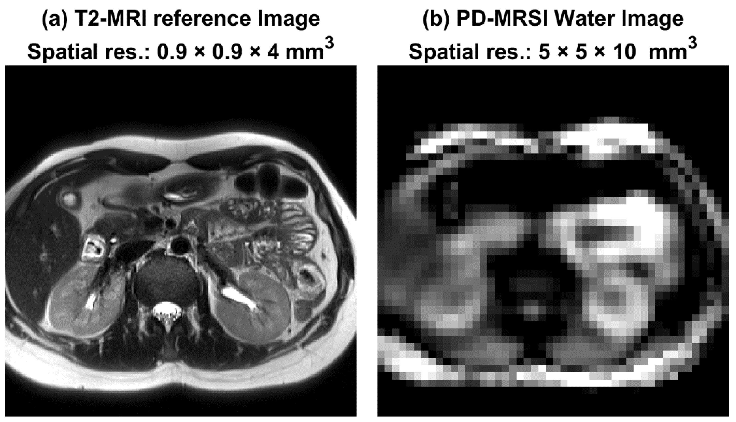
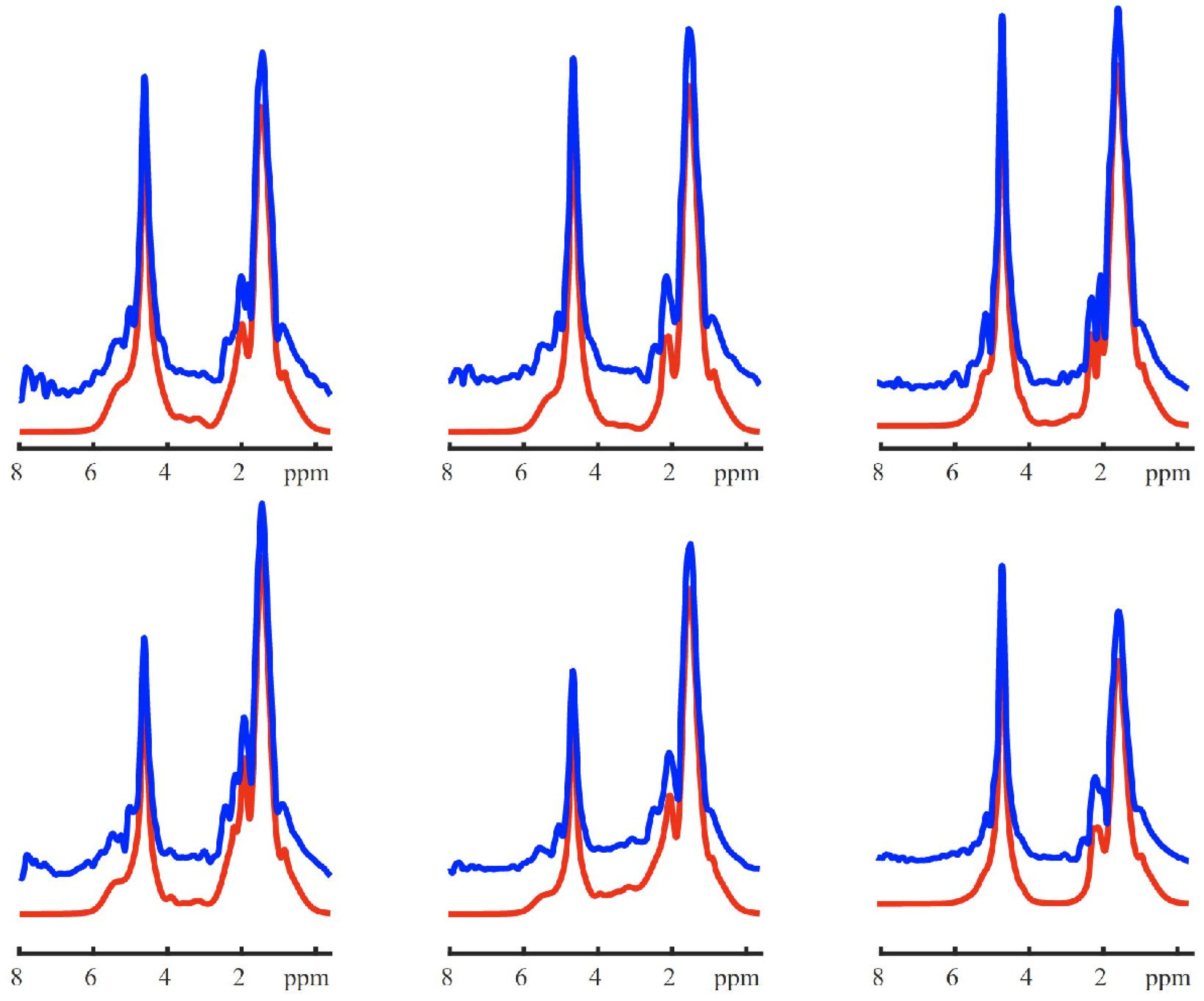
2. Results
3. Discussion
4. Materials and Methods
4.1. Human Subjects
4.2. Test–Retest Study
4.3. MRSI Post-Processing
4.4. Fat Fraction Quantification and Mapping
4.5. ROI Assignment and Statistical Analysis
5. Conclusions
Author Contributions
Funding
Institutional Review Board Statement
Informed Consent Statement
Data Availability Statement
Conflicts of Interest
References
- Bobulescu, I.A. Renal lipid metabolism and lipotoxicity. Curr. Opin. Nephrol. Hypertens. 2010, 19, 393–402. [Google Scholar] [CrossRef] [PubMed] [Green Version]
- De Vries, A.P.J.; Ruggenenti, P.; Ruan, X.Z.; Praga, M.; Cruzado, J.M.; Bajema, I.M.; D’Agati, V.D.; Lamb, H.J.; Barlovic, D.P.; Hojs, R.; et al. Fatty kidney: Emerging role of ectopic lipid in obesity-related renal disease. Lancet Diabetes Endocrinol. 2014, 2, 417–426. [Google Scholar] [CrossRef]
- Opazo-Ríos, L.; Mas, S.; Marín-Royo, G.; Mezzano, S.; Gómez-Guerrero, C.; Moreno, J.A.; Egido, J. Lipotoxicity and diabetic nephropathy: Novel mechanistic insights and therapeutic opportunities. Int. J. Mol. Sci. 2020, 21, 2632. [Google Scholar] [CrossRef] [PubMed] [Green Version]
- Takahashi, T.; Wang, F.; Quarles, C.C. Current MRI techniques for the assessment of renal disease. Curr. Opin. Nephrol. Hypertens 2015, 24, 217–223. [Google Scholar] [CrossRef] [Green Version]
- Mende, C.W.; Einhorn, D. Fatty kidney disease: A new renal and endocrine clinical entity? Describing the role of the kidney in obesity, metabolic syndrome, and type 2 diabetes. Endocr. Pract. 2019, 25, 854–858. [Google Scholar] [CrossRef]
- Wang, Y.C.; Feng, Y.; Lu, C.Q.; Ju, S. Renal fat fraction and diffusion tensor imaging in patients with early-stage diabetic nephropathy. Eur. Radiol. 2018, 28, 3326–3334. [Google Scholar] [CrossRef]
- Notohamiprodjo, M.; Goepfert, M.; Will, S.; Lorbeer, R.; Schick, F.; Rathmann, W.; Martirosian, P.; Peters, A.; Müller-Peltzer, K.; Helck, A.; et al. Renal and renal sinus fat volumes as quantified by magnetic resonance imaging in subjects with prediabetes, diabetes, and normal glucose tolerance. PLoS ONE 2020, 15, e0216635. [Google Scholar] [CrossRef] [Green Version]
- Yang, J.; Zhang, L.J.; Wang, F.; Hong, T.; Liu, Z. Molecular imaging of diabetes and diabetic complications: Beyond pancreatic β-cell targeting. Adv. Drug Deliv. Rev. 2019, 139, 32–50. [Google Scholar] [CrossRef]
- Jonker, J.T.; De Heer, P.; Engelse, M.A.; Van Rossenberg, E.H.; Klessens, C.Q.F.; Baelde, H.J.; Bajema, I.M.; Koopmans, S.J.; Coelho, P.G.; Streefland, T.C.M.; et al. Metabolic imaging of fatty kidney in diabesity: Validation and dietary intervention. Nephrol. Dial. Transplant. 2018, 33, 224–230. [Google Scholar] [CrossRef] [Green Version]
- Pokharel, S.S.; Macura, K.J.; Kamel, I.R.; Zaheer, A. Current MR imaging lipid detection techniques for diagnosis of lesions in the abdomen and pelvis. Radiographics 2013, 33, 681–702. [Google Scholar] [CrossRef]
- Sijens, P.E.; Edens, M.A.; Bakker, S.J.L.; Stolk, R.P. MRI-determined fat content of human liver, pancreas and kidney. World J. Gastroenterol. 2010, 16, 1993–1998. [Google Scholar] [CrossRef] [PubMed]
- Zhang, Y.; Udayakumar, D.; Cai, L.; Hu, Z.; Kapur, P.; Kho, E.Y.; Pavía-Jiménez, A.; Fulkerson, M.; de Leon, A.D.; Yuan, Q.; et al. Addressing metabolic heterogeneity in clear cell renal cell carcinoma with quantitative Dixon MRI. JCI Insight 2017, 2, e94278. [Google Scholar] [CrossRef] [PubMed]
- Rosenkrantz, A.B.; Raj, S.; Babb, J.S.; Chandarana, H. Comparison of 3D two-point Dixon and standard 2D dual-echo breath-hold sequences for detection and quantification of fat content in renal angiomyolipoma. Eur. J. Radiol. 2012, 81, 47–51. [Google Scholar] [CrossRef] [PubMed]
- Wu, Y.; Kwon, Y.S.; Labib, M.; Foran, D.J.; Singer, E.A. Magnetic Resonance Imaging as a Biomarker for Renal Cell Carcinoma. Dis. Markers 2015, 2015, 1–9. [Google Scholar] [CrossRef] [Green Version]
- Moosavi, B.; Shabana, W.M.; El-Khodary, M.; Van Der Pol, C.B.; Flood, T.A.; McInnes, M.D.F.; Schieda, N. Intracellular lipid in clear cell renal cell carcinoma tumor thrombus and metastases detected by chemical shift (in and opposed phase) MRI: Radiologic-pathologic correlation. Acta Radiol. 2016, 57, 241–248. [Google Scholar] [CrossRef]
- Outwater, E.K.; Bhatia, M.; Siegelman, E.S.; Burke, M.A.; Mitchell, D.G. Lipid in renal clear cell carcinoma: Detection on opposed-phase gradient-echo MR images. Radiology 1997, 205, 103–107. [Google Scholar] [CrossRef]
- Karlo, C.A.; Donati, O.F.; Burger, I.A.; Zheng, J.; Moskowitz, C.S.; Hricak, H.; Akin, O. MR imaging of renal cortical tumours: Qualitative and quantitative chemical shift imaging parameters. Eur. Radiol. 2013, 23, 1738–1744. [Google Scholar] [CrossRef]
- Pedrosa, I.; Alsop, D.C.; Rofsky, N.M. Magnetic resonance imaging as a biomarker in renal cell carcinoma. Cancer 2009, 115, 2334–2345. [Google Scholar] [CrossRef]
- Ali, H.A.; Couch, M.J.; Menezes, R.; Evans, A.J.; Finelli, A.; Jewett, M.A.; Jhaveri, K.S. Predictive value of in vivo MR spectroscopy with semilocalization by adiabatic selective refocusing in differentiating clear cell renal cell carcinoma from other subtypes. Am. J. Roentgenol. 2020, 214, 817–824. [Google Scholar] [CrossRef]
- Sevcenco, S.; Krssak, M.; Javor, D.; Ponhold, L.; Kuehhas, F.E.; Fajkovic, H.; Haitel, A.; Shariat, S.F.; Baltzer, P.A. Diagnosis of renal tumors by in vivo proton magnetic resonance spectroscopy. World J. Urol. 2014, 33, 17–23. [Google Scholar] [CrossRef]
- Katz-Brull, R.; Rofsky, N.M.; Morrin, M.M.; Pedrosa, I.; George, D.J.; Michaelson, M.D.; Marquis, R.P.; Maril, M.; Noguera, C.; Lenkinski, R.E. Decreases in free cholesterol and fatty acid unsaturation in renal cell carcinoma demonstrated by breath-hold magnetic resonance spectroscopy. Am. J. Physiol. Physiol. 2005, 288, F637–F641. [Google Scholar] [CrossRef] [PubMed] [Green Version]
- Kim, D.Y.; Kim, K.B.; Kim, O.D.; Kim, J.K. Localized in vivo proton spectroscopy of renal cell carcinoma in human kidney. J. Korean Med. Sci. 1998, 13, 49–53. [Google Scholar] [CrossRef] [PubMed] [Green Version]
- Süllentrop, F.; Hahn, J.; Moka, D. In Vitro and in vivo 1H-MR spectroscopic examination of the renal cell carcinoma. Int. J. Biomed. Sci. 2012, 8, 94–108. [Google Scholar] [PubMed]
- Al-Iedani, O.; Lechner-Scott, J.; Ribbons, K.; Ramadan, S. Fast magnetic resonance spectroscopic imaging techniques in human brain-applications in multiple sclerosis. J. Biomed. Sci. 2017, 24, 1–19. [Google Scholar] [CrossRef]
- Öz, G.; Alger, J.R.; Barker, P.B.; Bartha, R.; Bizzi, A.; Boesch, C.; Bolan, P.J.; Brindle, K.M.; Cudalbu, C.; Dinçer, A. Clinical proton MR spectroscopy in central nervous system disorders. Radiology 2014, 270, 658–679. [Google Scholar] [CrossRef] [Green Version]
- Vidya Shankar, R.; Chang, J.C.; Hu, H.H.; Kodibagkar, V.D. Fast data acquisition techniques in magnetic resonance spectroscopic imaging. NMR Biomed. 2019, 32, e4046. [Google Scholar] [CrossRef]
- Alhulail, A.A.; Patterson, D.A.; Xia, P.; Zhou, X.; Lin, C.; Thomas, M.A.; Dydak, U.; Emir, U.E. Fat–water separation by fast metabolite cycling magnetic resonance spectroscopic imaging at 3 T: A method to generate separate quantitative distribution maps of musculoskeletal lipid components. Magn. Reson. Med. 2020, 84, 1126–1139. [Google Scholar] [CrossRef]
- Chiew, M.; Jiang, W.; Burns, B.; Larson, P.; Steel, A.; Jezzard, P.; Albert Thomas, M.; Emir, U.E. Density-weighted concentric rings k-space trajectory for1H magnetic resonance spectroscopic imaging at 7 T. NMR Biomed. 2018, 31, e3838. [Google Scholar] [CrossRef] [Green Version]
- Bogner, W.; Otazo, R.; Henning, A. Accelerated MR spectroscopic imaging—A review of current and emerging techniques. NMR Biomed. 2021, 34, 1–32. [Google Scholar] [CrossRef]
- Emir, U.E.; Burns, B.; Chiew, M.; Jezzard, P.; Thomas, M.A. Non-water-suppressed short-echo-time magnetic resonance spectroscopic imaging using a concentric ring k-space trajectory. NMR Biomed. 2017, 30, e3714. [Google Scholar] [CrossRef] [Green Version]
- Katz-Brull, R.; Rofsky, N.M.; Lenkinski, R.E. Breathhold abdominal and thoracic proton MR spectroscopy at 3T. Magn. Reson. Med. 2003, 50, 461–467. [Google Scholar] [CrossRef] [PubMed]
- Bobulescu, I.A.; Lotan, Y.; Zhang, J.; Rosenthal, T.R.; Rogers, J.T.; Adams-Huet, B.; Sakhaee, K.; Moe, O.W. Triglycerides in the human kidney cortex: Relationship with body size. PLoS ONE 2014, 9, e101285. [Google Scholar] [CrossRef] [PubMed] [Green Version]
- Righi, V.; Mucci, A.; Schenetti, L.; Tosi, M.R.; Grigioni, W.F.; Corti, B.; Bertaccini, A.; Franceschelli, A.; Sanguedolce, F.; Schiavina, R.; et al. Ex vivo HR-MAS magnetic resonance spectroscopy of normal and malignant human renal tissues. Anticancer Res. 2007, 27, 3195–3204. [Google Scholar] [PubMed]
- Krššák, M.; Lindeboom, L.; Schrauwen-Hinderling, V.; Szczepaniak, L.S.; Derave, W.; Lundbom, J.; Befroy, D.; Schick, F.; Machann, J.; Kreis, R.; et al. Proton magnetic resonance spectroscopy in skeletal muscle: Experts’ consensus recommendations. NMR Biomed. 2021, 34, 1–20. [Google Scholar] [CrossRef] [Green Version]
- Lee, S.S.; Park, S.H. Radiologic evaluation of nonalcoholic fatty liver disease. World J. Gastroenterol. 2014, 20, 7392–7402. [Google Scholar] [CrossRef]
- Hasegawa, N.; Kurihara, T.; Sato, K.; Homma, T.; Fujie, S.; Fujita, S.; Sanada, K.; Hamaoka, T.; Iemitsu, M. Intramyocellular and Extramyocellular Lipids Are Associated with Arterial Stiffness. Am. J. Hypertens 2015, 28, 1473–1479. [Google Scholar] [CrossRef] [Green Version]
- Krssak, M.; Falk Petersen, K.; Dresner, A.; DiPietro, L.; Vogel, S.M.; Rothman, D.L.; Shulman, G.I.; Roden, M. Intramyocellular lipid concentrations are correlated with insulin sensitivity in humans: A 1H NMR spectroscopy study. Diabetologia 1999, 42, 113–116. [Google Scholar] [CrossRef] [Green Version]
- Jacob, S.; Machann, J.; Rett, K.; Brechtel, K.; Volk, A.; Renn, W.; Maerker, E.; Matthaei, S.; Schick, F.; Claussen, C.D.; et al. Association of increased intramyocellular lipid content with insulin resistance in lean nondiabetic offspring of type 2 diabetic subjects. Diabetes 1999, 48, 1113–1119. [Google Scholar] [CrossRef]
- Golla, S.; Ren, J.; Malloy, C.R.; Pascual, J.M. Intramyocellular lipid excess in the mitochondrial disorder MELAS. Neurol. Genet. 2017, 3, e160. [Google Scholar] [CrossRef] [Green Version]
- Dekkers, I.A.; Bizino, M.B.; Paiman, E.H.M.; Smit, J.W.; Jazet, I.M.; de Vries, A.P.J.; Lamb, H.J. The Effect of Glycemic Control on Renal Triglyceride Content Assessed by Proton Spectroscopy in Patients With Type 2 Diabetes Mellitus: A Single-Center Parallel-Group Trial. J. Ren. Nutr. 2021, 31, 611–619. [Google Scholar] [CrossRef]
- Steel, A.; Chiew, M.; Jezzard, P.; Voets, N.L.; Plaha, P.; Thomas, M.A.; Stagg, C.J.; Emir, U.E. Metabolite-cycled density-weighted concentric rings k-space trajectory (DW-CRT) enables high-resolution 1 H magnetic resonance spectroscopic imaging at 3-Tesla. Sci. Rep. 2018, 8, 7792. [Google Scholar] [CrossRef] [PubMed]
- Fessler, J.A.; Sutton, B.P. Nonuniform fast Fourier transforms using min-max interpolation. IEEE Trans. Signal Process. 2003, 51, 560–574. [Google Scholar] [CrossRef] [Green Version]
- Alhulail, A.A.; Xia, P.; Shen, X.; Nichols, M.; Volety, S.; Farley, N.; Thomas, M.A.; Nagel, A.M.; Dydak, U.; Emir, U.E. Fast in vivo 23Na imaging and T2∗ mapping using accelerated 2D-FID UTE magnetic resonance spectroscopic imaging at 3 T: Proof of concept and reliability study. Magn. Reson. Med. 2021, 85, 1783–1794. [Google Scholar] [CrossRef] [PubMed]
- Provencher, S.W. Estimation of metabolite concentrations from localized in vivo proton NMR spectra. Magn. Reson. Med. 1993, 30, 672–679. [Google Scholar] [CrossRef]


| Subject | Mean FF (%) | CV (%) |
|---|---|---|
| 1 | 1.01 ± 0.05 | 4.90 |
| 2 | 1.60 ± 0.02 | 1.30 |
| 3 | 1.11 ± 0.06 | 5.80 |
| 4 | 1.69 ± 0.03 | 2.00 |
| 5 | 2.00 ± 0.15 | 7.40 |
Publisher’s Note: MDPI stays neutral with regard to jurisdictional claims in published maps and institutional affiliations. |
© 2022 by the authors. Licensee MDPI, Basel, Switzerland. This article is an open access article distributed under the terms and conditions of the Creative Commons Attribution (CC BY) license (https://creativecommons.org/licenses/by/4.0/).
Share and Cite
Alhulail, A.A.; Servati, M.; Ooms, N.; Akin, O.; Dincer, A.; Thomas, M.A.; Dydak, U.; Emir, U.E. In Vivo Renal Lipid Quantification by Accelerated Magnetic Resonance Spectroscopic Imaging at 3T: Feasibility and Reliability Study. Metabolites 2022, 12, 386. https://doi.org/10.3390/metabo12050386
Alhulail AA, Servati M, Ooms N, Akin O, Dincer A, Thomas MA, Dydak U, Emir UE. In Vivo Renal Lipid Quantification by Accelerated Magnetic Resonance Spectroscopic Imaging at 3T: Feasibility and Reliability Study. Metabolites. 2022; 12(5):386. https://doi.org/10.3390/metabo12050386
Chicago/Turabian StyleAlhulail, Ahmad A., Mahsa Servati, Nathan Ooms, Oguz Akin, Alp Dincer, M. Albert Thomas, Ulrike Dydak, and Uzay E. Emir. 2022. "In Vivo Renal Lipid Quantification by Accelerated Magnetic Resonance Spectroscopic Imaging at 3T: Feasibility and Reliability Study" Metabolites 12, no. 5: 386. https://doi.org/10.3390/metabo12050386