
Polyelectrolyte Membrane Nanocoatings Aimed at Personal Protective and Medical Equipment Surfaces to Reduce Coronavirus Spreading
 , , , and
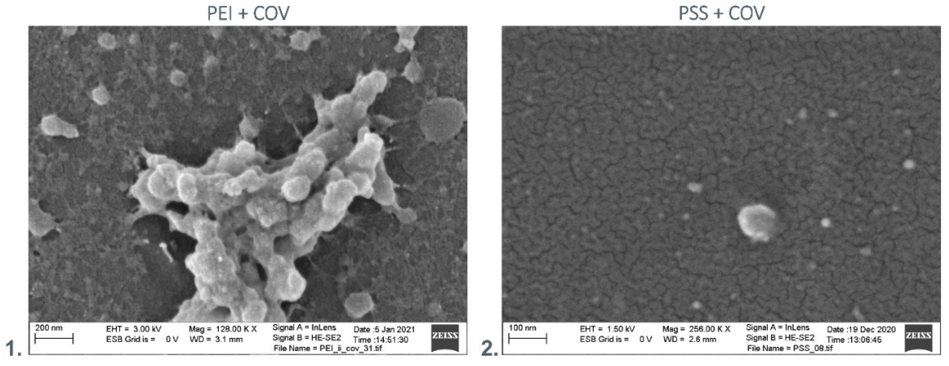
Abstract
:1. Introduction (Semi-Review)
2. Materials and Methods
2.1. Materials
2.2. Methods
2.2.1. FeNPs Synthesis
2.2.2. Preparation of Polyelectrolyte Membranes Deposited on Polystyrene Support for SEM/TEM Studies
2.2.3. Preparation of the Polyelectrolyte Membranes
- (1)
- Polyethylenimine (PEI) incorporating AuNPs (PEI-Au),
- (2)
- Polyethylenimine incorporating AgNPs (PEI-Ag),
- (3)
- Polyethylenimine incorporating CuNPs (PEI-Cu),
- (4)
- Polyethylenimine incorporating FeNPs (PEI-Fe).
- (1)
- Polystyrene sulfonate (PSS) incorporating AuNPs (PSS-Au)
- (2)
- Polystyrene sulfonate incorporating AgNPs (PSS-Ag)
- (3)
- Polystyrene sulfonate incorporating CuNPs (PSS-Cu)
- (4)
- Polystyrene sulfonate incorporating AgNPs (PSS-Fe)
2.2.4. A549 Cell Line Culture on Glass Coverslips Covered by Polyelectrolyte Membranes with Adsorbed COV
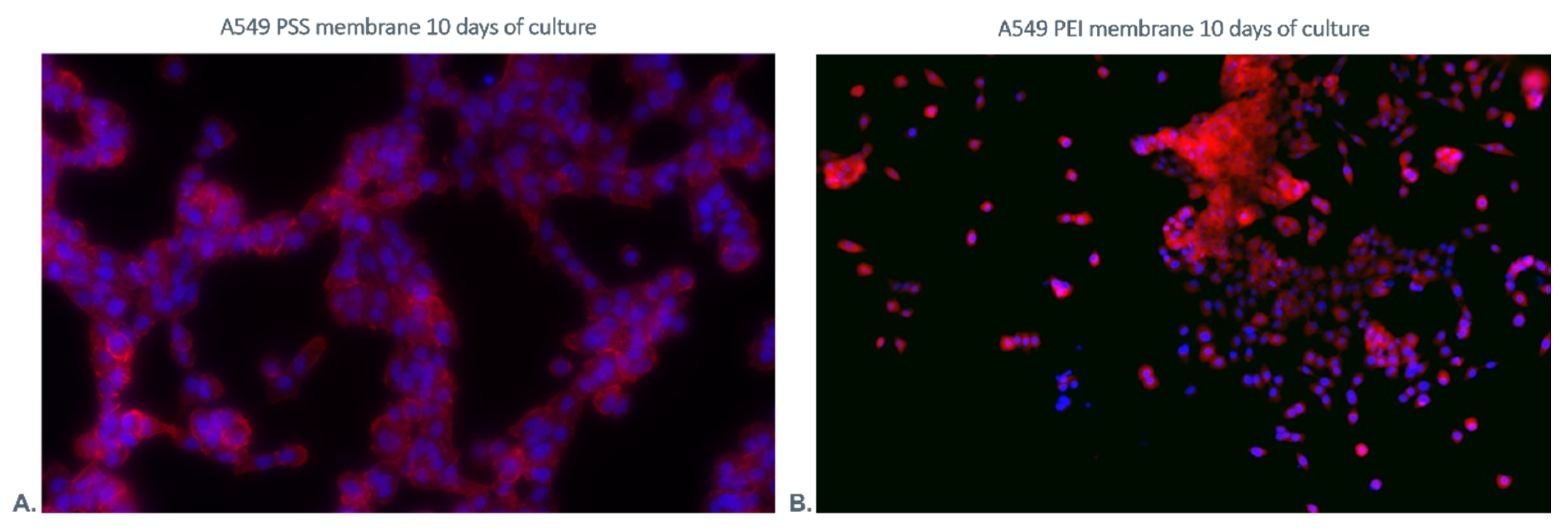
2.2.5. Fluorescence Staining
2.2.6. Flow Cytometric Analysis
2.2.7. MTT Assay
2.2.8. Scanning Electron Microscopy Analysis
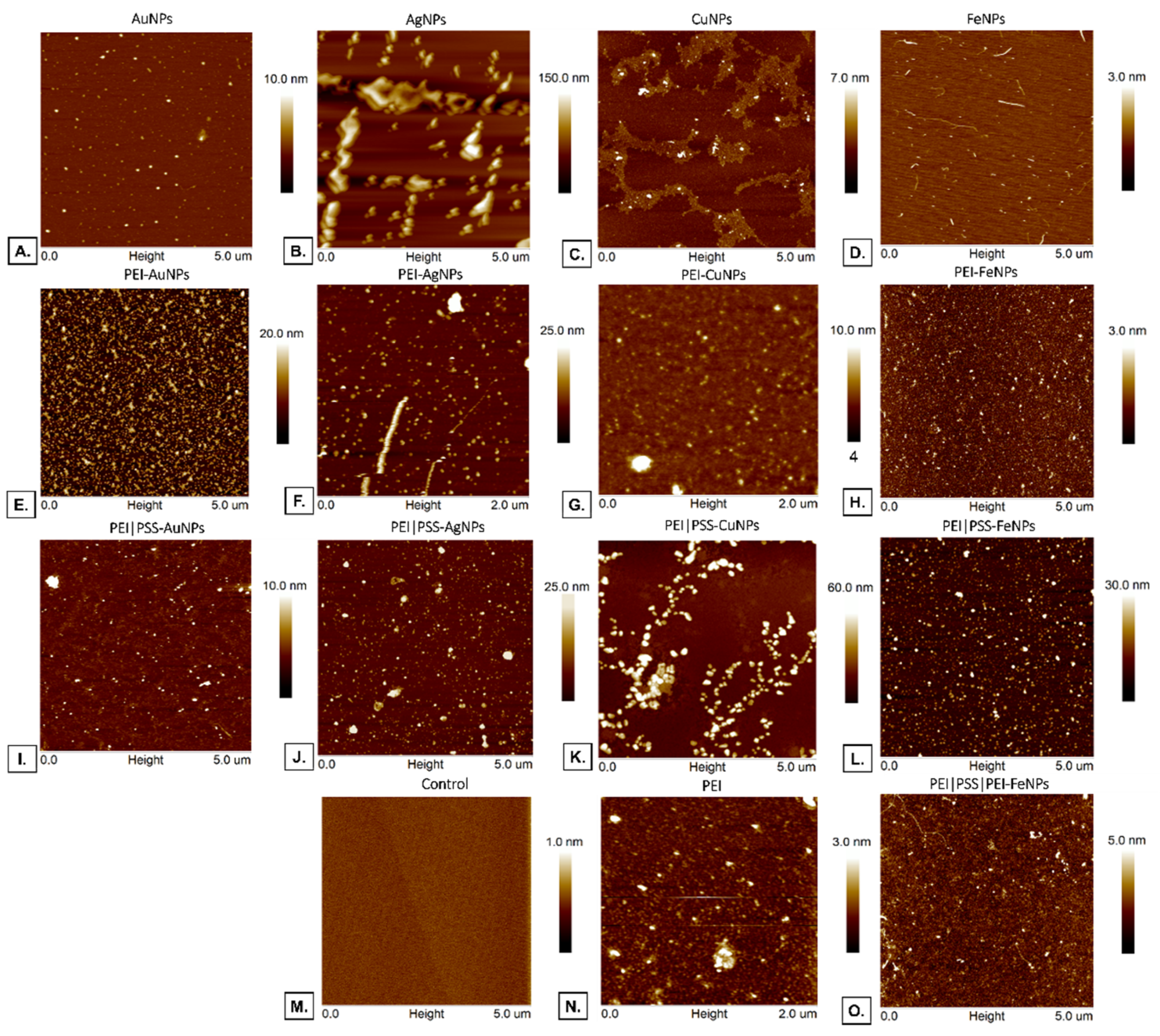
2.2.9. Atomic Forces Microscopy Evaluation
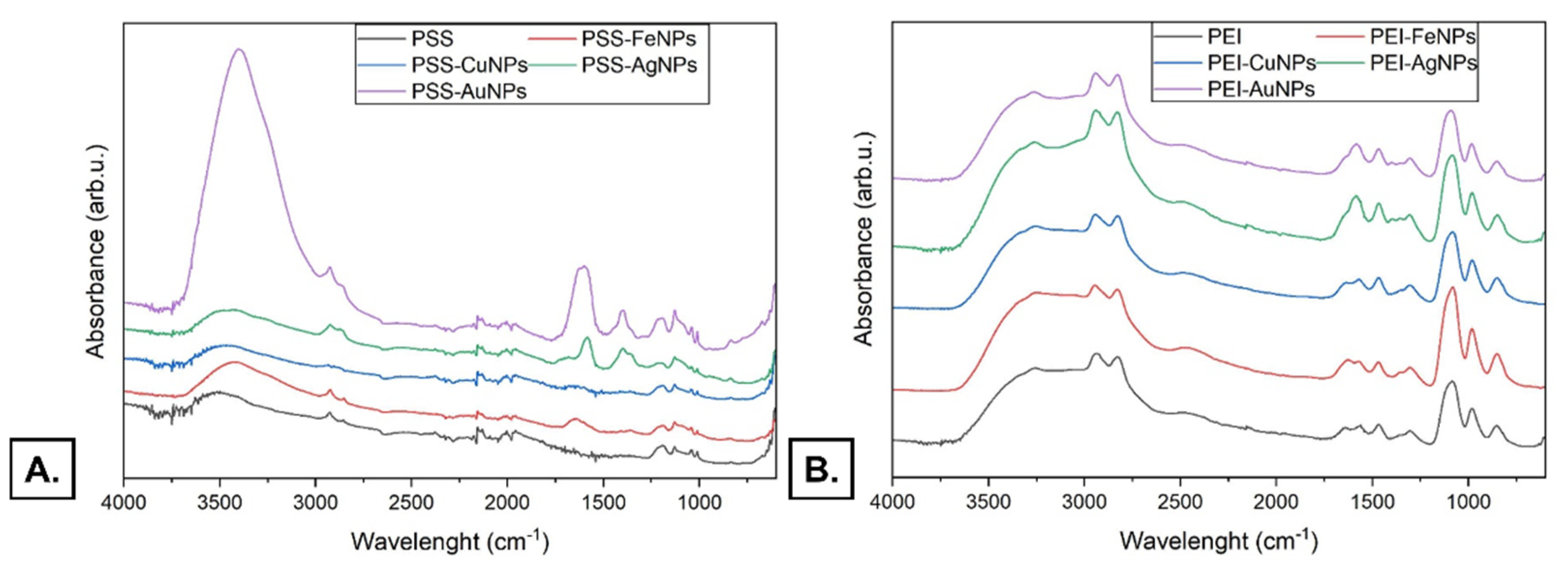
2.2.10. Fourier Transform Infrared (FT-IR) Spectroscopy
2.2.11. Statistical Analysis
3. Results and Discussion
3.1. Studies of Virus Adhesion to the Developed Membranes
Scanning Electron Microscopy Analysis
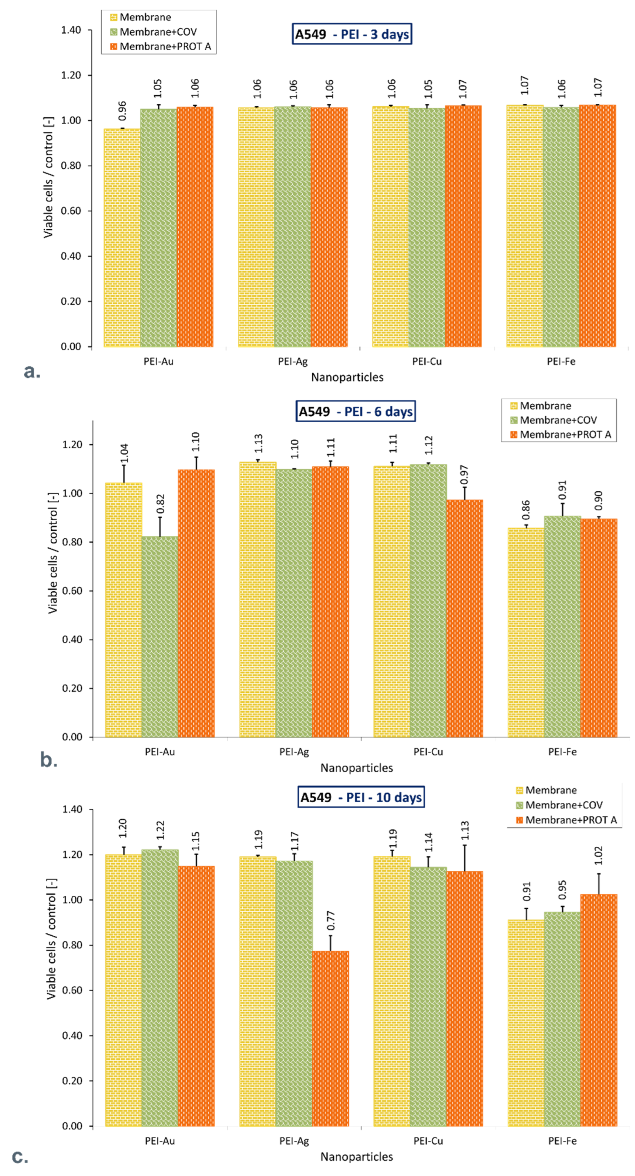
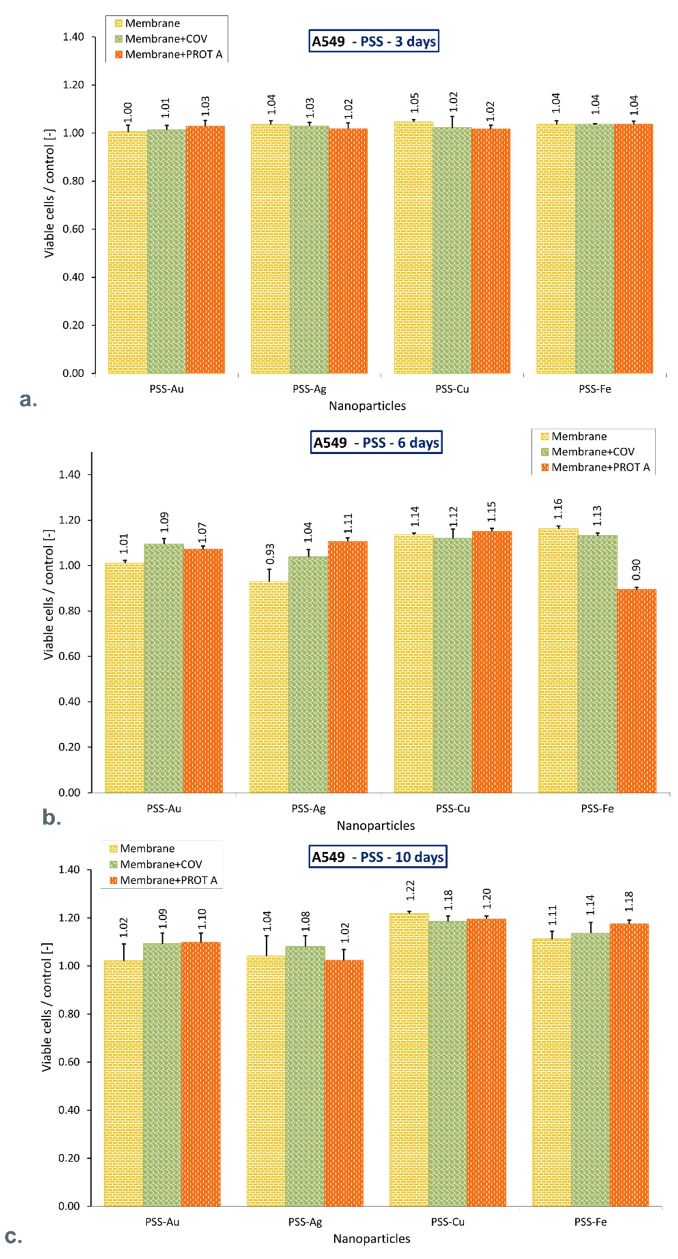
3.2. Analysis of the Functioning of Cells Immobilized within Membranes in the Virus Presence
3.2.1. Flow Cytometry
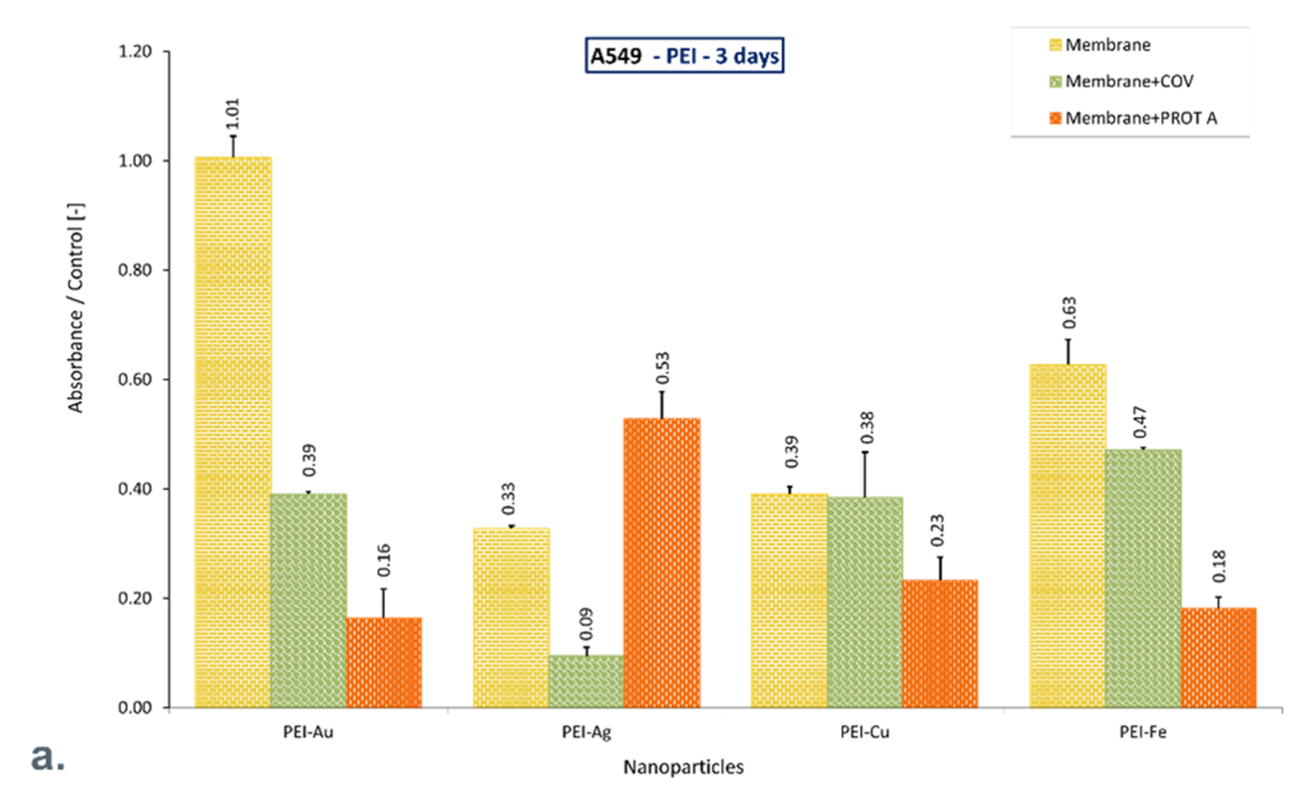
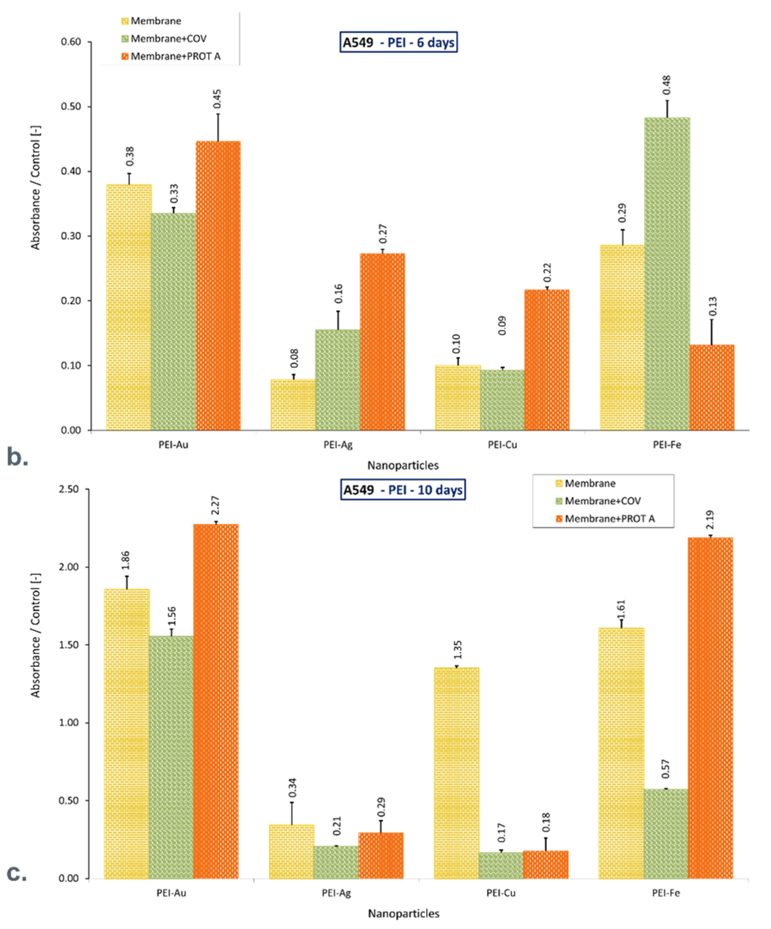
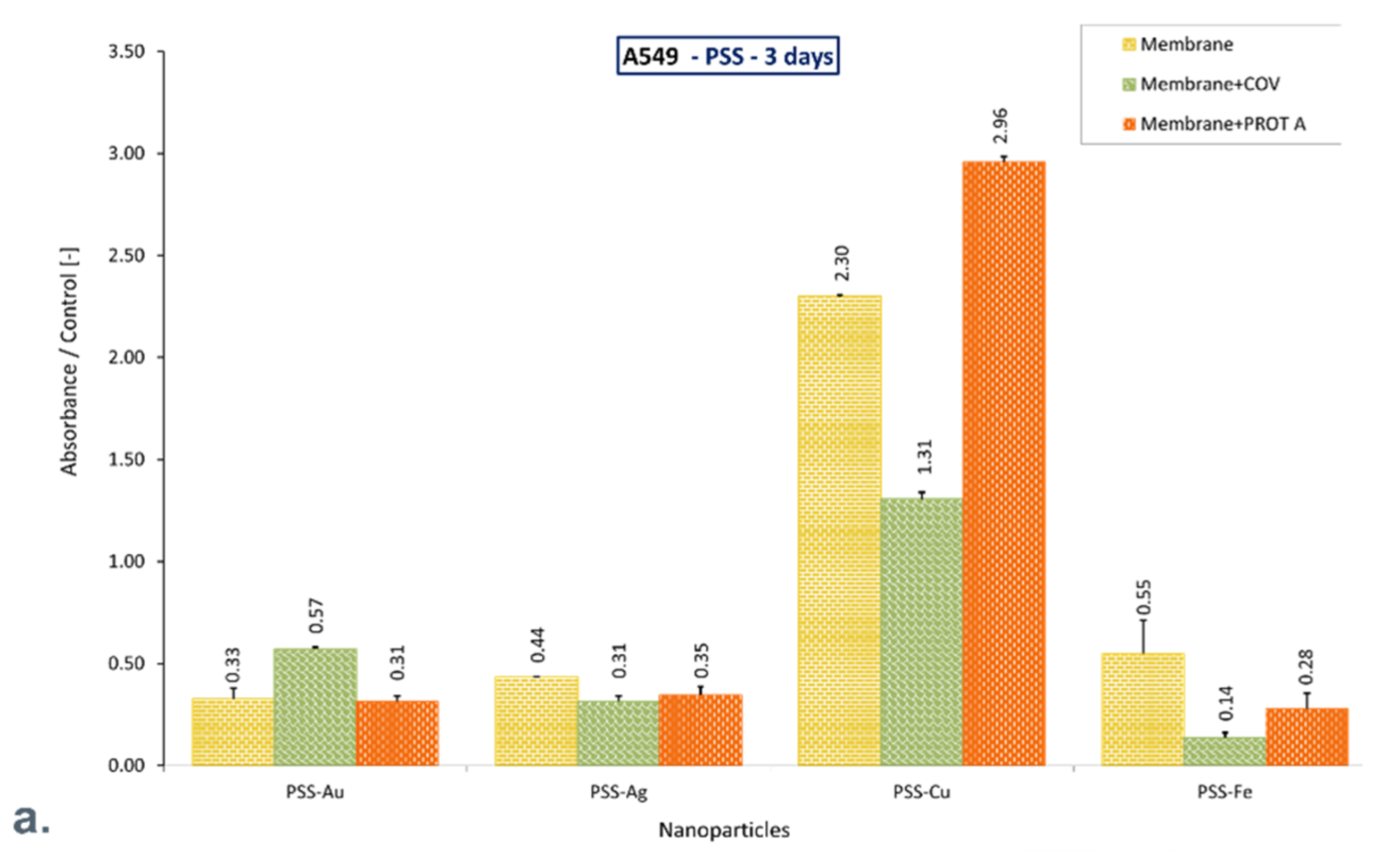
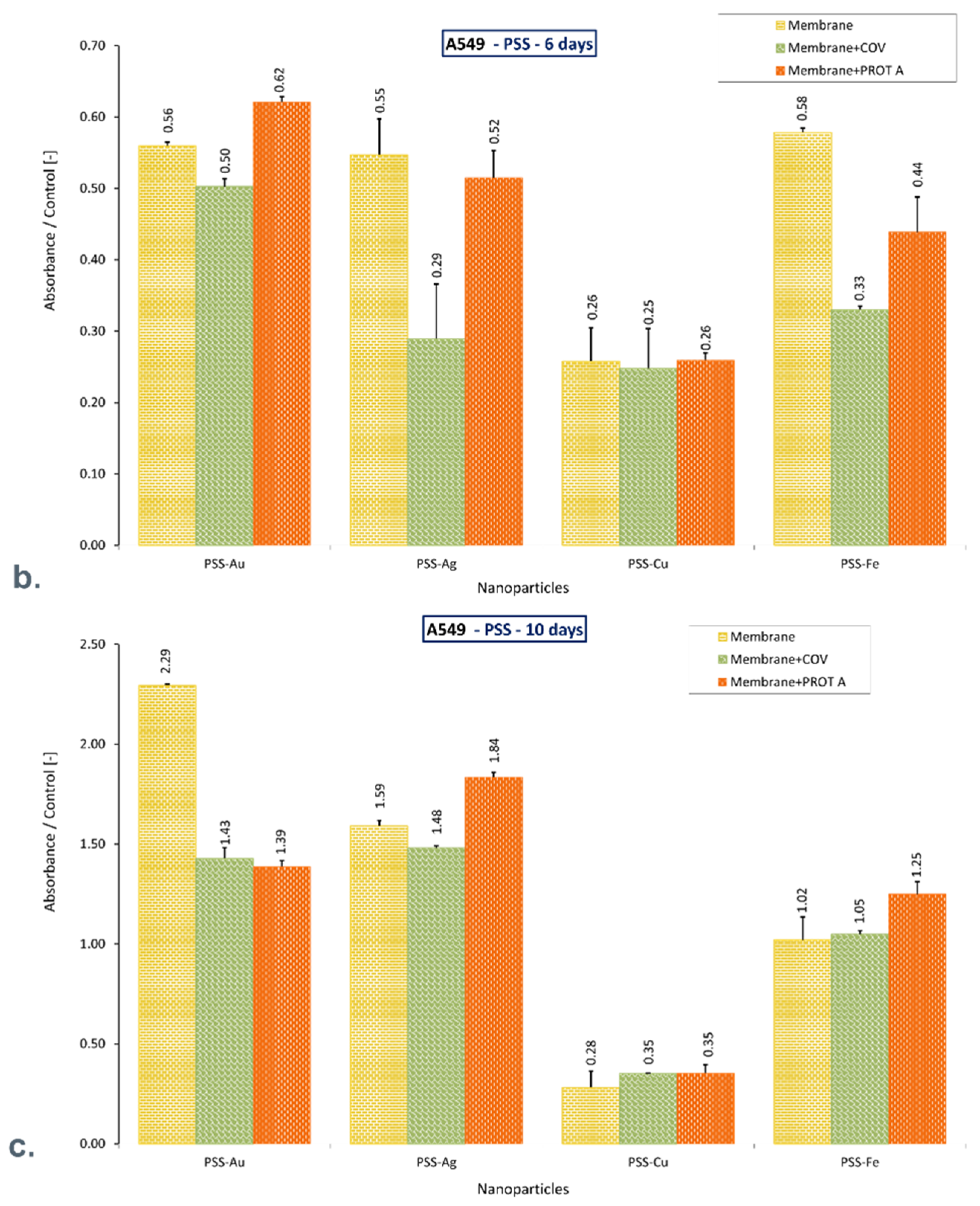
3.2.2. Assessment of the Cell Mitochondrial Metabolic Activity Using MTT Assay
3.3. Analysis of the Adhesion of Cells Cultured in the Virus Presence
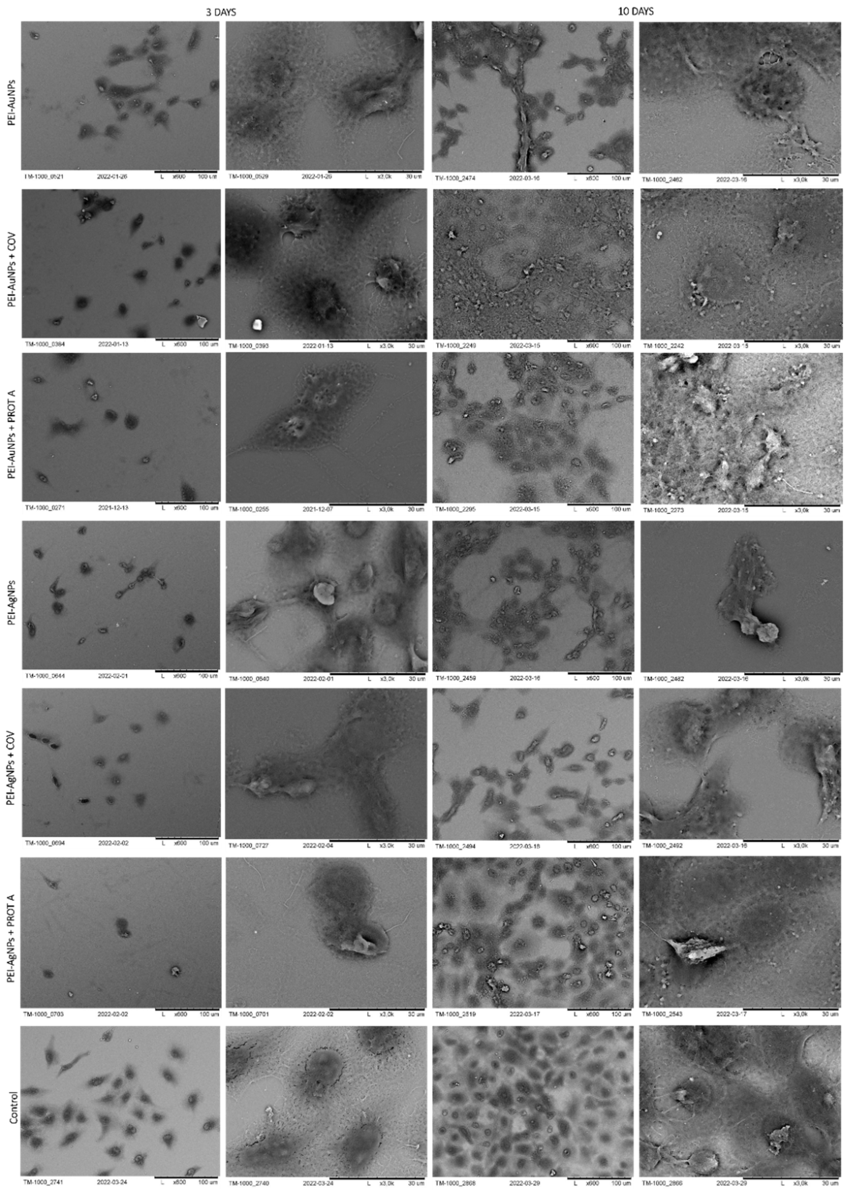
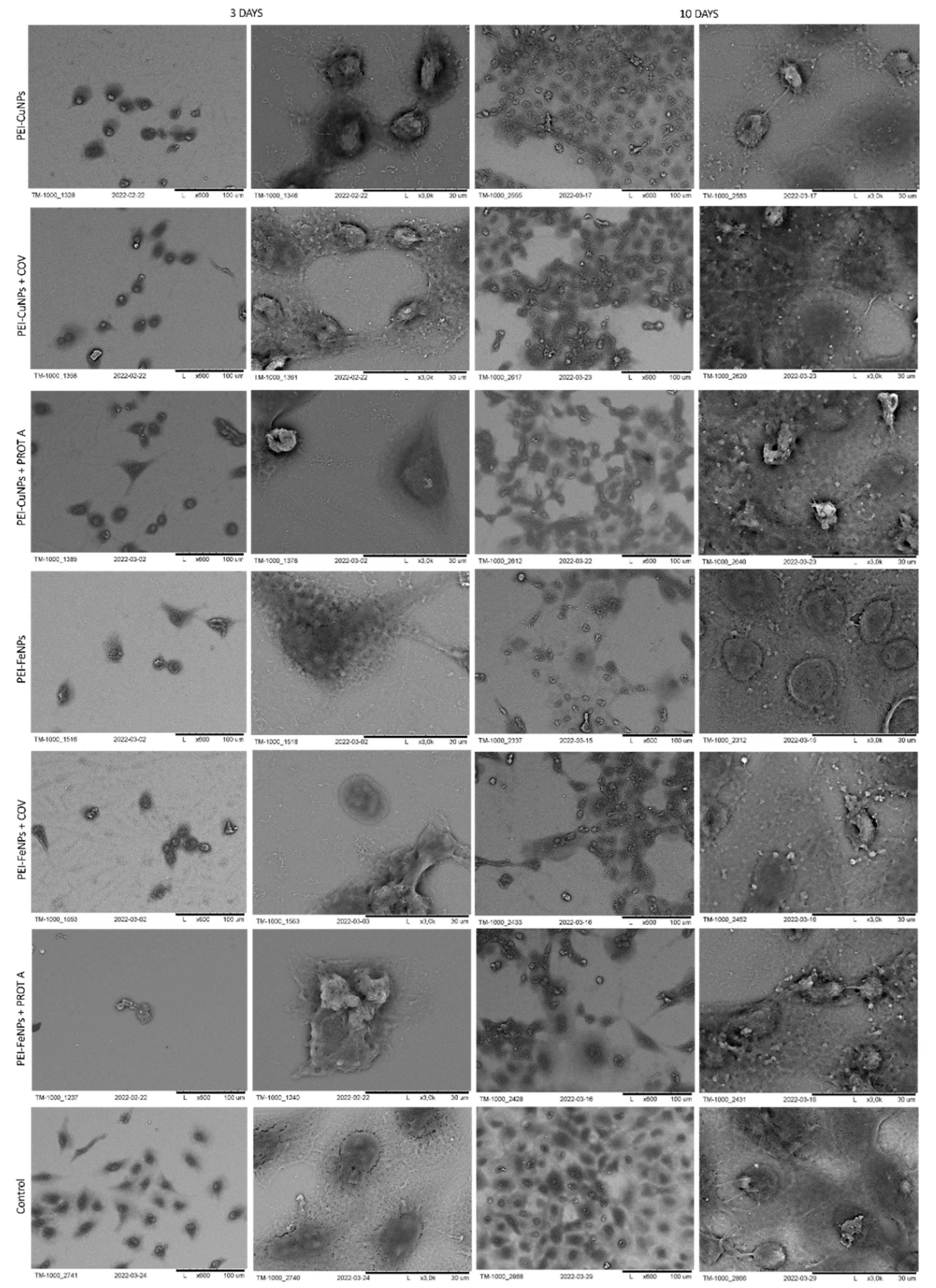
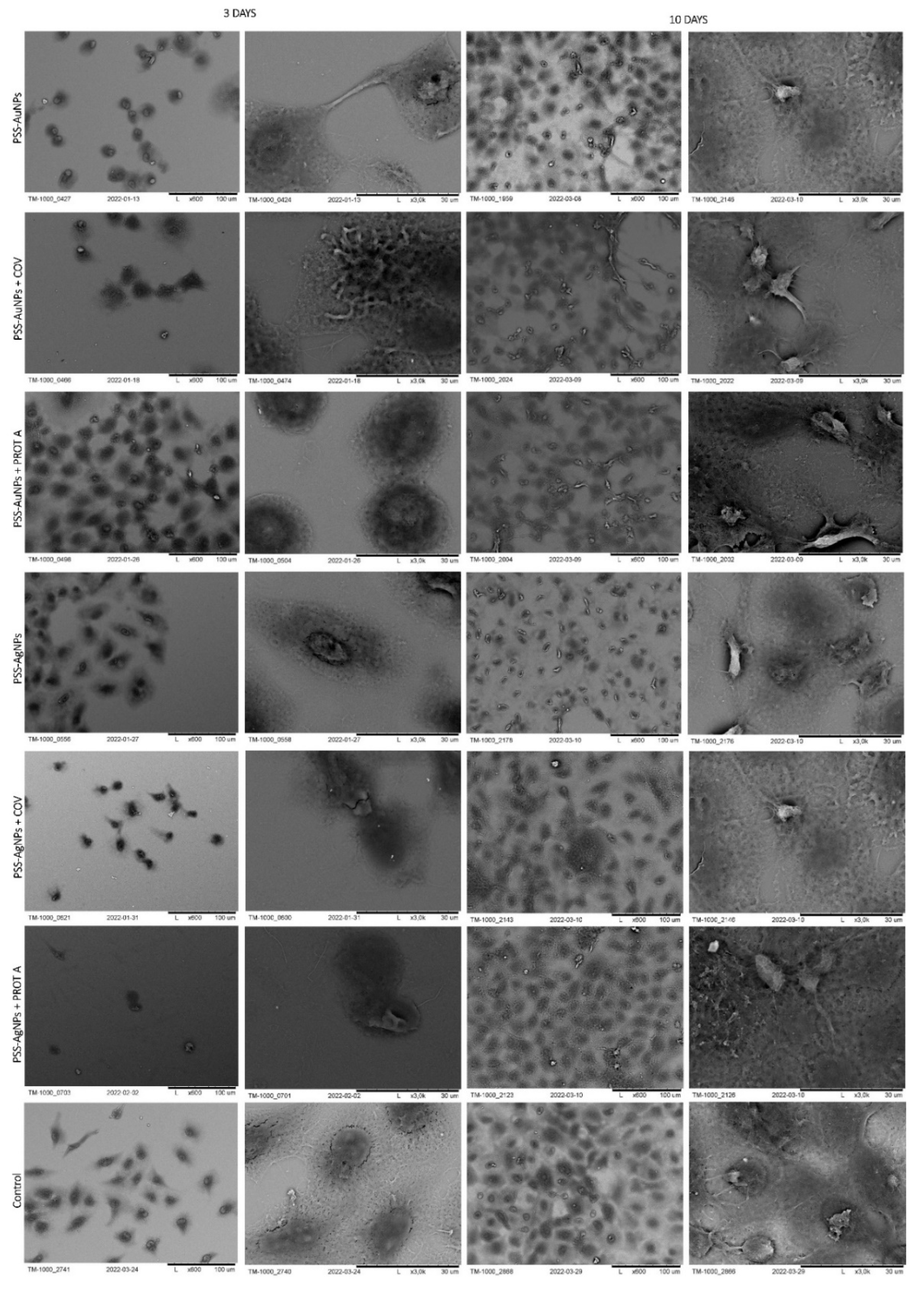
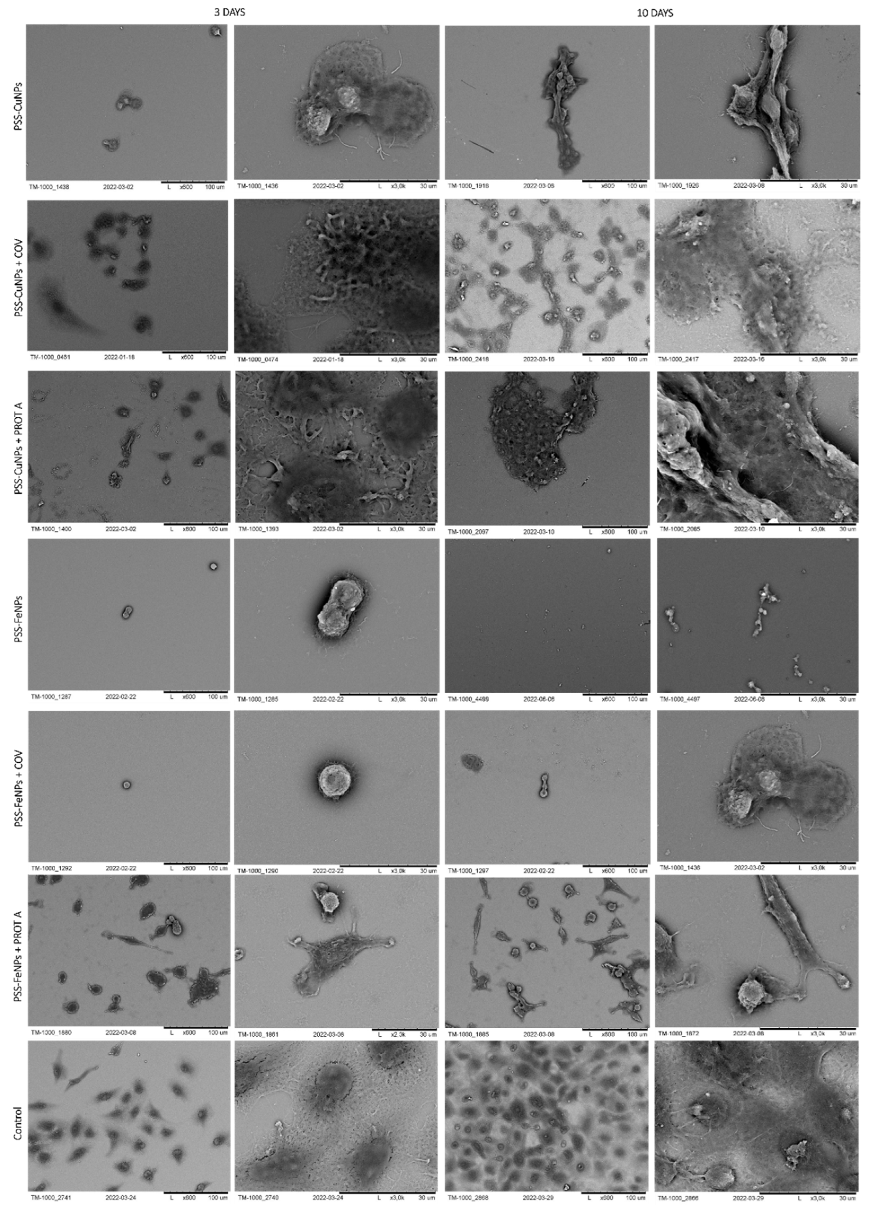
3.3.1. Scanning Electron Microscopy Analysis
3.3.2. Atomic Force Microscopy Analysis
3.3.3. Fourier Transform Infrared (FT-IR) Spectroscopy
3.3.4. Fluorescence Staining
4. Conclusions
Author Contributions
Funding
Institutional Review Board Statement
Data Availability Statement
Conflicts of Interest
References
- Matsubayashi, T.; Ishikawa, Y.; Ueda, M. Economic crisis and mental health during the COVID-19 pandemic in Japan. J. Affect. Disord. 2022, 306, 28–31. [Google Scholar] [CrossRef] [PubMed]
- Thomson, S.; García-Ramírez, J.A.; Akkazieva, B.; Habicht, T.; Cylus, J.; Evetovits, T. How resilient is health financing policy in Europe to economic shocks? Evidence from the first year of the COVID-19 pandemic and the 2008 global financial crisis. Health Policy 2022, 126, 7–15. [Google Scholar] [CrossRef]
- Kok, T.W.; Chan, P.K.S. Emergent human coronaviruses–History informs the future. J. Virol. Methods 2021, 290, 114095. [Google Scholar] [CrossRef] [PubMed]
- Chen, K.; Pun, C.S.; Wong, H.Y. Efficient social distancing during the COVID-19 pandemic: Integrating economic and public health considerations. Eur. J. Oper. Res. 2021, 304, 84. [Google Scholar] [CrossRef] [PubMed]
- World Health Organization. Social Distancing, Surveillance, and Stronger Health Systems as Keys to Controlling COVID-19 Pandemic, PAHO Director Says-PAHO/WHO|Pan American Health Organization. 2020. Available online: https://www.paho.org/en/news/2-6-2020-social-distancing-surveillance-and-stronger-health-systems-keys-controlling-covid-19 (accessed on 27 March 2022).
- Behl, A.; Nair, A.; Mohagaonkar, S.; Yadav, P.; Gambhir, K.; Tyagi, N.; Sharma, R.K.; Butola, B.S.; Sharma, N. Threat, challenges, and preparedness for future pandemics: A descriptive review of phylogenetic analysis based predictions. Infect. Genet. Evol. 2022, 98, 105217. [Google Scholar] [CrossRef] [PubMed]
- Cortes, A.A.; Zuñiga, J.M. The use of copper to help prevent transmission of SARS-coronavirus and influenza viruses. A general review. Diagn. Microbiol. Infect. Dis. 2020, 98, 115176. [Google Scholar] [CrossRef] [PubMed]
- Stagi, L.; De Forni, D.; Malfatti, L.; Caboi, F.; Salis, A.; Poddesu, B.; Cugia, G.; Lori, F.; Galleri, G.; Innocenzi, P. Effective SARS-CoV-2 antiviral activity of hyperbranched polylysine nanopolymers. Nanoscale 2021, 13, 16465–16476. [Google Scholar] [CrossRef] [PubMed]
- Pısıl, Y.; Shida, H.; Miura, T. A Neutralization Assay Based on Pseudo-Typed Lentivirus with SARS CoV-2 Spike Protein in ACE2-Expressing CRFK Cells. Pathogens 2021, 10, 153. [Google Scholar] [CrossRef]
- Rakowska, P.D.; Tiddia, M.; Faruqui, N.; Bankier, C.; Pei, Y.; Pollard, A.J.; Zhang, J.; Gilmore, I.S. Antiviral surfaces and coatings and their mechanisms of action. Commun. Mater. 2021, 2, 53. [Google Scholar] [CrossRef]
- Zhang, Q.; Honko, A.; Zhou, J.; Gong, H.; Downs, S.N.; Vasquez, J.H.; Fang, R.H.; Gao, W.; Griffiths, A.; Zhang, L. Cellular Nanosponges Inhibit SARS-CoV-2 Infectivity. Nano Lett. 2020, 20, 5570–5574. [Google Scholar] [CrossRef]
- Jiang, X.; Li, Z.; Young, D.J.; Liu, M.; Wu, C.; Wu, Y.L.; Loh, X.J. Toward the prevention of coronavirus infection: What role can polymers play? Mater. Today. Adv. 2021, 10, 100140. [Google Scholar] [CrossRef]
- Jang, Y.; Shin, H.; Lee, M.K.; Kwon, O.S.; Shin, J.S.; Kim, Y.i.; Kim, C.W.; Lee, H.R.; Kim, M. Antiviral activity of lambda-carrageenan against influenza viruses and severe acute respiratory syndrome coronavirus 2. Sci. Rep. 2021, 11, 821. [Google Scholar] [CrossRef]
- Jicsinszky, L.; Martina, K.; Cravotto, G. Cyclodextrins in the antiviral therapy. J. Drug Deliv. Sci. Technol. 2021, 64, 102589. [Google Scholar] [CrossRef]
- Garrido, P.F.; Calvelo, M.; Blanco-González, A.; Veleiro, U.; Suárez, F.; Conde, D.; Cabezón, A.; Piñeiro, Á.; Garcia-Fandino, R. The Lord of the NanoRings: Cyclodextrins and the battle against SARS-CoV-2. Int. J. Pharm. 2020, 588, 119689. [Google Scholar] [CrossRef]
- Gasbarri, M.; V’kovski, P.; Torriani, G.; Thiel, V.; Stellacci, F.; Tapparel, C.; Cagno, V. SARS-CoV-2 Inhibition by Sulfonated Compounds. Microorganisms 2020, 8, 1894. [Google Scholar] [CrossRef]
- Jaber, N.; Al-Remawi, M.; Al-Akayleh, F.; Al-Muhtaseb, N.; Al-Adham, I.S.I.; Collier, P.J. A review of the antiviral activity of Chitosan, including patented applications and its potential use against COVID-19. J. Appl. Microbiol. 2022, 132, 41–58. [Google Scholar] [CrossRef]
- Umar, Y.; Al-Batty, S.; Rahman, H.; Ashwaq, O.; Sarief, A.; Sadique, Z.; Sreekumar, P.A.; Haque, S.K.M. Polymeric Materials as Potential Inhibitors Against SARS-CoV-2. J. Polym. Environ. 2021, 30, 1244–1263. [Google Scholar] [CrossRef]
- Hashmi, A.; Nayak, V.; Singh, K.R.; Jain, B.; Baid, M.; Alexis, F.; Singh, A.K. Potentialities of graphene and its allied derivatives to combat against SARS-CoV-2 infection. Mater. Today Adv. 2022, 13, 100208. [Google Scholar] [CrossRef]
- Putro, J.N.; Lunardi, V.B.; Soetaredjo, F.E.; Yuliana, M.; Santoso, S.P.; Wenten, I.G.; Ismadji, S. A Review of Gum Hydrocolloid Polyelectrolyte Complexes (PEC) for Biomedical Applications: Their Properties and Drug Delivery Studies. Processes 2021, 9, 1796. [Google Scholar] [CrossRef]
- Vijitha, R.; Reddy, N.S.; Nagaraja, K.; Sudha Vani, T.J.; Hanafiah, M.M.; Venkateswarlu, K.; Lakkaboyana, S.K.; Krishna Rao, K.S.V.; Rao, K.M. Fabrication of Polyelectrolyte Membranes of Pectin Graft-Copolymers with PVA and Their Composites with Phosphomolybdic Acid for Drug Delivery, Toxic Metal Ion Removal, and Fuel Cell Applications. Membranes 2021, 11, 792. [Google Scholar] [CrossRef]
- Solomevich, S.O.; Dmitruk, E.I.; Bychkovsky, P.M.; Salamevich, D.A.; Kuchuk, S.V.; Yurkshtovich, T.L. Biodegradable polyelectrolyte complexes of chitosan and partially crosslinked dextran phosphate with potential for biomedical applications. Int. J. Biol. Macromol. 2021, 169, 500–512. [Google Scholar] [CrossRef] [PubMed]
- Vijitha, R.; Nagaraja, K.; Hanafiah, M.M.; Rao, K.M.; Venkateswarlu, K.; Lakkaboyana, S.K.; Krishna Rao, K.S.V. Fabrication of Eco-Friendly Polyelectrolyte Membranes Based on Sulfonate Grafted Sodium Alginate for Drug Delivery, Toxic Metal Ion Removal and Fuel Cell Applications. Polymers 2021, 13, 3293. [Google Scholar] [CrossRef] [PubMed]
- Peddinti, B.S.T.; Scholle, F.; Vargas, M.G.; Smith, S.D.; Ghiladi, R.A.; Spontak, R.J. Inherently self-sterilizing charged multiblock polymers that kill drug-resistant microbes in minutes. Mater. Horiz. 2019, 6, 2056–2062. [Google Scholar] [CrossRef]
- Haldar, J.; An, D.; De Cienfuegos, L.Á.; Chen, J.; Klibanov, A.M. Polymeric coatings that inactivate both influenza virus and pathogenic bacteria. Proc. Natl. Acad. Sci. USA 2006, 103, 17667–17671. [Google Scholar] [CrossRef]
- Antosiak-Iwańska, M.; Bącal, P.; Kazimierczak, B.; Kwiatkowska, A.; Godlewska, E.; Grzeczkowicz, A.; Stachowiak, R.; Bielecki, J.; Granicka, L. Polyelectrolyte Membrane with Hydroxyapatite and Silver Nanoparticles as a Material for Modern Wound Dressings. J. Biomed. Nanotechnol. 2020, 16, 702–714. [Google Scholar] [CrossRef]
- Zorov, D.B.; Juhaszova, M.; Sollott, S.J. Mitochondrial reactive oxygen species (ROS) and ROS-induced ROS release. Physiol. Rev. 2014, 94, 909–950. [Google Scholar] [CrossRef]
- Gallud, A.; Klöditz, K.; Ytterberg, J.; Östberg, N.; Katayama, S.; Skoog, T.; Gogvadze, V.; Chen, Y.Z.; Xue, D.; Moya, S.; et al. Cationic gold nanoparticles elicit mitochondrial dysfunction: A multi-omics study. Sci. Rep. 2019, 9, 4366. [Google Scholar] [CrossRef]
- Jin, X.; Yu, H.; Zhang, Z.; Cui, T.; Wu, Q.; Liu, X.; Gao, J.; Zhao, X.; Shi, J.; Qu, G.; et al. Surface charge-dependent mitochondrial response to similar intracellular nanoparticle contents at sublethal dosages. Part. Fibre Toxicol. 2021, 18, 36. [Google Scholar] [CrossRef]
- Adeyemi, J.A.; Machado, A.R.T.; Ogunjimi, A.T.; Alberici, L.C.; Antunes, L.M.G.; Barbosa, F. Cytotoxicity, mutagenicity, oxidative stress and mitochondrial impairment in human hepatoma (HepG2) cells exposed to copper oxide, copper-iron oxide and carbon nanoparticles. Ecotoxicol. Environ. Saf. 2020, 189, 109982. [Google Scholar] [CrossRef]
- Skalska, J.; Dąbrowska-Bouta, B.; Frontczak-Baniewicz, M.; Sulkowski, G.; Strużyńska, L. A Low Dose of Nanoparticulate Silver Induces Mitochondrial Dysfunction and Autophagy in Adult Rat Brain. Neurotox. Res. 2020, 38, 650–664. [Google Scholar] [CrossRef]
- Yang, J.C.; Jablonsky, M.J.; Mays, J.W. NMR and FT-IR studies of sulfonated styrene-based homopolymers and copolymers. Polymer 2002, 43, 5125–5132. [Google Scholar] [CrossRef]
- Sundararajan, B.; Ranjitha Kumari, B.D. Novel synthesis of gold nanoparticles using Artemisia vulgaris L. leaf extract and their efficacy of larvicidal activity against dengue fever vector Aedes aegypti L. J. Trace Elem. Med. Biol. 2017, 43, 187–196. [Google Scholar] [CrossRef]
- Paulkumar, K.; Gnanajobitha, G.; Vanaja, M.; Pavunraj, M.; Annadurai, G. Green synthesis of silver nanoparticle and silver based chitosan bionanocomposite using stem extract of Saccharum officinarum and assessment of its antibacterial activity. Adv. Nat. Sci. Nanosci. Nanotechnol. 2017, 8, 035019. [Google Scholar] [CrossRef]
- Gong, W. A real time in situ ATR-FTIR spectroscopic study of linear phosphate adsorption on titania surfaces. Int. J. Miner. Process. 2001, 63, 147–165. [Google Scholar] [CrossRef]
- Grenda, K.; Idström, A.; Evenäs, L.; Persson, M.; Holmberg, K.; Bordes, R. An analytical approach to elucidate the architecture of polyethyleneimines. J. Appl. Polym. Sci. 2022, 139, 51657. [Google Scholar] [CrossRef]
- Grzeczkowicz, A.; Gruszczynska-Biegala, J.; Czeredys, M.; Kwiatkowska, A.; Strawski, M.; Szklarczyk, M.; Koźbiał, M.; Kuźnicki, J.; Granicka, L.H. Polyelectrolyte membrane scaffold sustains growth of neuronal cells. J. Biomed. Mater. Res. Part A 2019, 107, jbm.a.36599. [Google Scholar] [CrossRef] [Green Version]


Publisher’s Note: MDPI stays neutral with regard to jurisdictional claims in published maps and institutional affiliations. |
© 2022 by the authors. Licensee MDPI, Basel, Switzerland. This article is an open access article distributed under the terms and conditions of the Creative Commons Attribution (CC BY) license (https://creativecommons.org/licenses/by/4.0/).
Share and Cite
Grzeczkowicz, A.; Lipko, A.; Kwiatkowska, A.; Strawski, M.; Bącal, P.; Więckowska, A.; Granicka, L.H. Polyelectrolyte Membrane Nanocoatings Aimed at Personal Protective and Medical Equipment Surfaces to Reduce Coronavirus Spreading. Membranes 2022, 12, 946. https://doi.org/10.3390/membranes12100946
Grzeczkowicz A, Lipko A, Kwiatkowska A, Strawski M, Bącal P, Więckowska A, Granicka LH. Polyelectrolyte Membrane Nanocoatings Aimed at Personal Protective and Medical Equipment Surfaces to Reduce Coronavirus Spreading. Membranes. 2022; 12(10):946. https://doi.org/10.3390/membranes12100946
Chicago/Turabian StyleGrzeczkowicz, Anna, Agata Lipko, Angelika Kwiatkowska, Marcin Strawski, Paweł Bącal, Agnieszka Więckowska, and Ludomira H. Granicka. 2022. "Polyelectrolyte Membrane Nanocoatings Aimed at Personal Protective and Medical Equipment Surfaces to Reduce Coronavirus Spreading" Membranes 12, no. 10: 946. https://doi.org/10.3390/membranes12100946