Review

The Role of Laser Photocoagulation in Treating Diabetic Macular Edema in the Era of Intravitreal Drug Administration: A Descriptive Review

1
Department of Ophthalmology, Laser Eye Center, Nagoya City University East Medical Center, Nagoya 464-8547, Japan
2
Department of Ophthalmology and Visual Science, Nagoya City University Graduate School of Medical Sciences, Nagoya 467-8601, Japan
3
Department of Ophthalmology, Toyota Kosei Hospital, Toyota 470-0396, Japan
*
Author to whom correspondence should be addressed.
Medicina 2023, 59(7), 1319; https://doi.org/10.3390/medicina59071319
Submission received: 3 July 2023 / Revised: 11 July 2023 / Accepted: 15 July 2023 / Published: 17 July 2023
(This article belongs to the Special Issue Macular Edema: Pathogenesis, Diagnosis and Therapies)

Abstract

:
Background and Objectives: This study aimed to elucidate the role of laser photocoagulation therapy in the treatment of diabetic macular edema (DME) as an alternative to, or in conjunction with, the first-line treatment, anti-vascular endothelial growth factor (VEGF). Materials and Methods: A comprehensive literature search to identify studies that evaluated the efficacy of laser photocoagulation therapy in the management of DME was performed. The relevant findings of the efficacy of focal/grid laser therapy from data in randomized, controlled trials were synthesized, and the potential of new laser technologies, such as navigated laser systems, pattern scan lasers, and subthreshold lasers, was explored. The usefulness of multimodal imaging-guided laser therapy was also evaluated, with a focus on the potential contribution to anti-VEGF therapy. Results: Focal laser photocoagulation targeting microaneurysms remains an effective therapeutic approach to chronic refractory edema, despite the widespread use of anti-VEGF therapy. To achieve the best possible treatment outcomes, precise identification of microaneurysms is essential. This requires the use of multimodal imaging-guided, highly accurate, minimally invasive coagulation techniques. Subthreshold laser therapy can also reduce the frequency of anti-VEGF injections and minimize treatment burden. Conclusions: Further studies are needed to determine the optimal timing and settings for laser photocoagulation therapy and the potential of new laser technologies in the management of DME. Nevertheless, laser photocoagulation therapy plays an important role in the management of DME, in conjunction with anti-VEGF therapy.

1. Introduction

Diabetic macular edema (DME), rather than proliferative diabetic retinopathy, is an increasingly common cause of visual impairment [1]. Macular laser photocoagulation used to be the primary treatment option for DME since The Early Treatment Diabetic Retinopathy Study Research Group (ETDRS) showed that focal/grid laser therapy could stabilize visual acuity [2]. However, with the introduction of anti-vascular endothelial growth factor (VEGF) drugs, the use of macular laser photocoagulation has decreased significantly [3,4,5]. The EURETINA guideline notes that laser treatment is no longer considered the standard of care for DME in the current era of anti-VEGF drugs [6]. Whereas anti-VEGF therapy has proven to be effective in DME [7,8,9,10], real-world data have shown that relying solely on anti-VEGF injections may not be practical due to factors such as the economic burden of treatment, poor response to anti-VEGF, and systemic conditions that limit treatment options [11,12,13].
Fortunately, new technologies in laser photocoagulation have been developed to address these challenges. These technologies include navigated laser systems [14], subthreshold laser therapies [15,16], and pattern scan laser systems [17]. In addition, advances in multimodal imaging technology have had a transformative impact on laser photocoagulation procedures, enabling more accurate, targeted, minimally invasive treatment options for patients. These advances have led to renewed interest in the use of laser photocoagulation as a treatment for DME.
In this review, the new techniques and indications for laser therapy in the treatment of DME in the anti-VEGF era are discussed. The potential benefits of incorporating these new laser technologies into the treatment plan, and the new role that laser photocoagulation may play in the management of DME are explored. By considering these new developments in laser technology, it may be possible to improve treatment outcomes and offer more options for patients with DME.

2. Focal/Grid Laser Therapy

2.1. ETDRS

The Early Treatment Diabetic Retinopathy Study (ETDRS) used fluorescein angio- grams to classify DME eyes with focal leakage associated with microaneurysms as focal and DME eyes with less leakage associated with microaneurysms as diffuse [2,18].
In the ETDRS, focal laser photocoagulation was directly applied to microaneurysms to obtain closure of the leak, and grid laser photocoagulation was applied to areas of diffuse leakage or thickened retinae [2,18,19]; these techniques became the gold standard for treating DME. The ETDRS suggested that focal/grid laser could prevent severe vision loss compared to observation only [2]. However, focal/grid laser therapy might result in scar atrophy and subretinal fibrosis in the macula in approximately 21% of patients, leading to vision loss (Figure 1) [20]. To avoid these complications, DRCR.net (Diabetic Retinopathy Clinical Research Network) recommended “modified” ETDRS settings, in which the laser spots were slightly smaller (50 μm) and lighter (gray), and a color change was not required when treating the microaneurysms [21]. DRCR.net protocol A found the modified ETDRS laser to be better than the Mild Macular Grid laser for DME in terms of optical coherence tomography (OCT)-based retinal thickness measures, but best-corrected visual acuity (BCVA) measures in the two groups were not substantially different [21]. In Protocol A, researchers specifically evaluated the effect of modified ETDRS laser in eyes with non–center-involved clinically significant macular edema (CSME). Vision was stable, and the study concluded that modified ETDRS focal/grid laser therapy was still an appropriate treatment for extrafoveal DME [22]. The EURETINA guideline also recommended laser application as a treatment option, specifically targeting the vasogenic subform of DME, characterized by the presence of focal accumulation of microaneurysms and leaking capillaries [6].

2.2. DRCR Network

Protocol I conducted by DRCR.net compared the efficacy of ranibizumab with prompt or deferred focal/grid laser therapy in treating DME. Prompt laser therapy was defined as that within 3 to 10 days of the diagnosis or injection. Deferred laser therapy meant that performed more than 6 months after the initial injection. This study demonstrated the superiority of anti-VEGF treatment over focal/grid laser therapy [7,8]. Furthermore, in Protocol I, it was observed that, when the baseline visual acuity was worse than 20/50, the deferred laser group demonstrated superior BCVA at the 5-year follow-up compared to the prompt laser group [23]. Since 56% of the deferred laser group had never received focal/grid laser therapy in the 5 years, deferring focal/grid laser therapy may be associated with a greater chance of relatively greater improvements in visual acuity over 5 years compared with adding focal/grid laser therapy when initiating intravitreous ranibizumab, especially for eyes with worse visual acuity at baseline.

2.3. Can Focal/Grid Laser Therapy Reduce the Number of Anti-VEGF Injections?

The efficacy of anti-VEGF therapy has been well established in multiple randomized, controlled trials (RCTs), such as RISE/RIDE [24], RESTORE [10], and DA VINCI [25], which have all shown its superiority over laser photocoagulation. Recently approved anti-VEGF drugs have been compared to aflibercept in clinical studies [26,27], and no further studies have been conducted to compare the efficacy of focal/grid laser treatment alone.
Nonetheless, administering frequent anti-VEGF injections in line with the protocols used in RCTs poses a significant challenge in real-world clinical practice [11,12,13]. The combination therapy of laser photocoagulation and anti-VEGF drugs may have potential in reducing the number of injections needed for treatment. The READ-2 study compared the efficacy of ranibizumab (0.5 mg) with/without laser, and they found that combination therapy with focal/grid laser treatment might reduce the percentage of patients that require frequent ranibizumab injections. The number of injections in 2 years was 9.3 in the monotherapy group and 2.9 in the combination therapy group. However, they also found that the monotherapy group did not have patients with resolved or controlled edema who had poor visual acuity, whereas this was the case in 22% of combination therapy patients. These indicated that adding focal/grid laser therapy might sacrifice the visual outcome [9].
In Protocol I, the mean change in the visual acuity letter score from baseline to the 5-year follow-up tended to be superior in the ranibizumab with deferred laser group than in the ranibizumab with prompt laser group, with +9.8 letters versus +7.2 letters, respectively (p = 0.09). The median number of injections was 17 versus 13 in the deferred and prompt laser groups, respectively [23]. Although the RESTORE study showed the effectiveness of combining laser photocoagulation with ranibizumab (0.5 mg), there was no significant difference in the frequency of injections required [10]. Further studies will need to identify the best timing of adding laser photocoagulation to provide better anatomical and functional improvement and reduce the number of injections.

2.4. Steroid vs. Focal/Grid Laser

There is strong evidence supporting the involvement of chronic inflammation in the pathophysiology of DME, highlighting the beneficial effects of corticosteroids in improving DME [7]. However, in the current era of anti-VEGF treatment, the use of steroids as a therapeutic option for DME is considered second-line therapy. This is primarily due to the potential complications associated with steroids, including cataract formation and elevated intraocular pressure [7]. The EURETINA guidelines recommend considering the use of steroids as the first choice for patients with a history of a major cardiovascular event, since these patients were excluded from major anti-VEGF trials [6]. In addition, corticosteroids may be considered a first-line therapy option for patients who are unwilling to undergo monthly injections and monitoring during the initial 6 months of treatment [6]. Furthermore, due to cataract progression, pseudophakic patients are well-suited for steroid therapy [6,7]. The PLACID trial reported the results of a comparison of a dexamethasone implant (Ozurdex) with laser treatment versus laser monotherapy in patients with DME. Though there was no difference between the groups at 1 year, the percentage of patients gaining at least 10 letters was significantly higher in the Ozurdex plus laser group than in the laser monotherapy group at months 1 and 9 [28]. Although no specific study has directly compared fluocinolone acetonide (Iluvien) with laser treatment versus laser monotherapy, available evidence suggests that steroid therapy, in combination with laser treatment, appears to be superior to laser monotherapy [8,28].

3. New Laser Technologies

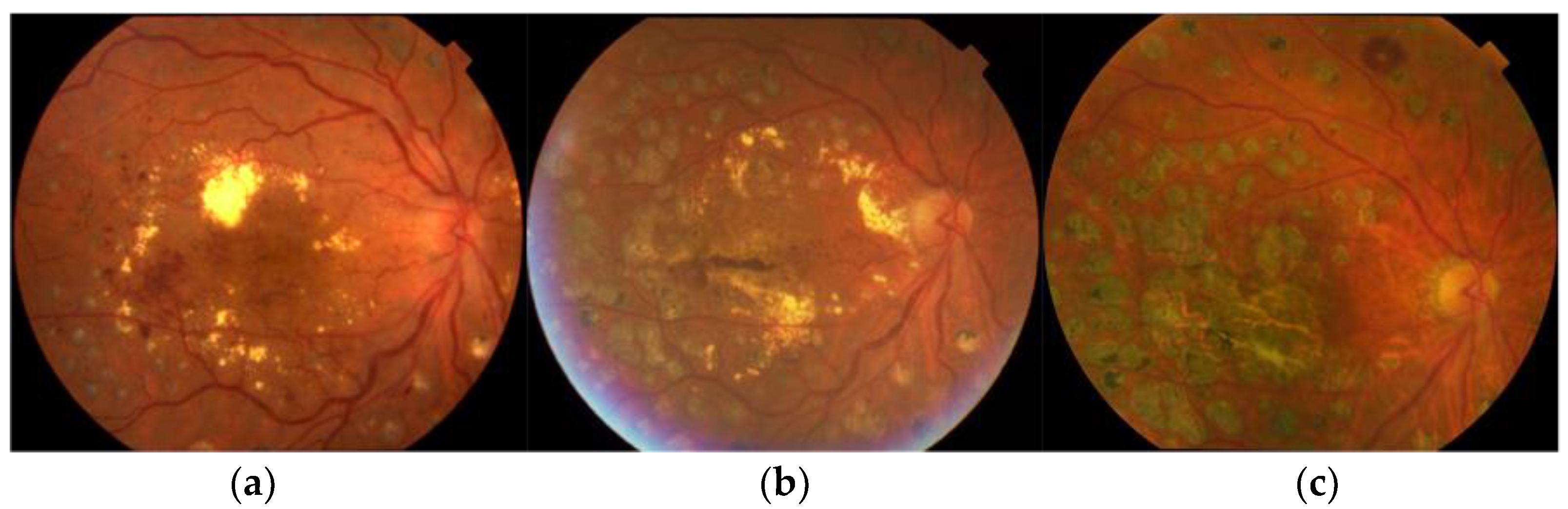
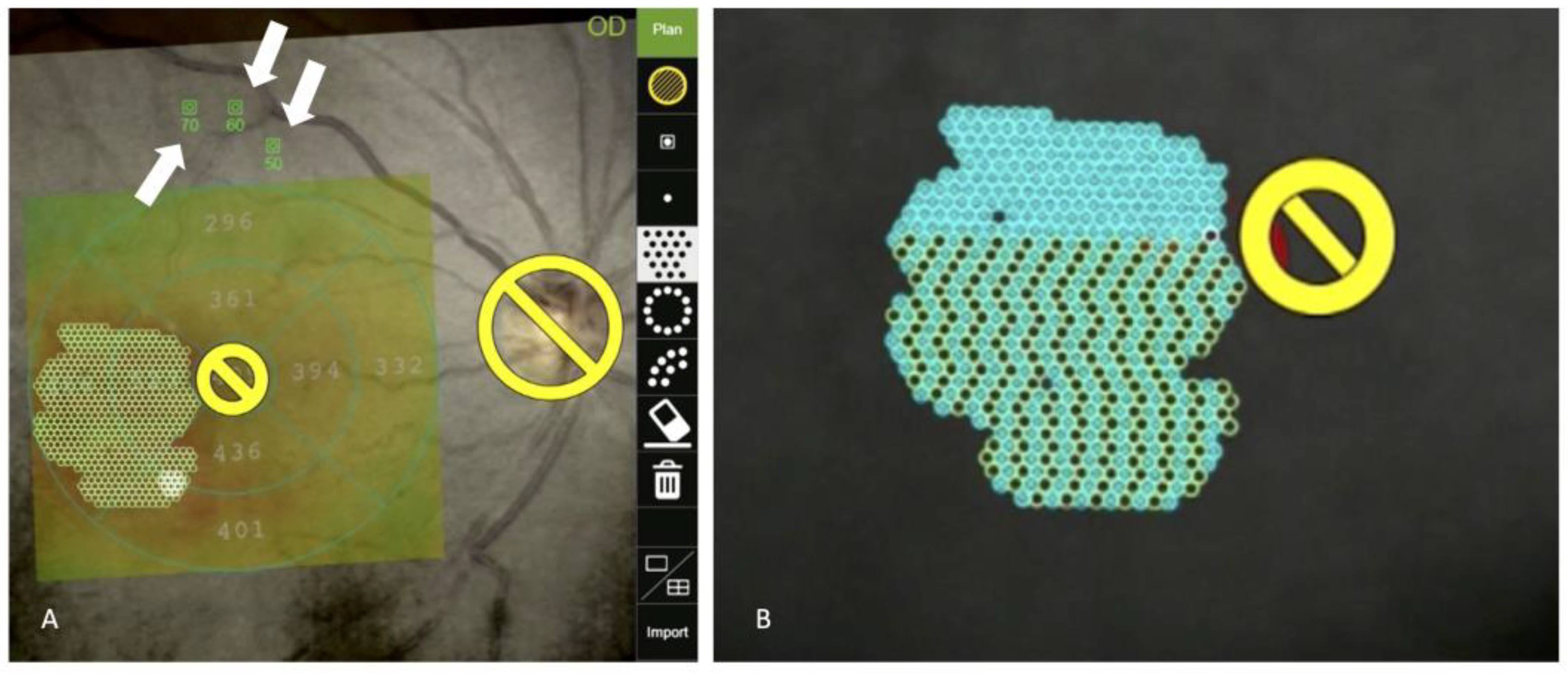
3.1. Navigated Laser Systems

The navigated laser photocoagulator (NAVILAS; OD-OS GmbH, Teltow, Germany) is a novel laser technology that incorporates a retinal eye-tracking laser delivery system and integrated digital fundus imaging. This system features advanced image processing to help treat retinal lesions by overlaying images onto the live image of the retina [14]. It was designed to improve accuracy and make it easier to treat specific lesions such as microaneurysms that require precise treatment [29]. Physicians create a treatment plan using fundus photography, fluorescein/indocyanine green angiography, or OCT, which is then displayed on a real-time image of the patient’s fundus during treatment to guide the process. By using NAVILAS, which incorporates an eye-tracking system and pre-registered laser planning, the hit rate for microaneurysms increased by 30% compared to the conventional slit-lamp delivery system [30]. In the study that compared the efficacy of the NAVILAS macular laser versus the conventional slit-lamp-based macular laser for treating DME, the NAVILAS group showed significantly better BCVA than the conventional laser group after 3 months. In addition, within the first 8 months, the NAVILAS group had a lower re-treatment rate (18%) compared to the conventional laser group (31%). These findings suggest that the NAVILAS macular laser may be a more effective treatment option for DME than the conventional slit-lamp-based macular laser [31]. Liegl et al. reported that the burden of anti-VEGF treatment for DME can be significantly reduced by the use of a precise focal laser with the NAVILAS laser system [32]. They found that NAVILAS laser combination therapy and ranibizumab monotherapy had similar effects on the mean BCVA letter score (8.41 vs. 6.31 letters, p = 0.258), but the combination group required significantly fewer injections after the loading phase (0.88 ± 1.23 vs. 3.88 ± 2.32, p ≤ 0.001). By month 12, 84% of patients in the monotherapy group needed additional ranibizumab injections, compared to 35% in the combination group (p ≤ 0.001) [31]. They also found that the effect lasted for 36 months. Patients who received the combination treatment required significantly fewer injections than those who received only anti-VEGF. Specifically, they needed two fewer injections in year 1 and 1.3-times fewer injections in years 2 and 3 (N = 24) [33]. However, in the TREX-DME study, which compared the long-term effects of treat-and-extend dosing of ranibizumab with and without navigated focal laser therapy for DME, there was no significant difference in BCVA or the number of ranibizumab injections needed (N = 109) [34]. Kato et al. reported that focal laser photocoagulation with the NAVILAS + 577 (wavelength 577 nm) was effective in treating refractory DME by reducing central retinal thickness and macular edema after 6 months. In their study, 11 eyes received an injection of aflibercept following initial navigated laser, and their number of injections for 6 months was 1.55 ± 1.0; for comparison, 23 eyes in their institution that were treated with anti-VEGF monotherapy received an average of 2.52 ± 1.0 injections during the same 6-month period [35]. More recently, an economic modeling study demonstrated that using the NAVILAS laser decreased patient injection burden while achieving comparable clinical outcomes [36]. Further studies involving a larger patient population are needed to determine whether the combination of precise focal laser photocoagulation using the NAVILAS laser is effective in reducing the number of anti-VEGF injections required.
Several studies have demonstrated the benefits of using the NAVILAS focal laser for treating refractory DME compared to other treatments [35,37]. The prospective study that treated 8 eyes with refractory DME showed that focal laser photocoagulation using NAVILAS significantly reduced central retinal thickness and macular volume, with a significant improvement in BCVA [36]. Hirano et al. reported that eyes with perifoveal leaking microaneurysms required a larger number of aflibercept injections, despite being treated with a combination of focal/grid laser therapy and aflibercept injections [38]. This is likely due to the limitation of conventional focal/grid laser treatment, which can only be applied outside the fovea (>500 μm). In that study, they demonstrated the efficacy of the NAVILAS focal laser for refractory DME; the mean distance from the center of the fovea to the closest microaneurysms was 624.8 ± 377.7 μm [37], and 63% had microaneurysms located at the perifovea, within less than 500 μm from the center of the fovea [37]. Based on the evidence presented, it can be inferred that conventional focal/grid laser treatment may have limited efficacy in treating refractory DME due to the proximity of microaneurysms to the fovea. Focal laser treatment using the NAVILAS laser may be a more suitable option for treating refractory DME due to its precise and accurate approach.
An additional advantage of the NAVILAS laser is the ability to plan laser treatment based on images obtained from multimodal imaging. This innovative feature enables more precise and customized treatment plans, tailored to the specific needs of each patient. The role of this technology will be discussed in greater detail in Section 4.

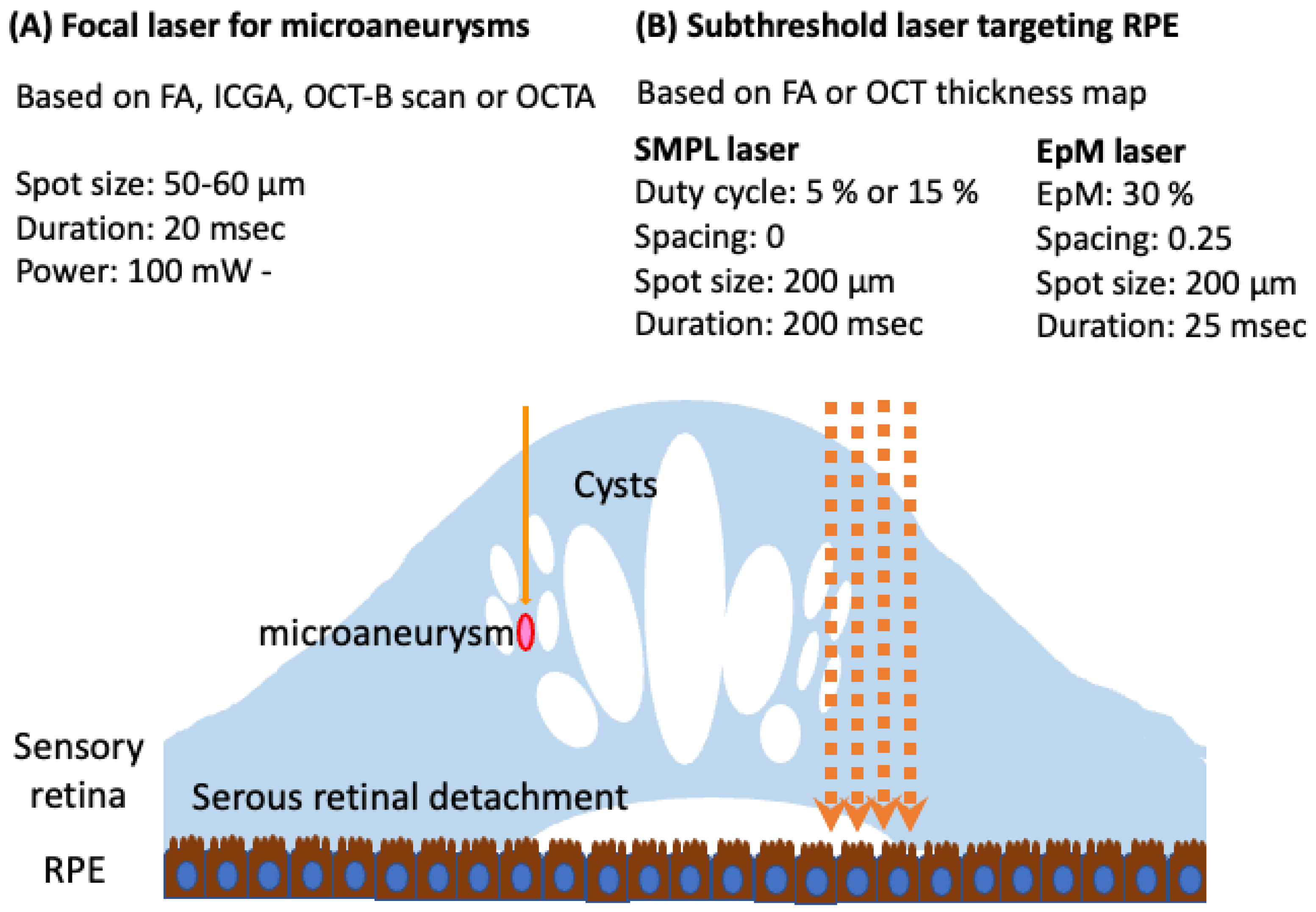
3.2. Subthreshold Laser

Conventional laser photocoagulation therapy poses a risk of RPE atrophy, which can lead to central scotoma or scar enlargement [20,39]. However, subthreshold micropulse laser (SMPL) therapy has been developed as a safer alternative, delivering laser energy below the threshold required for permanent tissue destruction. It was found that, by shortening the irradiation time, the RPE could be selectively coagulated [40]. The principle of subthreshold laser photocoagulation was then developed by controlling the on/off time of irradiation to achieve very short irradiation times only to the RPE. Friberg and Karatza first reported the usefulness of subthreshold laser treatment for macular edema using a micropulsed subthreshold laser with a diode laser wavelength of 810 nm [41]. Since then, numerous clinical studies have investigated the use of SMPL for the treatment of DME [42,43,44,45,46,47], and several reports have shown that combination therapy with SMPL might be effective to reduce the number of anti-VEGF injections in the treatment of DME [42,43,44,45,46]. Mansouri et al. [48] reported that SMPL was more effective in DME patients with central macular thickness (CMT) less than 400 μm. Other previous prospective studies also showed that SMPL was effective when used with intravitreal injections of aflibercept with the pretreatment CMT below 400 μm [44,46] or 450 μm [45] after at least 3 loading doses of aflibercept. Based on this evidence, initial anti-VEGF treatment with deferred SMPL laser therapy could be useful in reducing the number of anti-VEGF injections. According to Luttrull et al., transfoveal SMPL therapy has been shown to be safe and effective in patients with good visual acuity [49]. Their study also found that SMPL treatment yielded positive outcomes in eyes with CMT below 300 μm [49]. SMPL laser treatment could be considered a suitable option for DME patients with good preoperative visual acuity who are not suitable candidates for conventional photocoagulation or intravitreal injection [50]. The EURETINA guideline also recommends SMPL for eyes with higher visual acuity affected by early diffuse DME, since it provides a cost-effective alternative that helps avoid collateral thermal diffusion and associated chorioretinal damage [6].
However, the variability in the use of wavelengths, laser powers, and duty cycles of SMPL lasers presents a challenge in comparing outcomes across clinical trials. Therefore, establishing optimal laser parameters and standardizing SMPL laser settings are critical steps towards achieving wider acceptance and integration of SMPL lasers into clinical practice.
Other laser devices are also equipped with a subthreshold laser mode (Table 1). The NAVILAS laser has a “Microsecond Pulsing Mode” that controls the irradiation time for subthreshold laser coagulation, similar to the micropulse laser (Figure 2) [51]. Since the subthreshold laser is designed to be “invisible,” using the NAVILAS system to retain laser location information and settings may provide a significant advantage.
Selective Retina Laser Therapy (SRT) is another new subthreshold laser [52]. Unlike conventional laser photocoagulation therapy, SRT’s mechanism of action involves the generation of microbubbles around melanosomes in the RPE, which rupture and damage only the RPE while leaving the surrounding neural retina or choroid unaffected [53,54]. Subthreshold lasers do not produce visible laser spots, which can make it difficult to ensure effective treatment and increase the possibility of under-treatment. However, SRT uses optoacoustic techniques to provide real-time feedback on the degree of RPE damage, which overcomes this limitation. With this feedback, the treating physician can avoid both under-treatment and over-treatment, ensuring optimal treatment outcomes [55]. Several groups have reported the efficacy of SRT in treating DME [56,57,58]. It is interesting to note that, similar to the SMPL, SRT resulted in significant improvement in visual acuity and retinal thickness in patients with pre-treatment central foveal retinal thickness less than 400 μm, and there was almost no need for additional anti-VEGF drug injections in this group. However, in patients with pre-treatment retinal thickness of 400 μm or greater, there was no significant improvement in visual acuity or retinal thickness, and 73% of these patients required rescue treatment with anti-VEGF drugs [58]. Therefore, performing SRT as a deferred laser therapy after initiating anti-VEGF drug treatment may be useful. 2RT is an innovative nanopulse laser technology that delivers only around 0.2% of the energy per pulse compared to standard photocoagulation techniques [59]. With 2RT laser treatment, the effects are localized within the RPE, ensuring minimal impact on surrounding retinal tissues. A pilot, randomized trial evaluating the use of 2RT in patients with DME demonstrated its non-inferiority compared to conventional laser treatment [59]. Further studies are warranted to explore the efficacy and safety of 2RT in a larger population.

3.3. Pattern Scan Laser

The PASCAL® (Topcon Medical Laser systems, Santa Clara, CA, USA) is a semi-automated laser system that uses pattern scanning technology to deliver multiple laser burns in a single application [17]. This technology allows for precise and controlled delivery of arrays with predetermined parameters, resulting in reduced procedural time. To deliver multiple laser burns in a single application, the duration of laser photocoagulation with the PASCAL laser is set to 0.02 s, which is much shorter than the conventional laser setting of 0.2 s. This short-pulse setting offers several advantages over conventional laser treatment. Short-pulse laser treatment is faster, generates less heat, and causes less discomfort to the eyes [17,60]. Furthermore, research suggests that short-pulse laser treatment may induce less inflammation, fewer inflammatory cytokines in the sensory retina, and less macular thickening in patients with diabetic retinopathy compared to use of the conventional pulse duration [61,62]. Despite the advantages of short-pulse laser treatment, studies suggest that it may be less effective than conventional laser treatment for high-risk proliferative diabetic retinopathy. These studies propose that the total area of panretinal photocoagulation (PRP) scars generated by the conventional laser exceeds that of the short-pulse laser, despite both groups receiving the same number of laser spots [63]. This difference may be due to the tendency of photocoagulation scars generated by the conventional laser to expand after treatment [64]. However, other reports suggest that the expansion rate of photocoagulation scars generated by the short-pulse laser is lower than that of the conventional laser [65]. Conversely, the limited scar expansion observed with short-pulse lasers has led to the development of new modified settings for focal/grid lasers. In contrast to the “modified” ETDRS technique, which has been widely used for years, these newly modified settings now use shorter pulse durations, such as 0.02–0.03 s [35,37,66,67,68].
Endpoint Management (EpM) is another subthreshold laser method provided by the short-pulse pattern scan laser system PASCAL [16]. The EpM laser uses a continuous wave, and the EpM algorithm uses the Arrhenius integral formula to adjust the laser power and pulse duration, enabling the achievement of therapeutic effects while minimizing damage to surrounding tissues [16,69]. There have been several reports that the EpM laser was safe and effective for DME treatment. Hamada et al. reported that EpM laser monotherapy was effective in reducing central macular thickness, but visual acuity, macular sensitivity, and macular volume were not significantly improved (N = 10) [70]. The setting that this study used was 50% of the threshold (size, 100 μm; duration, 0.015 s; spacing, 0.5; and energy, 4.5–7.8 mJ). Several prospective studies aimed to investigate whether adding the EpM laser to anti-VEGF drug therapy could reduce the number of anti-VEGF injections required to treat DME. Though one study, the END-DME study, demonstrated that combination therapy with the EpM laser could reduce the number of anti-VEGF injections [71], another study did not find such an effect [72]. Both studies, however, used the same EpM threshold setting of 40%. Another study compared the efficacy of EpM 30%, EpM 50%, and SMPL (810 nm) for treating DME. The results showed that, though the micropulse system with a 15% duty cycle improved functional outcomes, neither EpM treatment was effective [73].
The effectiveness of EpM lasers for the treatment of DME is still under investigation. Clinical studies have been conducted under various conditions, but the lack of consistency in laser settings and protocols makes it challenging to compare results. Therefore, there is an urgent need to establish optimal laser conditions and indications for EpM therapy to improve the effectiveness and reproducibility of the treatment.

4. Multimodal Imaging-Guided Laser Therapy

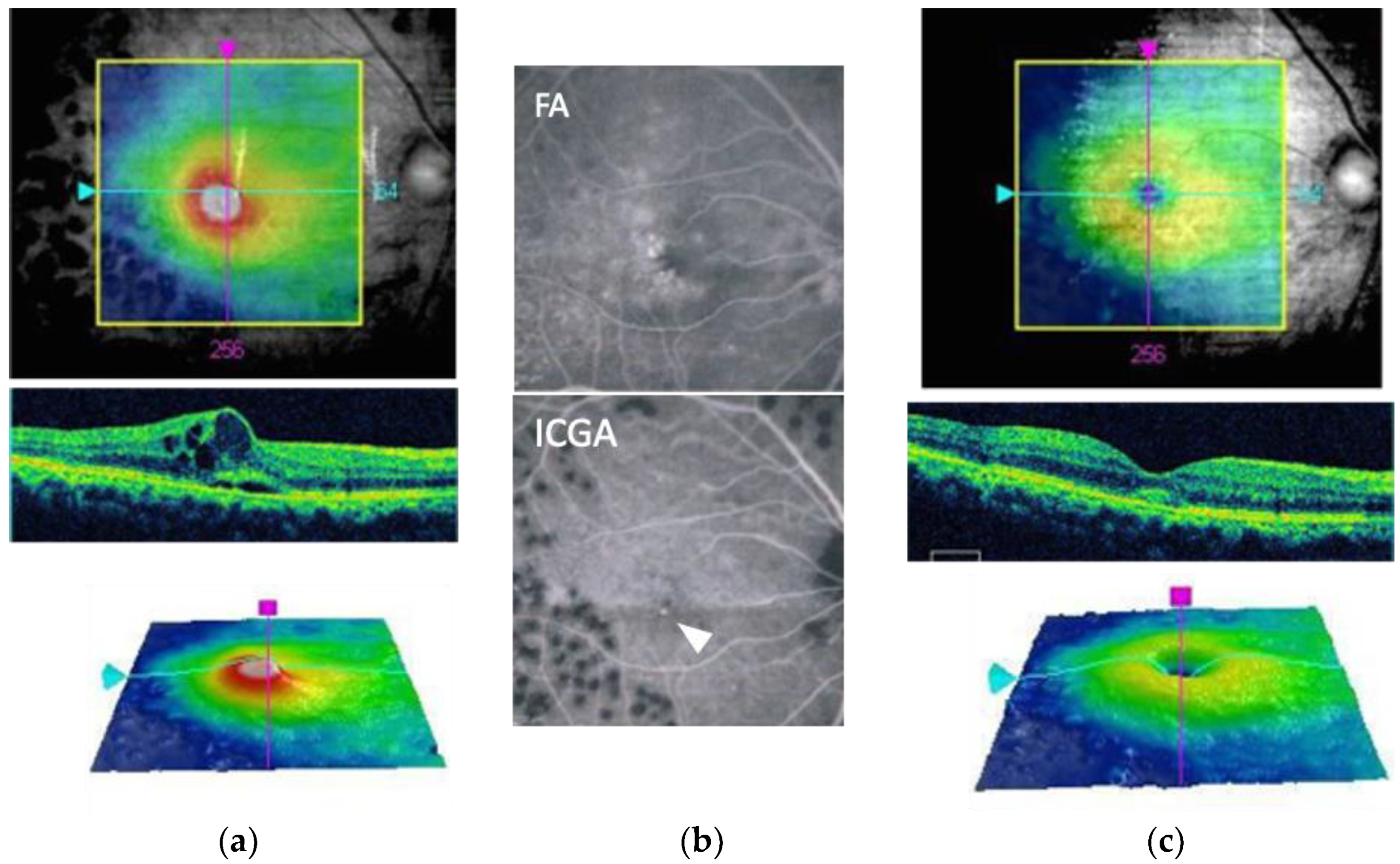
4.1. Indocyanine Green Angiography-Guided Laser Therapy

Fluorescein angiography (FA) has commonly been used to identify microaneurysms in patients with DME. However, several previous reports have suggested the superiority of indocyanine green fundus angiography (ICGA) for detecting the microaneurysms that contribute to the pathogenesis of DME [74,75]. Indocyanine green (ICG) dye binds mostly to serum proteins such as albumin and lipoproteins [76]. Therefore, the dye barely leaks through blood vessels, with the ICGA-positive microaneurysms persisting after the fading of the plasmatic fluorescence on the late-phase ICGA images. The mechanism underlying the superiority of ICGA in detecting microaneurysms that contribute to the pathogenesis of DME may involve the amphiphilic nature of ICG, which enables it to bind to hydrophobic intraluminal materials such as fibrin or lipids. In contrast, fluorescein, being hydrophilic, may be blocked by such intraluminal material. This may explain the preferential detectability of leakage points responsible for macular edema and the enlargement of spots in the late phase of ICGA. In cases of diffuse DME initially diagnosed using FA, ICGA imaging has proven useful in detecting microaneurysms or focal leakage spots [74]. ICGA-guided focal laser therapy has an additional benefit because it leads to significantly fewer targeted microaneurysms than FA-guided focal laser therapy, and ICGA-guided focal laser therapy might be helpful for reducing damage to the retina (Figure 3) [37,74]. Paques et al. identified the relatively large ICGA-positive area as telangiectatic capillaries (defined by a diameter larger than 150 µm on late-phase ICGA) [75,77]. Telangiectatic capillaries may be associated with chronic refractory macular edema [78,79,80], and focal direct laser photocoagulation of telangiectatic capillaries seems to be effective and safe for reducing the burden of anti-VEGF drugs in patients with refractory ME [81].

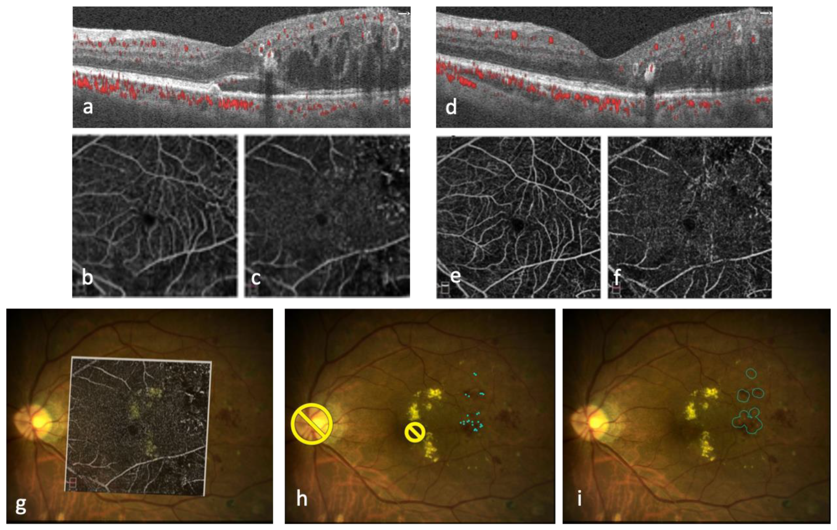
4.2. OCT-Guided Laser

Conventionally, to perform focal/grid laser treatment, dye-based fundus angiography has been necessary to determine the location of the treatment site. However, this procedure can be invasive and uncomfortable for patients. Using OCT instead of dye-based angiography would eliminate the need for an invasive procedure and improve the patient’s comfort during the treatment. OCT thickness maps can provide the topographic location and morphologic pattern of edema, and many physicians use OCT to guide laser photocoagulation, especially for grid laser photocoagulation [82,83]. However, Kozak et al. demonstrated improved accuracy of treatment planning by incorporating information from FA and OCT overlays using the NAVILAS system [84]. Takamura et al. recommended a novel laser photocoagulation protocol, named merged image-guided photocoagulation, for focal direct laser therapy. This protocol involves merging images of the fundus, OCT thickness map, and FA to identify the microaneurysms responsible for macular edema [82]. OCT angiography (OCTA) has emerged as a potential alternative imaging modality for detecting microaneurysms [85]. OCTA-guided focal laser treatment using the NAVILAS system holds promise as a new non-invasive approach for treating microaneurysms responsible for DME [35] (Figure 4). However, the reliability of microaneurysm detection with OCTA varies [86,87,88] and requires further investigation before it can be used to guide focal laser treatment targeting leaky microaneurysms.
OCT can also visualize leaky microaneurysms via B-scan [89,90] as circular structures that encroach upon and disrupt the integrity of the synaptic (inner) portion of the outer plexiform layer (OPL) and induce focal fluid collection, predominantly in the OPL/outer nuclear layer. Furthermore, en face OCT in the deep capillary plexus slab can also identify microaneurysms [91]. OCT-guided focal direct laser photocoagulation showed similar anatomical and functional outcomes compared to conventional laser based on FA [84,85], with significantly less retinal damage [92].
The use of OCT-guided focal laser technique offers several advantages for the management of leaky microaneurysms, including non-invasiveness and real-time imaging capabilities. OCT-guided focal laser therapy may allow precise determination of the proximity of the laser to the microaneurysms, thus increasing the effectiveness of the treatment [93].

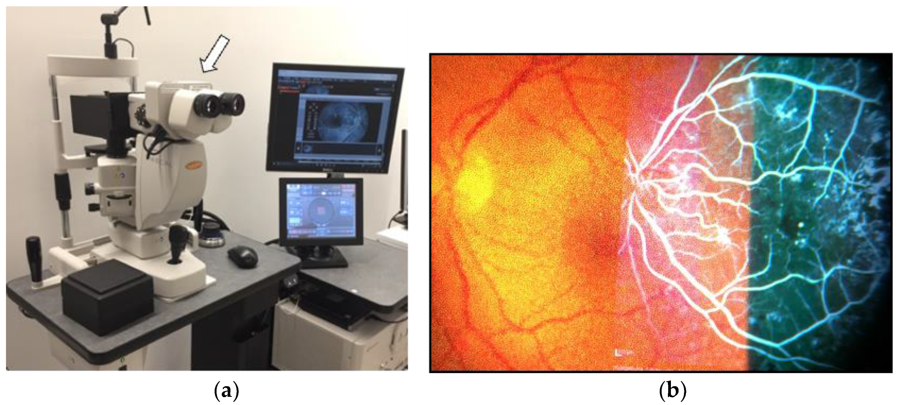
4.3. Multimodal Imaging Integrated System

The key to performing accurate and minimally invasive laser photocoagulation would be planning laser photocoagulation based on multimodal imaging. Takamura et al. reported the usefulness of merged retinal images, which are obtained from OCT thickness maps and FA and color fundus images, but their technique requires Adobe software (Photoshop CS6 Extended, Adobe Systems Inc., San Jose, CA, USA) to merge the images, and the merged images with the microaneurysms to treat marked were displayed on the computer screen next to the patient and used as a guide while performing laser treatment [85]. Both OCT-guided laser and ICGA-guided focal laser techniques also require a manual step to merge various images onto color fundus images [74,85]. The NAVILAS system is an ideal choice for laser procedures based on multimodal imaging because it allows precise planning of laser treatment by overlaying various digital fundus images on a live fundus image [14].
Recently, PASCAL synthesis (Topcon Medical Laser systems, Santa Clara, CA, USA) developed HUD-1, which is an ophthalmic image projector used as an accessory to PASCAL Synthesis (Figure 5). HUD-1 lets us compare the target area of treatment with a side-by-side view of a reference image previously obtained from the patient’s fundus for location and assessment. With HUD-1, the laser treatment can be performed while maintaining direct visualization of the treatment target area, without requiring the operators to divert their gaze from the slit-lamp microscope.

5. The Current Role of Laser Photocoagulation in the Era of Intravitreal Drug Administration

5.1. Comparison of Anti-VEGF/Steroid and Laser Treatment

The efficacy of anti-VEGF therapy has been well established in multiple RCTs [10,24,25], all of which have demonstrated its superiority over laser photocoagulation. Clinical studies have recently compared newly approved anti-VEGF drugs to aflibercept [26,27], but no further studies have been conducted to compare the efficacy of focal/grid laser treatment alone. Laser monotherapy is no longer performed as a treatment option for treatment-naive eyes with center-involved DME. It should be noted, however, that current laser photocoagulation techniques encompass various types and settings, such as SMPL, navigation laser, or multimodal imaging-guided laser.
One of the disadvantages of anti-VEGF therapy is the burden of frequent injections and clinic visits. In comparison, laser treatment is generally considered more cost-effective than pharmacotherapy, including both anti-VEGF and steroid treatments [94]. Laser treatment also offers the advantage of a longer-lasting effect compared to anti-VEGF therapy [95]. Moreover, several studies have demonstrated that the combination of laser photocoagulation with anti-VEGF treatment has the potential to reduce the frequency of injections (Table 2) [96]. However, it is important to note that the optimal timing and protocol for combining laser treatment with anti-VEGF injections have not yet been firmly established. Further studies are needed to determine the most effective approach to reduce the treatment burden for patients.
In terms of safety, anti-VEGF injections generally cause fewer topical side effects compared to steroid therapy and laser treatment. Steroid-related side effects may include increased intraocular pressure, cataract formation or progression, and the potential risk of developing steroid-induced glaucoma. On the other hand, advancements in laser treatment have led to the development of current laser settings that aim to minimize side effects [96], such as atrophic creep and the development of scotomata [20]. However, longer-term follow-up studies are still necessary to thoroughly evaluate the absence of side effects of modern retinal laser procedures.

5.2. A Real-World Approach: Laser Photocoagulation in Combination with Intravitreal Drug Administration

The types and settings of current laser are summarized in Figure 6. Focal/grid laser photocoagulation is crucial in treating non-center-involved, clinically significant DME [6,22,81]. For center-involved DME, anti-VEGF therapy has become the gold standard treatment. However, despite the benefits seen in RCTs of treating patients more frequently than in real-world practice, the results have shown that approximately 30–40% of patients exhibit a poor response to anti-VEGF treatment [97,98]. The next step would involve considering a switch of intravitreal medication, such as an alternative anti-VEGF agent or steroids [99]. However, if these measures prove unsuccessful, laser therapy could be considered a potential third-line option for managing refractory DME. Whereas microaneurysms in the deep capillary plexus or those detected by ICGA may show resistance to anti-VEGF drugs [37,74,75,76,100], it is generally observed that anti-VEGF therapy effectively reduces microaneurysms [78,101,102]. In addition, steroids have also demonstrated the ability to reduce microaneurysms [103]. Therefore, the current approach involves initiating pharmacotherapy, such as anti-VEGF or steroids, as the initial treatment. Subsequently, deferred focal laser therapy is considered for any remaining microaneurysms that contribute to residual edema, in accordance with the findings from Protocol I [104,105]. In refractory chronic DME cases, ICGA-guided/OCT-guided focal direct laser therapy might be useful for targeting residual microaneurysms. In addition, focal laser therapy using the NAVILAS system may provide benefit in chronic refractory DME cases, because of microaneurysms resistant to anti-VEGF therapy located in the perifoveal vascular network [37,66]. Subthreshold laser techniques have also shown promise in reducing the frequency of anti-VEGF injections for DME treatment. For DME patients with good preoperative visual acuity who are not candidates for intravitreal injection, subthreshold laser treatment could be indicated as a less expensive option [6,49,50].
However, further studies are needed to determine the optimal timing and settings used in these techniques. Nonetheless, laser photocoagulation remains an important tool for treating DME, even in the era of intravitreal drug administration.

6. Conclusions

In the future, novel treatments such as gene therapy [106] or anti-inflammatory drugs [99] might be poised to play a crucial role in the management of DME. Nevertheless, recent advancements in laser technology and multimodal imaging have demonstrated new potential applications for laser photocoagulation, even in the era of widespread use of anti-VEGF drugs and steroids. These innovative techniques aim to optimize treatment outcomes while minimizing adverse events and alleviating the burden of frequent injections, thereby offering significant value to DME management. These developments instill hope for patients with DME and underscore the importance of ongoing research in the field of laser photocoagulation.

Author Contributions

Conceptualization and writing the draft, M.N. Review and editing, R.A., T.K., F.K. and T.Y. All authors have read and agreed to the published version of the manuscript.

Funding

This article was supported by Grants-in-Aid for Scientific Research from the Japan Society for the Promotion of Science, Tokyo, Japan (Grant numbers: 20K09777 to M.N. and 22H03243 to T.Y.).

Institutional Review Board Statement

Not applicable.

Data Availability Statement

The datasets generated during and/or analyzed during the current study are available from the corresponding author on reasonable request.

Acknowledgments

The authors would like to thank Fumie Shibuya, Kazuyo Hayashi, and Yui Kaneko for their technical assistance.

Conflicts of Interest

The authors declare no conflict of interest.

References

  1. Sabanayagam, C.; Yip, W.; Ting, D.S.W.; Tan, G.; Wong, T.Y. Ten Emerging Trends in the Epidemiology of Diabetic Retinopathy. Ophthalmic Epidemiol. 2016, 23, 209–222. [Google Scholar] [CrossRef] [PubMed]
  2. Early Treatment Diabetic Retinopathy Study Research Group: Photocoagulation for Diabetic Macular Edema. Early Treatment Diabetic Retinopathy Study report number 1. Arch. Ophthalmol. 1985, 103, 1796–1806. [Google Scholar] [CrossRef]
  3. Ogura, Y.; Shiraga, F.; Terasaki, H.; Ohji, M.; Ishida, S.; Sakamoto, T.; Hirakata, A.; Ishibashi, T. Clinical practice pattern in management of diabetic macular edema in Japan: Survey results of Japanese retinal specialists. Jpn. J. Ophthalmol. 2017, 61, 43–50. [Google Scholar] [CrossRef]
  4. Sugimoto, M.; Tsukitome, H.; Okamoto, F.; Oshika, T.; Ueda, T.; Niki, M.; Mitamura, Y.; Ishikawa, H.; Gomi, F.; Kitano, S.; et al. Clinical preferences and trends of anti-vascular endothelial growth factor treatments for diabetic macular edema in Japan. J. Diabetes Investig. 2019, 10, 475–483. [Google Scholar] [CrossRef] [PubMed] [Green Version]
  5. Moulin, T.A.; Boakye, E.A.; Wirth, L.S.; Chen, J.; Burroughs, T.E.; Vollman, D.E. Yearly Treatment Patterns for Patients with Recently Diagnosed Diabetic Macular Edema. Ophthalmol. Retin. 2019, 3, 362–370. [Google Scholar] [CrossRef]
  6. Schmidt-Erfurth, U.; Garcia-Arumi, J.; Bandello, F.; Berg, K.; Chakravarthy, U.; Gerendas, B.S.; Jonas, J.; Larsen, M.; Tadayoni, R.; Loewenstein, A. Guidelines for the Management of Diabetic Macular Edema by the European Society of Retina Specialists (EURETINA). Ophthalmologica 2017, 237, 185–222. [Google Scholar] [CrossRef]
  7. Diabetic Retinopathy Clinical Research Network; Elman, M.J.; Aiello, L.P.; Beck, R.W.; Bressler, N.M.; Bressler, S.B.; Edwards, A.R.; Ferris, F.L., 3rd; Friedman, S.M.; Glassman, A.R.; et al. Randomized Trial Evaluating Ranibizumab Plus Prompt or Deferred Laser or Triamcinolone Plus Prompt Laser for Diabetic Macular Edema. Ophthalmology 2010, 117, 1064–1077.e35. [Google Scholar] [CrossRef] [PubMed] [Green Version]
  8. Elman, M.J.; Bressler, N.M.; Qin, H.; Beck, R.W.; Ferris, F.L.; Friedman, S.M., 3rd; Glassman, A.R.; Scott, I.U.; Stockdale, C.R.; Sun, J.K. Expanded 2-Year Follow-up of Ranibizumab Plus Prompt or Deferred Laser or Triamcinolone Plus Prompt Laser for Diabetic Macular Edema. Ophthalmology 2011, 118, 609–614. [Google Scholar] [CrossRef] [Green Version]
  9. Nguyen, Q.D.; Shah, S.M.; Khwaja, A.A.; Channa, R.; Hatef, E.; Do, D.V.; Boyer, D.; Heier, J.S.; Abraham, P.; Thach, A.B.; et al. Two-Year Outcomes of the Ranibizumab for Edema of the mAcula in Diabetes (READ-2) Study. Ophthalmology 2010, 117, 2146–2151. [Google Scholar] [CrossRef]
  10. Mitchell, P.; Bandello, F.; Schmidt-Erfurth, U.; Lang, G.E.; Massin, P.; Schlingemann, R.O.; Sutter, F.; Simader, C.; Burian, G.; Gerstner, O.; et al. The RESTORE Study: Ranibizumab monotherapy or combined with laser versus laser monotherapy for diabetic macular edema. Ophthalmology 2011, 118, 615–625. [Google Scholar] [CrossRef] [Green Version]
  11. Holekamp, N.M.; Campbell, J.; Almony, A.; Ingraham, H.; Marks, S.; Chandwani, H.; Cole, A.L.; Kiss, S. Vision Outcomes Following Anti–Vascular Endothelial Growth Factor Treatment of Diabetic Macular Edema in Clinical Practice. Am. J. Ophthalmol. 2018, 191, 83–91. [Google Scholar] [CrossRef] [Green Version]
  12. Kawasaki, R.; Bauer, M.; Bezlyak, V.; Ogura, Y. Treatment patterns for retinal diseases in patients newly-treated with anti-VEGF agents: A retrospective analysis of claims data from the Japan Medical Data Center database. Jpn. J. Ophthalmol. 2021, 65, 215–226. [Google Scholar] [CrossRef]
  13. Sakamoto, T.; Shimura, M.; Kitano, S.; Ohji, M.; Ogura, Y.; Yamashita, H.; Suzaki, M.; Mori, K.; Ohashi, Y.; Yap, P.S.; et al. Impact on visual acuity and psychological outcomes of ranibizumab and subsequent treatment for diabetic macular oedema in Japan (MERCURY). Graefes Arch. Clin. Exp. Ophthalmol. 2022, 260, 477–487. [Google Scholar] [CrossRef]
  14. Kernt, M.; Cheuteu, R.; Vounotrypidis, E.; Haritoglou, C.; Kampik, A.; Ulbig, M.W.; Neubauer, A.S. Focal and panretinal photocoagulation with a navigated laser (NAVILAS®). Acta Ophthalmol. 2011, 89, e662–e664. [Google Scholar] [CrossRef]
  15. Ohkoshi, K.; Yamaguchi, T. Subthreshold Micropulse Diode Laser Photocoagulation for Diabetic Macular Edema in Japanese Patients. Am. J. Ophthalmol. 2010, 149, 133–139.e1. [Google Scholar] [CrossRef]
  16. Lavinsky, D.; Sramek, C.; Wang, J.B.; Huie, P.M.; Dalal, R.M.; Mandel, Y.; Palanker, D. Subvisible retinal laser therapy: Titration algorithm and tissue response. Retina 2014, 34, 87–97. [Google Scholar] [CrossRef]
  17. Blumenkranz, M.S.; Yellachich, D.; Andersen, D.E.; Wiltberger, M.W.; Mordaunt, D.; Marcellino, G.R.; Palanker, D. Semiautomated patterned scanning laser for retinal photocoagulation. Retina 2006, 26, 370–376. [Google Scholar] [CrossRef]
  18. Early Treatment Diabetic Retinopathy Study Research Group. Focal photocoagulation treatment of diabetic macular edema. Relationship of treatment effects to fluorescein angiographic and other retinal characteristics at baseline: ETDRS report no. 19. Arch Ophthalmol. 1995, 113, 1144–1155. [Google Scholar] [CrossRef]
  19. McDONALD, H.R.; Schatz, H. Grid photocoagulation for diffuse macular edema. Retina 1985, 5, 65–72. [Google Scholar] [CrossRef]
  20. Lövestam-Adrian, M.; Agardh, E. Photocoagulation of diabetic macular oedema; complications and visual outcome. Acta Ophthalmol. Scand. 2000, 78, 667–671. [Google Scholar] [CrossRef]
  21. Fong, D.S.; Strauber, S.F.; Aiello, L.P.; Beck, R.W.; Callanan, D.G.; Danis, R.P.; Davis, M.D.; Feman, S.S.; Ferris, F.; Writing Committee for the Diabetic Retinopathy Clinical Research Network; et al. Comparison of the Modified Early Treatment Diabetic Retinopathy Study and Mild Macular Grid Laser Photocoagulation Strategies for Diabetic Macular Edema. Arch. Ophthalmol. 2007, 125, 469–480. [Google Scholar] [CrossRef] [Green Version]
  22. Scott, I.U.; Danis, R.P.; Bressler, S.B.; Bressler, N.M.; Browning, D.J.; Qin, H.; Diabetic Retinopathy Clinical Research Network. Effect of focal/grid photocoagulation on visual acuity and retinal thickening in eyes with non–center-involved diabetic macular edema. Retina 2009, 29, 613–617. [Google Scholar] [CrossRef] [Green Version]
  23. Elman, M.J.; Ayala, A.; Bressler, N.M.; Browning, D.; Flaxel, C.J.; Glassman, A.R.; Jampol, L.M.; Stone, T.W.; Diabetic Retinopathy Clinical Research Network. Intravitreal Ranibizumab for Diabetic Macular Edema with Prompt versus Deferred Laser Treatment: 5-Year Randomized Trial Results. Ophthalmology 2015, 122, 375–381. [Google Scholar] [CrossRef] [Green Version]
  24. Bressler, N.M.; Varma, R.; Suñer, I.J.; Dolan, C.M.; Ward, J.; Ehrlich, J.S.; Colman, S.; Turpcu, A.; RIDE and RISE Research Groups. Vision-related function after ranibizumab treatment for diabetic macular edema: Results from RIDE and RISE. Ophthalmology 2014, 121, 2461–2472. [Google Scholar] [CrossRef]
  25. Do, D.V.; Schmidt-Erfurth, U.; Gonzalez, V.H.; Gordon, C.M.; Tolentino, M.; Berliner, A.J.; Vitti, R.; Rückert, R.; Sandbrink, R.; Stein, D.; et al. The DA VINCI Study: Phase 2 Primary Results of VEGF Trap-Eye in Patients with Diabetic Macular Edema. Ophthalmology 2011, 118, 1819–1826. [Google Scholar] [CrossRef]
  26. Wykoff, C.C.; Abreu, F.; Adamis, A.P.; Basu, K.; Eichenbaum, D.A.; Haskova, Z.; Lin, H.; Loewenstein, A.; Mohan, S.; Pearce, I.A.; et al. Efficacy, durability, and safety of intravitreal faricimab with extended dosing up to every 16 weeks in patients with diabetic macular oedema (YOSEMITE and RHINE): Two randomised, double-masked, phase 3 trials. Lancet 2022, 399, 741–755. [Google Scholar] [CrossRef]
  27. Brown, D.M.; Emanuelli, A.; Bandello, F.; Barranco, J.J.E.; Figueira, J.; Souied, E.; Wolf, S.; Gupta, V.; Ngah, N.F.; Liew, G.; et al. KESTREL and KITE: 52-Week Results from Two Phase III Pivotal Trials of Brolucizumab for Diabetic Macular Edema. Am. J. Ophthalmol. 2022, 238, 157–172. [Google Scholar] [CrossRef]
  28. Callanan, D.G.; Gupta, S.; Boyer, D.S.; Ciulla, T.A.; Singer, M.A.; Kuppermann, B.D.; Liu, C.-C.; Li, X.-Y.; Hollander, D.A.; Schiffman, R.M.; et al. Dexamethasone Intravitreal Implant in Combination with Laser Photocoagulation for the Treatment of Diffuse Diabetic Macular Edema. Ophthalmology 2013, 120, 1843–1851. [Google Scholar] [CrossRef]
  29. Chalam, K.V.; Murthy, R.K.; Brar, V.; Radhakrishnan, R.; Khetpal, V.; Grover, S. Evaluation of a novel, non contact, automated focal laser with integrated (NAVILAS®) fluorescein angiography for diabetic macular edema. Middle East. Afr. J. Ophthalmol. 2012, 19, 158–162. [Google Scholar] [CrossRef]
  30. Kozak, I.; Oster, S.F.; Cortes, M.A.; Dowell, D.; Hartmann, K.; Kim, J.S.; Freeman, W.R. Clinical Evaluation and Treatment Accuracy in Diabetic Macular Edema Using Navigated Laser Photocoagulator NAVILAS. Ophthalmology 2011, 118, 1119–1124. [Google Scholar] [CrossRef]
  31. Kernt, M.; Kampik, A.; Neubauer, A.S.; Langer, W.; Kozak, S.; Ulbig, F.; Langer, J. Navigated macular laser decreases retreatment rate for diabetic macular edema: A comparison with conventional macular laser. Clin. Ophthalmol. 2013, 7, 121–128. [Google Scholar] [CrossRef] [PubMed] [Green Version]
  32. Liegl, R.; Langer, J.; Seidensticker, F.; Reznicek, L.; Haritoglou, C.; Ulbig, M.W.; Neubauer, A.S.; Kampik, A.; Kernt, M. Comparative Evaluation of Combined Navigated Laser Photocoagulation and Intravitreal Ranibizumab in the Treatment of Diabetic Macular Edema. PLoS ONE 2014, 9, e113981. [Google Scholar] [CrossRef] [Green Version]
  33. Herold, T.R.; Langer, J.; Vounotrypidis, E.; Kernt, M.; Liegl, R.; Priglinger, S.G. 3-year-data of combined navigated laser photocoagulation (Navilas) and intravitreal ranibizumab compared to ranibizumab monotherapy in DME patients. PLoS ONE 2018, 13, e0202483. [Google Scholar] [CrossRef]
  34. Payne, J.F.; Wykoff, C.C.; Clark, W.L.; Bruce, B.B.; Boyer, D.S.; Brown, D.M. TREX-DME Study Group. Long-term outcomes of treat-and-extend ranibizumab with and without navigated laser for diabetic macular oedema: TREX-DME 3-year results. Br. J. Ophthalmol. 2021, 105, 253–257. [Google Scholar] [CrossRef]
  35. Kato, F.; Nozaki, M.; Kato, A.; Hasegawa, N.; Morita, H.; Yoshida, M.; Ogura, Y. Evaluation of Navigated Laser Photocoagulation (Navilas 577+) for the Treatment of Refractory Diabetic Macular Edema. J. Ophthalmol. 2018, 2018, 3978514. [Google Scholar] [CrossRef]
  36. Menzler, J.; Neubauer, A.; Ziemssen, F. Navigated laser in diabetic macular edema: The impact of reduced injection burden on patients and physicians-who wins and who loses? Int. J. Ophthalmol. 2019, 12, 342–345. [Google Scholar] [CrossRef]
  37. Nozaki, M.; Kato, A.; Yasukawa, T.; Suzuki, K.; Yoshida, M.; Ogura, Y. Indocyanine green angiography-guided focal navigated laser photocoagulation for diabetic macular edema. Jpn. J. Ophthalmol. 2019, 63, 243–254. [Google Scholar] [CrossRef]
  38. Hirano, T.; Toriyama, Y.; Takamura, Y.; Sugimoto, M.; Nagaoka, T.; Sugiura, Y.; Okamoto, F.; Saito, M.; Noda, K.; Yoshida, S.; et al. Outcomes of a 2-year treat-and-extend regimen with aflibercept for diabetic macular edema. Sci. Rep. 2021, 11, 1–8. [Google Scholar] [CrossRef]
  39. Schatz, H.; Madeira, D.; McDonald, H.R.; Johnson, R.N. Progressive Enlargement of Laser Scars Following Grid Laser Photocoagulation for Diffuse Diabetic Macular Edema. Arch. Ophthalmol. 1991, 109, 1549–1551. [Google Scholar] [CrossRef]
  40. Pankratov, M.M. Pulsed delivery of laser energy in experimental thermal retinal photocoagulation. Proc. Soc. Photo Opt. Instrum. Eng. 1990, 1202, 205–213. [Google Scholar] [CrossRef]
  41. Friberg, T.R.; Karatza, E.C. The Treatment of Macular Disease Using a Micropulsed and Continuous Wave 810-nm Diode Laser. Ophthalmology 1997, 104, 2030–2038. [Google Scholar] [CrossRef] [PubMed]
  42. Moisseiev, E.; Abbassi, S.; Thinda, S.; Yoon, J.; Yiu, G.; Morse, L.S. Subthreshold micropulse laser reduces anti-VEGF injection burden in patients with diabetic macular edema. Eur. J. Ophthalmol. 2018, 28, 68–73. [Google Scholar] [CrossRef] [Green Version]
  43. Inagaki, K.; Hamada, M.; Ohkoshi, K. Minimally invasive laser treatment combined with intravitreal injection of anti-vascular endothelial growth factor for diabetic macular oedema. Sci. Rep. 2019, 9, 7585. [Google Scholar] [CrossRef] [Green Version]
  44. Khattab, A.M.; Hagras, S.M.; AbdElhamid, A.; Torky, M.A.; Awad, E.A.; Abdelhameed, A.G. Aflibercept with adjuvant micropulsed yellow laser versus aflibercept monotherapy in diabetic macular edema. Graefes Arch. Clin. Exp. Ophthalmol. 2019, 257, 1373–1380. [Google Scholar] [CrossRef]
  45. Kanar, H.S.; Arsan, A.; Altun, A.; Akı, S.F.; Hacısalihoglu, A. Can subthreshold micropulse yellow laser treatment change the anti-vascular endothelial growth factor algorithm in diabetic macular edema? A randomized clinical trial. Indian. J. Ophthalmol. 2020, 68, 145–151. [Google Scholar] [CrossRef]
  46. Abouhussein, M.A.; Gomaa, A.R. Aflibercept plus micropulse laser versus aflibercept monotherapy for diabetic macular edema: 1-year results of a randomized clinical trial. Int. Ophthalmol. 2020, 40, 1147–1154. [Google Scholar] [CrossRef]
  47. Koushan, K.; Eshtiaghi, A.; Fung, P.; Berger, A.R.; Chow, D.R. Treatment of Diabetic Macular Edema with Aflibercept and Micropulse Laser (DAM Study). Clin. Ophthalmol. 2022, 16, 1109–1115. [Google Scholar] [CrossRef] [PubMed]
  48. Mansouri, A.; Sampat, K.M.; Malik, K.J.; Steiner, J.N.; Glaser, B.M. Efficacy of subthreshold micropulse laser in the treatment of diabetic macular edema is influenced by pre-treatment central foveal thickness. Eye 2014, 28, 1418–1424. [Google Scholar] [CrossRef] [PubMed] [Green Version]
  49. Luttrull, J.K.; Sinclair, S.H. Safety of transfoveal subthreshold diode micropulse laser for fovea-involving diabetic macular edema in eyes with good visual acuity. Retina 2014, 34, 2010–2020. [Google Scholar] [CrossRef] [Green Version]
  50. Busch, C.; Fraser-Bell, S.; Zur, D.; Rodríguez-Valdés, P.J.; Cebeci, Z.; Lupidi, M.; Fung, A.T.; Gabrielle, P.-H.; Giancipoli, E.; International Retina Group; et al. Real-world outcomes of observation and treatment in diabetic macular edema with very good visual acuity: The OBTAIN study. Acta Diabetol. 2019, 56, 777–784. [Google Scholar] [CrossRef] [Green Version]
  51. Chhablani, J.; Roh, Y.J.; Jobling, A.I.; Fletcher, E.L.; Lek, J.J.; Bansal, P.; Guymer, R.; Luttrull, J.K. Restorative retinal laser therapy: Present state and future directions. Surv. Ophthalmol. 2018, 63, 307–328. [Google Scholar] [CrossRef]
  52. Roider, J.; Michaud, N.A.; Flotte, T.J.; Birngruber, R. Response of the Retinal Pigment Epithelium to Selective Photocoagulation. Arch. Ophthalmol. 1992, 110, 1786–1792. [Google Scholar] [CrossRef]
  53. Roider, J.; Brinkmann, R.; Wirbelauer, C.; Laqua, H.; Birngruber, R. Retinal sparing by selective retinal pigment epithelial photocoagulation. Arch. Ophthalmol. 1999, 117, 1028–1034. [Google Scholar] [CrossRef]
  54. Brinkmann, R.; Roider, J.; Birngruber, R.; Lin, C.P. Origin of retinal pigment epithelium cell damage by pulsed laser irradiance in the nanosecond to microsecond time regimen. Lasers Surg. Med. 2000, 27, 451–464. [Google Scholar] [CrossRef]
  55. Roider, J.; Liew, S.H.M.; Klatt, C.; Elsner, H.; Poerksen, E.; Hillenkamp, J.; Brinkmann, R.; Birngruber, R. Selective retina therapy (SRT) for clinically significant diabetic macular edema. Graefes Arch. Clin. Exp. Ophthalmol. 2010, 248, 1263–1272. [Google Scholar] [CrossRef] [PubMed]
  56. Park, Y.G.; Kim, J.R.; Kang, S.; Seifert, E.; Theisen-Kunde, D.; Brinkmann, R.; Roh, Y.-J. Safety and efficacy of selective retina therapy (SRT) for the treatment of diabetic macular edema in Korean patients. Graefes Arch. Clin. Exp. Ophthalmol. 2016, 254, 1703–1713. [Google Scholar] [CrossRef]
  57. Yamamoto, M.; Miura, Y.; Hirayama, K.; Kohno, T.; Kabata, D.; Theisen-Kunde, D.; Brinkmann, R.; Honda, S. Predictive factors of outcome of selective retina therapy for diabetic macular edema. Int. Ophthalmol. 2020, 40, 1221–1232. [Google Scholar] [CrossRef] [PubMed]
  58. Kim, M.; Park, Y.G.; Jeon, S.H.; Choi, S.Y.; Roh, Y.-J. The efficacy of selective retina therapy for diabetic macular edema based on pretreatment central foveal thickness. Lasers Med. Sci. 2020, 35, 1781–1790. [Google Scholar] [CrossRef] [PubMed]
  59. Casson, R.J.; Raymond, G.; Newland, H.S.; Gilhotra, J.S.; Gray, T.L. Pilot randomized trial of a nanopulse retinal laser versus conventional photocoagulation for the treatment of diabetic macular oedema. Clin. Exp. Ophthalmol. 2012, 40, 604–610. [Google Scholar] [CrossRef] [PubMed]
  60. Nagpal, M.; Marlecha, S.; Nagpal, K. Comparison of laser photocoagulation for diabetic retinopathy using 532-nm standard laser versus multispot pattern scan laser. Retina 2010, 30, 452–458. [Google Scholar] [CrossRef]
  61. Ito, A.; Hirano, Y.; Nozaki, M.; Ashikari, M.; Sugitani, K.; Ogura, Y. Short Pulse Laser Induces Less Inflammatory Cytokines in the Murine Retina after Laser Photocoagulation. Ophthalmic Res. 2015, 53, 65–73. [Google Scholar] [CrossRef]
  62. Takamura, Y.; Arimura, S.; Miyake, S.; Matsumura, T.; Gozawa, M.; Iwasaki, K.; Inatani, M. Panretinal Photocoagulation Using Short-Pulse Laser Induces Less Inflammation and Macular Thickening in Patients with Diabetic Retinopathy. J. Ophthalmol. 2017, 2017, 8530261. [Google Scholar] [CrossRef]
  63. Chappelow, A.V.; Tan, K.; Waheed, N.K.; Kaiser, P.K. Panretinal Photocoagulation for Proliferative Diabetic Retinopathy: Pattern Scan Laser Versus Argon Laser. Am. J. Ophthalmol. 2012, 153, 137–142.e2. [Google Scholar] [CrossRef]
  64. Maeshima, K.; Utsugi-Sutoh, N.; Otani, T.; Kishi, S. Progressive enlargement of scattered photocoagulation scars in diabetic retinopathy. Retina 2004, 24, 507–511. [Google Scholar] [CrossRef]
  65. Higaki, M.; Nozaki, M.; Yoshida, M.; Ogura, Y. Less Expansion of Short-Pulse Laser Scars in Panretinal Photocoagulation for Diabetic Retinopathy. J. Ophthalmol. 2018, 2018, 9371895. [Google Scholar] [CrossRef] [Green Version]
  66. Hirano, T.; Toriyama, Y.; Iesato, Y.; Imai, A.; Hirabayashi, K.; Nagaoka, T.; Takamura, Y.; Sugimoto, M.; Murata, T. Effect of leaking perifoveal microaneurysms on resolution of diabetic macular edema treated by combination therapy using anti-vascular endothelial growth factor and short pulse focal/grid laser photocoagulation. Jpn. J. Ophthalmol. 2017, 61, 51–60. [Google Scholar] [CrossRef]
  67. Datlinger, F.; Datlinger, A.; Pollreisz, A.; Sacu, S.; Schmidt-Erfurth, U.; Datlinger, P. Intraprocedural OCT monitoring of the immediate treatment response during indocyanine green angiography-guided laser therapy of teleangiectatic capillaries in diabetic macular edema. Sci. Rep. 2022, 12, 2315. [Google Scholar] [CrossRef]
  68. Ikegami, Y.; Shiraya, T.; Araki, F.; Ueta, T.; Toyama, T.; Yanagita, T.; Numaga, J.; Shoji, N.; Kato, S. Microperimetric analysis of diabetic macular edema after navigated direct photocoagulation with short-pulse laser for microaneurysms. Int. J. Retin. Vitr. 2023, 9, 1–7. [Google Scholar] [CrossRef]
  69. Lavinsky, D.; Wang, J.; Huie, P.; Dalal, R.; Lee, S.J.; Lee, D.Y.; Palanker, D. Nondamaging Retinal Laser Therapy: Rationale and Applications to the Macula. Investig. Opthalmol. Vis. Sci. 2016, 57, 2488–2500. [Google Scholar] [CrossRef]
  70. Hamada, M.; Ohkoshi, K.; Inagaki, K.; Ebihara, N.; Murakami, A. Subthreshold Photocoagulation Using Endpoint Management in the PASCAL® System for Diffuse Diabetic Macular Edema. J. Ophthalmol. 2018, 2018, 7465794. [Google Scholar] [CrossRef]
  71. Nozaki, M.; Wong, I.Y.; Kawasaki, R.; Lee, J.E.; Takamura, Y.; Lee, J.E.; Yoshida, S.; Shin, J.P.; Kida, T.; Chang, W.; et al. Anti-VEGF Monotherapy versus Combined Anti-VEGF and Endpoint Management Laser for Diabetic Macular Edema (END-DME Study). J. Retin. 2022, 7, 65–74. [Google Scholar] [CrossRef]
  72. Tatsumi, T.; Takatsuna, Y.; Oshitari, T.; Kaiho, T.; Kawasaki, Y.; Shiko, Y.; Sugawara, T.; Baba, T.; Yamamoto, S. Randomized clinical trial comparing intravitreal aflibercept combined with subthreshold laser to intravitreal aflibercept monotherapy for diabetic macular edema. Sci. Rep. 2022, 12, 10672. [Google Scholar] [CrossRef]
  73. Al-Barki, A.; Al-Hijji, L.; High, R.; Schatz, P.; Do, D.; Nguyen, Q.D.; Luttrull, J.K.; Kozak, I. Comparison of short-pulse subthreshold (532 nm) and infrared micropulse (810 nm) macular laser for diabetic macular edema. Sci. Rep. 2021, 11, 14. [Google Scholar] [CrossRef] [PubMed]
  74. Ogura, S.; Yasukawa, T.; Kato, A.; Kuwayama, S.; Hamada, S.; Hirano, Y.; Uemura, A.; Yoshida, M.; Ogura, Y. Indocyanine Green Angiography-Guided Focal Laser Photocoagulation for Diabetic Macular Edema. Ophthalmologica 2015, 234, 139–150. [Google Scholar] [CrossRef] [PubMed]
  75. Paques, M.; Philippakis, E.; Bonnet, C.; Falah, S.; Ayello-Scheer, S.; Zwillinger, S.; Girmens, J.-F.; Dupas, B. Indocyanine-green-guided targeted laser photocoagulation of capillary macroaneurysms in macular oedema: A pilot study. Br. J. Ophthalmol. 2017, 101, 170–174. [Google Scholar] [CrossRef] [PubMed]
  76. Baker, K.J. Binding of Sulfobromophthalein (BSP) Sodium and Indocyanine Green (ICG) by Plasma 1 Lipoproteins. Exp. Biol. Med. 1966, 122, 957–963. [Google Scholar] [CrossRef] [PubMed]
  77. Farías, D.C.; Serrano, R.M.; Gancharov, J.B.; Cuadras, U.D.D.; Sahel, J.; Wiechers, F.G.; Dupas, B.; Paques, M. Indocyanine green angiography for identifying telangiectatic capillaries in diabetic macular oedema. Br. J. Ophthalmol. 2020, 104, 509–513. [Google Scholar] [CrossRef]
  78. Mori, K.; Yoshida, S.; Kobayashi, Y.; Ishikawa, K.; Nakao, S.; Hisatomi, T.; Haruta, M.; Isihibashi, T.; Sonoda, K.-H. Decrease in the number of microaneurysms in diabetic macular edema after anti-vascular endothelial growth factor therapy: Implications for indocyanine green angiography-guided detection of refractory microaneurysms. Graefes Arch. Clin. Exp. Ophthalmol. 2020, 258, 735–741. [Google Scholar] [CrossRef]
  79. Chaperon, M.; Kodjikian, L.; Agard, E.; Mathis, T.; Billant, J.; El-Chehab, H.; Pradat, P.; Dot, C. Screening of telangiectatic capillaries in chronic macular edema based on multimodal imaging: A study of 101 eyes. LyoMAC1 study. Graefes Arch. Clin. Exp. Ophthalmol. 2022, 260, 2501–2508. [Google Scholar] [CrossRef]
  80. Itou, J.; Furushima, K.; Haruta, M.; Kato, N.; Arai, R.; Mori, K.; Ishikawa, K.; Yoshida, S. Reduced size of telangiectatic capillaries after intravitreal injection of anti-vascular endothelial growth factor agents in diabetic macular edema. Clin. Ophthalmol. 2023, 17, 239–245. [Google Scholar] [CrossRef]
  81. Séjournet, L.; Kodjikian, L.; Elbany, S.; Allignet, B.; Agard, E.; Chaperon, M.; Billant, J.; Denis, P.; Mathis, T.; Burillon, C.; et al. Focal Photocoagulation as an Adjunctive Therapy to Reduce the Burden of Intravitreal Injections in Macula Edema Patients, the LyoMAC2 Study. Pharmaceutics 2023, 15, 308. [Google Scholar] [CrossRef]
  82. Diaz-Llopis, M.; Gallego-Pinazo, R.; Cogollos, S.; Marco, D.; Arevalo, J.F.; Garcia-Delpech, S.; Mullor, J. Macular laser photocoagulation guided by spectral-domain optical coherence tomography versus fluorescein angiography for diabetic macular edema. Clin. Ophthalmol. 2011, 5, 613–617. [Google Scholar] [CrossRef] [Green Version]
  83. Hirano, T.; Iesato, Y.; Toriyama, Y.; Imai, A.; Murata, T. Detection of Fovea-Threatening Diabetic Macular Edema by Optical Coherence Tomography to Maintain Good Vision by Prophylactic Treatment. Ophthalmic Res. 2014, 52, 65–73. [Google Scholar] [CrossRef]
  84. Kozak, I.; El-Emam, S.Y.; Cheng, L.; Bartsch, D.-U.; Chhablani, J.; Freeman, W.R.; Arevalo, J.F.M. Fluorescein angiography versus optical coherence tomography-guided planning for macular laser photocoagulation in diabetic macular edema. Retina 2014, 34, 1600–1605. [Google Scholar] [CrossRef] [PubMed] [Green Version]
  85. Takamura, Y.; Matsumura, T.; Arimura, S.; Gozawa, M.; Morioka, M.; Inatani, M.; Yamada, Y. Direct Photocoagulation Guided by Merged Retinal Images for the Treatment of Focal Diabetic Macular Edema. Int. J. Endocrinol. 2018, 2018, 2401094. [Google Scholar] [CrossRef] [Green Version]
  86. Hasegawa, N.; Nozaki, M.; Takase, N.; Yoshida, M.; Ogura, Y. New Insights into Microaneurysms in the Deep Capillary Plexus Detected by Optical Coherence Tomography Angiography in Diabetic Macular Edema. Investig. Opthalmol. Vis. Sci. 2016, 57, OCT348–OCT355. [Google Scholar] [CrossRef] [Green Version]
  87. Kaizu, Y.; Nakao, S.; Wada, I.; Arima, M.; Yamaguchi, M.; Ishikawa, K.; Akiyama, M.; Kishimoto, J.; Hisatomi, T.; Sonoda, K.-H. Microaneurysm Imaging Using Multiple En Face OCT Angiography Image Averaging: Morphology and Visualization. Ophthalmol. Retin. 2020, 4, 175–186. [Google Scholar] [CrossRef] [PubMed]
  88. Parrulli, S.; Corvi, F.; Cozzi, M.; Monteduro, D.; Zicarelli, F.; Staurenghi, G. Microaneurysms visualisation using five different optical coherence tomography angiography devices compared to fluorescein angiography. Br. J. Ophthalmol. 2020, 105, 526–530. [Google Scholar] [CrossRef] [PubMed]
  89. Horii, T.; Murakami, T.; Nishijima, K.; Sakamoto, A.; Ota, M.; Yoshimura, N. Optical Coherence Tomographic Characteristics of Microaneurysms in Diabetic Retinopathy. Am. J. Ophthalmol. 2010, 150, 840–848.e1. [Google Scholar] [CrossRef] [Green Version]
  90. Byeon, S.H.; Chu, Y.K.; Hong, Y.T.; Kim, M.; Kang, H.M.; Kwon, O.W. New insights into the pathoanatomy of diabetic macular edema: Angiographic patterns and optical coherence tomography. Retina 2012, 32, 1087–1099. [Google Scholar] [CrossRef]
  91. Maltsev, D.S.; Kulikov, A.N.; Burnasheva, M.A.; Kazak, A.A.; Chhablani, J. Structural en face optical coherence tomography imaging for identification of leaky microaneurysms in diabetic macular edema. Int. Ophthalmol. 2020, 40, 787–794. [Google Scholar] [CrossRef] [PubMed]
  92. Shin, J.Y.; Byeon, S.H.; Kwon, O.W. Optical coherence tomography-guided selective focal laser photocoagulation: A novel laser protocol for diabetic macular edema. Graefes Arch. Clin. Exp. Ophthalmol. 2015, 253, 527–535. [Google Scholar] [CrossRef] [PubMed]
  93. Maltsev, D.S.; Kulikov, A.N.; Burnasheva, M.A.; Kazak, A.A.; Chhablani, J. Efficacy of navigated focal laser photocoagulation in diabetic macular edema planned with en face optical coherence tomography versus fluorescein angiography. Int. Ophthalmol. 2020, 40, 1913–1921. [Google Scholar] [CrossRef] [PubMed]
  94. Mulligan, K.; Kim, J.; Tysinger, B.; Blim, J.; Emerson, G.; Ferrone, P.J.; Kim, J.E.; Seabury, S.; Hahn, P. The Broader Economic Value of Treatment for Diabetic Macular Edema. Diabetes Care 2023, 46, dc222527. [Google Scholar] [CrossRef] [PubMed]
  95. Chen, J.; Wang, H.; Qiu, W. Intravitreal anti-vascular endothelial growth factor, laser photocoagulation, or combined therapy for diabetic macular edema: A systematic review and network meta-analysis. Front. Endocrinol. 2023, 14, 1096105. [Google Scholar] [CrossRef]
  96. Everett, L.A.; Paulus, Y.M. Laser Therapy in the Treatment of Diabetic Retinopathy and Diabetic Macular Edema. Curr. Diabetes Rep. 2021, 21, 35. [Google Scholar] [CrossRef]
  97. Bressler, S.B.; Ayala, A.R.; Bressler, N.M.; Melia, M.; Qin, H.; Ferris, F.; Flaxel, C.J.; Friedman, S.M.; Glassman, A.R.; Jampol, L.M.; et al. Persistent Macular Thickening After Ranibizumab Treatment for Diabetic Macular Edema with Vision Impairment. JAMA Ophthalmol. 2016, 134, 278–285. [Google Scholar] [CrossRef] [Green Version]
  98. Dugel, P.U.; Campbell, J.H.; Kiss, S.; Loewenstein, A.; Shih, V.; Xu, X.; Holekamp, N.M.; Augustin, A.J.; Ho, A.C.; Gonzalez, V.H.; et al. Association between early anatomic response to anti–vascular endothelial growth factor therapy and long-term outcome in diabetic macular edema. Retina 2019, 39, 88–97. [Google Scholar] [CrossRef]
  99. Kuroiwa, D.A.K.; Malerbi, F.K.; Regatieri, C.V.S. New Insights in Resistant Diabetic Macular Edema. Ophthalmologica 2021, 244, 485–494. [Google Scholar] [CrossRef]
  100. Lee, J.; Gil Moon, B.; Cho, A.R.; Yoon, Y.H. Optical Coherence Tomography Angiography of DME and Its Association with Anti-VEGF Treatment Response. Ophthalmology 2016, 123, 2368–2375. [Google Scholar] [CrossRef] [Green Version]
  101. Sugimoto, M.; Ichio, A.; Mochida, D.; Tenma, Y.; Miyata, R.; Matsubara, H.; Kondo, M. Multiple Effects of Intravitreal Aflibercept on Microvascular Regression in Eyes with Diabetic Macular Edema. Ophthalmol. Retin. 2019, 3, 1067–1075. [Google Scholar] [CrossRef] [PubMed]
  102. Takamura, Y.; Yamada, Y.; Inatani, M. Role of Microaneurysms in the Pathogenesis and Therapy of Diabetic Macular Edema: A Descriptive Review. Medicina 2023, 59, 435. [Google Scholar] [CrossRef]
  103. Kato, F.; Nozaki, M.; Kato, A.; Yasukawa, T. Retinal Microvascular Changes after Intravitreal Triamcinolone Acetonide in Diabetic Macular Edema. J. Clin. Med. 2023, 12, 3475. [Google Scholar] [CrossRef]
  104. Yoshida, S.; Murakami, T.; Nozaki, M.; Suzuma, K.; Baba, T.; Hirano, T.; Sawada, O.; Sugimoto, M.; Takamura, Y.; Tsuiki, E. Review of clinical studies and recommendation for a therapeutic flow chart for diabetic macular edema. Graefes Arch. Clin. Exp. Ophthalmol. 2021, 259, 815–836. [Google Scholar] [CrossRef] [PubMed]
  105. Yamada, Y.; Takamura, Y.; Morioka, M.; Gozawa, M.; Matsumura, T.; Inatani, M. Microaneurysm density in residual oedema after anti-vascular endothelial growth factor therapy for diabetic macular oedema. Acta Ophthalmol. 2021, 99, e876–e883. [Google Scholar] [CrossRef] [PubMed]
  106. Gelfman, C.M.; Grishanin, R.; Bender, K.O.; Nguyen, A.; Greengard, J.; Sharma, P.; Nieves, J.; Kiss, S.; Gasmi, M. Comprehensive Preclinical Assessment of ADVM-022, an Intravitreal Anti-VEGF Gene Therapy for the Treatment of Neovascular AMD and Diabetic Macular Edema. J. Ocul. Pharmacol. Ther. 2021, 37, 181–190. [Google Scholar] [CrossRef] [PubMed]
Figure 1. Representative fundus images of atrophic creep after focal/grid laser therapy for DME. This case received focal/grid laser therapy, and (a) accumulation and a circinate pattern of hard exudates with numerous microaneurysms are apparent before focal/grid laser therapy. Visual acuity is 20/200; (b) Seven years later, macular edema has resolved, and visual acuity is 20/100; (c) Fifteen years later, macular edema has resolved completely, but retinal pigment epithelium (RPE) atrophy is noted at the fovea, and the visual acuity has decreased to worse than 20/200.
Medicina 59 01319 g001
Figure 2. A sample case of subthreshold laser using the NAVILAS microsecond pulsing mode (A,B). An OCT thickness map is overlaid on a color fundus image. Yellow circles indicate the “no-laser” area, laser planning is set, and titration spots are also planned (white arrows) (A). During the procedure, the subthreshold laser may be invisible, but the NAVILAS system can accurately deliver the laser spots as planned with the eye tracking system. This mode delivers the laser in short pulses, alternating among different spots to prevent tissue temperature increase. Blue circles represent spots where the laser treatment has been performed. On the other hand, black circles indicate spots where the laser treatment is yet to be performed.
Medicina 59 01319 g002
Figure 3. A sample case of Indocyanine Green Angiography (ICGA) -guided NAVILAS focal laser therapy for refractory edema. A 67-year-old man received 4 anti-vascular endothelial growth factor (VEGF) injections, without resolution of edema. (a) Before NAVILAS focal laser, subretinal fluid and cystoid macular space are noted in optical coherence tomography (OCT). His visual acuity is 20/25; (b) Fluorescein angiography (FA) shows focal leakage from multiple microaneurysms, but ICGA shows one lesion (arrowhead). NAVILAS focal laser therapy is targeted to this ICGA-positive microaneurysm; (c) Three months later, OCT shows macular edema has resolved, and his visual acuity remains at 20/25.
Medicina 59 01319 g003
Figure 4. A sample case of Optical Coherence Tomography Angiography (OCTA)-guided NAVILAS focal laser therapy for refractory edema (ai). A 54-year-old man received 2 anti-vascular endothelial growth factor (VEGF) injections and conventional focal laser therapy, without resolution of the edema. Before NAVILAS focal laser treatment, subretinal fluid and sponge-like retinal swelling are noted (a). His visual acuity is 20/20, and central retinal thickness (CRT) is 405 μm. He has an allergy to fluorescein angiography. Compared to the superficial capillary plexus (SCP) (b), more microaneurysms are detected in the deep capillary plexus (DCP) by OCTA (c). Four months after OCTA-guided NAVILAS focal laser treatment, there are residual microaneurysms, but subretinal fluid has resolved (d). His visual acuity is 20/20, and CRT is 290 μm. The number of microaneurysms is decreased in SCP (e) and DCP (f). The DCP image (c) is overlaid on a color fundus image for planning focal laser using NAVILAS (g). Yellow circles indicate the “no-laser” area in the fundus color image. Planned laser spots to individual microaneurysms are seen as blue dots (h). Following delivery of focal laser treatment, the treated microaneurysms are visible as faint white dots, delineated by blue circles (i).
Medicina 59 01319 g004
Figure 5. A photo of HUD-1 (arrow), which is mounted on PASCAL Synthesis (a); (b) physician’s image view with HUD-1. The physician can compare the reference image (fluorescein angiography in this figure) displayed in the binocular microscope view. The reference image can be turned on/off manually.
Medicina 59 01319 g005
Figure 6. The types and settings of current laser therapies are summarized. (A) For focal laser (solid orange arrow) targeting microaneurysms, a short duration of laser application is recommended. For planning laser, the image of fluorescein angiography (FA), indocyanine green angiography (ICGA), optical coherence tomography (OCT) B-scan, or OCT angiography (OCTA) would be used. (B) Subthreshold laser (orange dotted arrow) targets the retinal pigment epithelium (RPE). For planning laser, the image of FA or OCT thickness map would be used.
Medicina 59 01319 g006
Table 1. Types of laser system for diabetic macular edema (DME).
Type of Laser SystemType of TreatmentAdvantageDisadvantageDevice
Navigation laserFocal laser
Microsecond pulsing laser
Accurate image-guided laser with eye trackingEvaluate laser only via live
monitor
Navilas (OD-OS)
Micropulse laserMicropulse laserNon-damaging laser with pattern
Numerous clinical studies have been reported
Central retinal thickness >400 μm cannot respond IQ 577, IQ 532
(IRIDEX)
Selective Retina Laser (SRT)Microsecond laserReal-time feedback by detecting the pressure wave of microbubbles generated after irradiationDistribution of the laser device is limitedR: GEN® (Lutronic vision)
Nanosecond laserNanosecond laserNon-damaging laserOne study report in DME 2RT (Ellex)
Pattern Scan (multisport)
laser
Focal laser
Endpoint management (EpM) laser (PASCAL)
SubLuminal laser (EasyRet)
Micropulse laser (Supra Scan 577)
SmartPulse (Lumenis)
Short-pulse laser shows less expansion of spotsNo eye-tracking system
Ideal setting has not yet been determined (EpM laser)
PASCAL (IRIDEX/Topcon)
EasyRet (Quantel)
Supra Scan 577 (Quantel)
Smart532 (Lumenis)
DME, diabetic macular edema.
Table 2. Summary of studies of combination therapy with anti-VEGF and laser.
First Author, Study Name and YearTotal EyesFollow-UpInterventionClinical ResultsNumber of Injections
Nguyen et al., READ-2,
2010 [9]
IVR = 33, Laser = 33, IVR + laser = 3424 monthsIVR at baseline and months 1, 3, and 5.
Focal/grid laser at baseline and month 3 (if needed)
IVR and focal/grid laser at baseline and month 3
Starting at month 6, IVR could be given to all groups.
There were no statistically significant differences in BCVA and CMT among 3 groups. However, the monotherapy group did not include patients with resolved or controlled edema who had poor visual acuity, whereas this was the case in 22% of combination therapy patients.9.3 (IVR monotherapy)
4.4 (laser)
2.9 (combination therapy)
Mitchell et al., RESTORE, 2011 [10]IVR = 116,
IVR + laser = 118, sham + laser = 111
12 months3 monthly injections followed by as needed.
Laser at baseline and as needed.
IVR monotherapy and IVR + laser showed significantly better clinical outcome compared to sham + laser.
However, there was no difference between IVR monotherapy and IVR + laser therapy.
7.0 (IVR monotherapy)
6.8 (IVR + laser)
7.3 (laser + sham)
Elman et al.,
Protocol I
2015 [23]
IVR + deferred laser = 111, IVR + prompt laser = 1245 years3 monthly injections followed by as needed.
Prompt laser given 7–10 days after initial IVR.
Deferred laser given if needed after 6 months
+9.8 letters in IVR + deferred laser and +7.2 letters in IVR + prompt laser (p = 0.09)
38% in IVR + deferred laser and 27% in IVR + prompt laser gained at least a 15-letter improvement (p = 0.03)
17 (IVR + deferred laser)
14 (IVR + prompt laser)
Liegl et al.
2014 [32]
IVR monotherapy = 32
IVR + Navilas laser = 34
12 months3 monthly injections followed by as needed.
Navilas laser given after 3 loading doses.
Navigated laser combination therapy and IVR monotherapy similarly improved mean BCVA letter score (+8.41 vs. +6.31 letters, p = 0.258)6.88 (IVR)
3.88 (IVR + Navilas laser) (p < 0.001)
Payne et al., TREX-DME, 2021 [34]IVR (0.3 mg) monthly = 24
IVR (0.3 mg) TAE = 40
IVR (0.3 mg) TAE + Navilas laser = 45
3 years4 monthly injections followed by TAE regimen.
Navilas laser given at week 4 and again every 3 months if microaneurysm leakage was present on fluorescein angiography
For 3rd year, IVR given as needed.
There were no significant differences among 3 groups.Third year
3.0 (monthly)
3.1 (IVR TAE)
2.4 (IVR TAE + Navilas laser)
Khattab et al.,
2019 [44]
IVA = 27
IVA + SMPL = 27
18 months3 monthly injections followed by as needed
3 IVAs followed by SPML (within 1 week after the 3rd injection)
Comparable anatomical and visual outcomes between 2 groups.7.3 (IVA monotherapy)
4.1 (IVA + SMPL) (p < 0.005)
Koushan et al.,
DAM, 2022 [47]
IVA = 15
IVA + SMPL = 15
48 weeks1 injection followed by as needed
SMPL given on the day of the first injection
Eyes that received SMPL showed a numerically greater improvement in BCVA, although this was not statistically significant.8.5 (IVA monotherapy)
7.9 (IVA + SMPL) (p = 0.61)
IVR, intravitreal ranibizumab; BCVA, best corrected visual acuity; CMT, central macular thickness; TAE, treat and extended; IVA, intravitreal aflibercept; SMPL, subthreshold micropulse laser.
Disclaimer/Publisher’s Note: The statements, opinions and data contained in all publications are solely those of the individual author(s) and contributor(s) and not of MDPI and/or the editor(s). MDPI and/or the editor(s) disclaim responsibility for any injury to people or property resulting from any ideas, methods, instructions or products referred to in the content.

