
Rare Cause of Severe Mitral Regurgitation after TAVI: Case Report and Literature Review

 ,
,  ,
, 
 ,
, 
Abstract
:1. Introduction
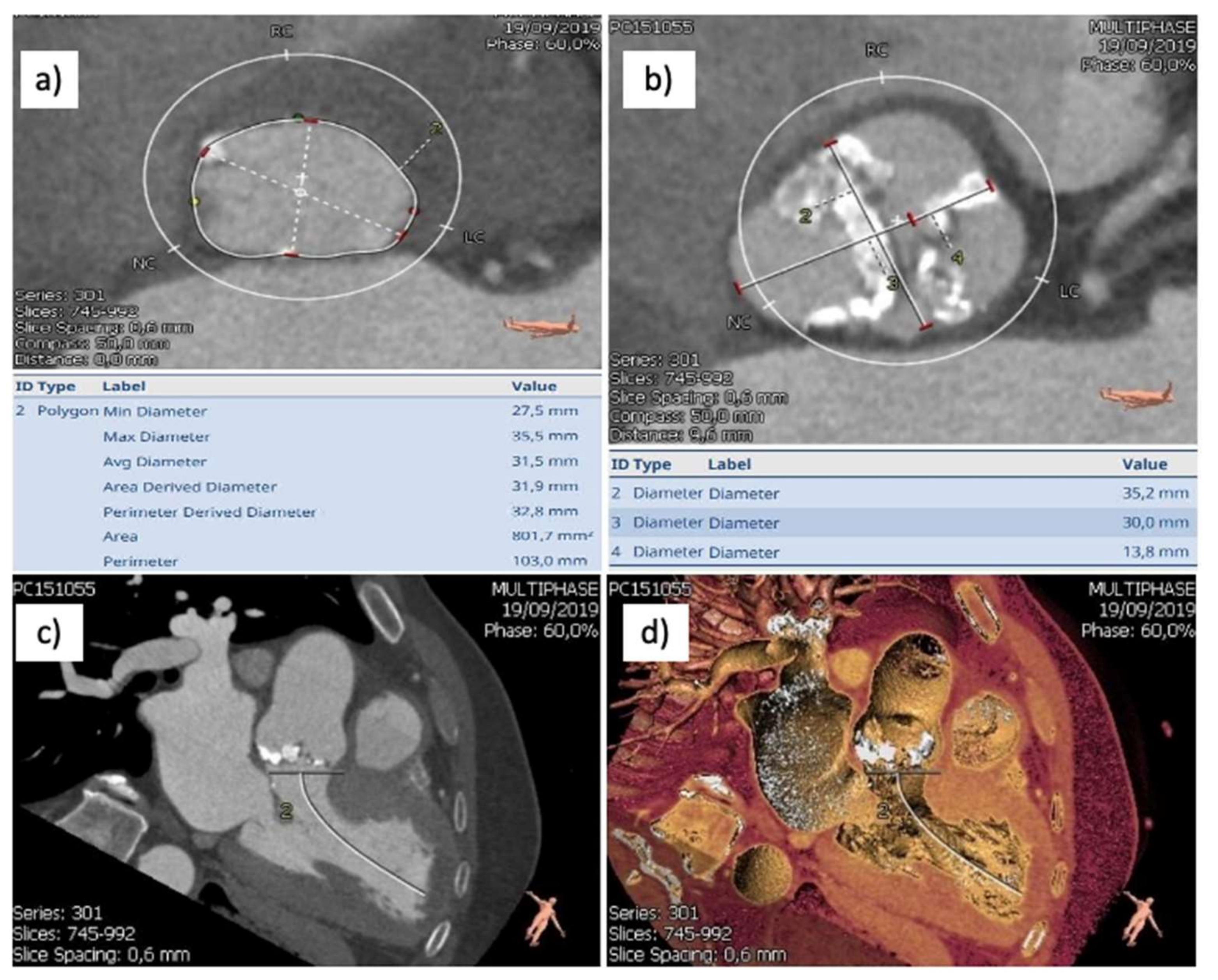
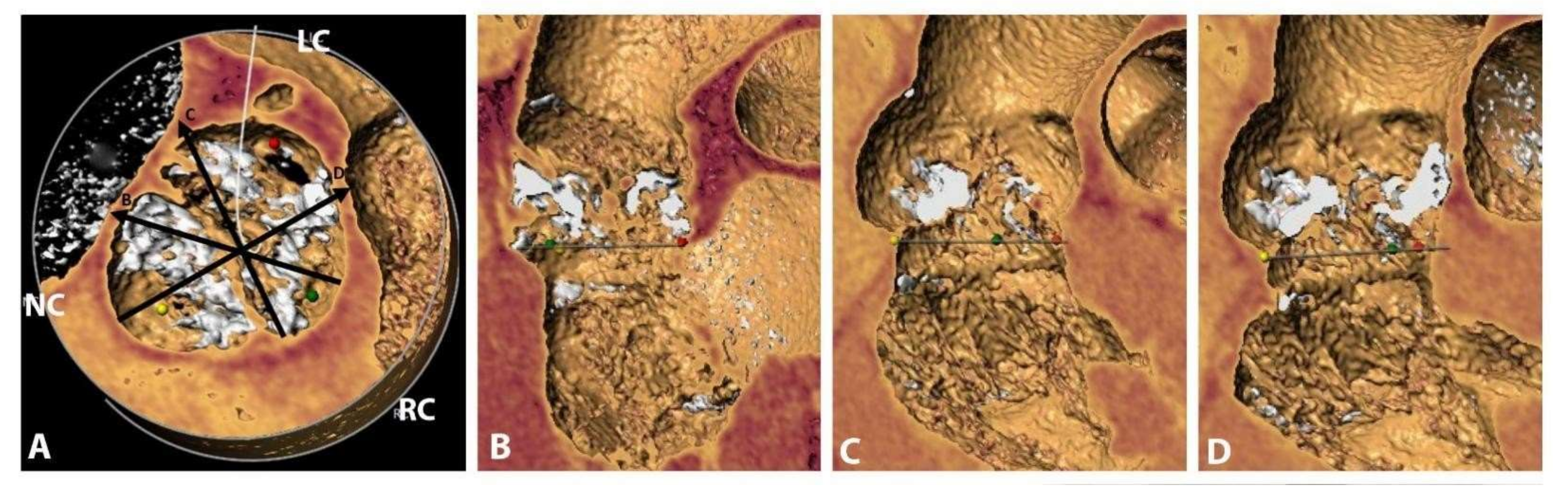
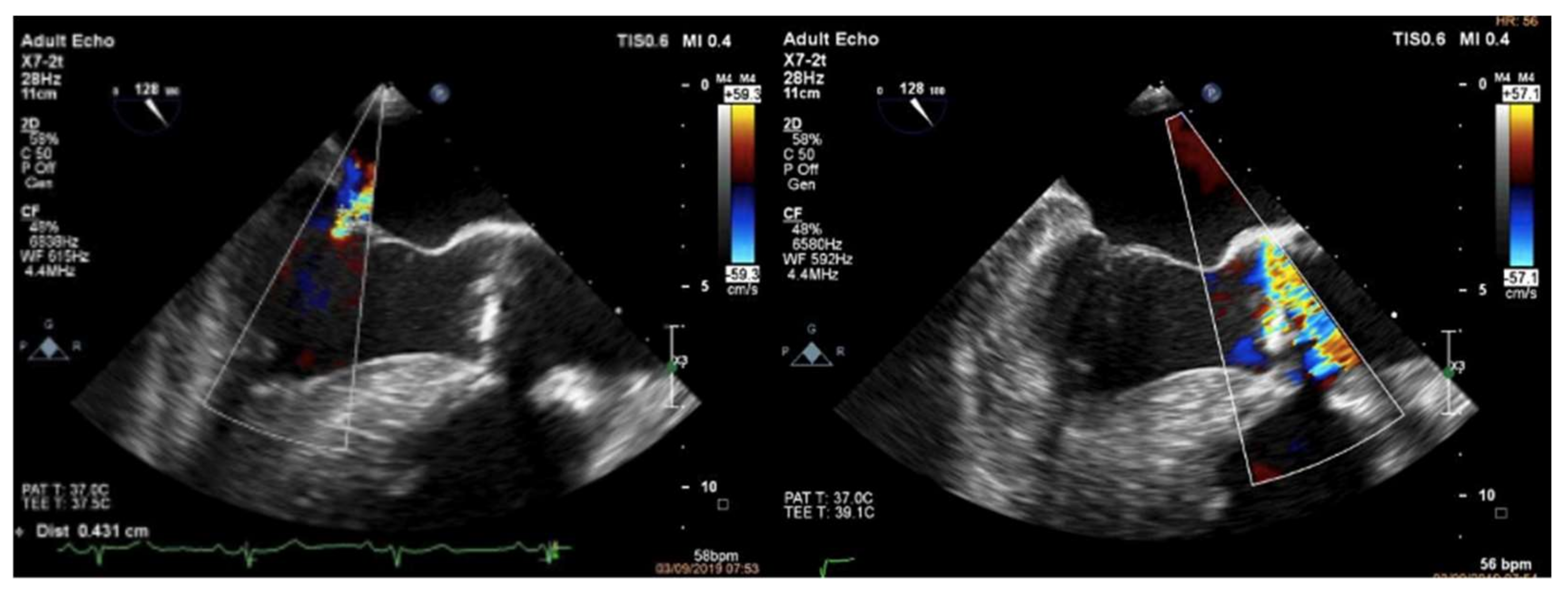
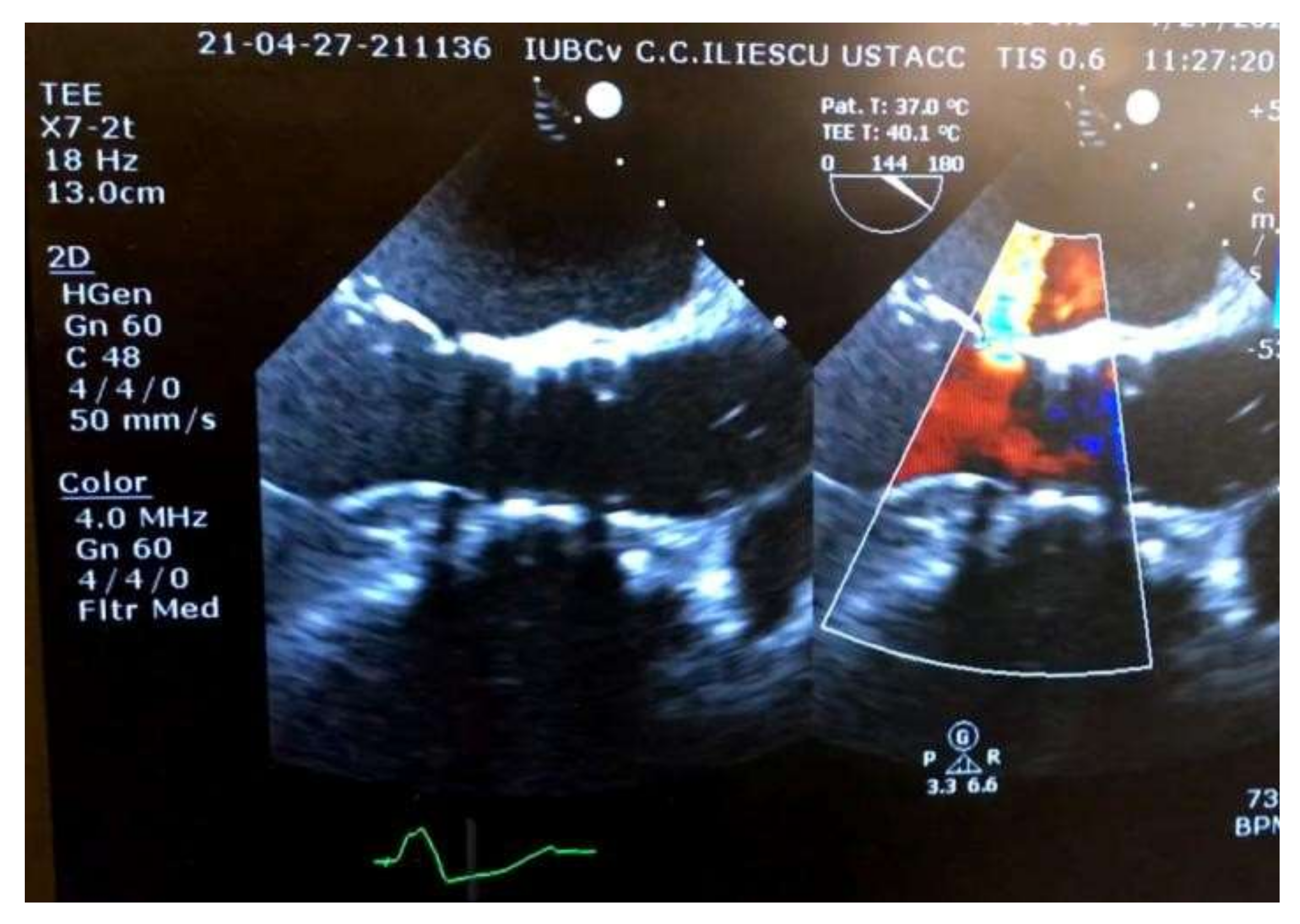
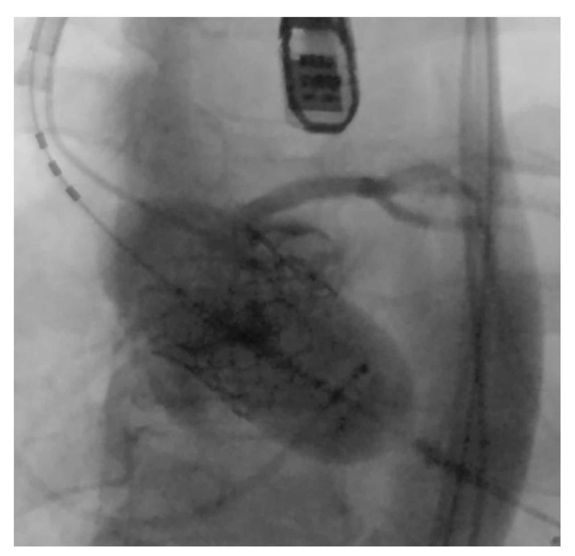
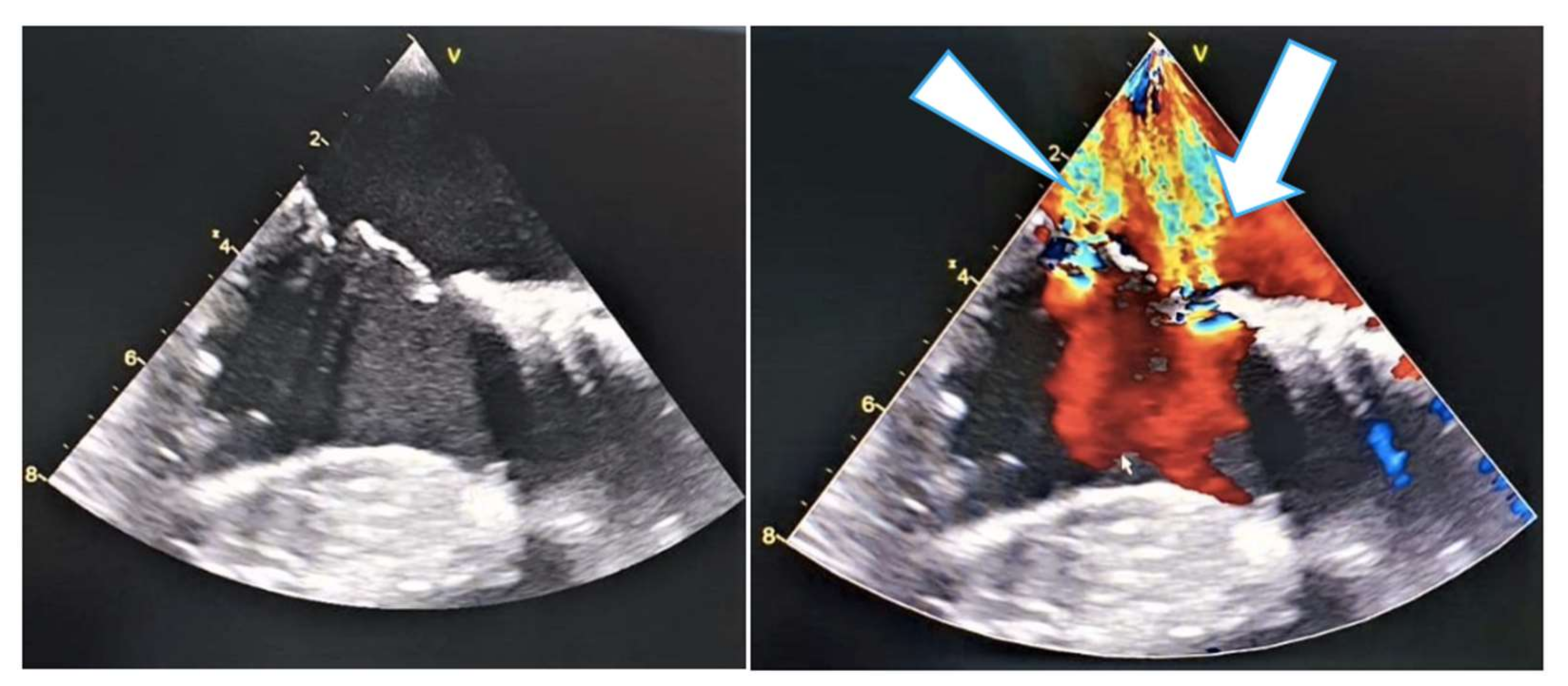
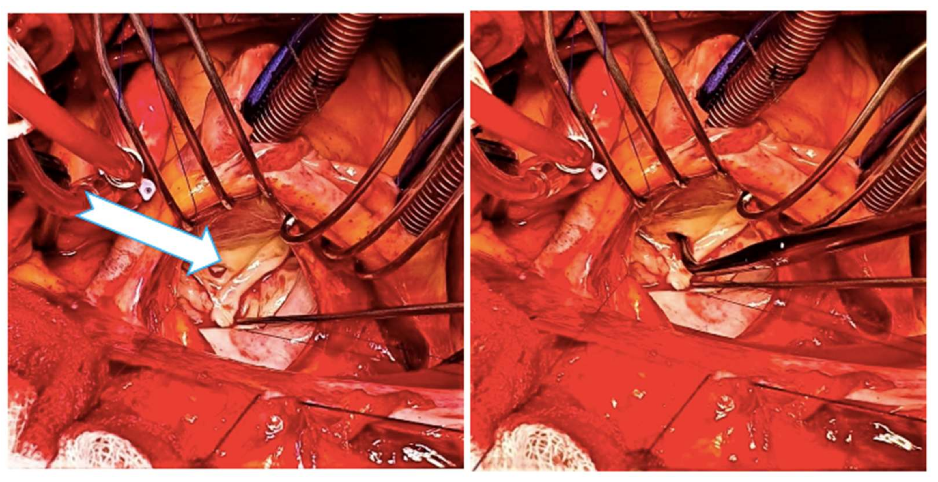
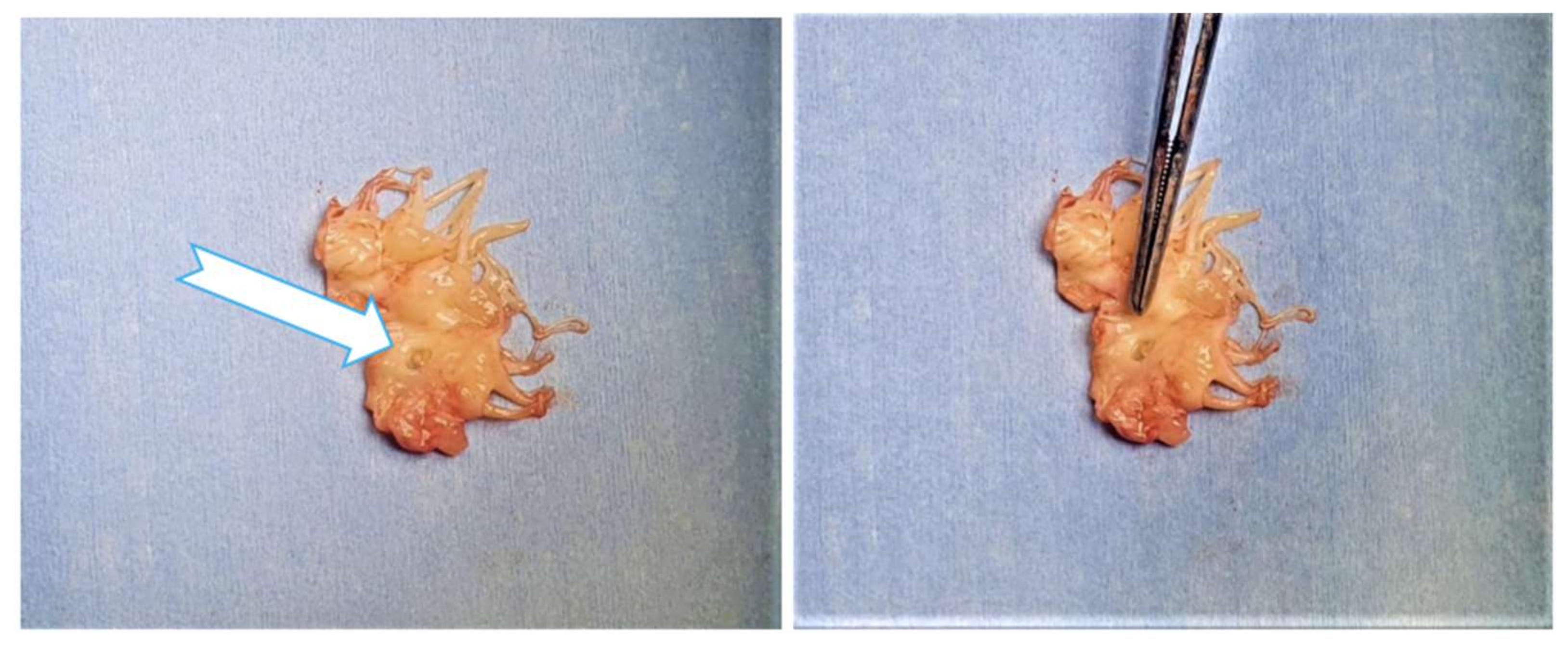
2. Case Report
3. Discussion
4. Conclusions
Author Contributions
Funding
Institutional Review Board Statement
Informed Consent Statement
Data Availability Statement
Conflicts of Interest
Abbreviations and Acronyms
| AS | aortic stenosis |
| IE | infective endocarditis |
| HF | heart failure |
| LBBB | left bundle-branch block |
| LV | left ventricular |
| MDCT | multidetector computed tomography |
| MR | mitral regurgitation |
| TAVR | transcatheter aortic valve replacement |
| TEE | transesophageal echocardiography |
| TTE | transthoracic echocardiography |
References
- Almasood, A.; Al Ahmari, S.; El-Shurafa, H.; Alotaibi, M.; Al Kasab, S.; Alabdallah, M.; Al-Moghairi, A.; Al Khushail, A.; Al-Amri, H. The change in mitral regurgitation severity after trans-catheter aortic valve implantation. J. Saudi Heart Assoc. 2014, 27, 10–17. [Google Scholar] [CrossRef] [PubMed] [Green Version]
- Toggweiler, S.; Boone, R.H.; Rodés-Cabau, J.; Humphries, K.H.; Lee, M.; Nombela-Franco, L.; Bagur, R.; Willson, A.B.; Binder, R.K.; Gurvitch, R.; et al. Transcatheter aortic valve replacement: Outcomes of patients with moderate or severe mitral regurgitation. J. Am. Coll. Cardiol. 2012, 59, 2068–2074. [Google Scholar] [CrossRef] [Green Version]
- Fojt, R.; Moťovská, Z.; Budera, P.; Malý, M.; Straka, Z. Prognostic impact and change of concomitant mitral regurgitation after surgical or transcatheter aortic valve replacement for aortic stenosis. J. Cardiol. 2016, 67, 526–530. [Google Scholar] [CrossRef] [PubMed] [Green Version]
- Szymański, P.; Hryniewiecki, T.; Dąbrowski, M.; Sorysz, D.; Kochman, J.; Jastrzębski, J.; Kukulski, T.; Zembala, M. Mitral and aortic regurgitation following transcatheter aortic valve replacement. Heart 2016, 102, 701–706. [Google Scholar] [CrossRef] [PubMed] [Green Version]
- David, T.E.; Armstrong, S.; Maganti, M.; Ihlberg, L. Clinical outcomes of combined aortic root replacement with mitral valve surgery. J. Thorac. Cardiovasc. Surg. 2008, 136, 82–87. [Google Scholar] [CrossRef] [PubMed] [Green Version]
- Barreiro, C.J.; Patel, N.D.; Fitton, T.P.; Williams, J.A.; Bonde, P.N.; Chan, V.; Alejo, D.E.; Gott, V.L.; Baumgartner, W.A. Aortic valve replacement and concomitant mitral valve regurgitation in the elderly: Impact on survival and functional outcome. Circulation 2005, 112, I443–I447. [Google Scholar] [CrossRef] [PubMed]
- Ruel, M.; Kapila, V.; Price, J.; Kulik, A.; Burwash, I.G.; Mesana, T.G. Natural History and Predictors of Outcome in Patients With Concomitant Functional Mitral Regurgitation at the Time of Aortic Valve Replacement. Circulation 2006, 114, I541–I546. [Google Scholar] [CrossRef] [PubMed] [Green Version]
- Khawaja, M.Z.; Williams, R.; Hung, J.; Arri, S.; Asrress, K.N.; Bolter, K.; Wilson, K.; Young, C.P.; Bapat, V.; Hancock, J.; et al. Impact of preprocedural mitral regurgitation upon mortality after transcatheter aortic valve implantation (TAVI) for severe aortic stenosis. Heart 2014, 100, 1799–1803. [Google Scholar] [CrossRef] [PubMed]
- Moldovan, H.; Popescu, D.; Buliga, T.; Filip, A.; Antoniac, I.; Gheorghiţă, D.; Molnar, A. Gastric Adenocarcinoma Associated with Acute Endocarditis of the Aortic Valve and Coronary Artery Disease in a 61-Year-Old Male with Multiple Comorbidities—Combined Surgical Management—Case Report. Medicina 2019, 55, 242. [Google Scholar] [CrossRef] [Green Version]
- Webb, J.G.; Pasupati, S.; Humphries, K.; Thompson, C.; Altwegg, L.; Moss, R.; Sinhal, A.; Carere, R.G.; Munt, B.; Ricci, D.; et al. Percutaneous Transarterial Aortic Valve Replacement in Selected High-Risk Patients With Aortic Stenosis. Circulation 2007, 116, 755–763. [Google Scholar] [CrossRef] [PubMed] [Green Version]
- Gurvitch, R.; Wood, D.A.; Tay, E.L.; Leipsic, J.; Ye, J.; Lichtenstein, S.V.; Thompson, C.R.; Carere, R.G.; Wijesinghe, N.; Nietlispach, F.; et al. Transcatheter aortic valve implantation: Durability of clinical and hemodynamic outcomes beyond 3 years in a large patient cohort. Circulation 2010, 122, 1319–1327. [Google Scholar] [CrossRef] [Green Version]
- Leon, M.B.; Smith, C.R.; Mack, M.; Miller, D.C.; Moses, J.W.; Svensson, L.G.; Tuzcu, E.M.; Webb, J.G.; Fontana, G.P.; Makkar, R.R.; et al. Transcatheter Aortic-Valve Implantation for Aortic Stenosis in Patients Who Cannot Undergo Surgery. N. Engl. J. Med. 2010, 363, 1597–1607. [Google Scholar] [CrossRef] [Green Version]
- Clavel, M.A.; Webb, J.G.; Rodés-Cabau, J.; Masson, J.B.; Dumont, E.; De Larochellière, R.; Doyle, D.; Bergeron, S.; Baumgartner, H.; Burwash, I.G.; et al. Comparison between transcatheter and surgical prosthetic valve implantation in patients with severe aortic stenosis and reduced left ventricular ejection fraction. Circulation 2010, 122, 1928–1936. [Google Scholar] [CrossRef] [PubMed] [Green Version]
- Webb, J.G.; Altwegg, L.; Boone, R.H.; Cheung, A.; Ye, J.; Lichtenstein, S.; Lee, M.; Masson, J.B.; Thompson, C.; Moss, R.; et al. Transcatheter Aortic Valve Implantation: Impact on Clinical and Valve-Related Outcomes. Circulation 2009, 119, 3009–3016. [Google Scholar] [CrossRef] [PubMed] [Green Version]
- Bleiziffer, S.; Ruge, H.; Mazzitelli, D.; Schreiber, C.; Hutter, A.; Laborde, J.-C.; Bauernschmitt, R.; Lange, R. Results of percutaneous and transapical transcatheter aortic valve implantation performed by a surgical team. Eur. J. Cardiothorac. Surg. 2009, 35, 615–621, discussion 620–621. [Google Scholar] [CrossRef] [PubMed]
- Iosifescu, A.G.; Moldoyan, H.; Iliescu, V.A. Aortic Prosthesis-Patient Mismatch Strongly Affects Early Results of Double Valve Replacement. J. Heart Valve Dis. 2014, 23, 149–157. [Google Scholar]
- Stähli, B.E.; Bünzli, R.; Grünenfelder, J.; Bühler, I.; Felix, C.; Bettex, D.; Biaggi, P.; Tanner, F.C.; Nguyen-Kim, T.D.L.; Plass, A.; et al. Transcatheter aortic valve implantation (TAVI) outcome according to standardized endpoint definitions by the Valve Academic Research Consortium (VARC). J. Invasive Cardiol. 2011, 23, 307–312. [Google Scholar] [PubMed]
- Costache, V.S.; Moldovan, H.; Arsenescu, C.; Costache, A. Aortic valve surgery of the 21st century: Sutureless AVR versus TAVI. Minerva Cardioangiol. 2018, 66, 191–197. [Google Scholar] [CrossRef] [PubMed]
- Rodés-Cabau, J. Transcatheter aortic valve implantation: Current and future approaches. Nat. Rev. Cardiol. 2011, 9, 15–29. [Google Scholar] [CrossRef] [PubMed]
- Nappi, F.; Nenna, A.; Timofeeva, I.; Mihos, C.; Gentile, F.; Chello, M. Mitral regurgitation after transcatheter aortic valve replacement. J. Thorac. Dis. 2020, 12, 2926–2935. [Google Scholar] [CrossRef] [PubMed]
- Sanna, G.D.; Moccia, E.; Pepi, M.; Parodi, G. Anterior mitral leaflet perforation and infective endocarditis following transcatheter aortic valve replacement in a patient presenting with heart failure. J. Cardiovasc. Echogr. 2020, 30, 44–46. [Google Scholar] [CrossRef] [PubMed]
- Miura, M.; Isotani, A.; Murata, K.; Kawaguchi, T.; Hayashi, M.; Arai, Y.; Shirai, S.; Hanyu, M.; Ando, K. Perforation of Anterior Mitral Leaflet Due to Mechanical Stimulation Late After Transcatheter Aortic Valve Replacement. JACC Cardiovasc. Interv. 2016, 9, e233–e234. [Google Scholar] [CrossRef]
- Bourezg, A.; Prieur, C.; Finet, G.; Rioufol, G. Percutaneous Management of Mitral Perforation During Transcatheter Aortic Valve Replacement. JACC Cardiovasc. Interv. 2017, 10, 1710–1711. [Google Scholar] [CrossRef] [PubMed]
- Raschpichler, M.; Seeburger, J.; Strasser, R.H.; Misfeld, M. Corevalve prosthesis causes anterior mitral leaflet perforation resulting in severe mitral regurgitation and subsequent endocarditis. Eur. Hear. J. 2013, 35, 1587. [Google Scholar] [CrossRef] [PubMed] [Green Version]
- Amat-Santos, I.J.; Cortés, C.; Revilla, A.; San Román, J.A. Infective endocarditis: Cause or consequence of delayed anterior mitral leaflet perforation after transcatheter aortic valve implantation? Rev. Esp. Cardiol. Engl. Ed. 2016, 69, 87. [Google Scholar] [CrossRef]
- Amat-Santos, I.J.; Cortés, C.; Varela-Falcón, L.H. Delayed left anterior mitral leaflet perforation and infective endocarditis after transapical aortic valve implantation-Case report and systematic review. Catheter. Cardiovasc. Interv. 2017, 89, 951–954. [Google Scholar] [CrossRef]
- Jurcut, R.; Savu, O.; Popescu, B.A.; Florian, A.; Herlea, V.; Moldovan, H.; Ginghina, C. Primary cardiac leiomyosarcoma when valvular disease becomes a vascular surgical emergency. Circulation 2010, 121, E415–E418. [Google Scholar] [CrossRef] [Green Version]
- Masson, J.-B.; Kovac, J.; Schuler, G.; Ye, J.; Cheung, A.; Kapadia, S.; Tuzcu, M.E.; Kodali, S.; Leon, M.B.; Webb, J.G. Transcatheter Aortic Valve Implantation: Review of the Nature, Management, and Avoidance of Procedural Complications. JACC Cardiovasc. Interv. 2009, 2, 811–820. [Google Scholar] [CrossRef] [Green Version]
- Saji, M.; Ailawadi, G.; Ragosta, M.; Fowler, D.E.; Dent, J.M.; Lim, D.S. Anterior Mitral Leaflet Perforation During Transcatheter Aortic Valve Replacement in a Patient With Mitral Annular Calcification. JACC Cardiovasc. Interv. 2015, 8, e215–e216. [Google Scholar] [CrossRef] [Green Version]
- Franco, E.; De Agustin, J.A.; Hernandez-Antolín, R.; Garcia, E.; Silva, J.; Maroto, L.; Olmos, C.; Fortuny, E.; Viliani, D.; Macaya, C.; et al. Acute Mitral Stenosis After Transcatheter Aortic Valve Implantation. J. Am. Coll. Cardiol. 2012, 60, e35. [Google Scholar] [CrossRef] [Green Version]
- Yamashita, Y.; Sonoda, H.; Ushijima, T.; Shiose, A. Acute torrential mitral regurgitation during transcatheter aortic valve replacement: A case report. Surg. Case Rep. 2018, 4, 35. [Google Scholar] [CrossRef] [PubMed] [Green Version]
- López-Aguilera, J.; Mesa-Rubio, D.; Ruiz-Ortiz, M.; Delgado-Ortega, M.; Villanueva-Fernández, E.; Romo-Peña, E.; Pan Álvarez-Ossorio, M.; Suárez de Lezo, J. Mitral regurgitation during transcatheter aortic valve implantation: The same complication with a different mechanism. J. Invasive Cardiol. 2014, 26, 603–608. [Google Scholar] [PubMed]
- Cortés, C.; Amat-Santos, I.J.; Nombela-Franco, L.; Muñoz-Garcia, A.J.; Gutiérrez-Ibanes, E.; De La Torre Hernandez, J.M.; Córdoba-Soriano, J.G.; Jimenez-Quevedo, P.; Hernández-García, J.M.; Gonzalez-Mansilla, A.; et al. Mitral Regurgitation After Transcatheter Aortic Valve Replacement: Prognosis, Imaging Predictors, and Potential Management. JACC Cardiovasc. Interv. 2016, 9, 1603–1614. [Google Scholar] [CrossRef] [PubMed]
- Kapadia, S.R.; Leon, M.B.; Makkar, R.R.; Tuzcu, E.M.; Svensson, L.G.; Kodali, S.; Webb, J.G.; Mack, M.J.; Douglas, P.S.; Thourani, V.H.; et al. 5-year outcomes of transcatheter aortic valve replacement compared with standard treatment for patients with inoperable aortic stenosis (PARTNER 1): A randomised controlled trial. Lancet 2015, 385, 2485–2491. [Google Scholar] [CrossRef]
- Mack, M.J.; Leon, M.B.; Smith, C.R.; Miller, D.C.; Moses, J.W.; Tuzcu, E.M.; Webb, J.G.; Douglas, P.S.; Anderson, W.N.; Blackstone, E.H.; et al. 5-year outcomes of transcatheter aortic valve replacement or surgical aortic valve replacement for high surgical risk patients with aortic stenosis (PARTNER 1): A randomised controlled trial. Lancet 2015, 385, 2477–2484. [Google Scholar] [CrossRef]
- Adams, D.H.; Popma, J.J.; Reardon, M.J.; Yakubov, S.J.; Coselli, J.S.; Deeb, G.M.; Gleason, T.G.; Buchbinder, M.; Hermiller, J., Jr.; Kleiman, N.S.; et al. Transcatheter Aortic-Valve Replacement with a Self-Expanding Prosthesis. N. Engl. J. Med. 2014, 370, 1790–1798. [Google Scholar] [CrossRef] [PubMed] [Green Version]


Publisher’s Note: MDPI stays neutral with regard to jurisdictional claims in published maps and institutional affiliations. |
© 2022 by the authors. Licensee MDPI, Basel, Switzerland. This article is an open access article distributed under the terms and conditions of the Creative Commons Attribution (CC BY) license (https://creativecommons.org/licenses/by/4.0/).
Share and Cite
Moldovan, H.; Popescu, B.-Ş.; Nechifor, E.; Badea, A.; Ciomaga, I.; Nica, C.; Zaharia, O.; Gheorghiță, D.; Broască, M.; Diaconu, C.; et al. Rare Cause of Severe Mitral Regurgitation after TAVI: Case Report and Literature Review. Medicina 2022, 58, 464. https://doi.org/10.3390/medicina58040464
Moldovan H, Popescu B-Ş, Nechifor E, Badea A, Ciomaga I, Nica C, Zaharia O, Gheorghiță D, Broască M, Diaconu C, et al. Rare Cause of Severe Mitral Regurgitation after TAVI: Case Report and Literature Review. Medicina. 2022; 58(4):464. https://doi.org/10.3390/medicina58040464
Chicago/Turabian StyleMoldovan, Horațiu, Bogdan-Ştefan Popescu, Elena Nechifor, Aida Badea, Irina Ciomaga, Claudia Nica, Ondin Zaharia, Daniela Gheorghiță, Marian Broască, Camelia Diaconu, and et al. 2022. "Rare Cause of Severe Mitral Regurgitation after TAVI: Case Report and Literature Review" Medicina 58, no. 4: 464. https://doi.org/10.3390/medicina58040464