High SPOCK1 Expression Is Associated with Advanced Stage, T Value, and Gleason Grade in Prostate Cancer

 ,
,
Abstract
:1. Introduction
2. Methods
2.1. Patients
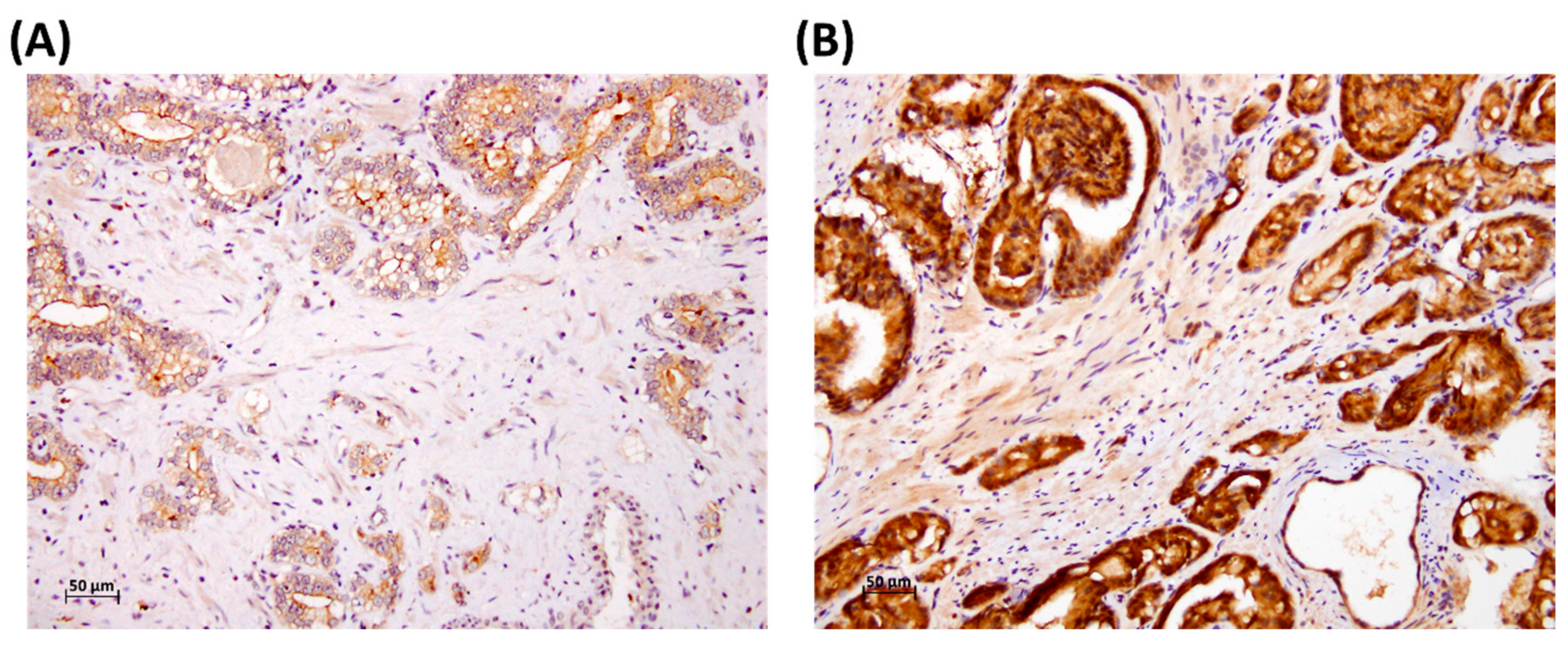
2.2. Immunohistochemical Staining of SPOCK1
2.3. Patient and Public Involvement
2.4. Statistical Analysis
3. Results
4. Discussion
5. Conclusions
Author Contributions
Funding
Conflicts of Interest
Abbreviations
References
- Attard, G.; Parker, C.; Eeles, R.A.; Schroder, F.; Tomlins, S.A.; Tannock, I.; Drake, C.G.; de Bono, J.S. Prostate cancer. Lancet 2016, 387, 70–82. [Google Scholar] [CrossRef]
- Chen, S.L.; Wang, S.C.; Ho, C.J.; Kao, Y.L.; Hsieh, T.Y.; Chen, W.J.; Chen, C.J.; Wu, P.R.; Ko, J.L.; Lee, H.; Sung, W.W. Prostate Cancer Mortality-To-Incidence Ratios Are Associated with Cancer Care Disparities in 35 Countries. Sci Rep. 2017, 7, 40003. [Google Scholar] [CrossRef] [Green Version]
- Siegel, R.; Naishadham, D.; Jemal, A. Cancer statistics, 2013. CA Cancer J. Clin. 2013, 63, 11–30. [Google Scholar] [CrossRef] [PubMed] [Green Version]
- Gunjur, A. Personalising the treatment of prostate cancer. Lancet Oncol. 2015, 16, e592. [Google Scholar] [CrossRef]
- Chien, M.H.; Lin, Y.W.; Wen, Y.C.; Yang, Y.C.; Hsiao, M.; Chang, J.L.; Huang, H.C.; Lee, W.J. Targeting the SPOCK1-snail/slug axis-mediated epithelial-to-mesenchymal transition by apigenin contributes to repression of prostate cancer metastasis. J. Exp. Clin. Cancer Res. 2019, 38, 246. [Google Scholar] [CrossRef] [PubMed]
- Okato, A.; Arai, T.; Kojima, S.; Koshizuka, K.; Osako, Y.; Idichi, T.; Kurozumi, A.; Goto, Y.; Kato, M.; Naya, Y.; Ichikawa, T.; Seki, N. Dual strands of pre-miR150 (miR1505p and miR1503p) act as antitumor miRNAs targeting SPOCK1 in naive and castration-resistant prostate cancer. Int. J. Oncol. 2017, 51, 245–256. [Google Scholar] [CrossRef] [PubMed]
- Bradshaw, A.D. Diverse biological functions of the SPARC family of proteins. Int. J. Biochem. Cell Biol. 2012, 44, 480–488. [Google Scholar] [CrossRef] [PubMed] [Green Version]
- Tai, I.T.; Tang, M.J. SPARC in cancer biology: Its role in cancer progression and potential for therapy. Drug Resist. Updat. 2008, 11, 231–246. [Google Scholar] [CrossRef] [PubMed]
- Colin, C.; Baeza, N.; Bartoli, C.; Fina, F.; Eudes, N.; Nanni, I.; Martin, P.M.; Ouafik, L.; Figarella-Branger, D. Identification of genes differentially expressed in glioblastoma versus pilocytic astrocytoma using Suppression Subtractive Hybridization. Oncogene 2006, 25, 2818–2826. [Google Scholar] [CrossRef] [PubMed]
- Li, Y.; Chen, L.; Chan, T.H.; Liu, M.; Kong, K.L.; Qiu, J.L.; Li, Y.; Yuan, Y.F.; Guan, X.Y. SPOCK1 is regulated by CHD1L and blocks apoptosis and promotes HCC cell invasiveness and metastasis in mice. Gastroenterology 2013, 144, 179–191. [Google Scholar] [CrossRef] [PubMed]
- Ma, L.J.; Wu, W.J.; Wang, Y.H.; Wu, T.F.; Liang, P.I.; Chang, I.W.; He, H.L.; Li, C.F. SPOCK1 Overexpression Confers a Poor Prognosis in Urothelial Carcinoma. J. Cancer 2016, 7, 467–476. [Google Scholar] [CrossRef] [PubMed] [Green Version]
- Chen, Q.; Yao, Y.T.; Xu, H.; Chen, Y.B.; Gu, M.; Cai, Z.K.; Wang, Z. SPOCK1 promotes tumor growth and metastasis in human prostate cancer. Drug Des. Devel. Ther. 2016, 10, 2311–2321. [Google Scholar] [PubMed]
- Chang, C.F.; Chen, S.L.; Sung, W.W.; Hsieh, M.J.; Hsu, H.T.; Chen, L.H.; Chen, M.K.; Ko, J.L.; Chen, C.J.; Chou, M.C. PBK/TOPK Expression Predicts Prognosis in Oral Cancer. Int. J. Mol. Sci. 2016, 17, 1007. [Google Scholar] [CrossRef] [PubMed]
- Sung, W.W.; Lin, Y.M.; Wu, P.R.; Yen, H.H.; Lai, H.W.; Su, T.C.; Huang, R.H.; Wen, C.K.; Chen, C.Y.; Chen, C.J.; Yeh, K.T. High nuclear/cytoplasmic ratio of Cdk1 expression predicts poor prognosis in colorectal cancer patients. BMC Cancer 2014, 14, 951. [Google Scholar] [CrossRef] [PubMed]
- Sung, W.W.; Wang, Y.C.; Cheng, Y.W.; Lee, M.C.; Yeh, K.T.; Wang, L.; Wang, J.; Chen, C.Y.; Lee, H. A polymorphic -844T/C in FasL promoter predicts survival and relapse in non-small cell lung cancer. Clin. Cancer Res. 2011, 17, 5991–5999. [Google Scholar] [CrossRef] [PubMed]
- Baetke, S.C.; Adriaens, M.E.; Seigneuric, R.; Evelo, C.T.; Eijssen, L.M. Molecular pathways involved in prostate carcinogenesis: Insights from public microarray datasets. PLoS ONE 2012, 7, e49831. [Google Scholar] [CrossRef] [PubMed]
- Wlazlinski, A.; Engers, R.; Hoffmann, M.J.; Hader, C.; Jung, V.; Muller, M.; Schulz, W.A. Downregulation of several fibulin genes in prostate cancer. Prostate 2007, 67, 1770–1780. [Google Scholar] [CrossRef] [PubMed]
Sample Availability: The data that support the findings of this study are available on request from the first author, Mei-Ling Chen. |


| SPOCK1 Expression | ||||
|---|---|---|---|---|
| Parameters | Case Number | Low | High | p Value |
| Age (years) | 73.2 ± 5.8 | 75.5 ± 7.4 | 0.151 | |
| Gleason grade group | ||||
| 1 + 2 | 38 | 19 (50.0) | 19 (50.0) | 0.370 |
| 3 + 4 + 5 | 33 | 13 (39.4) | 20 (60.6) | |
| Stage | ||||
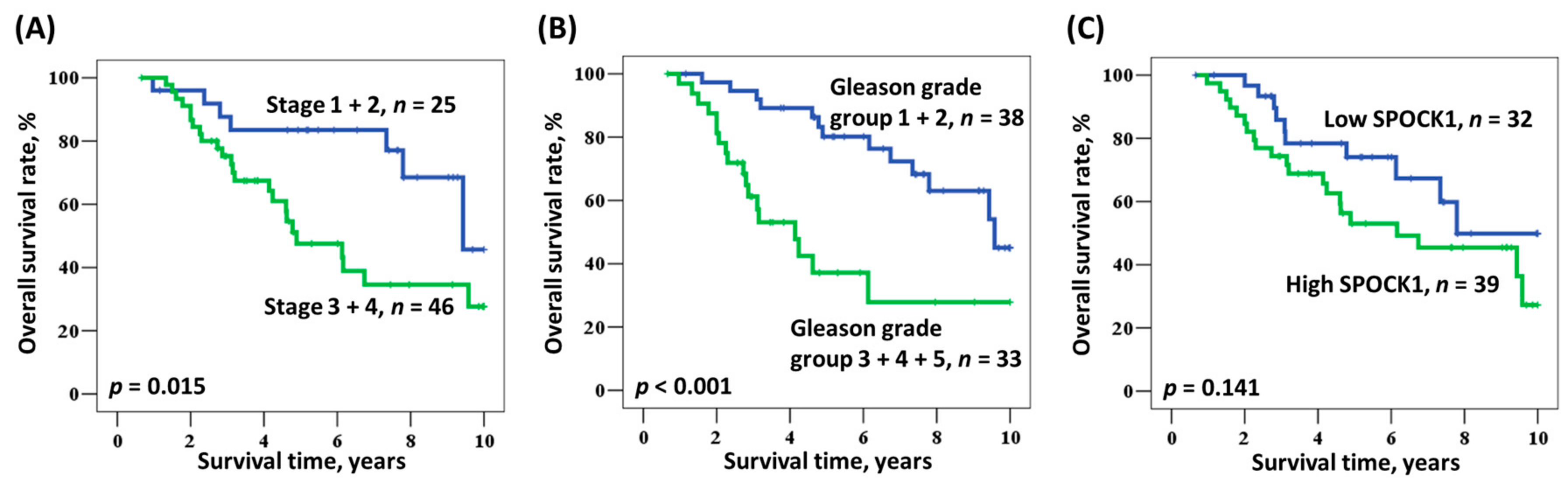
| I + II | 25 | 16 (64.0) | 9 (36.0) | 0.018 |
| III + IV | 46 | 16 (34.8) | 30 (65.2) | |
| Tumor (T) value | ||||
| 1 | 13 | 10 (76.9) | 3 (23.1) | 0.014 * |
| 2 + 3 + 4 | 58 | 22 (37.9) | 36 (62.1) | |
| Node (N) value | ||||
| 0 | 55 | 26 (47.3) | 29 (52.7) | 0.489 |
| 1 + 2 | 16 | 6 (37.5) | 10 (62.5) | |
| Metastasis (M) value | ||||
| 0 | 42 | 21 (50.0) | 21 (50.0) | 0.315 |
| 1 | 29 | 11 (37.9) | 18 (62.1) | |
| Parameters | SPOCK1 Expression* | p Value |
|---|---|---|
| Stage | ||
| 1+2 | 216 ± 73 | Reference |
| 3+4 | 254 ± 60 | 0.020 |
| Gleason grade group | ||
| 1 | 229 ± 65 | Reference |
| 2 | 248 ± 65 | 0.375 |
| 3 | 270 ± 46 | 0.044 |
| 4 | 285 ± 17 | 0.003 |
| 5 | 210 ± 82 | 0.454 |
| Overall Survival | |||||
|---|---|---|---|---|---|
| Parameter | Category | Medium Survival (Years) | Hazard Ratio (HR) | 95% Confidence Interval (CI) | p Value |
| Age, years | ≥74/<74 | 7.8/11.3 | 1.335 | 0.651–2.738 | 0.430 |
| Gleason grade group | 3 + 4 + 5/1 + 2 | 4.1/9.6 | 3.700 | 1.769–7.737 | 0.001 |
| Stage | III + IV/I + II | 4.9/9.4 | 2.419 | 1.075–5.444 | 0.033 |
| SPOCK1 | High/Low | 6.2/7.8 | 1.750 | 0.823–3.720 | 0.146 |
| Overall Survival | |||||
|---|---|---|---|---|---|
| Parameter | Category | Mean Survival (Years) | HR | 95% CI | p Value |
| Age, years | ≥74/<74 | 7.8/11.3 | 1.193 | 0.555–2.562 | 0.651 |
| Gleason grade group | 3 + 4 + 5/1 + 2 | 4.1/9.6 | 3.322 | 1.531–7.209 | 0.002 |
| Stage | III + IV/I + II | 4.9/9.4 | 1.948 | 0.811–4.679 * | 0.136 * |
| SPOCK1 | High/Low | 6.2/7.8 | 1.411 | 0.627–3.174 | 0.405 |
© 2019 by the authors. Licensee MDPI, Basel, Switzerland. This article is an open access article distributed under the terms and conditions of the Creative Commons Attribution (CC BY) license (http://creativecommons.org/licenses/by/4.0/).
Share and Cite
Chen, M.-L.; Ho, C.-J.; Yeh, C.-M.; Chen, S.-L.; Sung, W.-W.; Wang, S.-C.; Chen, C.-J. High SPOCK1 Expression Is Associated with Advanced Stage, T Value, and Gleason Grade in Prostate Cancer. Medicina 2019, 55, 343. https://doi.org/10.3390/medicina55070343
Chen M-L, Ho C-J, Yeh C-M, Chen S-L, Sung W-W, Wang S-C, Chen C-J. High SPOCK1 Expression Is Associated with Advanced Stage, T Value, and Gleason Grade in Prostate Cancer. Medicina. 2019; 55(7):343. https://doi.org/10.3390/medicina55070343
Chicago/Turabian StyleChen, Mei-Ling, Cheng-Ju Ho, Chung-Min Yeh, Sung-Lang Chen, Wen-Wei Sung, Shao-Chuan Wang, and Chih-Jung Chen. 2019. "High SPOCK1 Expression Is Associated with Advanced Stage, T Value, and Gleason Grade in Prostate Cancer" Medicina 55, no. 7: 343. https://doi.org/10.3390/medicina55070343