
The Importance of Chitosan Coatings in Dentistry
by
 , ,
, ,
Anna Paradowska-Stolarz
1,* ,
,
Marcin Mikulewicz
1,
Joanna Laskowska
1,
Bożena Karolewicz
2 and
Artur Owczarek
2,* 1
Division of Dentofacial Anomalies, Department of Orthodontics and Dentofacial Orthopedics, Wroclaw Medical University, Krakowska 26, 50-425 Wroclaw, Poland
2
Department of Drug Forms Technology, Wroclaw Medical University, Borowska 211A, 50-556 Wroclaw, Poland
*
Authors to whom correspondence should be addressed.
Mar. Drugs 2023, 21(12), 613; https://doi.org/10.3390/md21120613
Submission received: 14 August 2023
/
Revised: 15 November 2023
/
Accepted: 21 November 2023
/
Published: 26 November 2023
(This article belongs to the Special Issue Collagen and Chitin from Marine Resources and Their Interdisciplinary Applications)
Abstract
:A Chitosan is a copolymer of N-acetyl-D-glucose amine and D-glucose amine that can be easily produced. It is a polymer that is widely utilized to create nanoparticles (NPs) with specific properties for applications in a wide range of human activities. Chitosan is a substance with excellent prospects due to its antibacterial, anti-inflammatory, antifungal, haemostatic, analgesic, mucoadhesive, and osseointegrative qualities, as well as its superior film-forming capacity. Chitosan nanoparticles (NPs) serve a variety of functions in the pharmaceutical and medical fields, including dentistry. According to recent research, chitosan and its derivatives can be embedded in materials for dental adhesives, barrier membranes, bone replacement, tissue regeneration, and antibacterial agents to improve the management of oral diseases. This narrative review aims to discuss the development of chitosan-containing materials for dental and implant engineering applications, as well as the challenges and future potential. For this purpose, the PubMed database (Medline) was utilised to search for publications published less than 10 years ago. The keywords used were “chitosan coating” and “dentistry”. After carefully selecting according to these keywords, 23 articles were studied. The review concluded that chitosan is a biocompatible and bioactive material with many benefits in surgery, restorative dentistry, endodontics, prosthetics, orthodontics, and disinfection. Furthermore, despite the fact that it is a highly significant and promising coating, there is still a demand for various types of coatings. Chitosan is a semi-synthetic polysaccharide that has many medical applications because of its antimicrobial properties. This article aims to review the role of chitosan in dental implantology.
1. Introduction
Polymer coatings in dentistry are widely used and, recently, materials obtained from natural polymers have been widely treated as matrices for drug delivery, especially in surgery and periodontics [1,2,3]. The coatings may also be used in other dental specialities. In stainless steel corrosion, the elution of metallic ions from the surface of materials used in orthodontics, and which can be protected by coating, has been proven by Mikulewicz et al. [4]. This phenomenon has also been shown to influence gene expression [5]. In dentistry, coatings are used to improve the quality and properties of dental devices [6].
1.1. Chitosan Structure
Chitosan, a naturally derived marine polymer with linear amino-polysaccharide structure, is widely used in dentistry. A D-glucosamine and N-acetyl glucosamine copolymer is obtained by deacetylation of the chitinous exoskeletons of crustaceans (also known as shellfish scaffolds). It also occurs naturally in mammalian cells. The main characteristics of chitosan are biocompatibility, non-toxicity, regenerative properties, natural availability, and the possibility of chemical treatment. According to the Food and Drug Administration agency (FDA), it has biological activity properties and wide spectrum of usage against all types of bacteria, including Gram-positive and Gram-negative. Due to those conditions, it is used willingly to treat damages in the tissues of the oral cavity. Additionally, chitosan has elastane-like properties [1,7]. Chitosan represents a group of multifunctional excipients [8]. It is used as an auxiliary substance and an antimicrobial agent. This is especially important in current times due to the possible elimination of the SARS-CoV2 virus from saliva [9,10,11]. It is used in preventive and conservative dentistry, endodontics, periodontal procedures, surgery, prosthodontics and orthodontics [7,12,13]. Although widely used, it is still unclear whether chitosan could potentially be an allergen for humans [7]. Although chitosan is more frequently a drug carrier component and active formulation agent, the variety of its use changes [14]. It shows a high percentage of antimicrobial reactions, especially against Streptococcus gordonii, which is the first bacteria to colonise surfaces in the oral cavity [15].
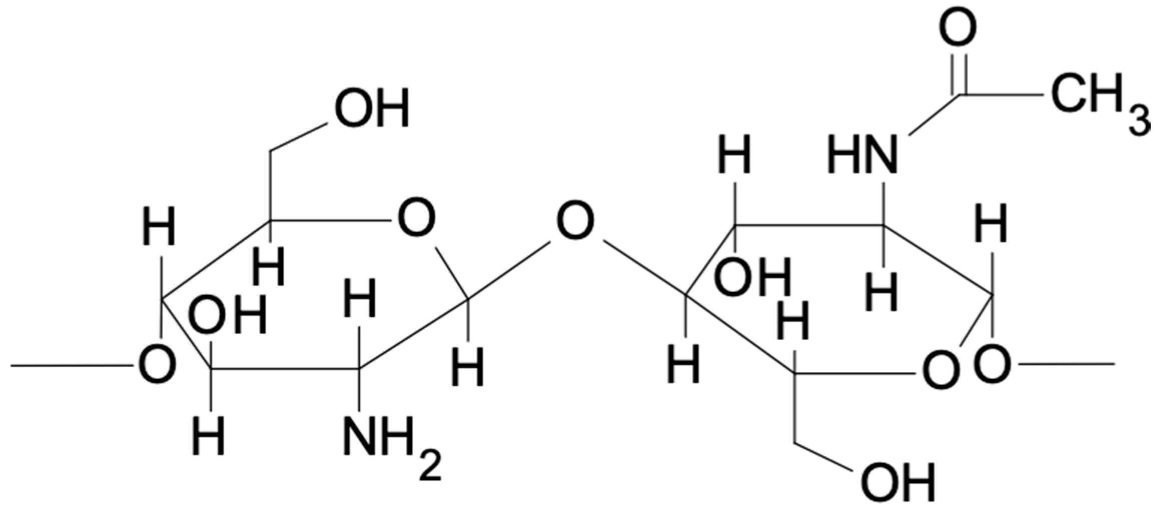
In a linearly built chitosan molecule, the relationship between the present hydroxyl and amino groups forms hydrogen bounds that influence the structure and flexibility of the polymer chain. Chitosan is a copolymer of glucosamine and N-acetyl-D-glucose amine units as shown in Figure 1.
Natural polymers and natural-based medicine are prominent topics of recent research. It is important to search for accessible materials which present biocompatibility and biocompetence. Chitosan is one of the natural materials which meets these criteria. The authors of the presented study are engaged in the two different fields of medicine–pharmacy (BK and AO) and dentistry (APS, MM and JL). We therefore tried to combine two points of view on chitosan: coatings–pharmaceutic and dental. We found this approach to be the advantageous to our research.
1.2. General Use
Chitosan belongs to the group of multifunctional polymers used in dentistry. It is used to design carriers and materials adapted to some specific conditions, as well as the specificity of the application site, which deliver active substances directly to diseased changed tissues. This includes intrapocket application and the release of the active substances in a timely manner. This specification allows for the improvement of the form of application and further development of therapeutic strategies. Its multifunctionality results from the function of the auxiliary substance that allows for the development of both the mucoadhesive carrier and the formulation component with biological activity. This phenomenon was confirmed in the biological activity tests, the mentioned antibacterial, haemostatic, but also antifungal and immunomodulating properties of the polymer and its derivatives, i.e., chitooligosaccharides. In the literature, chitosan, in addition to its antibacterial, antifungal, and anti-inflammatory properties mentioned before, is also reported to act as a biocompatible dental carrier that accelerates the process of periodontal regeneration. The biological activity of the polymer is caused by the cationic nature of the polymer and the interaction of the positively charged amino-group molecules with the negatively charged components of the Gram-negative bacteria cell membranes. As a result of the interaction, the properties of the cell membranes of the bacteria change-the transport of the nutrients inside the cell fluctuates, and its content starts leaking outside. Another explanation for the polymer activity is its penetration into the bacterial cell interior and binding to the DNA of the bacteria, which inhibits RNA transcription and, consequently, handicaps the protein synthesis. The biological activity of the polymer depends on its physicochemical properties, e.g., molecular weight, degree of polymerisation, degree of deacetylation, and also depends on the type of the microorganism. The properties of the polymer are crucial for its activity. It has been proven to react strongly against the bacteria responsible for creating dental plaque, including Porphyronomas gingivalis, Prevotella intermedia, and Aggregatibacter actinomycetemcomitans (formerly Actinobacillus actinomycetemcomitans). Due to all of these characteristics, polymers find many biological applications in materials and carriers used in the treatment of periodontitis, bone and tissue regeneration, drug delivery, and wound healing. However, the choice of appropriate molecular weight and degree of deacetylation affects the desired application properties as well as development of the carriers, e.g., matrices, bioadhesive tablets, films, microspheres, microparticles, nanoparticles, nanofibers, or gel forms.
1.3. Molecular Interaction
The use of chitosan in dentistry is also due to its increased induction of osteoblast growth and binding around polymer coated implants, which allows for the obtaining of bioactive surfaces ensuring osseointegration. Research has shown that chemical modifications of the polymer structure increase its antibacterial activity and osteoinductive properties. The polymer has the ability to bond to the metals surface, which can improve their mechanical strength and increase the durability of, e.g., titanium implants. Some researchers, however, emphasise that the adhesion of chitosan coatings to titanium surfaces is not suitable for clinical applications. Ferraresse et al. [16] proposed a chemical treatment of the titanium implant surface, using the method of ‘direct coating’, which is simply immersing the surface in an acid solution that allows a strong electrostatic attraction between the chitosan particles and the coated surface. In this condition, stable coatings are obtained mechanically and chemically, and chitosan-coated dental implants can have increased possibilities for osseointegration, which can lead to their commercial use. The preparation of the implant surface and bone preparation affects the stability and clinical success of the dental implant. To improve osseointegration, a number of methods of surface treatment and implant coatings have been tried, with promising results in coating them with chitosan. Chitosan coating can affect the surface that comes into contact with bone by changing the biological, mechanical and morphological properties of the surface. For example, considering the mechanical properties, the chitosan coating changes the modulus of elasticity, thus reducing the mismatch of the implant surface with the bone of the alveolar process and reducing stress concentration areas [17,18]. In addition, chitosan coatings can potentially be used to release drugs, such as antibiotics, for local delivery around the implant area. Additionally, chitosan can prevent damage to the tooth surface by organic acids by buffering saliva pH in the mouth, preventing adhesion pathogens, and stimulating the structured regeneration of oral soft tissues.
1.4. Biomechanical Features
Chitosan and chitosan-based coatings have relatively low stiffness and strength, especially in high humidity. Therefore, it is valid to obtain modified chitosan sol-gel preparations to improve their mechanical properties. The addition of chitosan improved the antibacterial activity of two types of subjects [9,19] of hybrid silica-chitosan coatings (50M50G and 45 M45G10T) and increased the proliferation rate of cells that grow on their surface. The proliferation indicators were the highest for coatings containing 5% and 10% chitosan. We can also conclude that the addition of chitosan and TEOS modulates the release of Si, which plays an important role in osteocompatibility. Tertiary coatings containing 5% and 10% chitosan have very good antibacterial properties [20]. They should be suitable for dental implants, as they protect against bone infection for a long time. The introduction of these materials into dental practice can increase the number of patients admitted to implantation.
A study by Tarsi et al. [21] showed that chitosan is an interesting candidate capable of preventing the adhesion of bacteria Streptococcus mutans to hydroxyapatite in teeth with tooth decay. This effect was attributed to the ability of chitosan to stimulate the orderly regeneration of soft tissues of the oral cavity, preventing the harmful effects of organic acids and the bactericidal effect. The desorption effect of chitosan was weaker when Streptococcus mutans adhered to saliva-coated hydroxyapatite in the presence of sucrose. These results indicate the possibility that the presence of small amounts of chitosan in the teeth, mouthwashes, or chewing gum may interfere with the bacterial colonisation of the surface of the teeth. The use of chitosan in dentistry is widespread and reflects many branches of this discipline. Table 1 presents examples of methodology and results of chitosan coatings used in dentistry. In summary, chitosan is a semisynthetic polysaccharide that has many medical applications due to its antimicrobial properties.
1.5. Aims
There are existing reviews on chitosan and its use in dentistry in general, while the literature is expected to focus on a more particular application in dental specialities. Existing reviews attempt to answer the question: What are the general results of dental treatments using chitosan and does the use of chitosan provide beneficial clinical results? [30] Furthermore, Hallmann et al. [31] evaluated chitosan’s properties for use in dental implantology, and Qu et al. [32] investigated the use of chitosan as a biomaterial for the prevention and treatment of dental caries. Researchers believe that developing chitosan-based solutions in dental implantology, orthodontics, and prosthetics will improve clinical practice. The purpose of this review was to identify gaps in existing studies for potential future research.
2. Search Criteria
Methods
Pubmed (Medline) database was searched by key words “chitosan coatings” and “dentistry” in August 2022. The search was carried out according to the PRISMA guidelines. In total, 135 articles were found, among them 131 full text articles were selected. To gain the most recent data, the filter excluding papers published earlier than 10 years ago and English-written papers was used, which gave us a number of 112 papers in total. After including the words “Covid-19” or “SARS-CoV-2”, no results were found for this investigation. We checked this because the pandemic was one of the most important topics of scientific research in the last 3 years. After skimming through the abstract, the papers that did not fit the topic were excluded, which gave us the final number of 92 papers. One of the articles was duplicated. Furthermore, the obtained full text articles were read, and additional articles were excluded, especially those concerning influence of chitosan coatings on the genome and compositions added to the dental materials. In the end, 23 papers were chosen that concerned the topic directly.
3. Biomedical Application of Chitosan Coatings
Chitosan is recognized as an exciting and promising excipient for the pharmaceutical industry. There is evidence that the addition of drugs, chitosan nanoparticles can improve absorption in the whole small intestine, increase survival time, reduce drug cardiotoxicity, reduce tumour cell dispersion, and even improve tumour cell destruction efficiency. Chitosan nanoparticles prevent proteins and DNA from being degraded by gastric enzymes and nucleases, thereby increasing the resident time in the gastrointestinal tract. This leads to greater control of drug release, improved protein biodegradation, and increased absorption of hydrophilic molecules by the epithelium layer. These possibilities have also been used for insulin, resulting in high drug entrapment efficiency, good stability, low outbreak, and consistent insulin release [33].
Moreover, chitosan-based nanoparticles are used to improve vaccine delivery due to their mucoadhesive and osmotic properties, which increase peptide adsorption and transport through the nasal epithelium. Chitosan nanoparticle glucuronidation significantly increased systemic (serum immunoglobulin G (IgG) titers), mucosal (secretory immunoglobulin A (IgA)), and cell-mediated (IL-2 and interferon- (IFN-)) immune responses. In addition, there is evidence that chitosan can stimulate an immune response on its own [34].
Furthermore, chitosan and its derivatives have been found to have haemostatic potential, antimicrobial activity, and biocompatibility. In addition, chitosan is effective in lowering plasma and liver lipid levels in rats fed high-fat diets and increasing lipase activities [35]. Furthermore, it prevented and improved the condition of hypercholesterolemia in rats fed a high-fat diet. Longer term feeding of chitosan resulted in a better hypolipidemic effect [36]. Chitosan induced the formation of granulation tissue or re-epithelialization in the early stages of wound healing, so it is commonly used as a wound care material [37].
4. Dental Application of Chitosan Coatings
4.1. Surgical Procedures
Chitosan coatings, because of their properties in increasing tissue healing and stabilisation of implants and membranes, are mainly used in surgery, and they could be used in other dental specialities. This is thanks to its potential to increase healing and antiseptic activity. Chitosan coatings are used especially during dental implantation. The search for a perfect coating agent that has a bacteriostatic effect, which also results in greater implant stabilisation, was implemented. The most common problem with implant loss is periimplantitis around the metal part of the implant and the need for better osteointegration pairs with the need for an antimicrobial reaction. It has been proven that chitosan/AgNPs and chitosan/HA coatings protect the surface more effectively, especially when used on porous surfaces [38,39,40]. VEGF (vascular endothelial growth factor) plays a role in vascularization and osteoblastic differentiation as well as bone regeneration. These procedures are key factors in the osteointegration of dental implants. There are serious trials to add a chitosan coating to the implant surface to increase the osteointegrative process, although the results of this research have not been published yet [41]. The addition of other substances, however, increases osteointegration with implants [42]. The addition of chitosan is even more effective when the implant surface is under osteoblast formation [43]. Recent interest in the application of chitosan in dental implantology is also the key to the attention of other researchers, which is presented in the recent review by Hallmann et al. [31]. One of the most specific branches where chitosan coatings are used is dental and orofacial surgery. The substances and the use of chitosan-based coatings in surgery are presented in Table S1, [15,16,44,45,46,47,48,49,50,51,52,53,54,55,56,57,58] which is in the supplementary files.
4.2. Disinfection
Chitosan finds a wide range of uses as a disinfection agent, especially when prosthetic restorations and devices should be disinfected to increase wound healing [50]. Recent studies show that alternative methods of disinfection are being researched [59]. Thanks to the possibility of creating a coating, chitosan might be beneficial in the use of that agent for a longer period of time when compared to conventional disinfection agents. Additionally, the addition of chitosan to hand sanitisers increases the bactericidal effect [60].
4.3. Dental Appliances
Among dental appliances, we could find removable prostheses, orthodontic appliances, and occlusal splints [1]. The influence of the archwire coating on the properties of the archwire and what follows: friction, duration of treatment, and risk of complications is widely known [61]. When it comes to chitosan, the benefits of using coatings are quite large. Chitosan coating reduces friction and therefore helps achieve better final bracket expression and reduce treatment time [62]. It could also be beneficial due to the bactericidal activity, because the orthodontically treated patient may have some problems achieving perfect oral hygiene.
Chitosan coatings could be used in postoperative prosthodontics, where the denture is applied to the patient’s mouth directly after the surgical procedure [63].
It had been proven that chitosan coating of the dental prostheses has its antifungal properties, especially against the most commonly observed in the oral cavity Candida spp. [64]. It may be extremely important not only on the surface of the appliance itself, but also when the impression is transferred between the dental office and the technician [59].
Except for antifungal, haemostatic and antibacterial properties, chitosan could be used to cover the tooth surfaces, e.g., as an ingredient of the toothpaste. As a coating on the surface of the tooth, it desensitises the tooth to external conditions, such as acids in food [61].
Although chitosan finds less obvious outside of surgical and periodontological procedures, the application of its coatings has been collected in Table 2.
4.4. Risk of Bias
It was not possible to assess any univocal statistics among the examined papers, as the papers apply to different methods and applications. For this reason, the article was prepared as a narrative review.
5. Advantages and Limitations of Chitosan Coatings in Dentistry
Due to the many possibilities of the use of chitosan in dental applications, its use in general is beneficial. By binding those two substances, we could gain perfect novel drug carriers or create new drug formulations. Biodegradability, biocompatibility, antimicrobial activity, and low immunogenicity are properties that are advantageous in terms of the prevention and treatment of inflammatory diseases and can accelerate the development of biological materials for wound healing and osteointegration in dentistry. The mixture of natural polymers with nanomaterials could be a future design of dental materials. The fact that the material is naturally-based makes it interesting to the researchers and keeps on developing the new trends to its use, not only separately, but also as a part of materials and matrices used in clinic. One of the examples of that use was the addition of binding nanomaterials with chitosan in endodontic treatment. By binding those two substances, we could gain perfect novel drug carriers or create new drug formulations. Although the use of chitosan itself showed a beneficial effect in endodontic treatment, its application with nanomaterials also seems to have great possibilities [12,66]. This proves that the cooperation of material engineers, pharmacists and dentists is crucial to introducing new potential applications of chitosan.
The problem of the biocompatibility of dental materials used in dentistry is very large, especially due to the fact that this could prolong the retention of teeth in the dental socket. According to mimicking, this is the most natural way of healing with such materials [67,68]. Also, the trend to use more natural substances and the development of green dentistry make naturally-found substances like chitosan important [69]. The development of 3D printing in dentistry also adds new perspectives in the world today, as the materials are biocompatible and more precise in preparation [70]. In addition, the porous structure of the material creates a similar structure to natural matrices that mimics the adhesive properties of the cells. This structural construction emphasizes the three-dimensional distribution of the substance particles, creating a healing process and allowing for bone regeneration and new bone formation. The three-dimensional distribution refers not only to dentistry but also to all aspects of medicine and science [71,72,73,74]. Chitosan has many applications in almost every field because of its unique properties, but it also has some limitations. It is insoluble in water and most organic solvents, limiting its applications in a variety of fields. It is insoluble in water as a result of the presence of intermolecular and intramolecular H-bonding. It can only be dissolved in dilute acids like 1% acetic acid, formic acid, lactic acid, and so on. However, several modifications can be made to improve solubility [75].
6. Future Directions
Given that the surfaces of dental materials used after coating may change, one has to predict how to make the use of the dental materials as repeatable as possible. The nanostructure and microstructure of the materials changes. Even a 1 m layer of 1 µm of the coating can have an effect on properties and act in an antibacterial manner, which is a promising for dental implantology [76,77,78]. Other natural polymers could also be searched to be used as coatings, which could also be promising in the treatment of serious conditions, such as oral cancer [1,79]. Because of the high biocompatibility, the scaffolds could heal into the tissues and give great potential in tissue restorations. The experiments performed on rabbits show that Mg-CD/CH coated Ti-6Al-4V helps in bone regeneration procedures and reduces the bone defects. This could open the door to future bone scaffolds in tissue engineering and gives prospects for the production of novel bone substitutes [80]. Although chitosan is a very important and promising coating, different types of coatings to help heal and osteointegrate are still needed and being researched. Recent studies have hydroxyapatites (HAPs) are even more effective in fighting implant loss and that the bacteriostatic reaction is greater compared to chitosan [81]. Chitosan coatings could be added to help heal alveolar bone loss and during implantation to increase the possibility of implant adaptation and healing. In the authors’ opinion, the studies with chitosan coatings should go into the fractal dimension analysis of bone to visualise the actual bone condition around the dental implant [81].
7. Conclusions
The study presented shows that chitosan has many applications in dentistry. Due to its antimicrobial effect and natural origin, it is a perfect natural disinfection agent. It is used not only in dental surgery, including implantology, but also in other dental specialities, including conservative dentistry, orthodontics, prosthetics etc. Chitosan is a multifactorial naturally-derived polymer that is very useful in pharmaceutical technology. Due to the multidisciplinary approach of this paper, we have shown that the cooperation between pharmacists and dentists might be useful in fully understanding the development of its use. Its natural origin makes chitosan a perfect candidate for multiple applications in the environmentally-conscious thinking of the world today.
Supplementary Materials
The following supporting information can be downloaded at: https://www.mdpi.com/article/10.3390/md21120613/s1, Table S1:Presentation of the substances and their activity in organism to chitosan-based coatings used in dental surgery.
Author Contributions
Conceptualization, A.P.-S. and A.O.; methodology, A.P.-S., A.O., M.M. and B.K.; validation, M.M. and B.K.; formal analysis, A.P.-S., A.O. and J.L.; investigation, A.P.-S., A.O. and J.L.; resources, A.P.-S. and A.O.; data curation, A.P.-S., A.O. and J.L.; writing—original draft preparation, A.P.-S. and A.O.; writing—review and editing, M.M. and B.K.; visualization, A.O.; supervision, A.P.-S.; project administration, A.P.-S. and A.O.; funding acquisition, A.P.-S. and A.O. All authors have read and agreed to the published version of the manuscript.
Funding
This research received no external funding.
Data Availability Statement
No new data were created or analyzed in this study. Data sharing is not applicable to this article.
Conflicts of Interest
The authors declare no conflict of interests.
References
- Paradowska-Stolarz, A.; Wieckiewicz, M.; Owczarek, A.; Wezgowiec, J. Natural Polymers for the Maintenance of Oral Health: Review of Recent Advances and Perspectives. Int. J. Mol. Sci. 2021, 22, 10337. [Google Scholar] [CrossRef] [PubMed]
- Kida, D.; Karolewicz, B.; Junka, A.; Sender-Janeczek, A.; Duś, I.; Marciniak, D.; Szulc, M. Metronidazole-Loaded Porous Matrices for Local Periodontitis Treatment: In Vitro Evaluation and In Vivo Pilot Study. Appl. Sci. 2019, 9, 4545. [Google Scholar] [CrossRef]
- Kida, D.; Zakrzewska, A.; Zborowski, J.; Szulc, M.; Karolewicz, B. Polymer-Based Carriers in Dental Local Healing-Review and Future Challenges. Materials 2021, 14, 3948. [Google Scholar] [CrossRef] [PubMed]
- Mikulewicz, M.; Wołowiec, P.; Michalak, I.; Chojnacka, K.; Czopor, W.; Berniczei-Royko, A.; Vegh, A.; Gedrange, T. Mapping chemical elements on the surface of orthodontic appliance by SEM-EDX. Med. Sci. Monit. 2014, 20, 860–865. [Google Scholar] [CrossRef] [PubMed]
- Kochanowska, I.E.; Chojnacka, K.; Pawlak-Adamska, E.; Mikulewicz, M. Metallic Orthodontic Materials Induce Gene Expression and Protein Synthesis of Metallothioneins. Materials 2021, 14, 1922. [Google Scholar] [CrossRef] [PubMed]
- Kielan-Grabowska, Z.; Bącela, J.; Zięty, A.; Seremak, W.; Gawlik-Maj, M.; Kawala, B.; Borak, B.; Detyna, J.; Sarul, M. Improvement of Properties of Stainless Steel Orthodontic Archwire Using TiO2:Ag Coating. Symmetry 2021, 13, 1734. [Google Scholar] [CrossRef]
- Wieckiewicz, M.; Boening, K.W.; Grychowska, N.; Paradowska-Stolarz, A. Clinical Application of Chitosan in Dental Specialities. Mini Rev. Med. Chem. 2017, 17, 401–409. [Google Scholar] [CrossRef]
- Grimling, B.; Karolewicz, B.; Nawrot, U.; Włodarczyk, K.; Górniak, A. Physicochemical and Antifungal Properties of Clotrimazole in Combination with High-Molecular Weight Chitosan as a Multifunctional Excipient. Mar. Drugs 2020, 18, 591. [Google Scholar] [CrossRef]
- Atay, H.Y. Antibacterial Activity of Chitosan-Based Systems. In Functional Chitosan; Springer: Singapore, 2020; pp. 457–489. [Google Scholar] [CrossRef]
- Duś-Ilnicka, I.; Krala, E.; Cholewińska, P.; Radwan-Oczko, M. The Use of Saliva as a Biosample in the Light of COVID-19. Diagnostics 2021, 11, 1769. [Google Scholar] [CrossRef]
- Safarzadeh, M.; Sadeghi, S.; Azizi, M.; Rastegari-Pouyani, M.; Pouriran, R.; Haji Molla Hoseini, M. Chitin and chitosan as tools to combat COVID-19: A triple approach. Int. J. Biol. Macromol. 2021, 183, 235–244. [Google Scholar] [CrossRef]
- Skoskiewicz-Malinowska, K.; Kaczmarek, U.; Malicka, B.; Walczak, K.; Zietek, M. Application of Chitosan and Propolis in Endodontic Treatment: A Review. Mini Rev. Med. Chem. 2017, 17, 410–434. [Google Scholar] [CrossRef] [PubMed]
- Costa-Pinto, A.R.; Lemos, A.L.; Tavaria, F.K.; Pintado, M. Chitosan and Hydroxyapatite Based Biomaterials to Circumvent Periprosthetic Joint Infections. Materials 2021, 14, 804. [Google Scholar] [CrossRef] [PubMed]
- Ganguly, A.; Ian, C.K.; Sheshala, R.; Sahu, P.S.; Al-Waeli, H.; Meka, V.S. Application of diverse natural polymers in the design of oral gels for the treatment of periodontal diseases. J. Mater. Sci. Mater. Med. 2017, 28, 39. [Google Scholar] [CrossRef] [PubMed]
- Govindharajulu, J.P.; Chen, X.; Li, Y.; Rodriguez-Cabello, J.C.; Battacharya, M.; Aparicio, C. Chitosan-Recombinamer Layer-by-Layer Coatings for Multifunctional Implants. Int. J. Mol. Sci. 2017, 18, 369. [Google Scholar] [CrossRef] [PubMed]
- de Lima, T.M.; Arias, L.S.; Afanaci, L.F.; Ferraresse, R.F.; de SNeto, F.N.; de Lima, B.H.; Straioto, F.G.; de Camargo, E.R.; Pessan, J.P.; Monteiro, D.R. Assembly and antifungal effect of a new fluconazole-carrier nanosystem. Future Microbiol. 2020, 15, 273–285. [Google Scholar] [CrossRef]
- Aryaei, A.; Jayatissa, A.H.; Jayasuriya, A.C. Nano and micro mechanical properties of uncross-linked and cross-linked chitosan films. J. Mech. Behav. Biomed. Mater. 2012, 5, 82–89. [Google Scholar] [CrossRef]
- Adamski, R.; Siuta, D. Mechanical, Structural, and Biological Properties of Chitosan/Hydroxyapatite/Silica Composites for Bone Tissue Engineering. Molecules 2021, 26, 1976. [Google Scholar] [CrossRef]
- Gouveia, Z.; Perinpanayagam, H.; Zhu, J. Development of Robust Chitosan–Silica Class II Hybrid Coatings with Antimicrobial Properties for Titanium Implants. Coatings 2020, 10, 534. [Google Scholar] [CrossRef]
- Praveen, M.; Aarthi, G.; Meenapriya, P.K.; Kumar, S.S.; Kumar, N.S.M.; Karunakaran, J.V. A Comparative Evaluation of Intraradicular Smear Removal Efficacy of 2% Chitosan (Low Molecular Weight), 4% Chitosan Citrate, and 10% Citric Acid when Used as Final Rinse in Irrigation Protocols: A Field Emission Scanning Electron Microscopic Study. J. Pharm. Bioallied Sci. 2017, 9 (Suppl. S1), S73–S78. [Google Scholar] [CrossRef]
- Tarsi, R.; Muzzarelli, R.A.; Guzman, C.A.; Pruzzo, C. Inhibition of Streptococcus mutansadsorption to hydroxyapatite by low-molecular-weight chitosans. J. Dent. Res. 1997, 7, 665–672. [Google Scholar] [CrossRef]
- Norowski, P.A.; Courtney, H.S.; Babu, J.; Haggard, W.O.; Bumgardner, J.D. Chitosan Coatings Deliver Antimicrobials from Titanium Implants: A Preliminary Study. Implant. Dent. 2011, 20, 56–67. [Google Scholar] [CrossRef]
- Bumgardner, J.D.; Chesnutt, B.M.; Yuan, Y.; Yang, Y.; Appleford, M.; Oh, S.; McLaughlin, R.; Elder, S.H.; Ong, J.L. The Integration of Chitosan-Coated Titanium in Bone: An In Vivo Study in Rabbits. Implant. Dent. 2007, 16, 66–79. [Google Scholar] [CrossRef] [PubMed]
- López-Valverde, N.; López-Valverde, A.; Cortés, M.P.; Rodríguez, C.; Macedo De Sousa, B.; Aragoneses, J.M. Bone Quantification Around Chitosan-Coated Titanium Dental Implants: A Preliminary Study by Micro-CT Analysis in Jaw of a Canine Model. Front. Bioeng. Biotechnol. 2022, 10, 858786. [Google Scholar] [CrossRef] [PubMed]
- Pistone, S.; Rykke, M.; Smistad, G.; Hiorth, M. Polysaccharide-coated liposomal formulations for dental targeting. Int. J. Pharm. 2017, 516, 106–115. [Google Scholar] [CrossRef] [PubMed]
- Kalyoncuoglu, U.T.; Yilmaz, B.; Gungor, S.; Evis, Z.; Uyar, P.; Akca, G.; Kansu, G. Evaluation of the chitosan-coating effectiveness on a dental titanium alloy in terms of microbial and fibroblastic attachment and the effect of aging. Mater. Technol. 2015, 49, 925–931. [Google Scholar] [CrossRef]
- Soares, Í.; Faria, J.; Marques, A.; Ribeiro, I.A.C.; Baleizão, C.; Bettencourt, A.; Ferreira, I.M.M.; Baptista, A.C. Drug Delivery from PCL/Chitosan Multilayer Coatings for Metallic Implants. ACS Omega 2022, 7, 23096–23106. [Google Scholar] [CrossRef]
- Takeuchi, I.; Kamiki, Y.; Makino, K. Therapeutic efficacy of rebamipide-loaded PLGA nanoparticles coated with chitosan in a mouse model for oral mucositis induced by cancer chemotherapy. Colloids Surf. B Biointerfaces 2018, 167, 468–473. [Google Scholar] [CrossRef] [PubMed]
- Chittratan, P.; Chalitangkoon, J.; Wongsariya, K.; Mathaweesansurn, A.; Detsri, E.; Monvisade, P. New Chitosan-Grafted Thymol Coated on Gold Nanoparticles for Control of Cariogenic Bacteria in the Oral Cavity. ACS Omega 2022, 7, 26582–26590. [Google Scholar] [CrossRef]
- Cicciù, M.; Fiorillo, L.; Cervino, G. Chitosan Use in Dentistry: A Systematic Review of Recent Clinical Studies. Mar. Drugs 2019, 17, 417. [Google Scholar] [CrossRef]
- Hallmann, L.; Gerngroß, M.D. Chitosan and its application in dental implantology. J. Stomatol. Oral. Maxillofac. Surg. 2022, 123, e701–e707. [Google Scholar] [CrossRef]
- Qu, S.; Ma, X.; Yu, S.; Wang, R. Chitosan as a biomaterial for the prevention and treatment of dental caries: Antibacterial effect, biomimetic mineralization, and drug delivery. Front. Bioeng. Biotechnol. 2023, 11, 1234758. [Google Scholar] [CrossRef]
- Feng, C.; Li, J.; Kong, M.; Liu, Y.; Cheng, X.J.; Li, Y.; Park, H.J.; Chen, X.G. Surface charge effect on mucoadhesion of chitosan based nanogels for local anti-colorectal cancer drug delivery. Colloids Surf. B 2015, 128, 439–447. [Google Scholar] [CrossRef] [PubMed]
- Harde, H.; Agrawal, A.K.; Jain, S. Development of stabilized glucomannosylated chitosan nanoparticles using tandem crosslinking method for oral vaccine delivery. Nanomedicine 2014, 9, 2511–2529. [Google Scholar] [CrossRef] [PubMed]
- Gheorghiță, D.; Moldovan, H.; Robu, A.; Bița, A.-I.; Grosu, E.; Antoniac, A.; Corneschi, I.; Antoniac, I.; Bodog, A.D.; Băcilă, C.I. Chitosan-Based Biomaterials for Hemostatic Applications: A Review of Recent Advances. Int. J. Mol. Sci. 2023, 24, 10540. [Google Scholar] [CrossRef] [PubMed]
- Liu, S.H.; Cai, F.Y.; Chiang, M.T. Long-Term Feeding of Chitosan Ameliorates Glucose and Lipid Metabolism in a High-Fructose-Diet-Impaired Rat Model of Glucose Tolerance. Mar. Drugs 2015, 13, 7302–7313. [Google Scholar] [CrossRef]
- Feng, P.; Luo, Y.; Ke, C.; Qiu, H.; Wang, W.; Zhu, Y.; Hou, R.; Xu, L.; Wu, S. Chitosan-Based Functional Materials for Skin Wound Repair: Mechanisms and Applications. Front. Bioeng. Biotechnol. 2021, 9, 650598. [Google Scholar] [CrossRef] [PubMed]
- Pawłowski, Ł.; Bartmański, M.; Mielewczyk-Gryń, A.; Cieślik, B.M.; Gajowiec, G.; Zieliński, A. Electrophoretically Deposited Chitosan/Eudragit E 100/AgNPs Composite Coatings on Titanium Substrate as a Silver Release System. Materials 2021, 14, 4533. [Google Scholar] [CrossRef]
- Kubasiewicz-Ross, P.; Fleischer, M.; Pitułaj, A.; Hadzik, J.; Nawrot-Hadzik, I.; Bortkiewicz, O.; Dominiak, M.; Jurczyszyn, K. Evaluation of the three methods of bacterial decontamination on implants with three different surfaces. Adv. Clin. Exp. Med. 2020, 29, 177–182. [Google Scholar] [CrossRef]
- García-Cabezón, C.; Godinho, V.; Salvo-Comino, C.; Torres, Y.; Martín-Pedrosa, F. Improved Corrosion Behavior and Biocompatibility of Porous Titanium Samples Coated with Bioactive Chitosan-Based Nanocomposites. Materials 2021, 14, 6322. [Google Scholar] [CrossRef]
- Leedy, M.R.; Jennings, J.A.; Haggard, W.O.; Bumgardner, J.D. Effects of VEGF-loaded chitosan coatings. J. Biomed. Mater. Res. A 2014, 12, 752–759. [Google Scholar] [CrossRef]
- Anggani, H.S.; Perdana, R.G.; Siregar, E.; Bachtiar, E.W. The effect of coating chitosan on Porphyromonas gingivalis biofilm formation in the surface of orthodontic mini-implant. J. Adv. Pharm. Technol. Res. 2021, 12, 84–88. [Google Scholar] [CrossRef] [PubMed]
- Alnufaiy, B.M.; Lambarte, R.N.A.; Al-Hamdan, K.S. The Osteogenetic Potential of Chitosan Coated Implant: An In Vitro Study. J. Stem Cells. Regen. Med. 2020, 16, 44–49. [Google Scholar] [CrossRef] [PubMed]
- Karimi, S.; Salahinejad, E.; Sharifi, E.; Nourian, A.; Tayebi, L. Bioperformance of chitosan/fluoride-doped diopside nanocomposite coatings deposited on medical stainless steel. Carbohydr. Polym. 2018, 202, 600–610. [Google Scholar] [CrossRef]
- Paulino-Gonzalez, A.D.; Sakagami, H.; Bandow, K.; Kanda, Y.; Nagasawa, Y.; Hibino, Y.; Nakajima, H.; Yokose, S.; Amano, O.; Nakaya, G.; et al. Biological Properties of the Aggregated Form of Chitosan Magnetic Nanoparticle. In Vivo 2020, 34, 1729–1738. [Google Scholar] [CrossRef]
- Caldeirão, A.C.M.; Araujo, H.C.; Arias, L.S.; Ramírez Carmona, W.; Miranda, G.P.; Oliveira, S.H.P.; Pessan, J.P.; Monteiro, D.R. Nanocarriers of Miconazole or Fluconazole: Effects on Three-Species Candida Biofilms and Cytotoxic Effects In Vitro. J. Fungi 2021, 7, 500. [Google Scholar] [CrossRef] [PubMed]
- Arias, L.S.; Pessan, J.P.; de Souza Neto, F.N.; Lima, B.H.R.; de Camargo, E.R.; Ramage, G.; Delbem, A.C.B.; Monteiro, D.R. Novel nanocarrier of miconazole based on chitosan-coated iron oxide nanoparticles as a nanotherapy to fight Candida biofilms. Colloids Surf. B Biointerfaces 2020, 192, 111080. [Google Scholar] [CrossRef]
- Lin, M.H.; Wang, Y.H.; Kuo, C.H.; Ou, S.F.; Huang, P.Z.; Song, T.Y.; Chen, Y.C.; Chen, S.T.; Wu, C.H.; Hsueh, Y.H.; et al. Hybrid ZnO/chitosan antimicrobial coatings with enhanced mechanical and bioactive properties for titanium implants. Carbohydr. Polym. 2021, 257, 117639. [Google Scholar] [CrossRef]
- Liu, W.C.; Ballenger, B.; Algarni, A.; Velez, M.; GChu, T.M. FTIR characterization and release of bovine serum albumin from bioactive glasses. J. Appl. Biomater. Funct. Mater. 2017, 15, e347–e355. [Google Scholar] [CrossRef]
- Walczak, K.; Thiele, J.; Geisler, D.; Boening, K.; Wieckiewicz, M. Effect of Chemical Disinfection on Chitosan Coated PMMA and PETG Surfaces-An In Vitro Study. Polymers 2018, 10, 536. [Google Scholar] [CrossRef]
- Chai, M.; An, M.; Zhang, X. Construction of a TiO2/MoSe2/CHI coating on dental implants for combating Streptococcus mutans infection. Mater. Sci. Eng. C Mater. Biol. Appl. 2021, 129, 112416. [Google Scholar] [CrossRef]
- Campos, D.M.; Toury, B.; D’Almeida, M.; Attik, G.N.; Ferrand, A.; Renoud, P.; Grosgogeat, B. Acidic pH resistance of grafted chitosan on dental implant. Odontology 2015, 103, 210–217. [Google Scholar] [CrossRef] [PubMed]
- Kalyoncuoglu, U.T.; Yilmaz, B.; Koc, S.G.; Evis, Z.; Arpaci, P.U.; Kansu, G. Investigation of surface structure and biocompatibility of chitosan-coated zirconia and alumina dental abutments. Clin. Implant. Dent. Relat. Res. 2018, 20, 1022–1029. [Google Scholar] [CrossRef] [PubMed]
- Fernandes, R.C.; Damasceno, M.I.; Pimentel, G.; Mendonça, J.S.; Gelfuso, M.V.; da Silva Pereira, S.R.L.; Passos, V.F. Development of a membrane for guided tissue regeneration: An in vitro study. Indian J. Dent. Res. 2020, 763–767. [Google Scholar] [CrossRef]
- Yoon, S.W.; Kim, M.J.; Paeng, K.W.; Yu, K.A.; Lee, C.K.; Song, Y.W.; Cha, J.K.; Sanz, M.; Jung, U.W. Locally Applied Slow-Release of Minocycline Microspheres in the Treatment of Peri-Implant Mucositis: An Experimental In Vivo Study. Pharmaceutics 2020, 1, 668. [Google Scholar] [CrossRef] [PubMed]
- Mukherjee, S.; Sharma, S.; Soni, V.; Joshi, A.; Gaikwad, A.; Bellare, J.; Kode, J. Improved osteoblast function on titanium implant surfaces coated with nanocomposite Apatite-Wollastonite-Chitosan- an experimental in-vitro study. J. Mater. Sci. Mater. Med. 2022, 33, 25. [Google Scholar] [CrossRef]
- Tonglairoum, P.; Ngawhirunpat, T.; Rojanarata, T.; Panomsuk, S.; Kaomongkolgit, R.; Opanasopit, P. Fabrication of mucoadhesive chitosan coated polyvinylpyrrolidone/cyclodextrin/clotrimazole sandwich patches for oral candidiasis. Carbohydr. Polym. 2015, 132, 173–179. [Google Scholar] [CrossRef]
- Takanche, J.S.; Kim, J.E.; Kim, J.S.; Lee, M.H.; Jeon, J.G.; Park, I.S.; Yi, H.K. Chitosan-gold nanoparticles mediated gene delivery of c-myb facilitates osseointegration of dental implants in ovariectomized rat. Artif. Cells Nanomed. Biotechnol. 2018, 46 (Suppl. S3), S807–S817. [Google Scholar] [CrossRef]
- Wezgowiec, J.; Wieczynska, A.; Wieckiewicz, M.; Czarny, A.; Malysa, A.; Seweryn, P.; Zietek, M.; Paradowska-Stolarz, A. Evaluation of Antimicrobial Efficacy of UVC Radiation, Gaseous Ozone, and Liquid Chemicals Used for Disinfection of Silicone Dental Impression Materials. Materials 2022, 15, 2553. [Google Scholar] [CrossRef]
- Wulandari, I.O.; Pebriatin, B.E.; Valiana, V.; Hadisaputra, S.; Ananto, A.D.; Sabarudin, A. Green Synthesis of Silver Nanoparticles Coated by Water Soluble Chitosan and Its Potency as Non-Alcoholic Hand Sanitizer Formulation. Materials 2022, 15, 4641. [Google Scholar] [CrossRef]
- Słonik, K.; Mikulewicz, M.; Sarul, M. Influence of Aesthetic Archwire Coatings on Bacterial Adhesion. Coatings 2022, 12, 1120. [Google Scholar] [CrossRef]
- Elhelbawy, N.; Ellaithy, M. Comparative evaluation of Stainless-steel wires and brackets coated with nanoparticles of Chitosan or Zinc oxide upon friction: An in vitro study. Int. Orthod. 2021, 9, 274–280. [Google Scholar] [CrossRef]
- Więckiewicz, M.; Wolf, E.; Walczak, K.; Meissner, H.; Boening, K. Chitosan Coating on Silica- Modified Polymethyl Methacrylate for Dental Applications. Coatings 2017, 7, 168. [Google Scholar] [CrossRef]
- Jung, J.; Li, L.; Yeh, C.K.; Ren, X.; Sun, Y. Amphiphilic quaternary ammonium chitosan/sodium alginate multilayer coatings kill fungal cells and inhibit fungal biofilm on dental biomaterials. Mater. Sci. Eng. C Mater. Biol. Appl. 2019, 104, 109961. [Google Scholar] [CrossRef]
- Asadi, S.; Mortezagholi, B.; Hadizadeh, A.; Borisov, V.; Ansari, M.J.; Shaker Majdi, H.; Nishonova, A.; Adelnia, H.; Farasati Far, B.; Chaiyasut, C. Ciprofloxacin-Loaded Titanium Nanotubes Coated with Chitosan: A Promising Formulation with Sustained Release and Enhanced Antibacterial Properties. Pharmaceutics 2022, 14, 1359. [Google Scholar] [CrossRef] [PubMed]
- Diogo, P.; Amparo FFaustino, M.; Palma, P.J.; Rai, A.; Graça PM SNeves, M.; Miguel Santos, J. May carriers at nanoscale improve the Endodontic’s future? Adv. Drug Deliv. Rev. 2023, 195, 114731. [Google Scholar] [CrossRef] [PubMed]
- Rady, D.; Abdel Rahman, M.H.; El-Mallah, S.; Khalil, M.M. Biocompatibility assessment of different root-end filling materials implanted subcutaneously in rats: An in vivo study. Dent. Med. Probl. 2021, 58, 525–532. [Google Scholar] [CrossRef] [PubMed]
- Palma, P.J.; Ramos, J.C.; Martins, J.B.; Diogenes, A.; Figueiredo, M.H.; Ferreira, P.; Viegas, C.; Santos, J.M. Histologic Evaluation of Regenerative Endodontic Procedures with the Use of Chitosan Scaffolds in Immature Dog Teeth with Apical Periodontitis. J. Endod. 2017, 43, 1279–1287. [Google Scholar] [CrossRef] [PubMed]
- Mazur, M.; Ndokaj, A.; Bietolini, S.; Nisii, V.; Duś-Ilnicka, I.; Ottolenghi, L. Green dentistry: Organic toothpaste formulations. A literature review. Dent. Med. Probl. 2022, 59, 461–474. [Google Scholar] [CrossRef] [PubMed]
- Paradowska-Stolarz, A.; Malysa, A.; Mikulewicz, M. Comparison of the Compression and Tensile Modulus of Two Chosen Resins Used in Dentistry for 3D Printing. Materials 2022, 15, 8956. [Google Scholar] [CrossRef] [PubMed]
- Murphy, C.M.; O’Brien, F.J. Understanding the effect of mean pore size on cell activity in collagen-glycosaminoglycan scaffolds. Cell Adh. Migr. 2010, 4, 377–381. [Google Scholar] [CrossRef]
- Palma, P.; Matos, S.; Ramos, J.; Guerra, F.; Figueiredo, M.; Kauser, J. New formulations for space provision and bone regeneration. Biodental Eng. I 2010, 1, 71–76. [Google Scholar]
- Černý, M.; Petruš, J.; Chamradová, I. The Influence of Porosity on Mechanical Properties of PUR-Based Composites: Experimentally Derived Mathematical Approach. Polymers 2023, 15, 1960. [Google Scholar] [CrossRef] [PubMed]
- Michurov, D.A.; Makhina, T.K.; Siracusa, V.; Bonartsev, A.P.; Lozinsky, V.I.; Iordanskii, A.L. Cryo-Structuring of Polymeric Systems. Poly(Vinyl Alcohol)-Based Cryogels Loaded with the Poly(3-hydroxybutyrate) Microbeads and the Evaluation of Such Composites as the Delivery Vehicles for Simvastatin. Polymers 2022, 14, 2196. [Google Scholar] [CrossRef] [PubMed]
- Pardo-Castaño, C.; Bolaños, G. Solubility of chitosan in aqueous acetic acid and pressurized carbon dioxide-water: Experimental equilibrium and solubilization kinetics. J. Supercrit. Fluids 2019, 151, 63–74. [Google Scholar] [CrossRef]
- Sarul, M.; Kozakiewicz, M.; Jurczyszyn, K. Surface Evaluation of Orthodontic Wires Using Texture and Fractal Dimension Analysis. Materials 2021, 14, 3688. [Google Scholar] [CrossRef] [PubMed]
- Sarul, M.; Amm, E. Bioactive Coatings in Dentistry—What Is the Future? Coatings 2022, 12, 842. [Google Scholar] [CrossRef]
- Wezgowiec, J.; Wieczynska, A.; Wieckiewicz, W.; Kulbacka, J.; Saczko, J.; Pachura, N.; Wieckiewicz, M.; Gancarz, R.; Wilk, K.A. Polish Propolis-Chemical Composition and Biological Effects in Tongue Cancer Cells and Macrophages. Molecules 2020, 25, 2426. [Google Scholar] [CrossRef]
- Tsai, C.H.; Hung, C.H.; Kuo, C.N.; Chen, C.Y.; Peng, Y.N.; Shie, M.Y. Improved Bioactivity of 3D Printed Porous Titanium Alloy Scaffold with Chitosan/Magnesium-Calcium Silicate Composite for Orthopaedic Applications. Materials 2019, 12, 203. [Google Scholar] [CrossRef]
- Alhazmi, A.S.; Syame, S.M.; Mohamed, W.S.; Hakim, A.S. Incorporation of Plant Extracted Hydroxyapatite and Chitosan Nanoparticles on the Surface of Orthodontic Micro-Implants: An In-Vitro Antibacterial Study. Microorganisms 2022, 10, 581. [Google Scholar] [CrossRef]
- Jurczyszyn, K.; Kubasiewicz-Ross, P.; Nawrot-Hadzik, I.; Gedrange, T.; Dominiak, M.; Hadzik, J. Fractal dimension analysis a supplementary mathematical method for bone defect regeneration measurement. Ann. Anat. 2018, 219, 83–88. [Google Scholar] [CrossRef]
Figure 1.
Chemical structure of chitosan.


Table 1.
Obtaining chitosan coating: methods and basic uses.
| Coating | Material | Results | References |
|---|---|---|---|
| Chitosan coatings (80.7% deacetylated polymer, 108 kDa) with incorporated tetracycline (20%) or digluconate chlorhexidine (0.02%) applied to the titanium implants in reactions silane |
|
| Norowski et al. [22] |
| Chitosan coating (92.3% deacetylated polymer) |
|
| Bumgardner et al. [23] |
| chitosan coating |
|
| López-Valverde et al. [24] |
| Chitosan coated liposomes |
|
| Pistone [25] |
| Titanium alloy implant (Ti6Al4V) coated chitosan |
|
| Kalyoncuoglu et al. [26] |
| Chitosan and polycaprolactone (PCL) multilayers coating for metallic implants with incorporated vancomycin or daptomycin in microspheres with poly (methyl methacrylate) (PMMA) for treatment of periimplantitis |
|
| Soares et al. [27] |
| Polylactic-co-glycolic acid (PLGA) nanoparticles coated with chitosan hydroxypropyltrimonium chloride as a carrier for rebamipid |
|
| Takeuchi et al. [28] |
| Gold nanoparticles (AuNP) coated chitosan-grafted thymol (CST) |
|
| Pakawat et al. [29] |
Table 2.
The use of chitosan coatings in other than surgery and periodontology specialities.
| Use | Author (Reference) | |
|---|---|---|
| Prosthodontics | Antifungal properties | Jung et al. [64] |
| Conservative dentistry | Desensitizer, used to coved tooth surfaces in tooth pastes | Cicciù [30] |
| Orthodontics | Helping with healing after miniimplants placement | Anggani [42] |
| Lower friction and therefore faster tooth movement in fixed appliances; better root control movement; reduced treatment time; better anchorage; reduced risk of root resorption | Elhelbawy [62] | |
| Endodontics | Improved reaction anti-bacterial species Peptostreptococcus and Fusobacterium | Asadi [65] |
Disclaimer/Publisher’s Note: The statements, opinions and data contained in all publications are solely those of the individual author(s) and contributor(s) and not of MDPI and/or the editor(s). MDPI and/or the editor(s) disclaim responsibility for any injury to people or property resulting from any ideas, methods, instructions or products referred to in the content. |
© 2023 by the authors. Licensee MDPI, Basel, Switzerland. This article is an open access article distributed under the terms and conditions of the Creative Commons Attribution (CC BY) license (https://creativecommons.org/licenses/by/4.0/).
Share and Cite
MDPI and ACS Style
Paradowska-Stolarz, A.; Mikulewicz, M.; Laskowska, J.; Karolewicz, B.; Owczarek, A. The Importance of Chitosan Coatings in Dentistry. Mar. Drugs 2023, 21, 613. https://doi.org/10.3390/md21120613
AMA Style
Paradowska-Stolarz A, Mikulewicz M, Laskowska J, Karolewicz B, Owczarek A. The Importance of Chitosan Coatings in Dentistry. Marine Drugs. 2023; 21(12):613. https://doi.org/10.3390/md21120613
Chicago/Turabian StyleParadowska-Stolarz, Anna, Marcin Mikulewicz, Joanna Laskowska, Bożena Karolewicz, and Artur Owczarek. 2023. "The Importance of Chitosan Coatings in Dentistry" Marine Drugs 21, no. 12: 613. https://doi.org/10.3390/md21120613
Note that from the first issue of 2016, this journal uses article numbers instead of page numbers. See further details here.