
New Secondary Metabolites from the Marine-Derived Fungus Talaromyces mangshanicus BTBU20211089
by
 ,
,
Kai Zhang
1,†, 
Xinwan Zhang
2,†,
Rui Lin
2,
Haijin Yang
2,
Fuhang Song
1,*,
Xiuli Xu
2,* and
Long Wang
3,* 1
School of Light Industry, Beijing Technology and Business University, Beijing 100048, China
2
School of Ocean Sciences, China University of Geosciences, Beijing 100083, China
3
Key Laboratory of Mycology, Institute of Microbiology, Chinese Academy of Sciences, Beijing 100101, China
*
Authors to whom correspondence should be addressed.
†
These authors contributed equally to this work.
Mar. Drugs 2022, 20(2), 79; https://doi.org/10.3390/md20020079
Submission received: 24 December 2021
/
Revised: 12 January 2022
/
Accepted: 12 January 2022
/
Published: 18 January 2022
(This article belongs to the Special Issue Bioactive Compounds from Marine Sediment Derived Fungi)
Abstract
:Seven new compounds, namely talaromanloid A (1), talaromydene (2), 10-hydroxy-8-demethyltalaromydine and 11-hydroxy-8-demethyltalaromydine (3 and 4), talaromylectone (5), and ditalaromylectones A and B (6 and 7), together with seven known compounds were identified from a marine-derived fungus, Talaromyces mangshanicus BTBU20211089, which was isolated from a sediment sample collected from the South China Sea. Their chemical structures were determined using spectroscopic data, including HRESIMS, 1D, and 2D NMR techniques. The absolute configurations of 1 and 2 were elucidated by comparing experimental and calculated ECD spectra. Compounds 1, 2, 6, and 7 are new compounds possessing a novel carbon skeleton. Compound 6 is a dimeric molecule of 3 and 9. Compound 7 shared a unique structure of the cyclized dimer of 3 and 4. All the compounds were tested for their bioactivities against Staphylococcus aureus, Escherichia coli, and Candida albicans.
1. Introduction
Marine-derived fungi represent the most prolific source of new chemical entries with diverse bioactivities [1,2]. Talaromyces species had been included in the Penicillium subgenus Biverticillium and were classified as a valid genus by McNeill [3]. The Talaromyces fungus, widely distributed in marine and terrestrial environments, is an important natural resource producing enzymes and pigments of industrial importance, and sometimes can cause invasive mycosis. Different classes of secondary metabolites, such as polyene and isocoumarin [4], oxaones and oxaphenalenones [5], meroterpenoids [6], oxaphenalenone [7], diphenyl ether derivatives, and sesquiterpene-conjugated amino acids [8,9], have been characterized from marine-derived Talaromyces strains.
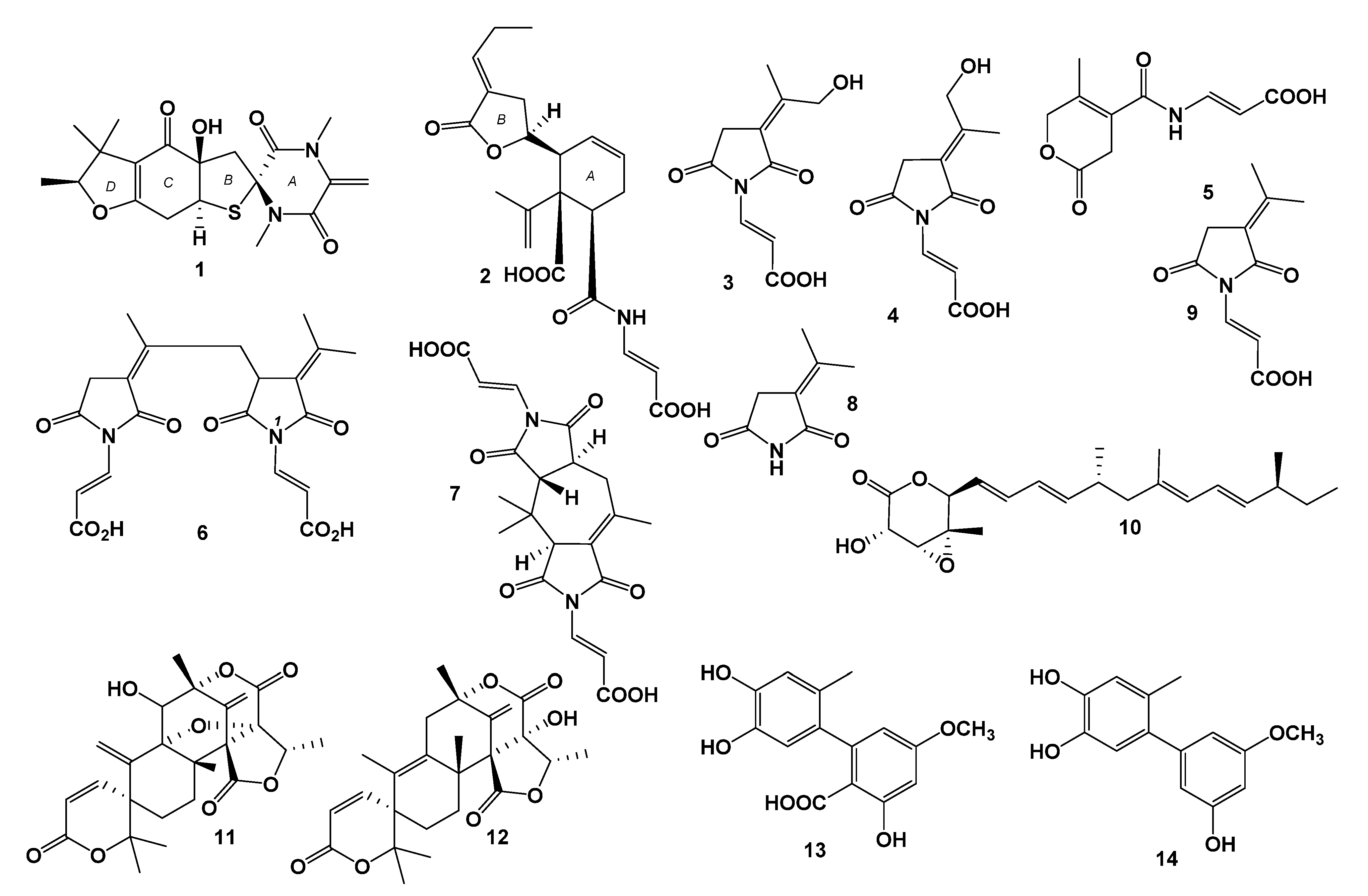
During our ongoing investigations into bioactive natural products from marine-derived microorganisms [10,11,12,13], a fungal strain of Talaromyces mangshanicus BTBU20211089, which was isolated from a sediment sample collected from the South China Sea, was found to be active against Candida albicans. Chemical investigation of this fungus cultured in rice solid media led to the isolation and identification of seven new compounds, namely, talaromanloid A (1), talaromydene (2), 10-hydroxy-8-demethyltalaromydine and 11-hydroxyd-8-emethyltalaromydine (3 and 4), talaromylectone (5), and ditalaromylectones A and B (6 and 7), together with seven known compounds. The known compounds were determined to be 3-(propan-2-ylidene)-pyrrolidine-2, 5-dione (8) [14], (E)-3-(2,5-dioxo-3-(propan-2-ylidene)pyrrolidin-1-yl)acrylic acid (9) [15], nafuredin (10) [16], dehydroaustinol (11) [17], austinolide (12) [18], altenusin (13), and 5′-methoxy-6-methyl-biphenyl-3,4,3′-triol (14) [19] (Figure 1). Here, we report the isolation, structure elucidation, and bioactivities of these compounds.
2. Results
2.1. Structure Elucidation
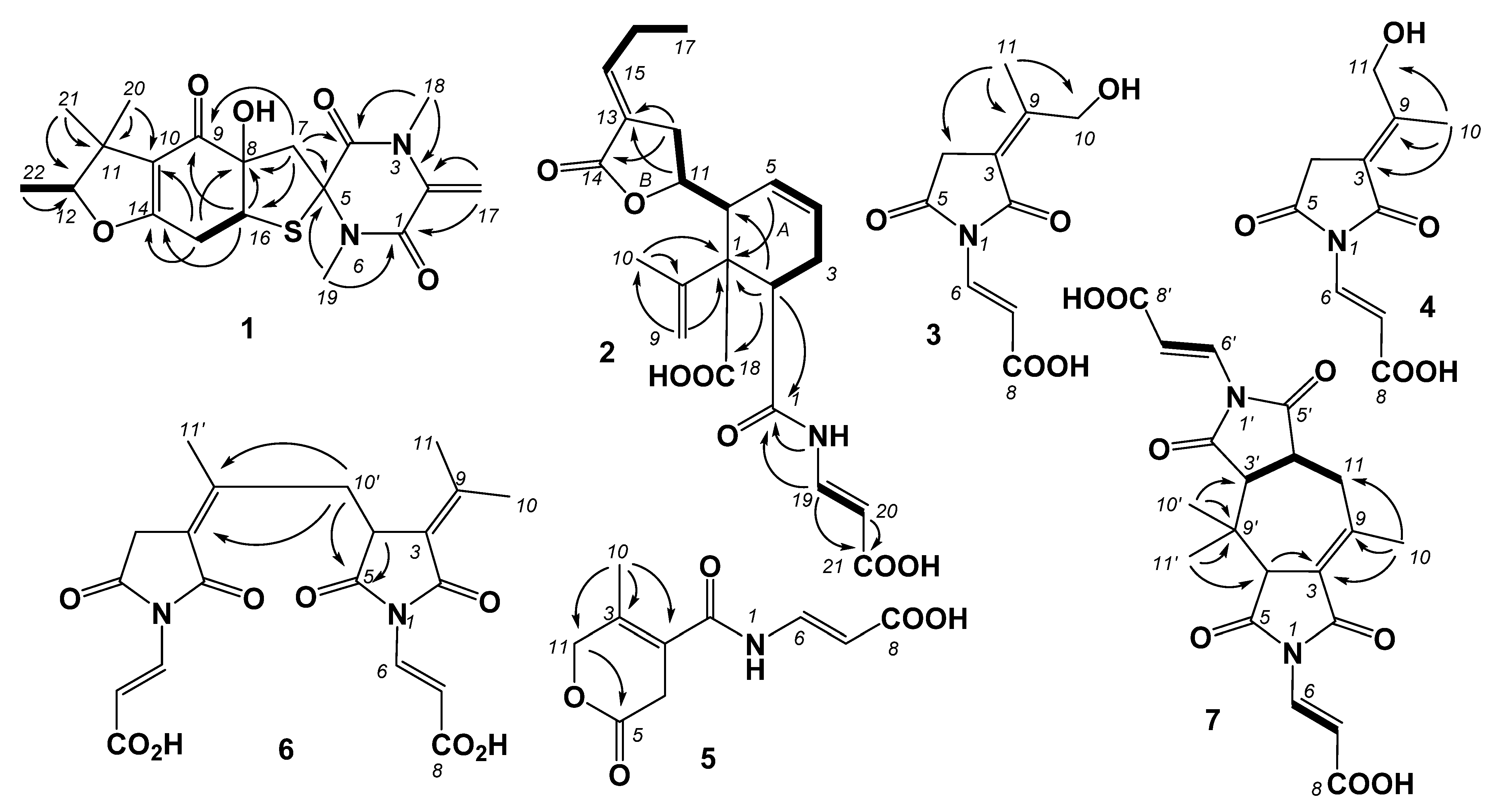
Compound 1 was isolated as a light-yellow powder. The molecular formula of 1 was determined to be C19H24N2O5S based on the HRESIMS spectrum (m/z [M + H]+ 393.1481, calcd for C19H25N2O5S, 393.1479), accounting for nine degrees of unsaturation (Figure S1). The 1H NMR, 13C, and HSQC spectra of 1 (Figures S2–S4, Table 1) demonstrated signals for one terminal double bond (δH 5.57 (1H, H-17a) and 5.08 (1H, H-17b)/ δC 136.0 (C-3) and 102.8 (C-17)), two sp3 methylenes (δH 3.37 (1H, H-7a) and 2.86 (1H, H-7b)/δC 42.6 (C-7), 3.02 (1H, H-15a) and 2.47 (1H, H-15b)/δC 21.6 (C-15)), one sp3 oxygenated methine (δH 4.42 (1H, H-12)/δC 90.7 (C-12)), one sp3 methine (δH 4.18 (1H, H-16)/δC 50.9 (C-16)), four singlet methyl groups (δH 3.19 (3H, H-18)/δC 30.7 (C-18), δH 2.83 (3H, H-19)/δC 25.9 (C-19), δH 1.01 (3H, H-20)/δC 20.2 (C-20), δH 1.29 (3H, H-21)/δC 24.8 (C-21)), one doublet methyl (δH 1.28 (3H, H-22)/δC 13.9 (C-22)), one ketone carbonyl at δC 190.8 (C-9), two amide carbonyls at δC 158.0 (C-1) and 165.7 (C-3), one oxygenated sp2 quaternary carbon at δC 171.1 (C-14), two sp2 quaternary carbons at δC 136.0 (C-2) and 116.9 (C-10), and two sp3 quaternary carbons at δC 72.2 (C-5) and 81.6 (C-8). The downfield shift of oxygenated methine (C-12) in the 13C spectrum was consistent with that of phomalirazine [20]. Detailed analysis of the 2D NMR data (Figures S4–S6) confirmed the structure of 1. The HMBC correlations (Figure 2) between H-17b and C-2, H2-17 and C-1, H3-18 and C-2 and C-4, and H3-19 and C-1 and C-5 revealed the N, N-dimethyldiketopiperazine moiety of ring A. The long-range HMBC correlations between H-15b and C-8, H2-15 and C-10 and C-14, and H-16 and C-8, C-9, and C-14 indicated the presence of the cyclopentenone moiety of ring C. The HMBC crossing peaks from H-7a to C-16, and H2-7 to C-4, C-5, C-8, and C-9 revealed that rings A and C were linked from C-5 to C-8 through C-7. The HMBC correlations between H3-20 and H3-21 and C-10, C-11, and C-12, and H3-22 and C-11 and C-12 indicated that the 1,1-dimethylpropyl was attached to C-10 by C-11. By analyzing the downfield chemical shifts of C-5, C-12, and C-14 and the molecular formula, C-5 and C-16 were linked by a sulfur atom to form the tetrahydrothiophene moiety of ring B, and C-12 and C-14 were linked by an oxygen atom to form the dihydrofuran moiety of ring D. Therefore, the planar structure of 1 was assigned. The absolute configurations of 1 were established as 5S, 8R, 12S, and 16S by comparing the experimental and calculated ECD spectra (Figure 3). Thus, the structure of 1 was determined and named talaromanloid A.
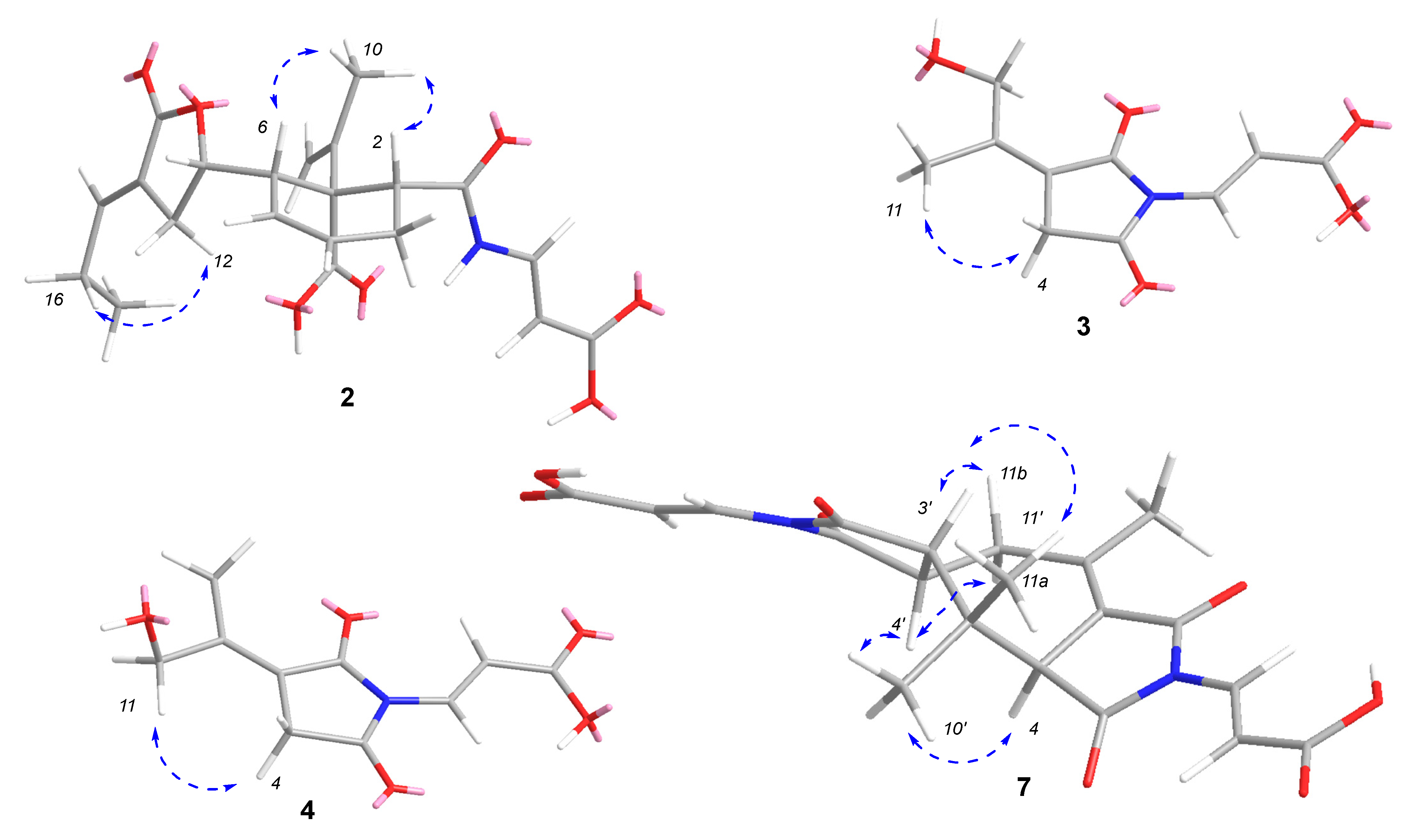
Compound 2 was isolated as a light-yellow powder. The molecular formula of 2 was determined to be C21H25NO7 based on the HRESIMS spectrum (m/z [M + H]+ 404.1701, calcd for C21H26NO7, 404.1704), accounting for ten degrees of unsaturation (Figure S8). The 1H and 13C NMR spectra, along with HSQC data (Figures S9–S11, Table 1), revealed the presence of two methyls (δH 1.84/δC 19.6 (C-10); δH 0.98/δC 13.1 (C-17)), four methylenes (δH 2.34/δC 25.1 (C-3); δH 5.05, 4.68/δC 115.0 (C-9); δH 2.56/δC 27.7 (C-12); δH 2.13/δC 21.9 (C-16)), eight methines (δH 2.90/δC 44.7 (C-2); δH 6.05/δC 129.7 (C-4); δH 5.82/δC 123.1 (C-5); δH 3.20/δC 45.5 (C-6); δH 4.47/δC 80.3 (C-11); δH 6.77/δC 146.7 (C-15); δH 7.70/δC 137.9 (C-19); δH 5.49/δC 101.6 (C-20)), two sp2 quaternary carbons (δC 140.8 (C-8), 127.6 (C-13)), one two sp3 quaternary carbon (δC 57.9 (C-7)), and four carbonyls (δC 169.9 (C-1), 168.5 (C-14), δC 176.5 (C-18), δC 168.5 (C-21)). The 1H-1H COSY correlations (Figure S12) between H-2 and H2-3, H-4, H-5, H-6, H-11, and H2-12 revealed the connections of C-2, C-3, C-4, C-5, C-6, C-11, and C-12. The 1H-1H COSY crossing peaks from H-15 through H2-16 to H3-17 suggested a side chain of C-15/C-16/C-17. The 1H-1H COSY correlations between H-NH and H-19 and H-20, along with the coupling constants (δH 10.26, d (11.0), H-NH, 7.70 (dd, J = 14.0, 11.0 Hz, H-19), 5.49 (d, J = 14.0 Hz, H-20)) demonstrated the trans-double bond attached to the amino of N/C-19/C-20. In the HMBC spectrum (Figure S13), the correlations between H-2 and C-6 and C-7, and H-5 and C-7 suggested the presence of ring A. The HMBC correlations (Figure 2) between H2-9 and C-7 and C-10, and between H3-10 and C-7, C-8, and C-9 indicated the connection between C-8 and C-7. The carbonyls of C-1 and C-18 were confirmed by HMBC correlations between H-2 and C-1 and C-18, and between H-NH and C-1. The acrylic acid moiety attached to NH was characterized by HMBC correlations between H-19 and C-1 and between H-19 and H-20 and C-21. Ring B was revealed by the HMBC correlations between H-11 and C-13, and between H-12 and C-13 and C-14. The HMBC correlations between H2-16 and C-13 and C-15, together with the crossing peak from H-15 to C-14, indicated the connection between C-13 and C-15. The ROESY correlations (Figure 4 and Figure S14) between H3-10 (δH 1.84, s) and H-2 (δH 2.90, t, J = 8.5 Hz) and H-6 (δH 3.20, m) indicated that they were on the same side of ring A. The conformation of the double bond of C-13/C-15 was deduced by the ROESY correlations between H2-12 and H2-16 (Figure 2 and Figure S14). By comparing the experimental and calculated ECD spectra (Figure 3), the absolute configurations of 2 were established as 2R, 6S, 7R, 8S, and 11S. Thus, the structure of 2 was determined and named talaromydene.
Compound 3 was isolated as a colorless powder. The molecular formula of 3 was determined to be C10H11NO5 based on the HRESIMS spectrum (m/z [M + H]+ 226.0716, calcd for C10H12NO5, 226.0710), accounting for six degrees of unsaturation (Figure S15). The 1H and 13C NMR spectra, along with HSQC data (Figures S16–S18, Table 2), revealed the presence of one trans-double bond (δH 7.55 (d, J = 14.5 Hz)/δC 131.3 (C-6); 6.73 (d, J = 14.5 Hz)/δC 109.2 (C-7)), one sp3 methylene (δH 3.40/δC 33.6 (C-4)), one sp3 oxygenated methylene (δH 4.66/δC 60.0 (C-10)), one methyl group (δH 1.91/δC 18.1 (C-11)), two sp2 quaternary carbons (δC 117.8 (C-3); 156.2 (C-9)), and three carbonyls (δC 166.5 (C-2), 172.5 (C-5), 167.8 (C-8)). The NMR data were similar to those of (E)-3-(2,5-dioxo-3-(propan-2-ylidene) pyrrolidin-1-yl)acrylic acid [15], although one of the methyl groups was replaced by a hydroxymethyl in 3. The hydroxyl of C-10 was confirmed by the HMBC correlations (Figure 2 and Figure S19) between H3-11 and C-3, C-9, and C-10, and ROESY correlation (Figure 4 and Figure S20) between H3-11 and H2-4. Thus, the structure of 3 was determined and named 10-hydroxy-8-demethyltalaromydine.
Compound 4 was isolated as a colorless powder. The molecular formula of 4 was determined to be C10H11NO5 based on the HRESIMS spectrum (m/z [M + H]+ 226.0716, calcd for C10H12NO5, 226.0710), accounting for six degrees of unsaturation (Figure S21). The 1H, 13C NMR, and HSQC data (Figures S22–S24, Table 2) shared high similarity with those of 3. The hydroxyl of C-11 was confirmed by the HMBC correlations (Figure 2 and Figure S25) between H3-10 and C-3, C-9, and C-11, and ROESY correlation (Figure 4 and Figure S26) between H2-11 and H2-4. Thus, the structure of 4 was determined and named 11-hydroxy-8-demethyltalaromydine.
Compound 5 was isolated as a colorless powder. The molecular formula of 5 was determined to be C10H11NO5 based on the HRESIMS spectrum (m/z [M + H]+ 226.0715, calcd for C10H12NO5, 226.0710), accounting for six degrees of unsaturation (Figure S27). The 1H, 13C, NMR, and HSQC data (Figures S28–S30, Table 2) shared high similarity with those of 3 and 4. The signal at δH 10.59 (brs, H-NH) in the 1H spectrum and 1H-1H COSY correlation between H-NH and H-6 (δH 7.55 (br d, J = 8.5 Hz)) suggested that the pyrrolidine-2,5-dione in 5 was replaced by a ring-opening moiety. The HMBC correlations (Figure 2 and Figure S32) between H2-11 and C-5 (δC 168.6) revealed that C-5 and C-11 formed a lactone unit. Thus, the structure of 5 was determined and named talaromylectone.
Compound 6 was isolated as a light-yellow powder. The molecular formula of 6 was determined to be C20H20N2O8 based on the HRESIMS spectrum (m/z [M + H]+ 417.1287, calcd for C20H21N2O8, 417.1292), accounting for twelve degrees of unsaturation (Figure S33). The 1H and 13C NMR spectra, along with the HSQC data (Figures S34–S36, Table 3), revealed that 6 is a dimeric analog of 3 and (E)-3-(2,5-dioxo-3-(propan-2-ylidene) pyrrolidin-1-yl)acrylic acid (9). The proton NMR spectrum showed signals for three singlet methyl groups at δH 2.30 (H3-10), 2.03 (H3-11), and 1.95 (H3-11′), one singlet methylene at δH 3.46 (H2-4′), and one coupled methylene at δH 3.31 (dd, J = 13.0, 10.0 Hz, H-10′a) and 3.01 (dd, J = 13.0, 6.5 Hz, H-10′b) with one methine at δH 3.83 (dd, J = 10.0, 6.5 Hz, H-4), which revealed that one of the methyl groups in the monomer was replaced by methylene and attached to the methine of the other monomer. The linkage of C-4 and C-10′ was confirmed by the HMBC correlations (Figure 2 and Figure S37) between H2-10′ and C-3′ (δC 121.2) and C-9′ (δC 150.5), and between H2-10′ and H-4 and C-5 (δC 173.7). Therefore, the structure of 6 was determined and named ditalaromylectone A.
Compound 7 was isolated as a light-yellow powder. The molecular formula of 7 was determined to be C20H20N2O8 based on the HRESIMS spectrum (m/z [M + H]+ 417.1289, calcd for C20H21N2O8, 417.1292), accounting for twelve degrees of unsaturation (Figure S38). The 1H and 13C NMR spectra, along with the HSQC data (Figures S39–S41, Table 3), revealed that 7 is a dimeric analog of 4, which possessed a different skeleton to that of 6. The 1H-1H COSY correlations (Figure S42) between H2-11 and H-4′, and between H-3′ and H-4’, indicated the connection of C-11/C-4′/C-3′. The HMBC correlations (Figure 2 and Figure S43) between H-4 and C-3, H3-10 and C-3, C-9, and C-11, and H3-10′ and H3-11′ and C-4, C-3′, and C-9′ confirmed the presence of the cycloheptene moiety. In the ROESY spectra (Figure S44), the correlations between H-3′ (δH 2.90) and H-11b (δH 2.71)/H3-11′ (δH 1.06), between H-4′ (δH 3.62) and H-11a (δH 3.07)/H3-10’ (δH 1.31), and between H-4 (δH 3.70) and H3-10’ suggested the relative configurations of 7. The optical rotation is near zero, so 7 was assigned as a racemic mixture and named ditalaromylectone B.
Seven known compounds, including 3-(propan-2-ylidene)-pyrrolidine-2, 5-dione (8), (E)-3-(2,5-dioxo-3-(propan-2-ylidene)pyrrolidin-1-yl)acrylic acid (9), nafuredin (10), dehydroaustinol (11), austinolide (12), altenusin (13), and 5′-methoxy-6-methyl-biphenyl-3,4,3′-triol (14), were identified by comparing the NMR data with the corresponding reported data.
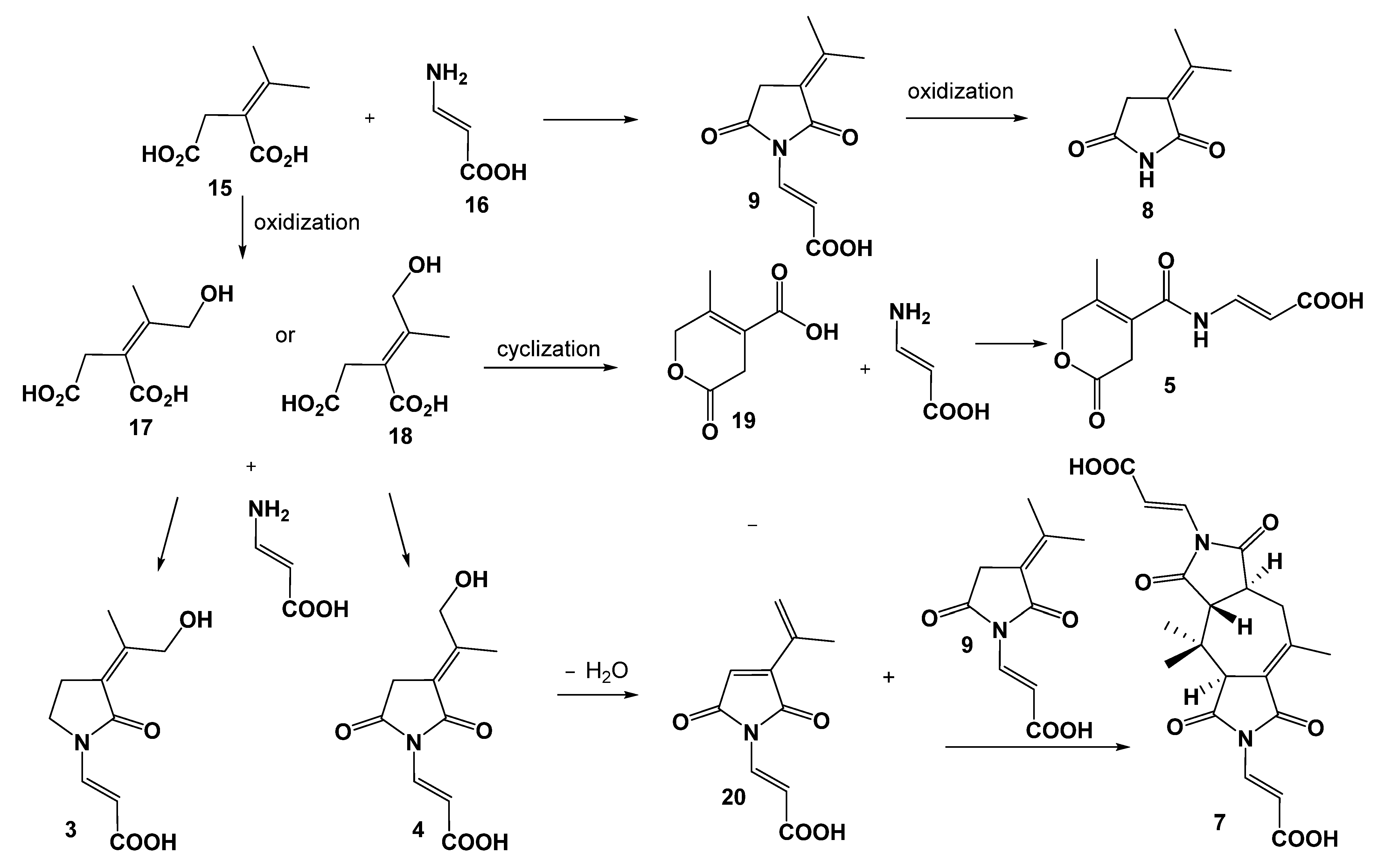
The biosynthesis of 2–9 most likely proceeds via the same precursors, and plausible biosynthetic relationships of 3–5 and 7–9 are presented in Figure 5. Precursors 15 and 16 may be derived from the tricarboxylic acid cycle [21] and then form 9 by an amidation reaction. Compound 8 is produced by the oxidization of 9. Compound 5 is proposed to be generated after the oxidization, cyclization, and amidation of 15 or 16. Compound 7 is proposed to be derived from the cyclization of 9 and dehydration of 4.
2.2. Biological Activity
These compounds were evaluated for their antibacterial activities against Candida albicans ATCC 10231, Staphylococcus aureus ATCC 25923, and Escherichia coli ATCC 25923. Compounds 6 and 13 showed an inhibitory effect against C. albicans with an MIC value of 200 μg/mL. Compounds 13 and 14 exhibited antibacterial activity against S. aureus with MIC values of 50 μg/mL.
3. Materials and Methods
3.1. General Experimental Procedures
Optical rotations ([α) were measured on an Anton Paar MCP 200 Modular Circular Polarimeter (Austria) in a 100 × 2 mm cell. CD spectra were recorded on an Applied Photophysics Chirascan spectropolarimeter (Surrey, UK). NMR spectra were obtained on a Bruker Avance 500 spectrometer with residual solvent peaks as references (DMSO-d6: δH 2.50, δC 39.52). High-resolution ESIMS measurements were obtained on an Accurate-Mass-Q-TOF LC/MS 6520 instrument (Santa Clara, CA, USA) in the positive ion mode. HPLC was performed using an Agilent 1200 Series separation module equipped with an Agilent 1200 Series diode array, Agilent 1260 Series fraction collector, and Agilent ZORBAX SB-C18 column (250 × 9.4 mm, 5 µm).
3.2. Microbial Material, Fermentation, Extraction and Purification
Strain Talaromyces mangshanicus BTBU20211089 was isolated from a mud sample collected from a sediment sample collected from the South China Sea and grown on a potato dextrose agar plate at 28 °C. The genomic DNA of BTBU20211089 was extracted using a Fungi Genomic DNA Extraction Kit (Solarbio Life Sciences, Beijing, China). The ITS region was amplified by using a conventional primer pair of ITS4 (5′ -TCCTCCGCTTATTGATATGC -3′) and ITS5 (5′-GGAAGTAAAAGTCGTAACAAGG -3′). PCR products were sent to Beijing Qingke Biotechnology Co., Ltd. (Beijing, China) for DNA sequencing and deposited in GenBank (accession number, OL905958). BTBU20211089 was identified as Talaromyces mangshanicus by comparing the internal transcribed spacer (ITS) region sequence with the GenBank database using the BLAST program. A neighbor-joining (NJ) tree (Figure S45) was constructed using the software package Mega version 5 [22]. The fungus was assigned the accession number BTBU20211089 in the culture collection at Beijing Technology and Business University, Beijing. The strain BTBU20211089 was inoculated on a potato dextrose agar plate and cultured for 7 days. Then, a slit of agar with the fungus was cut from the plate and inoculated in ten 1 L conical flasks, each containing a solid medium consisting of rice (200 g in 200 mL distilled water), and the flasks were incubated under static conditions at 28 °C for 30 days. The cultures were extracted three times with a mixture of EtOAc:MeOH (80:20), and the combined extracts were evaporated to dryness in vacuo. The residue was suspended in distilled water and partitioned with EtOAc. Then, the EtOAc layer was dried in vacuo to yield a dark residue (14.3 g). The EtOAc fraction was fractionated by vacuum liquid silica gel chromatography (80 × 80 mm column, Silica gel 60 H for thin-layer chromatography) using a stepwise gradient of hexane/CH2Cl2 (4:1, 7:3, 1:1, 1:4, 1:9, 3:97, and 0:100, v/v) and then a stepwise gradient of MeOH/CH2Cl2 (1:99, 2:98, 3:97, 5:95, 5:45, 1:4, and 100:0 v/v) to afford 14 fractions. Fraction G was purified by HPLC (Agilent ZORBAX SB-C18, 250 × 9.4 mm, 5 μm column, 3.0 mL/min, elution with 60% to 100% acetonitrile/H2O in 15 min) to yield 10 (5.8 mg). Fraction I was fractionated on a Sephadex LH-20 column using an isocratic elution of CH2Cl2:MeOH (2:1) to yield seven subfractions (I1–I7), and subfraction I2 was further purified by HPLC (Agilent ZORBAX SB-C18, 250 × 9.4 mm, 5 μm column, 3.0 mL/min, elution with 30% to 55% acetonitrile/H2O (0–20 min), and then to 80% acetonitrile/H2O (20–25 min)) to yield 1 (2.0 mg), 11 (10.4 mg), and 12 (14.3 mg). Subfraction I4 was further fractionated by HPLC (Agilent ZORBAX SB-C18, 250 × 9.4 mm, 5 μm column, 3.0 mL/min, elution with 30% to 100% acetonitrile/H2O) in 15 min to 8 (Rt 5.7 min, 3.6 mg) and 9 (20.5 mg). Fraction K was fractionated on a Sephadex LH-20 column using an isocratic elution of CH2Cl2:MeOH (2:1) to yield eight subfractions (K1–K8). Subfraction K3 was further fractionated by HPLC (Agilent ZORBAX SB-C18, 250 × 9.4 mm, 5 μm column, 3.0 mL/min, with 30% to 55% acetonitrile/H2O) in 15 min to yield 2 (Rt 13.4 min, 3.5 mg). Subfraction K4 was further fractionated by HPLC (Agilent ZORBAX SB-C18, 250 × 9.4 mm, 5 μm column, 3.0 mL/min, elution with 30% to 100% acetonitrile/H2O) in 15 min to yield 6 (1.7 mg). Subfraction K5 was further fractionated by HPLC (Agilent ZORBAX SB-C18, 250 × 9.4 mm, 5 μm column, 3.0 mL/min, elution with 30% acetonitrile/H2O) in 15 min to yield 7 (1.8 mg). Subfraction K6 was further fractionated by HPLC (Agilent ZORBAX SB-C18, 250 × 9.4 mm, 5 μm column, 3.0 mL/min, elution with 10% to 27% acetonitrile/H2O) in 15 min to yield 3 (1.8 mg), 4 (1.2 mg), and 5 (3.3 mg). Subfraction K8 was further fractionated by HPLC (Agilent ZORBAX SB-C18, 250 × 9.4 mm, 5 μm column, 3.0 mL/min, elution with 40% to 50% acetonitrile/H2O) in 15 min to yield 13 (6.8 mg) and 14 (5.6 mg).
- Talaromanloid A (1): Light-yellow powder; [α + 43.0 (c 0.1, MeOH); CD (c 5.0 × 10⁻⁵, CH3OH), λmax(∆ε) 259 nm (+9.14) and 290 nm (−2.20); 1H and 13C NMR data, Table 1; HRESIMS m/z 393.1481 [M + H]+ (calcd for C19H25N2O5S, 393.1479).
- Talaromydene (2): Light-yellow powder; [α −67.0 (c 0.1, MeOH); CD (c 5.0 × 10⁻⁵, CH3OH), λmax(∆ε) 256 nm (−4.88); 1H and 13C NMR data, Table 1; HRESIMS m/z 404.1701 [M + H]+ (calcd for C21H26NO7, 404.1704).
- 10-Hydroxydemethyltalaromydine (3): Colorless powder; 1H and 13C NMR data, Table 2; HRESIMS m/z 226.0716 [M + H]+ (calcd for C10H12NO5, 226.0710).
- 11. -hydroxydemethyltalaromydine (4): Colorless powder; 1H and 13C NMR data, Table 2; HRESIMS m/z 226.0716 [M + H]+ (calcd for C10H12NO5, 226.0710).
- Talaromylectone (5): Colorless powder; 1H and 13C NMR data, Table 2; HRESIMS m/z 226.0715 [M + H]+ (calcd for C10H12NO5, 226.0710).
- Ditalaromylectone A (6): Light-yellow powder; [α +6.0 (c 0.1, MeOH); 1H and 13C NMR data, Table 3; HRESIMS m/z 417.1287 [M + H]+ (calcd for C20H21N2O8, 417.1292).
- Ditalaromylectone B (7): Light-yellow powder; [α 0.0 (c 0.1, MeOH); 1H and 13C NMR data, Table 3; HRESIMS m/z 417.1289 [M + H]+ (calcd for C20H21N2O8, 417.1292).
3.3. ECD Computation Method
Conformation searching was performed using OpenBabel by a genetic algorithm (GA) with the default settings [23]. The conformers were subsequently optimized using the DFT method at the B3LYP/TZVP level with GAUSSIAN 09 [24]. The TDDFT calculations of their low-energy conformations within 0–2.5 kcal/mol were performed at the same level with 40 single excited states. The solvent effect was taken into account by using the polarizable continuum model (PCM).
3.4. Biological Activity
Compounds 1–14 were evaluated for their antimicrobial activities in 96 well plates according to the Antimicrobial Susceptibility Testing Standards outlined by the Clinical and Laboratory Standards Institute document M07-A7 (CLSI) and our previous report [13]. The MIC was defined as the minimum concentration of the compound that prevented visible growth of the microbes.
4. Conclusions
Seven new compounds, talaromanloid A (1), talaromydene (2), 10-hydroxy-8-demethyltalaromydine and 11-hydroxy-8-demethyltalaromydine (3 and 4), talaromylectone (5), and ditalaromylectones A and B (6 and 7), and seven known compounds (8–14) were isolated from the marine-derived fungus Talaromyces mangshanicus BTBU20211089. The structures of the new compounds were elucidated by detailed spectroscopic analysis. The absolute configurations of 1 and 2 were elucidated by comparing experimental and calculated ECD spectra. Compound 6 was a dimeric molecule of 3 and 9 possessing a novel carbon skeleton. Compound 7 possessed a unique novel carbon skeleton structure of a cyclized dimer of 3 and 4. Compounds 6 and 13 showed an inhibitory effect against C. albicans with an MIC value of 200 μg/mL. Compounds 13 and 14 exhibited antibacterial activity against S. aureus with MIC values of 50 μg/mL.
Supplementary Materials
The following supporting information can be downloaded at https://www.mdpi.com/article/10.3390/md20020079/s1: Figures S1–S44: HRESIMS, 1D and 2D NMR for compounds 1–7; Figure S45: Neighbor-joining phylogenetic tree of strain BTBU20211089.
Author Contributions
Data curation, K.Z., X.Z. and H.Y.; Funding acquisition, F.S., X.X. and L.W.; Investigation, K.Z., X.Z., R.L. and H.Y.; Supervision, F.S. and X.X.; Writing—original draft, X.X.; Writing—review and editing, F.S., X.X. and L.W. All authors have read and agreed to the published version of the manuscript.
Funding
This work was funded by grants from the National Key R&D Program of China (2018YFC0311000), the Key Lab of Marine Bioactive Substance and Modern Analytical Technique, SOA (MBSMAT-2019-06), the National Natural Science Foundation of China (81973204), and Research Foundation for Advanced Talents of Beijing Technology and Business University (No. 19008021176).
Institutional Review Board Statement
Not applicable.
Informed Consent Statement
Not applicable.
Data Availability Statement
Not applicable.
Conflicts of Interest
The authors declare no conflict of interest.
References
- Carroll, A.R.; Copp, B.R.; Davis, R.A.; Keyzers, R.A.; Prinsep, M.R. Marine natural products. Nat. Prod. Rep. 2020, 37, 175–223. [Google Scholar] [CrossRef] [PubMed]
- Carroll, A.R.; Copp, B.R.; Davis, R.A.; Keyzers, R.A.; Prinsep, M.R. Marine natural products. Nat. Prod. Rep. 2021, 38, 362–413. [Google Scholar] [CrossRef] [PubMed]
- McNeill, J.B.F.; Buck, W.R.; Demoulin, V.; Greuter, W.; Hawksworth, D.L.H.P.; Knapp, S.; Marhold, K.; Prado, J.; Smith, G.F.; Wiersem, J.H. International Code of Nomenclature for Algae, Fungi, and Plants (Melbourne Code); Regnum Vegetabile 154; Koeltz Scientific Books: Koenigstein, Germany, 2012. [Google Scholar]
- Ma, M.Z.; Yi, W.W.; Qin, L.; Lian, X.Y.; Zhang, Z.Z. Talaromydien a and talaroisocoumarin A, new metabolites from the marine-sourced fungus Talaromyces sp. ZZ1616. Nat. Prod. Res. 2020, 36, 460–465. [Google Scholar] [CrossRef]
- Liang, X.; Huang, Z.H.; Shen, W.B.; Lu, X.H.; Zhang, X.X.; Ma, X.; Qi, S.H. Talaromyoxaones A and B: Unusual oxaphenalenone spirolactones as phosphatase inhibitors from the marine-derived fungus Talaromyces purpureogenus SCSIO 41517. J. Org. Chem. 2021, 86, 12831–12839. [Google Scholar] [CrossRef] [PubMed]
- Huang, Z.H.; Liang, X.; Li, C.J.; Gu, Q.; Ma, X.; Qi, S.H. Talaromynoids A-I, highly oxygenated meroterpenoids from the marine-derived fungus Talaromyces purpureogenus SCSIO 41517 and their lipid accumulation inhibitory activities. J. Nat. Prod. 2021, 84, 2727–2737. [Google Scholar] [CrossRef]
- Wu, B.; Ohlendorf, B.; Oesker, V.; Wiese, J.; Malien, S.; Schmaljohann, R.; Imhoff, J.F. Acetylcholinesterase inhibitors from a marine fungus Talaromyces sp strain LF458. Mar. Biotechnol. 2015, 17, 110–119. [Google Scholar] [CrossRef]
- Lan, D.; Wu, B. Chemistry and bioactivities of secondary metabolites from the genus Talaromyces. Chem. Biodivers 2020, 17, e2000229. [Google Scholar] [CrossRef] [PubMed]
- Nicoletti, R.; Trincone, A. Bioactive compounds produced by strains of Penicillium and Talaromyces of marine origin. Mar. Drugs 2016, 14, 37. [Google Scholar] [CrossRef] [Green Version]
- Xu, X.; Han, J.; Lin, R.; Polyak, S.W.; Song, F. Two new piperazine-triones from a marine-derived Streptomycetes sp. strain SMS636. Mar. Drugs 2019, 17, 186. [Google Scholar] [CrossRef] [Green Version]
- Xu, X.; Han, J.; Wang, Y.; Lin, R.; Yang, H.; Li, J.; Wei, S.; Polyak, S.W.; Song, F. Two New Spiro-Heterocyclic gamma-Lactams from A Marine-Derived Aspergillus fumigatus Strain CUGBMF170049. Mar. Drugs 2019, 17, 289. [Google Scholar] [CrossRef] [Green Version]
- Song, F.; Lin, R.; Yang, N.; Jia, J.; Wei, S.; Han, J.; Li, J.; Bi, H.; Xu, X. Antibacterial secondary metabolites from marine-derived fungus Aspergillus sp. IMCASMF180035. Antibiotics 2021, 10, 377. [Google Scholar] [CrossRef] [PubMed]
- Xu, X.; Li, J.; Zhang, K.; Wei, S.; Lin, R.; Polyak, S.W.; Yang, N.; Song, F. New isocoumarin analogues from the marine-derived fungus Paraphoma sp. CUGBMF180003. Mar. Drugs 2021, 19, 313. [Google Scholar] [CrossRef] [PubMed]
- Li, J.W.; Duan, R.G.; Zou, J.H.; Chen, R.D.; Chen, X.G.; Dai, J.G. Meroterpenoids and isoberkedienolactone from endophytic fungus Penicillium sp. associated with Dysosma versipellis. Acta Pharm. Sin. 2014, 49, 913–920. [Google Scholar]
- Miao, F.; Yang, R.; Chen, D.D.; Wang, Y.; Qin, B.F.; Yang, X.J.; Zhou, L. Isolation, identification and antimicrobial activities of two secondary metabolites of Talaromyces verruculosus. Molecules 2012, 17, 14091–14098. [Google Scholar] [CrossRef] [Green Version]
- Ui, H.; Shiomi, K.; Yamaguchi, Y.; Masuma, R.; Nagamitsu, T.; Takano, D.; Sunazuka, T.; Namikoshi, M.; Omura, S. Nafuredin, a novel inhibitor of NADH-fumarate reductase, produced by Aspergillus niger FT-0554. J. Antibiot. 2001, 54, 234–238. [Google Scholar] [CrossRef] [PubMed] [Green Version]
- Xie, J.; Wu, Y.Y.; Zhang, T.Y.; Zhang, M.Y.; Peng, F.; Lin, B.; Zhang, Y.X. New antimicrobial compounds produced by endophytic Penicillium janthinellum isolated from Panax notoginseng as potential inhibitors of FtsZ. Fitoterapia 2018, 131, 35–43. [Google Scholar] [CrossRef] [PubMed]
- Lo, H.C.; Entwistle, R.; Guo, C.J.; Ahuja, M.; Szewczyk, E.; Hung, J.H.; Chiang, Y.M.; Oakley, B.R.; Wan, C.C. Two separate Gene clusters encode the biosynthetic pathway for the meroterpenoids austinol and dehydroaustinol in Aspergillus nidulans. J. Am. Chem. Soc. 2012, 134, 4709–4720. [Google Scholar] [CrossRef] [PubMed] [Green Version]
- Wang, Q.-X.; Bao, L.; Yang, X.-L.; Guo, H.; Yang, R.-N.; Ren, B.; Zhang, L.-X.; Dai, H.-Q.; Guo, L.-D.; Liu, H.-W. Polyketides with antimicrobial activity from the solid culture of an endolichenic fungus Ulocladium sp. Fitoterapia 2012, 83, 209–214. [Google Scholar] [CrossRef] [PubMed]
- Soledade, M.; Pedras, C.; Abrams, S.R. Phomalirazine, a novel toxin from the phytopathogenic fungus Phoma lingam. J. Am. Chem. Soc. 1989, 1, 1904–1905. [Google Scholar]
- Riko, R.; Nakamura, H.; Shindo, K. Studies on pyranonigrins–isolation of pyranonigrin E and biosynthetic studies on pyranonigrin A. J. Antibiot. 2014, 67, 179–181. [Google Scholar] [CrossRef] [PubMed]
- Tamura, K.; Peterson, D.; Peterson, N.; Stecher, G.; Nei, M.; Kumar, S. MEGA5: Molecular evolutionary genetics analysis us-ing maximum likelihood, evolutionary distance, and maximum parsimony methods. Mol. Biol. Evol. 2011, 28, 2731–2739. [Google Scholar] [CrossRef] [PubMed] [Green Version]
- O’Boyle, N.M.; Banck, M.; James, C.A.; Morley, C.; Vandermeersch, T.; Hutchison, G.R. Open Babel: An open chemical toolbox. J. Cheminform. 2011, 3, 33. [Google Scholar]
- Frisch, M.J.; Trucks, G.W.; Schlegel, H.B.; Scuseria, G.E.; Robb, M.A.; Cheeseman, J.R.; Scalmani, G.; Barone, V.; Mennucci, B.; Petersson, G.A.; et al. Gaussian 09, Revision E.01; Gaussian, Inc.: Wallingford, CT, USA, 2009. [Google Scholar]
Figure 1.
Chemical structures of 1–14.

Figure 2.
Key COSY (bold lines) and HMBC (arrows) correlations in 1–7.

Figure 3.
Calculated and experimental electronic circular dichroism (ECD) spectra of 1 and 2.

Figure 4.
ROESY correlations in 2–4 and 7.

Figure 5.
Plausible biosynthetic relationships of 3–5 and 7–9.


Table 1.
1H (500 MHz) and 13C NMR (125 MHz) data of 1 and 2 (DMSO-d6).
| Position | 1 | 2 | ||
|---|---|---|---|---|
| δC, Type | δH (J in Hz) | δC, Type | δH (J in Hz) | |
| 1 | 158.0, C | 169.9, C | ||
| 2 | 136.0, C | 44.7, CH | 2.90, t (8.5) | |
| 3 | 25.1, CH2 | 2.34, m | ||
| 4 | 165.7, C | 129.7, CH | 6.05, m | |
| 5 | 72.2, C | 123.1, CH | 5.82, dd (10.0, 1.5) | |
| 6 | 45.5, CH | 3.20, m | ||
| 7 | 42.6, CH2 | 3.37, d (14.5) 2.86, d (14.5) | 57.9, C | 5.05, s |
| 8 | 81.6, C | 140.8, C | ||
| 9 | 190.8, C | 115.0, CH2 | 5.05, s 4.68, s | |
| 10 | 116.9, C | 19.6, CH3 | 1.84, s | |
| 11 | 42.7, C | 80.3, CH | 4.47, ddd (10.0, 4.0, 4.0) | |
| 12 | 90.7, CH | 4.42, q (7.0) | 27.7, CH2 | 2.56, m |
| 13 | 127.6, C | |||
| 14 | 171.1, C | 168.5, C | ||
| 15 | 21.6, CH2 | 3.02, dd (19.5, 6.0) 2.47, overlap | 146.7, CH | 6.77, t (7.5) |
| 16 | 50.9 | 4.18, dd (6.0, 1.0) | 21.9, CH2 | 2.13, dq (7.5, 7.5) |
| 17 | 102.8, CH2 | 5.57, d (1.0) 5.08, d (1.0) | 13.1, CH3 | 0.98, t (7.5) |
| 18 | 30.7, CH3 | 3.19, s | 176.5, C | |
| 19 | 29.5, CH3 | 2.83, s | 137.9, CH | 7.70, dd (14.0, 11.0) |
| 20 | 20.2, CH3 | 1.01, s | 101.6, CH | 5.49, d (14.0) |
| 21 | 24.8, CH3 | 1.29, s | 168.5, C | |
| 22 | 13.9, CH3 | 1.28, s (5.5) | ||
| NH | 10.26, d (11.0) | |||
Table 2.
1H (500 MHz) and 13C NMR (125 MHz) data of 3–5 (DMSO-d6).
| Position | 3 | 4 | 5 | |||
|---|---|---|---|---|---|---|
| δC, Type | δH, (J in Hz) | δC, Type | δH, (J in Hz) | δC, Type | δH, (J in Hz) | |
| 2 | 166.5, C | 167.4, C | 164.7, C | |||
| 3 | 117.8, C | 117.2, C | 121.9, C | |||
| 4 | 33.6, CH2 | 3.40, br s | 32.9, CH2 | 3.41, br d, 1.0 | 30.7, CH2 | 3.29, br q (2.0) |
| 5 | 172.5, C | 172.5, C | 168.6, C | |||
| 6 | 131.3, CH | 7.55, d (14.5) | 131.3, CH | 7.57, d (15.0) | 137.8, CH | 7.81, br d (8.5) |
| 7 | 109.2, CH | 6.73, d (14.5) | 109.0, CH | 6.74, d (15.0) | 102.3, CH | 5.56, d (14.0) |
| 8 | 167.8, C | 167.8, C | 168.2, C | |||
| 9 | 156.2, C | 154.3, C | 136.6, C | |||
| 10 | 60.0, CH2 | 4.66, s | 15.5, CH3 | 2.27, s | 16.0, CH3 | 1.88, s |
| 11 | 18.1, CH3 | 1.91, s | 63.4, CH2 | 4.05, s | 71.2, CH2 | 4.83, br s |
| NH | 10.59, br s | |||||
Table 3.
1H (500 MHz) and 13C NMR (125 MHz) data of 6 and 7 (DMSO-d6).
| Position | 6 | 7 | ||
|---|---|---|---|---|
| δC, Type | δH, mult (J in Hz) | δC, Type | δH, mult (J in Hz) | |
| 2 | 166.8, C | 166.5, C | ||
| 3 | 122.1, C | 119.0, C | ||
| 4 | 42.1, CH | 3.83, dd (10.0, 6.5) | 49.9, CH | 3.70, s |
| 5 | 173.7, C | 173.3, C | ||
| 6 | 131.0, CH | 7.53, d (15.0) | 130.7, CH | 7.57, d (15.0) |
| 7 | 109.6, CH | 6.70, d (15.0) | 110.0, CH | 6.77, d (15.0) |
| 8 | 167.7, C | 167.5, C | ||
| 9 | 153.7, C | 155.6, C | ||
| 10 | 21.1, CH3 | 2.30, s | 21.2, CH3 | 2.31, s |
| 11 | 23.8, CH3 | 2.03, s | 37.8, CH2 | 3.07, dd (19.5, 7.5) 2.71, dd (19.5, 11.0) |
| 2′ | 166.3, C | 174.5, C | ||
| 3′ | 121.2, C | 52.0, CH | 2.90, d (8.5) | |
| 4′ | 33.8, CH2 | 3.46, s | 36.7, CH | 3.62, m |
| 5′ | 172.0, C | 175.9, C | ||
| 6′ | 131.0, CH | 7.51, d (15.0) | 130.7, CH | 7.46, d (15.0) |
| 7′ | 109.6, CH | 6.61, d (15.0) | 109.8, CH | 6.70, d (15.0) |
| 8′ | 166.7, C | 167.5, C | ||
| 9′ | 150.5, C | 39.1, C | ||
| 10′ | 35.4, CH2 | 3.31, dd (13.0, 10.0) 3.01, dd (13.0, 6.5) | 20.4, CH3 | 1.31, s |
| 11′ | 22.5, CH3 | 1.95, s | 22.1, CH3 | 1.06, s |
Publisher’s Note: MDPI stays neutral with regard to jurisdictional claims in published maps and institutional affiliations. |
© 2022 by the authors. Licensee MDPI, Basel, Switzerland. This article is an open access article distributed under the terms and conditions of the Creative Commons Attribution (CC BY) license (https://creativecommons.org/licenses/by/4.0/).
Share and Cite
MDPI and ACS Style
Zhang, K.; Zhang, X.; Lin, R.; Yang, H.; Song, F.; Xu, X.; Wang, L. New Secondary Metabolites from the Marine-Derived Fungus Talaromyces mangshanicus BTBU20211089. Mar. Drugs 2022, 20, 79. https://doi.org/10.3390/md20020079
AMA Style
Zhang K, Zhang X, Lin R, Yang H, Song F, Xu X, Wang L. New Secondary Metabolites from the Marine-Derived Fungus Talaromyces mangshanicus BTBU20211089. Marine Drugs. 2022; 20(2):79. https://doi.org/10.3390/md20020079
Chicago/Turabian StyleZhang, Kai, Xinwan Zhang, Rui Lin, Haijin Yang, Fuhang Song, Xiuli Xu, and Long Wang. 2022. "New Secondary Metabolites from the Marine-Derived Fungus Talaromyces mangshanicus BTBU20211089" Marine Drugs 20, no. 2: 79. https://doi.org/10.3390/md20020079
Note that from the first issue of 2016, this journal uses article numbers instead of page numbers. See further details here.