
Application of Direct Current Atmospheric Pressure Glow Microdischarge Generated in Contact with a Flowing Liquid Solution for Synthesis of Au-Ag Core-Shell Nanoparticles


Abstract
:1. Introduction
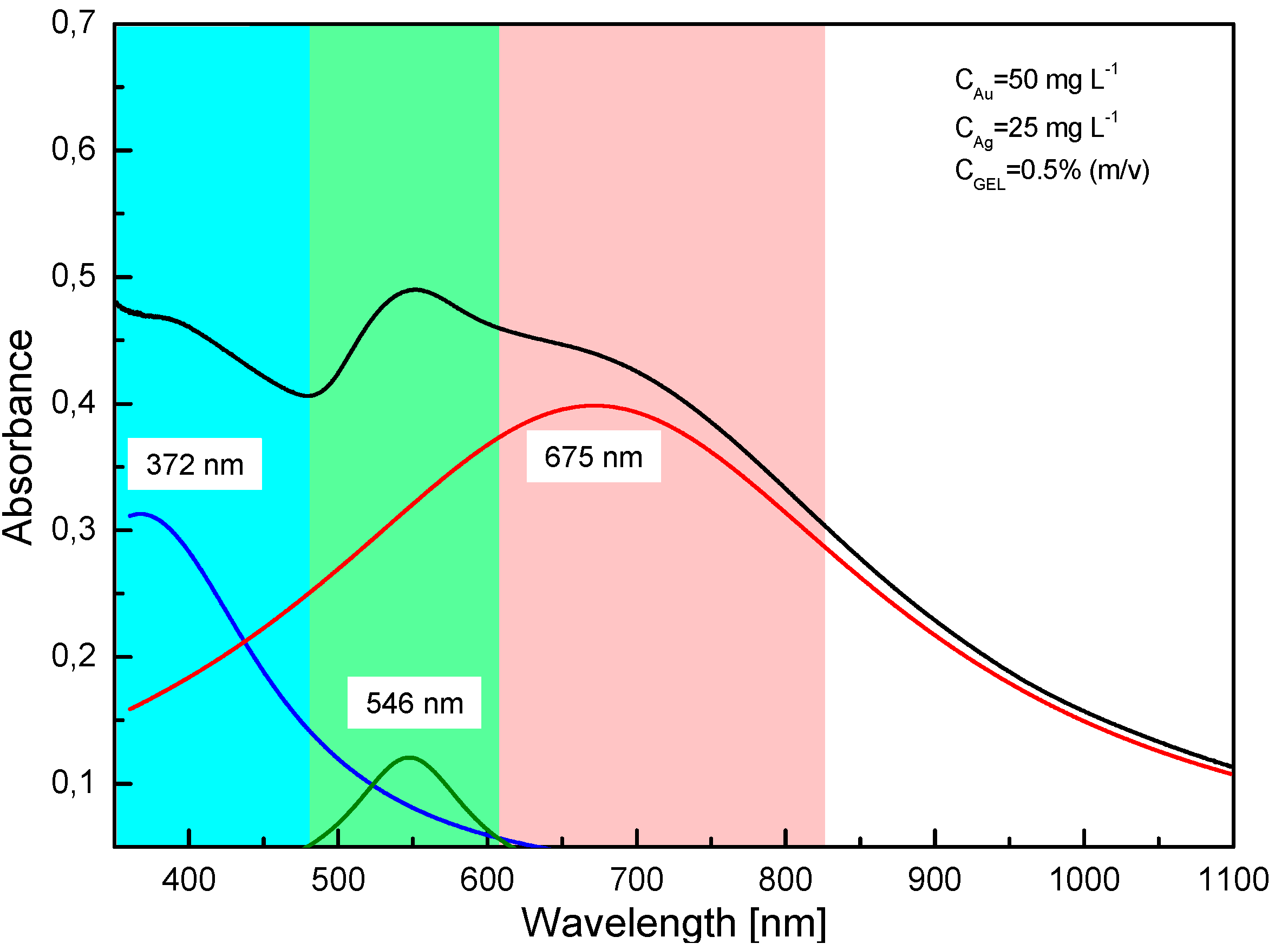
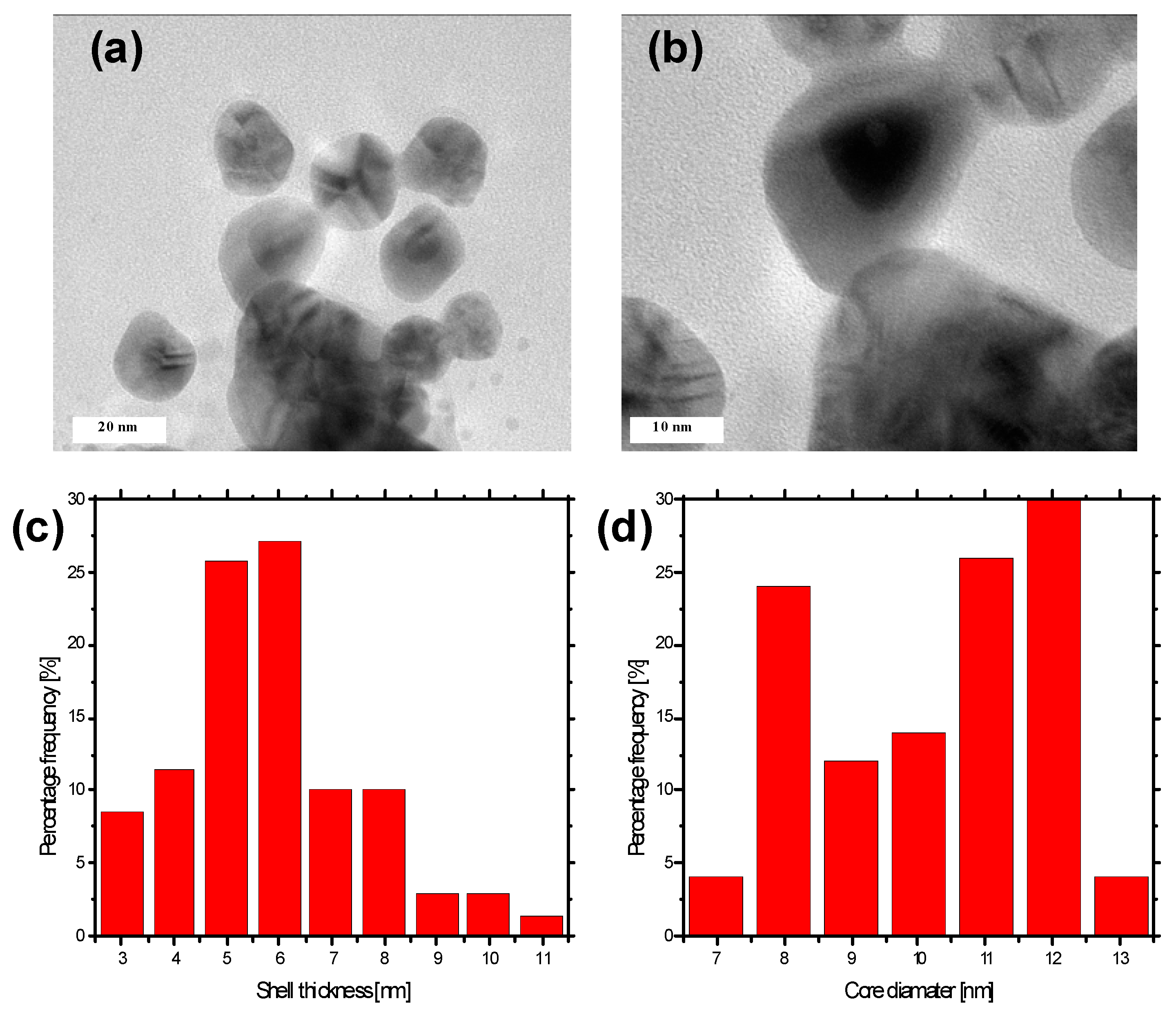
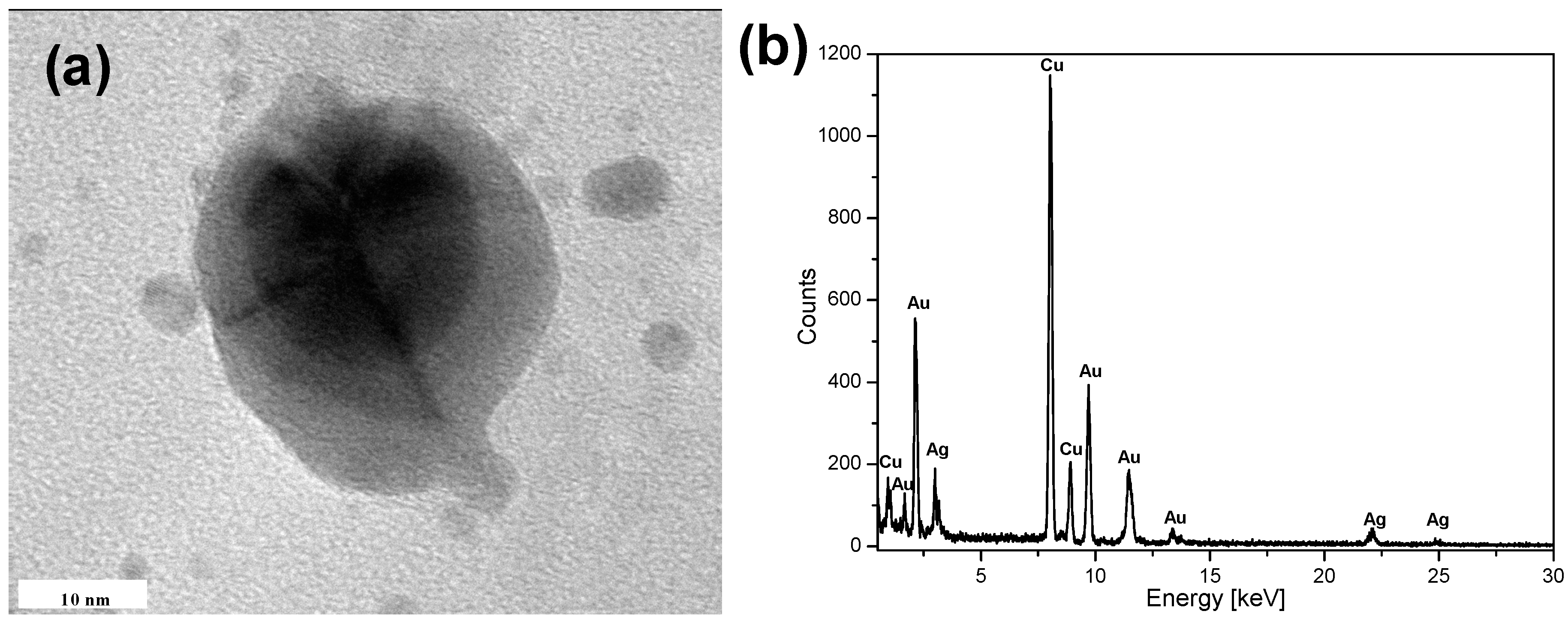
2. Results
3. Discussion
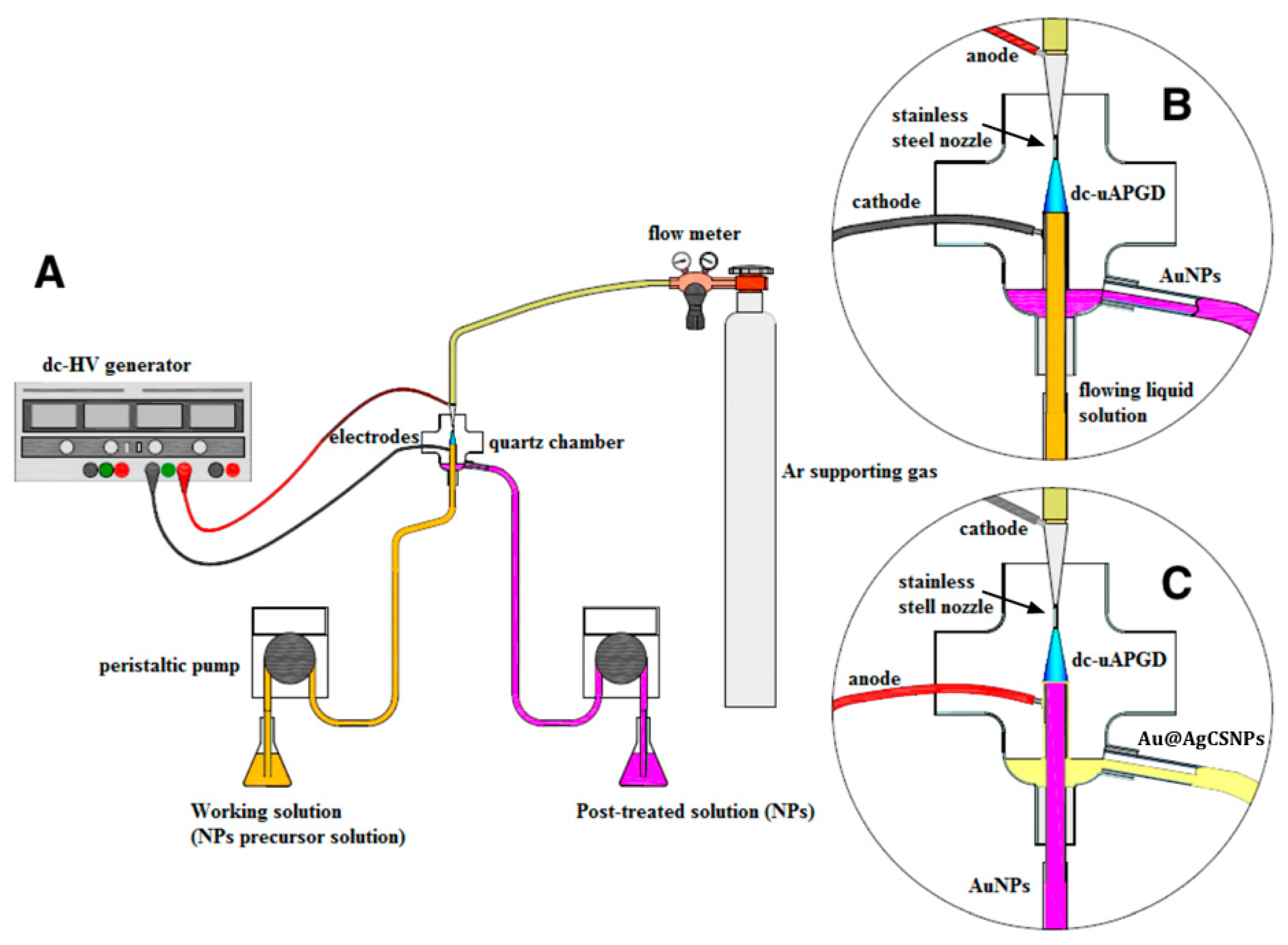
4. Materials and Methods
4.1. Reagents and Solutions
4.2. Synthesis of the AuNPs Core
4.3. Synthesis of the AgNPs Nanoshell on the AuNPs Core
4.4. Characterization of the Au@AgCSNPs
Acknowledgments
Author Contributions
Conflicts of Interest
Abbreviations
| Au@AgCSNPs | Au-Ag core-shell nanoparticles |
| AuNPs | gold nanoparticles |
| UV-Vis | ultraviolet-visible |
| TEM | transmission electron microscopy |
| EDS | energy-dispersive X-ray spectroscopy |
| SERS | surface-enhance Raman scattering |
| APPs | atmospheric pressure plasmas |
| ROS | reactive oxygen species |
| RNS | reactive nitrogen species |
| DoE | design of Experiments |
| LSPR | localized surface plasmon resonance |
| FLC | flowing liquid cathode |
| FLA | flowing liquid anode |
References
- Sivaraman, S.K.; Kumar, S.; Santhanam, V. Monodisperse sub-10 nm gold nanoparticles by reversing the order of addition in Turkevich method—The role of chloroauric acid. J. Colloid Interface Sci. 2011, 361, 543–547. [Google Scholar] [CrossRef] [PubMed]
- Lafon, A.; Treguer-Delapierre, M.; Bazin, D.; Faure, C. Deposit of UV- or γ-synthesized gold nanoparticles on TiO2 powder using lipid-based multilamellar vesicles. Colloid Polym. Sci. 2012, 290, 1015–1022. [Google Scholar] [CrossRef]
- Okitsu, K.; Mizukoshi, Y.; Yamamoto, T.A.; Maeda, Y.; Nagata, Y. Sonochemical synthesis of gold nanoparticles on chitosan. Mater. Lett. 2007, 61, 3429–3431. [Google Scholar] [CrossRef]
- Gupta, S.; Huda, S.; Kilpatrick, P.K.; Velev, O.D. Characterization and optimization of gold nanoparticle-based silver-enhanced immunoassays. Anal. Chem. 2007, 79, 3810–3820. [Google Scholar] [CrossRef] [PubMed]
- Jain, P.K.; Huang, X.; El-Sayed, I.H.; El-Sayed, M.A. Noble metals on the nanoscale: Optical and photothermal properties and some applications in imaging, sensing, biology, and medicine. Acc. Chem. Res. 2008, 41, 1578–1586. [Google Scholar] [CrossRef] [PubMed]
- West, J.L.; Halas, N. Applications of nanotechnology to biotechnology: Commentary. Curr. Opin. Biotechnol. 2000, 11, 215–217. [Google Scholar] [CrossRef]
- Rosi, N.L.; Mirkin, C.A. Nanostructures in biodiagnostics. Chem. Rev. 2005, 105, 1547–1562. [Google Scholar] [CrossRef] [PubMed]
- Saha, K.; Bajaj, A.; Duncan, B.; Rotello, V.M. Beauty is skin deep: A surface monolayer perspective on nanoparticle interactions with cells and bio-macromolecules. Small 2011, 7, 1903–1918. [Google Scholar] [CrossRef] [PubMed]
- Zeng, H.; Sun, S.; Li, J.; Liu, J.P.; Wang, Z.L. Tailoring magnetic properties of core/shell nanoparticles. Appl. Phys. Lett. 2004, 85, 792–794. [Google Scholar] [CrossRef]
- Park, H.Y.; Schadt, M.J.; Wang, L.; Lim, I.I.S.; Njoki, P.N.; Kim, S.H.; Zhong, C.J. Fabrication of magnetic core@ shell Fe oxide@Au nanoparticles for interfacial bioactivity and bio-separation. Langmuir 2007, 23, 9050–9056. [Google Scholar] [CrossRef] [PubMed]
- Rodríguez-González, B.; Burrows, A.; Watanabe, M.; Kiely, C.J.; Marzán, L.M.L. Multishell bimetallic AuAg nanoparticles: Synthesis, structure and optical properties. J. Mater. Chem. 2005, 15, 1755–1759. [Google Scholar] [CrossRef]
- Zhong, C.J.; Maye, M.M. Core-shell assembled nanoparticles as catalysts. Adv. Mater. 2001, 13, 1507–1511. [Google Scholar] [CrossRef]
- Xia, B.; He, F.; Li, L. Preparation of bimetallic nanoparticles using a facile green synthesis method and their application. Langmuir 2013, 29, 4901–4907. [Google Scholar] [CrossRef] [PubMed]
- Jiang, H.L.; Akita, T.; Ishida, T.; Haruta, M.; Xu, Q. Synergistic catalystis of Au@Ag core-shell nanoparticles stabilized on metal-organic framework. J. Am. Chem. Soc. 2011, 133, 1304–1306. [Google Scholar] [CrossRef] [PubMed]
- Valerini, D.; Creti, A.; Lomascolo, M.; Manna, L.; Cingolani, R.; Anni, M. Temperature dependence of the photoluminescence properties of colloidal CdSe/ZnS core/shell quantum dots embedded in a polystyrene matrix. Phys. Rev. B 2005, 71, 235409. [Google Scholar] [CrossRef]
- Malola, S.; Hakkinen, H. Electronic structure and bonding of icosahedral core-shell gold-silver nanoalloy clusters Au144–xAgx(SR)60. J. Phys. Chem. Lett. 2011, 2, 2316–2321. [Google Scholar] [CrossRef]
- Ghosh Chaudhuri, R.; Paria, S. Core/shell nanoparticles: Classes, properties, synthesis mechanisms, characterization, and applications. Chem. Rev. 2011, 112, 2373–2433. [Google Scholar] [CrossRef] [PubMed]
- Güzel, R.; Üstündağ, Z.; Ekşi, H.; Keskin, S.; Taner, B.; Durgun, Z.G.; Turan, A.A.I.; Solak, A.O. Effect of Au and Au@Ag core-shell nanoparticles on the SERS of bridging organic molecules. J. Colloid Interface Sci. 2010, 351, 35–42. [Google Scholar] [CrossRef] [PubMed]
- Feng, L.L.; Gao, G.; Huang, P.; Wang, K.; Wang, X.S.; Luo, T.; Zhang, C.L. Optical properties and catalytic activity of bimetallic gold-silver nanoparticles. Nano Biomed. Eng. 2010, 2, 258–267. [Google Scholar] [CrossRef]
- Wang, A.Q.; Chang, C.M.; Mou, C.Y. Evolution of catalytic activity of Au-Ag bimetallic nanoparticles on mesoporous support for CO oxidation. J. Phys. Chem. B 2005, 109, 18860–18867. [Google Scholar] [CrossRef] [PubMed]
- Lee, J.; Lee, Y.; Youn, J.K.; Na, H.B.; Yu, T.; Kim, H.; Chang, H.N. Simple synthesis of functionalized superparamagnetic magnetite/silica core/shell nanoparticles and their application as magnetically separable high-performance biocatalysts. Small 2008, 4, 143–152. [Google Scholar] [CrossRef] [PubMed]
- Samal, A.K.; Polavarapu, L.; Rodal-Cedeira, S.; Liz-Marzán, L.M.; Pérez-Juste, J.; Pastoriza-Santos, I. Size tunable Au@Ag core-shell nanoparticles: Synthesis and surface-enhanced Raman scattering properties. Langmuir 2013, 29, 15076–15082. [Google Scholar] [CrossRef] [PubMed]
- Liu, M.; Guyot-Sionnest, P. Synthesis and optical characterization of Au/Ag core/shell nanorods. J. Phys. Chem. B 2004, 108, 5882–5888. [Google Scholar] [CrossRef]
- Sinha, T.; Ahmaruzzaman, M. High-value utilization of egg shell to synthesize silver and gold-silver core shell nanoparticles and their application for the degradation of hazardous dyes from aqueous phase—A green approach. J. Colloid Interface Sci. 2015, 453, 115–131. [Google Scholar] [CrossRef] [PubMed]
- Anandan, S.; Grieser, F.; Ashokkumar, M. Sonochemical synthesis of Au-Ag core-shell bimetallic nanoparticles. J. Phys. Chem. C 2008, 112, 15102–15105. [Google Scholar] [CrossRef]
- Peng, Z.Q.; Spliethoff, B.; Tesche, B.; Walther, T.; Kleinermanns, K. Laser-assisted synthesis of Au-Ag alloy nanoparticles insolution. J. Phys. Chem. B 2006, 110, 2549–2554. [Google Scholar] [CrossRef] [PubMed]
- Furusho, H.; Kitano, K.; Hamaguchi, S.; Nagasaki, Y. Preparation of stable water-dispersible PEGylated gold nanoparticles assisted by nonequilibrium atmospheric-pressure plasma jets. Chem. Mater. 2009, 21, 3526–3535. [Google Scholar] [CrossRef]
- Zhu, Y.J.; Schnieders, A.; Alexander, J.D.; Beebe, T.P. Pit-templated synthesis and oxygen adsorption properties of gold nanostructures on highly oriented pyrolytic graphite. Langmuir 2002, 18, 5728–5733. [Google Scholar] [CrossRef]
- Richmonds, C.; Sankaran, R.M. Plasma-liquid electrochemistry: Rapid synthesis of colloidal metal nanoparticles by microplasma reduction of aqueous cations. Appl. Phys. Lett. 2008, 93, 131501. [Google Scholar] [CrossRef]
- Koo, I.G.; Lee, M.S.; Shim, J.H.; Ahn, J.H.; Lee, W.M. Platinum nanoparticles prepared by a plasma-chemical reduction method. J. Mater. Chem. 2005, 15, 4125–4128. [Google Scholar] [CrossRef]
- Sankaran, R.M.; Holunga, D.; Flagan, R.C.; Giapis, K.P. Synthesis of blue luminescent Si nanoparticles using atmospheric-pressure microdischarges. Nano Lett. 2005, 5, 537–541. [Google Scholar] [CrossRef] [PubMed]
- Shirai, N.; Uchida, S.; Tochikubo, F. Synthesis of metal nanoparticles by dual plasma electrolysis using atmospheric pressure glow discharge in contact with liquid. Jpn. J. Appl. Phys. 2014, 53, 046202. [Google Scholar] [CrossRef]
- Yan, T.; Zhong, X.; Rider, A.E.; Lu, Y.; Furman, S.A.; Ostrikov, K.K. Microplasma-chemical synthesis and tunable real-time plasmonic responses of alloyed AuxAg1−x nanoparticles. Chem. Commun. 2014, 50, 3144–3147. [Google Scholar] [CrossRef] [PubMed]
- Dzimitrowicz, A.; Jamroz, P.; Greda, K.; Nowak, P.; Nyk, M.; Pohl, P. The influence of stabilizers on the production of gold nanoparticles by direct current atmospheric pressure glow microdischarge generated in contact with liquid flowing cathode. J. Nanopart. Res. 2015, 17, 185. [Google Scholar] [CrossRef] [PubMed]
- Dzimitrowicz, A.; Lesniewicz, T.; Greda, K.; Jamroz, P.; Nyk, M.; Pohl, P. Production of gold nanoparticles using atmospheric pressure glow microdischarge generated in contact with a flowing liquid cathode—A design of experiments study. RSC Adv. 2015, 110, 90534–90541. [Google Scholar] [CrossRef]
- Jamroz, P.; Greda, K.; Pohl, P.; Zyrnicki, W. Atmospheric pressure glow discharges generated in contact with flowing liquid cathode: Production of active species and application in wastewater purification processes. Plasma Chem. Plasma Process. 2014, 34, 25–37. [Google Scholar] [CrossRef]
- Billaud, P.; Huntzinger, J.R.; Cottancin, E.; Lermé, J.; Pellarin, M.L.; Arnaud, L.; Vallée, F. Optical extinction spectroscopy of single silver nanoparticles. Eur. Phys. J. D 2007, 43, 271–274. [Google Scholar] [CrossRef]
- Pal, N.B.; Kryschi, C. A facile one-pot synthesis of blue and red luminescent thiol stabilized gold nanoclusters: A through optical and microscopy study. Phys. Chem. Chem. Phys. 2015, 17, 21423–21431. [Google Scholar] [CrossRef] [PubMed]
- Yang, Y.; Shi, J.; Kawamura, G.; Nogami, M. Preparation of Au-Ag, Ag-Au core-shell bimetallic nanoparticles for surface-enhanced Raman scattering. Scr. Mater. 2008, 58, 862–865. [Google Scholar] [CrossRef]
- Toma, H.E.; Zamarion, V.M.; Toma, S.H.; Araki, J. The coordination chemistry at gold nanoparticles. J. Braz. Chem. Soc. 2010, 21, 1158–1176. [Google Scholar] [CrossRef]
- Shankar, S.S.; Rai, A.; Ahmad, A.; Sastry, M. Rapid synthesis of Au, Ag, and bimetallic Au core-Ag shell nanoparticles using Neem (Azadirachtaindica) leaf broth. J. Colloid Interface Sci. 2004, 275, 496–502. [Google Scholar] [CrossRef] [PubMed]
- Neupane, M.P.; Lee, S.J.; Park, I.S.; Lee, M.H.; Bae, T.S.; Kuboki, Y.; Watari, F. Synthesis of gelatin-capped gold nanoparticles with variable gelatin concentration. J. Nanopart. Res. 2011, 13, 491–498. [Google Scholar] [CrossRef]


© 2016 by the authors; licensee MDPI, Basel, Switzerland. This article is an open access article distributed under the terms and conditions of the Creative Commons by Attribution (CC-BY) license (http://creativecommons.org/licenses/by/4.0/).
Share and Cite
Dzimitrowicz, A.; Jamroz, P.; Nyk, M.; Pohl, P. Application of Direct Current Atmospheric Pressure Glow Microdischarge Generated in Contact with a Flowing Liquid Solution for Synthesis of Au-Ag Core-Shell Nanoparticles. Materials 2016, 9, 268. https://doi.org/10.3390/ma9040268
Dzimitrowicz A, Jamroz P, Nyk M, Pohl P. Application of Direct Current Atmospheric Pressure Glow Microdischarge Generated in Contact with a Flowing Liquid Solution for Synthesis of Au-Ag Core-Shell Nanoparticles. Materials. 2016; 9(4):268. https://doi.org/10.3390/ma9040268
Chicago/Turabian StyleDzimitrowicz, Anna, Piotr Jamroz, Marcin Nyk, and Pawel Pohl. 2016. "Application of Direct Current Atmospheric Pressure Glow Microdischarge Generated in Contact with a Flowing Liquid Solution for Synthesis of Au-Ag Core-Shell Nanoparticles" Materials 9, no. 4: 268. https://doi.org/10.3390/ma9040268