
Logogenic Primary Progressive Aphasia or Alzheimer Disease: Contribution of Acoustic Markers in Early Differential Diagnosis
 ,
,
Abstract
:1. Introduction
2. Materials and Methods
2.1. Ethics
2.2. Population
2.3. Protocol Procedure
2.4. Material
2.5. Acoustic Analyses
2.6. Statistical Analyses
3. Results
3.1. Demographic and Clinical Information
3.1.1. Demographic and Psychometric Information
3.1.2. Neuroimaging and CSF Information
3.2. The SST Record Associated with Temporal and Prosodic Markers, an Efficient Task for Diagnosis
3.3. Acoustic Markers of Interest for AD and lvPPA Differential Diagnosis
3.4. Acoustic and Temporal Characteristics according to Biomarkers
3.4.1. Acoustic and Temporal Differences in lvPPA Group Sub-Types According to CSF Biomarkers Variations
3.4.2. Comparison of Acoustic and Temporal Features, Diagnostic Groups, and Their Biomarker Profiles
4. Discussion
5. Conclusions
Author Contributions
Funding
Institutional Review Board Statement
Informed Consent Statement
Data Availability Statement
Acknowledgments
Conflicts of Interest
Appendix A
Protocol Procedure

| Item | Span (Total Words) | Stimulus |
|---|---|---|
| 1 | 3 (5) | La fille mange un gâteau. “The girl eats a cake” |
| 2 | 3 (5) | Un étudiant fait ses devoirs. “A student does his homework”. |
| 3 | 4 (6) | L’enseignantpart et ferme la porte. “The teacher leaves and closes the door”. |
| 4 | 4 (7) | Le chat a chassé une PET-scanite souris. “The cat chased a little mouse”. |
| 5 | 5 (8) | Les oiseaux sont nichés sur un grand arbre vert. “The birds are nestled on a tall green tree”. |
| 6 | 5 (9) | La mère du garçon a acheté des amandes au marché. “The boy’s mother bought almonds at the market”. |
| 7 | 6 (11) | Une jeune fille a tenté un plongeon pendant ses vacances en juillet. “A young girl attempted a dive while on vacation in July”. |
| 8 | 6 (12) | Le vieil homme est en retard alors il veut prendre un taxi. “The old man is late so he wants to take a taxi” |
| 9 | 7 (13) | L’évierde la cuisine est très bouché mais le réparateur utilise une pompe. “The kitchen sink is very clogged but the repairman uses a pump”. |
| 10 | 7 (14) | Au restaurant la femme du professeur a mangé des nouilles asiatiques pour le déjeuner. “At the restaurant the teacher’s wife ate Asian noodles for lunch”. |
| 11 | 8 (16) | Le lapin s’est échappé très vite dans la forêt et le chasseur n’a pas réussi à l’attraper. “The rabbit escaped very quickly into the hood and the huntman failed to catch him”. |
| 12 | 8 (16) | Les touristes ont découvert les immenses sculptures de bronze du musée grâce à la visite guidée. “Tourists discovered the museum’s huge bronze sculptures through the guided tour”. |
| 13 | 9 (17) | Les chercheurs en archéologie ont découvert une grande tombe romaine dont des squelettes et des outils tranchants. “Archaeological researchers have discovered a large Roman tomb including skeletons and sharp tools”. |
| 14 | 9 (18) | Les océans deviendront nos ennemis si rien n’est fait pour freiner le réchauffement dû aux émissions de gaz. “The oceans will become our enemies if nothing is done to curb the warming caused by gas emissions”. |
References
- Routhier, S.; Gravel-Laflamme, K.; Macoir, J. Non-Pharmacological Therapies for Language Deficits in the Agrammatic and Logopenic Variants of Primary Progressive Aphasia: A Literature Review. Gériatrie Psychol. Neuropsychiatr. Vieil. 2013, 11, 87–97. [Google Scholar] [CrossRef] [PubMed]
- Harris, J.M.; Jones, M. Pathology in Primary Progressive Aphasia Syndromes. Curr. Neurol. Neurosci. Rep. 2014, 14, 466. [Google Scholar] [CrossRef] [PubMed]
- Matias-Guiu, J.A.; Díaz-Álvarez, J.; Cuetos, F.; Cabrera-Martín, M.N.; Segovia-Ríos, I.; Pytel, V.; Moreno-Ramos, T.; Carreras, J.L.; Matías-Guiu, J.; Ayala, J.L. Machine Learning in the Clinical and Language Characterisation of Primary Progressive Aphasia Variants. Cortex 2019, 119, 312–323. [Google Scholar] [CrossRef] [PubMed]
- Mouton, A.; Plonka, A.; Fabre, R.; Tran, M.; Robert, P.; Macoir, J.; Manera, V.; Gros, A. The Course of Primary Progressive Aphasia Diagnosis: A Cross-Sectional Study. Alzheimers Res. Ther. 2022, 14, 1–10. [Google Scholar] [CrossRef]
- Mesulam, M.-M. Primary Progressive Aphasia. Ann. Neurol. 2001, 49, 425–432. [Google Scholar] [CrossRef]
- Mesulam, M.-M. Primary Progressive Aphasia—A Language-Based Dementia. N. Engl. J. Med. 2003, 349, 1535–1542. [Google Scholar] [CrossRef]
- Grossman, M.; Ash, S. Primary Progressive Aphasia: A Review. Neurocase 2004, 10, 3–18. [Google Scholar] [CrossRef]
- Rhun, E.L.; Richard, F.; Pasquier, F. Natural History of Primary Progressive Aphasia. Neurology 2005, 65, 887–891. [Google Scholar] [CrossRef]
- Montembeault, M.; Brambati, S.M.; Gorno-Tempini, M.L.; Migliaccio, R. Clinical, Anatomical, and Pathological Features in the Three Variants of Primary Progressive Aphasia: A Review. Front. Neurol. 2018, 9, 692. [Google Scholar] [CrossRef] [Green Version]
- Vandenberghe, R. Classification of the Primary Progressive Aphasias: Principles and Review of Progress since 2011. Alzheimers Res. Ther. 2016, 8, 16. [Google Scholar] [CrossRef] [Green Version]
- Gorno-Tempini, M.L.; Hillis, A.E.; Weintraub, S.; Kertesz, A.; Mendez, M.; Cappa, S.F.; Ogar, J.M.; Rohrer, J.D.; Black, S.; Boeve, B.F.; et al. Classification of Primary Progressive Aphasia and Its Variants. Neurology 2011, 76, 1006–1014. [Google Scholar] [CrossRef] [PubMed] [Green Version]
- Josephs, K.A.; Duffy, J.R.; Strand, E.A.; Whitwell, J.L.; Layton, K.F.; Parisi, J.E.; Hauser, M.F.; Witte, R.J.; Boeve, B.F.; Knopman, D.S.; et al. Clinicopathological and Imaging Correlates of Progressive Aphasia and Apraxia of Speech. Brain 2006, 129, 1385–1398. [Google Scholar] [CrossRef] [PubMed] [Green Version]
- Weintraub, S.; Rubin, N.P.; Mesulam, M.-M. Primary Progressive Aphasia: Longitudinal Course, Neuropsychological Profile, and Language Features. Arch. Neurol. 1990, 47, 1329–1335. [Google Scholar] [CrossRef] [PubMed]
- Mesulam, M. Primary Progressive Aphasia: A Dementia of the Language Network. Dement. Neuropsychol. 2013, 7, 2–9. [Google Scholar] [CrossRef]
- Harris, J.M.; Gall, C.; Thompson, J.C.; Richardson, A.M.T.; Neary, D.; du Plessis, D.; Pal, P.; Mann, D.M.A.; Snowden, J.S.; Jones, M. Classification and Pathology of Primary Progressive Aphasia. Neurology 2013, 81, 1832–1839. [Google Scholar] [CrossRef]
- Watanabe, H.; Ikeda, M.; Mori, E. Primary Progressive Aphasia as a Prodromal State of Dementia with Lewy Bodies: A Case Report. Front. Neurol. 2020, 11, 49. [Google Scholar] [CrossRef]
- Mesulam, M.; Wicklund, A.; Johnson, N.; Rogalski, E.; Léger, G.C.; Rademaker, A.; Weintraub, S.; Bigio, E.H. Alzheimer and Frontotemporal Pathology in Subsets of Primary Progressive Aphasia. Ann. Neurol. 2008, 63, 709–719. [Google Scholar] [CrossRef]
- Gorno-Tempini, M.L.; Brambati, S.M.; Ginex, V.; Ogar, J.; Dronkers, N.F.; Marcone, A.; Perani, D.; Garibotto, V.; Cappa, S.F.; Miller, B.L. The Logopenic/Phonological Variant of Primary Progressive Aphasia. Neurology 2008, 71, 1227–1234. [Google Scholar] [CrossRef] [Green Version]
- Collette, F.; Van der Linden, M.; Bechet, S.; Salmon, E. Phonological Loop and Central Executive Functioning in Alzheimer’s Disease. Neuropsychologia 1999, 37, 905–918. [Google Scholar] [CrossRef]
- Gorno-Tempini, M.L.; Dronkers, N.F.; Rankin, K.P.; Ogar, J.M.; Phengrasamy, L.; Rosen, H.J.; Johnson, J.K.; Weiner, M.W.; Miller, B.L. Cognition and Anatomy in Three Variants of Primary Progressive Aphasia. Ann. Neurol. 2004, 55, 335–346. [Google Scholar] [CrossRef] [Green Version]
- Phillips, J.S.; Da Re, F.; Dratch, L.; Xie, S.X.; Irwin, D.J.; McMillan, C.T.; Vaishnavi, S.N.; Ferrarese, C.; Lee, E.B.; Shaw, L.M.; et al. Neocortical Origin and Progression of Gray Matter Atrophy in Nonamnestic Alzheimer’s Disease. Neurobiol. Aging 2018, 63, 75–87. [Google Scholar] [CrossRef] [PubMed]
- Dubois, B.; Feldman, H.H.; Jacova, C.; Cummings, J.L.; Dekosky, S.T.; Barberger-Gateau, P.; Delacourte, A.; Frisoni, G.; Fox, N.C.; Galasko, D.; et al. Revising the Definition of Alzheimer’s Disease: A New Lexicon. Lancet Neurol. 2010, 9, 1118–1127. [Google Scholar] [CrossRef]
- Spinelli, E.G.; Mandelli, M.L.; Miller, Z.A.; Santos-Santos, M.A.; Wilson, S.M.; Agosta, F.; Grinberg, L.T.; Huang, E.J.; Trojanowski, J.Q.; Meyer, M.; et al. Typical and Atypical Pathology in Primary Progressive Aphasia Variants. Ann. Neurol. 2017, 81, 430–443. [Google Scholar] [CrossRef] [PubMed]
- McGirr, S.; Venegas, C.; Swaminathan, A. Alzheimers Disease: A Brief Review. J. Exp. Neurol. 2020, 1, 89–98. [Google Scholar] [CrossRef]
- Norise, C.; Ungrady, M.; Halpin, A.; Jester, C.; McMillan, C.T.; Irwin, D.J.; Cousins, K.A.; Grossman, M. Clinical Correlates of Alzheimer’s Disease Cerebrospinal Fluid Analytes in Primary Progressive Aphasia. Front. Neurol. 2019, 10, 485. [Google Scholar] [CrossRef]
- Arslan, S.; Plonka, A.; Cogordan, M.P.; Manera, V.; Gros, A.; Meunier, F. Répéter s’il Vous Plait: Working Memory Intensive Sentence Repetition Deficits as a Sensitive Neuropsychological Marker of Primary Progressive Aphasia: Neuropsychology/Neuropsychological Profiles of Dementia: Valid Biomarkers? Alzheimers Dement. 2020, 16, e042842. [Google Scholar] [CrossRef]
- Madhavan, A.; Whitwell, J.L.; Weigand, S.D.; Duffy, J.R.; Strand, E.A.; Machulda, M.M.; Tosakulwong, N.; Senjem, M.L.; Gunter, J.L.; Lowe, V.J.; et al. FDG PET and MRI in Logopenic Primary Progressive Aphasia versus Dementia of the Alzheimer’s Type. PLoS ONE 2013, 8, e62471. [Google Scholar] [CrossRef]
- Rossini, P.M.; Di Iorio, R.; Vecchio, F.; Anfossi, M.; Babiloni, C.; Bozzali, M.; Bruni, A.C.; Cappa, S.F.; Escudero, J.; Fraga, F.J.; et al. Early Diagnosis of Alzheimer’s Disease: The Role of Biomarkers Including Advanced EEG Signal Analysis. Report from the IFCN-Sponsored Panel of Experts. Clin. Neurophysiol. 2020, 131, 1287–1310. [Google Scholar] [CrossRef]
- Olsson, B.; Lautner, R.; Andreasson, U.; Öhrfelt, A.; Portelius, E.; Bjerke, M.; Hölttä, M.; Rosén, C.; Olsson, C.; Strobel, G.; et al. CSF and Blood Biomarkers for the Diagnosis of Alzheimer’s Disease: A Systematic Review and Meta-Analysis. Lancet Neurol. 2016, 15, 673–684. [Google Scholar] [CrossRef]
- Janeiro, M.H.; Ardanaz, C.G.; Sola-Sevilla, N.; Dong, J.; Cortés-Erice, M.; Solas, M.; Puerta, E.; Ramírez, M.J. Biomarkers in Alzheimer’s Disease. Adv. Lab. Med. Av. En Med. Lab. 2021, 2, 27–37. [Google Scholar] [CrossRef]
- Josephs, K.A.; Duffy, J.R.; Strand, E.A.; Machulda, M.M.; Vemuri, P.; Senjem, M.L.; Perkerson, R.B.; Baker, M.C.; Lowe, V.; Jack, C.R.; et al. Progranulin-Associated PiB-Negative Logopenic Primary Progressive Aphasia. J. Neurol. 2014, 261, 604–614. [Google Scholar] [CrossRef] [PubMed] [Green Version]
- Matías-Guiu, J.A.; Cabrera-Martín, M.N.; Moreno-Ramos, T.; Valles-Salgado, M.; Fernandez-Matarrubia, M.; Carreras, J.L.; Matías-Guiu, J. Amyloid and FDG-PET Study of Logopenic Primary Progressive Aphasia: Evidence for the Existence of Two Subtypes. J. Neurol. 2015, 262, 1463–1472. [Google Scholar] [CrossRef] [PubMed]
- Teichmann, M.; Kas, A.; Boutet, C.; Ferrieux, S.; Nogues, M.; Samri, D.; Rogan, C.; Dormont, D.; Dubois, B.; Migliaccio, R. Deciphering Logopenic Primary Progressive Aphasia: A Clinical, Imaging and Biomarker Investigation. Brain 2013, 136, 3474–3488. [Google Scholar] [CrossRef]
- Whitwell, J.L.; Duffy, J.R.; Strand, E.A.; Machulda, M.M.; Senjem, M.L.; Schwarz, C.G.; Reid, R.; Baker, M.C.; Perkerson, R.B.; Lowe, V.J.; et al. Clinical and Neuroimaging Biomarkers of Amyloid-Negative Logopenic Primary Progressive Aphasia. Brain Lang. 2015, 142, 45–53. [Google Scholar] [CrossRef] [PubMed] [Green Version]
- Hampel, H.; Shaw, L.M.; Aisen, P.; Chen, C.; Lleó, A.; Iwatsubo, T.; Iwata, A.; Yamada, M.; Ikeuchi, T.; Jia, J.; et al. State-of-the-Art of Lumbar Puncture and Its Place in the Journey of Patients with Alzheimer’s Disease. Alzheimers Dement. 2022, 18, 159–177. [Google Scholar] [CrossRef] [PubMed]
- Hofmann, B.L.; Serrano, K.J.; Shifflett, B.; Gigliotti, C.; Little, E.A.; Salmon, D.P.; Peavy, G.M. Barriers to Recruitment for Research Procedures in Studies of Alzheimer’s Disease and Related Disorders. Alzheimers Dement. 2020, 16, e046450. [Google Scholar] [CrossRef]
- Stiffel, M.; Bergeron, D.; Amari, K.M.; Poulin, É.; Roberge, X.; Meilleur-Durand, S.; Sellami, L.; Molin, P.; Nadeau, Y.; Fortin, M.-P.; et al. Use of Alzheimer’s Disease Cerebrospinal Fluid Biomarkers in A Tertiary Care Memory Clinic. Can. J. Neurol. Sci. 2022, 49, 203–209. [Google Scholar] [CrossRef]
- Wild, K.; Howieson, D.; Webbe, F.; Seelye, A.; Kaye, J. Status of Computerized Cognitive Testing in Aging: A Systematic Review. Alzheimers Dement. J. Alzheimers Assoc. 2008, 4, 428–437. [Google Scholar] [CrossRef] [Green Version]
- Brown, L.J.E.; Adlam, T.; Hwang, F.; Khadra, H.; Maclean, L.M.; Rudd, B.; Smith, T.; Timon, C.; Williams, E.A.; Astell, A.J. Computer-Based Tools for Assessing Micro-Longitudinal Patterns of Cognitive Function in Older Adults. AGE 2016, 38, 335–350. [Google Scholar] [CrossRef] [Green Version]
- Ballard, K.J.; Savage, S.; Leyton, C.E.; Vogel, A.P.; Hornberger, M.; Hodges, J.R. Logopenic and Nonfluent Variants of Primary Progressive Aphasia Are Differentiated by Acoustic Measures of Speech Production. PLoS ONE 2014, 9, e89864. [Google Scholar] [CrossRef] [Green Version]
- Ammar, R.; Benayed, Y. Evaluation of Acoustic Features for Early Diagnosis of Alzheimer Disease. In Proceedings of the International Conference on Intelligent Systems Design and Applications, Online, 13–15 December 2021; pp. 172–181, ISBN 978-3-030-49341-7. [Google Scholar]
- Nagumo, R.; Zhang, Y.; Ogawa, Y.; Hosokawa, M.; Abe, K.; Ukeda, T.; Sumi, S.; Kurita, S.; Nakakubo, S.; Lee, S.; et al. Automatic Detection of Cognitive Impairments through Acoustic Analysis of Speech. Curr. Alzheimer Res. 2020, 17, 60–68. [Google Scholar] [CrossRef] [PubMed]
- Themistocleous, C.; Eckerström, M.; Kokkinakis, D. Identification of Mild Cognitive Impairment from Speech in Swedish Using Deep Sequential Neural Networks. Front. Neurol. 2018, 9, 975. [Google Scholar] [CrossRef] [PubMed]
- König, A.; Satt, A.; Sorin, A.; Hoory, R.; Toledo-Ronen, O.; Derreumaux, A.; Manera, V.; Verhey, F.; Aalten, P.; Robert, P.H.; et al. Automatic Speech Analysis for the Assessment of Patients with Predementia and Alzheimer’s Disease. Alzheimers Dement. Diagn. Assess. Dis. Monit. 2015, 1, 112–124. [Google Scholar] [CrossRef] [PubMed] [Green Version]
- Moon, K.R.; Chung, S.M.; Park, H.S.; Kim, H.S. Materials of Acoustic Analysis: Sustained Vowel Versus Sentence. J. Voice 2012, 26, 563–565. [Google Scholar] [CrossRef] [PubMed]
- DSM-5. Manuel Diagnostique et Statistique des Troubles Mentaux|Livre|9782294739293. Available online: https://www.elsevier-masson.fr/dsm-5-manuel-diagnostique-et-statistique-des-troubles-mentaux-9782294739293.html (accessed on 9 April 2022).
- Bézy, C.; Renard, A.; Pariente, J. GRÉMOTS: Évaluation du Langage Dans les Pathologies Neurodégénératives; De Boeck Superieur: Louvain-la-Neuve, Belgium, 2016; ISBN 978-2-35327-293-8. [Google Scholar]
- Macoir, J.; Fossard, M.; Lefebvre, L.; Monetta, L.; Renard, A.; Tran, T.M.; Wilson, M.A. Detection Test for Language Impairments in Adults and the Aged—A New Screening Test for Language Impairment Associated with Neurodegenerative Diseases: Validation and Normative Data. Am. J. Alzheimers Dis. Dement. 2017, 32, 382–392. [Google Scholar] [CrossRef] [Green Version]
- Findeis, M.A. The Role of Amyloid Beta Peptide 42 in Alzheimer’s Disease. Pharmacol. Ther. 2007, 116, 266–286. [Google Scholar] [CrossRef]
- Ibarra, R.; Radanovic, M.; Pais, M.V.; Talib, L.L.; Forlenza, O.V. AD-Related CSF Biomarkers Across Distinct Levels of Cognitive Impairment: Correlations with Global Cognitive State. J. Geriatr. Psychiatry Neurol. 2021, 34, 659–667. [Google Scholar] [CrossRef]
- Audacity Manual. Available online: https://manual.audacityteam.org (accessed on 25 January 2022).
- Praat: Doing Phonetics by Computer. Available online: https://www.fon.hum.uva.nl/praat (accessed on 10 February 2022).
- Martínez-Nicolás, I.; Llorente, T.E.; Martínez-Sánchez, F.; Meilán, J.J.G. Ten Years of Research on Automatic Voice and Speech Analysis of People with Alzheimer’s Disease and Mild Cognitive Impairment: A Systematic Review Article. Front. Psychol. 2021, 12, 620251. [Google Scholar] [CrossRef]
- Petti, U.; Baker, S.; Korhonen, A. A Systematic Literature Review of Automatic Alzheimer’s Disease Detection from Speech and Language. J. Am. Med. Inform. Assoc. JAMIA 2020, 27, 1784–1797. [Google Scholar] [CrossRef]
- Themistocleous, C.; Dimitrios, K.; Eckerström, M.; Fraser, K.; Lundholm Fors, K. Effects of Cognitive Impairment on Vowel Duration. In Proceedings of the 9th Tutorial & Research Workshop on Experimental Linguistics (ExLing 2018), Paris, France, 28–30 August 2018. [Google Scholar]
- Baker, J.; Ryalls, J.; Brice, A.; Whiteside, J. Voice Onset Time Production in Speakers with Alzheimer’s Disease. Clin. Linguist. Phon. 2009, 21, 859–867. [Google Scholar] [CrossRef]
- Pistono, A.; Pariente, J.; Bézy, C.; Lemesle, B.; Le Men, J.; Jucla, M. What Happens When Nothing Happens? An Investigation of Pauses as a Compensatory Mechanism in Early Alzheimer’s Disease. Neuropsychologia 2019, 124, 133–143. [Google Scholar] [CrossRef] [PubMed]
- Horley, K.; Reid, A.; Burnham, D. Emotional Prosody Perception and Production in Dementia of the Alzheimer’s Type. J. Speech Lang. Hear. Res. 2010, 53, 1132–1146. [Google Scholar] [CrossRef]
- Haley, K.L.; Jacks, A.; Jarrett, J.; Ray, T.; Cunningham, K.T.; Gorno-Tempini, M.L.; Henry, M.L. Speech Metrics and Samples That Differentiate Between Nonfluent/Agrammatic and Logopenic Variants of Primary Progressive Aphasia. J. Speech Lang. Hear. Res. 2021, 64, 754–775. [Google Scholar] [CrossRef]
- Meyer, A.M.; Snider, S.F.; Campbell, R.E.; Friedman, R.B. Phonological Short-Term Memory in Logopenic Variant Primary Progressive Aphasia and Mild Alzheimer’s Disease. Cortex J. Devoted Study Nerv. Syst. Behav. 2015, 71, 183–189. [Google Scholar] [CrossRef] [Green Version]
- Nevler, N.; Ash, S.; Irwin, D.J.; Liberman, M.; Grossman, M. Validated Automatic Speech Biomarkers in Primary Progressive Aphasia. Ann. Clin. Transl. Neurol. 2019, 6, 4–14. [Google Scholar] [CrossRef] [PubMed]
- Cho, S.; Cousins, K.; Shellikeri, S.; Ash, S.; Irwin, D.; Liberman, M.; Grossman, M.; Nevler, N. Lexical and Acoustic Features in Speech Relating to Alzheimer’s Disease Pathology. medRxiv 2021. [CrossRef]
- Bergeron, D.; Gorno-Tempini, M.L.; Rabinovici, G.D.; Santos-Santos, M.A.; Seeley, W.; Miller, B.L.; Pijnenburg, Y.; Keulen, M.A.; Groot, C.; van Berckel, B.N.M.; et al. Prevalence of Amyloid-β Pathology in Distinct Variants of Primary Progressive Aphasia. Ann. Neurol. 2018, 84, 729–740. [Google Scholar] [CrossRef] [Green Version]
- Martínez-Sánchez, F.; Meilán, J.J.G.; Carro, J.; Ivanova, O. A Prototype for the Voice Analysis Diagnosis of Alzheimer’s Disease. J. Alzheimers Dis. 2018, 64, 473–481. [Google Scholar] [CrossRef]
- Matias-Guiu, J.A.; Suárez-Coalla, P.; Pytel, V.; Cabrera-Martín, M.N.; Moreno-Ramos, T.; Delgado-Alonso, C.; Delgado-Álvarez, A.; Matías-Guiu, J.; Cuetos, F. Reading Prosody in the Non-Fluent and Logopenic Variants of Primary Progressive Aphasia. Cortex 2020, 132, 63–78. [Google Scholar] [CrossRef]
- Teichmann, M.; Ferrieux, S. Aphasia(s) in Alzheimer. Rev. Neurol. 2013, 169, 680–686. [Google Scholar] [CrossRef]
- Oh, M.J.; Kim, S.; Park, Y.H.; Suh, J.; Yi, S. Early Onset Alzheimer’s Disease Presenting as Logopenic Primary Progressive Aphasia. Dement. Neurocogn. Disord. 2018, 17, 66–70. [Google Scholar] [CrossRef] [PubMed]
- Yunusova, Y.; Graham, N.L.; Shellikeri, S.; Phuong, K.; Kulkarni, M.; Rochon, E.; Tang-Wai, D.F.; Chow, T.W.; Black, S.E.; Zinman, L.H.; et al. Profiling Speech and Pausing in Amyotrophic Lateral Sclerosis (ALS) and Frontotemporal Dementia (FTD). PLoS ONE 2016, 11, e0147573. [Google Scholar] [CrossRef] [PubMed] [Green Version]
- Nevler, N.; Ash, S.; Cho, S.; Shellikeri, S.; Parjane, N.; Irwin, D.J.; Liberman, M.Y.; Grossman, M. A Longitudinal Study of Automated Analysis of Acoustic Speech Markers in FTD and PPA. Alzheimers Dement. 2020, 16, e045315. [Google Scholar] [CrossRef]
- Nevler, N.; Ash, S.; Jester, C.; Irwin, D.J.; Liberman, M.; Grossman, M. Automatic Measurement of Prosody in Behavioral Variant FTD. Neurology 2017, 89, 650–656. [Google Scholar] [CrossRef]

| AD | lvPPA | −lvPPA | +lvPPA | p-Value | |
|---|---|---|---|---|---|
| N | 8 | 8 | 4 | 4 | |
| Female (%) * | 62.5 | 62.5 | 50 | 75 | 1 |
| Age range (y) | 67–86 | 52–77 | 52–77 | 61–77 | |
| mean age ** | 74.875 | 69.5 | 67.75 | 71.25 | 0.26 |
| SD age | 6.17 | 8.97 | 11.35 | 7.13 | |
| Laterality (Right %) * | 100 | 100 | 100 | 100 | 1 |
| Primary Education level (%) * | 25 | 25 | 0 | 50 | 1 |
| Secondary Education level (%) * | 62.5 | 62.5 | 75 | 50 | 1 |
| Higher Education level (%) * | 12.5 | 12.5 | 25 | 0 | 1 |
| range MMSE | 21–27 | 20–28 | 20–28 | 20–28 | |
| mean MMSE ** | 23 | 23.75 | 23 | 24.5 | 0.71 |
| SD MMSE | 2.32 | 3.41 | 3.82 | 3.31 | |
| Range DTLA score | 61–98 | 57–94 | 66–94 | 57–88 | |
| mean DTLA score ** | 86.75 | 78.00 | 78.25 | 77.75 | 0.13 |
| SD DTLA score | 12.25 | 12.05 | 11.67 | 14.24 | |
| range BREF score | 11–18 | 9–18 | 14–18 | 9–17 | |
| mean BREF score ** | 14.25 | 14.62 | 16.25 | 13.00 | 0.87 |
| SD BREF score | 3.23 | 2.95 | 1.48 | 3.16 | |
| Range categorical fluency | 4–29 | 18–40 | 24–40 | 18–29 | |
| mean categorical fluency ** | 19.00 | 29.38 | 34.75 | 24.00 | 0.07 |
| SD categorical fluency | 9.03 | 7.61 | 6.53 | 3.94 | |
| mean IADL score ** | 1.63 | 0.38 | 0.25 | 0.50 | 0.09 |
| SD IADL score | 1.49 | 0.70 | 0.43 | 0.87 |
| Features | Explanations | Category |
|---|---|---|
| Vocal reaction time | Latency time before initiating sentence repetition(s). [43] | Temporal |
| Vowel phonation time | Mean vowel phonation duration (s). [55] | Temporal |
| Consonant phonation time | Distinctive mean occlusive and fricative consonant voice onset time and sounding time (s). [56] | Temporal |
| Phonation time deviation from model | Phonation time distance compared to model’s phonation time (calculated on vowels, occlusive and fricative consonants). [44] | Temporal |
| Pause ratio | Number of total pauses per second. [53] | Temporal |
| Non-silent pause ratio | Number of non-silent pauses, considering hesitations, autocorrections and repetitions. [53] | Temporal |
| Silent pause ratio | Number of silent pauses, considering voiceless segments longer than 30 ms. [53,57] | Temporal |
| Speech rate | Number of pulses/words/syllables/phonemes per second. [53] | Prosodic |
| Intensity range | Distance between maximum and minimum intensity amplitude during sentence repetition (dB). | Prosodic |
| Fundamental frequency (F0) maximum range | Distance between maximum and minimum fundamental frequency during sentence repetition (Hz). [58] | Prosodic |
| Minimum F0 | Lowest fundamental frequency during sentence repetition (Hz). [58] | Prosodic |
| Maximum F0 | Highest fundamental frequency during sentence repetition (Hz). [58] | Prosodic |
| β 42-Amyloid (±SD) | Total Tau (±SD) | Phospho-Tau (±SD) | |
|---|---|---|---|
| AD | 636.5 (±173.64) | 874.625 (±234.59) | 166.625 (±38.28) |
| lvPPA | 901.375 (±687.29) | 495.5 (±447.25) | 90.375 (±128.46) |
| −lvPPA | 1371.75 (±747.24) | 494.25 (±571.69) | 79.75 (±170.58) |
| +lvPPA | 431 (±97.131) | 496.75 (±133.36) | 101 (±28.40) |
| p-value AD/lvPPA | 0.38 | 0.38 | 0.72 |
| p-value AD/+lvPPA | 0.36 | 0.21 | 0.68 |
| p-value AD/−lvPPA | 0.008 * | 0.93 | 0.93 |
| p-value +lvPPA/−lvPPA | 0.05 * | 0.68 | 1 |
| Diagnoses | N | Mean Content Words (±SD) | Mean Semantic Errors (±SD) | Mean Phonological Errors (±SD) | Mean Performance Score Ratio (±SD) |
|---|---|---|---|---|---|
| AD | 8 | 4.169 (±1.432) | 0.161 (±0.216) | 0.018 (±0.042) | 0.857 (±0.240) |
| lvPPA | 8 | 4.034 (±0.798) | 0.299 (±0.325) | 0.055 (±0.076) | 0.786 (±0.179) |
| +lvPPA | 4 | 4.452 (±0.855) | 0.457 (±0.501) | 0.054 (±0.099) | 0.857 (±0.857) |
| −lvPPA | 4 | 3.643 (±0.855) | 0.161 (±0.235) | 0.054 (±0.099) | 0.696 (±0.696) |
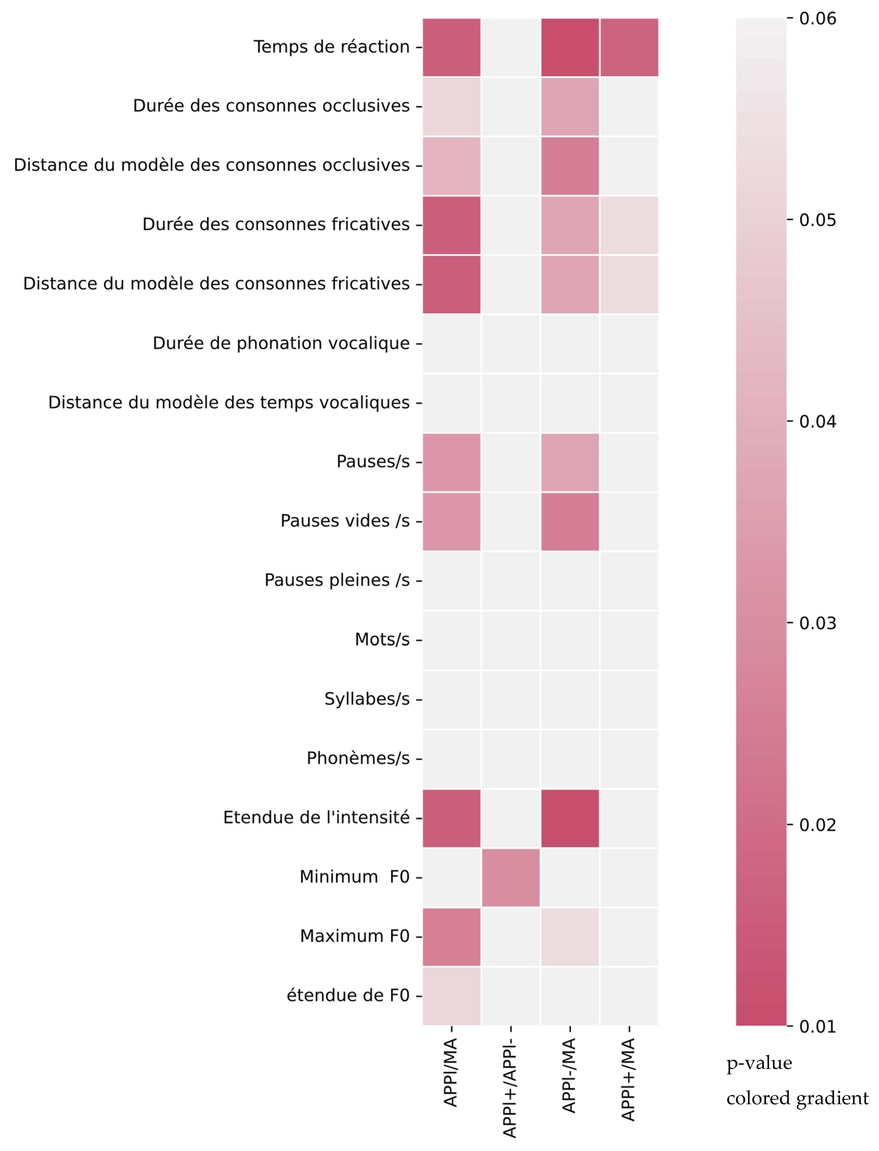
| Temporal Parameters | Significance (p-Value) | Diagnoses | Mean (±SD) |
|---|---|---|---|
| Vocal reaction time (s) | AD/lvPPA (0.016) ** | AD | 0.779 (±0.180) |
| AD/−lvPPA (0.010) ** | lvPPA | 1.379 (±0.727) | |
| AD/+lvPPA (0.017) ** | +lvPPA | 1.842 (±0.751) | |
| −lvPPA/+lvPPA (0.096) | −lvPPA | 1.916 (±0.253) | |
| Occlusive consonants duration (s) | AD/lvPPA (0.051) * | AD | 0.049 (±0.016) |
| AD/−lvPPA (0.037) ** | lvPPA | 0.067 (±0.017) | |
| AD/+lvPPA (0.097) | +lvPPA | 0.101 (±0.012) | |
| −lvPPA/+lvPPA (0.222) | −lvPPA | 0.113 (±0.016) | |
| Occlusive consonant distance from model | AD/lvPPA (0.042) ** | AD | 1.083 (±0.397) |
| AD/−lvPPA (0.025) ** | lvPPA | 1.481 (±0.389) | |
| AD/+lvPPA (0.156) | +lvPPA | 1.288 (±0.304) | |
| −lvPPA/+lvPPA (0.222) | −lvPPA | 1.674 (±0.368) | |
| Fricative consonant duration (s) | AD/lvPPA (0.016) ** | AD | 0.079 (±0.022) |
| AD/−lvPPA (0.037) ** | lvPPA | 0.107 (±0.015) | |
| AD/+lvPPA (0.097) | +lvPPA | 0.101 (±0.012) | |
| −lvPPA/+lvPPA (0.053) * | −lvPPA | 0.114 (±0.015) | |
| Fricative consonant distance from model | AD/lvPPA (0.016) ** | AD | 0.978 (±0.261) |
| AD/−lvPPA (0.037) ** | lvPPA | 1.274 (±0.155) | |
| AD/+lvPPA (0.235) | +lvPPA | 1.223 (±0.138) | |
| −lvPPA/+lvPPA (0.053) * | −lvPPA | 1.326 (±0.154) | |
| Vowel phonation time (s) | AD/lvPPA (0.357) | AD | 0.153 (±0.020) |
| AD/−lvPPA (0.222) | lvPPA | 0.154 (±0.022) | |
| AD/+lvPPA (0.156) | +lvPPA | 0.145 (±0.018) | |
| −lvPPA/+lvPPA (0.466) | −lvPPA | 0.162 (±0.022) | |
| Vowel phonation distance from model | AD/lvPPA (0.437) | AD | 1.195 (±0.148) |
| AD/−lvPPA (0.399) | lvPPA | 1.171 (±0.153) | |
| AD/+lvPPA (0.333) | +lvPPA | 1.133 (±0.156) | |
| −lvPPA/+lvPPA (0.466) | −lvPPA | 1.208 (±0.139) | |
| Pauses/s | AD/lvPPA (0.033) ** | AD | 2.175 (±0.565) |
| AD/−lvPPA (0.037) ** | lvPPA | 0.763 (±0.387) | |
| AD/+lvPPA (0.443) | +lvPPA | 0.7669 (±0.450) | |
| −lvPPA/+lvPPA (0.135) | −lvPPA | 0.759 (±0.309) | |
| Silent pauses/s | AD/lvPPA (0.033) ** | AD | 1.148 (±0.316) |
| AD/−lvPPA (0.037) ** | lvPPA | 0.699 (±0.410) | |
| AD/+lvPPA (0.442) | +lvPPA | 0.711 (±0.480) | |
| −lvPPA/+lvPPA (0.175) | −lvPPA | 0.688 (±0.325) | |
| Non-silent pauses/s | AD/lvPPA (0.054) * | AD | 0.227 (±0.311) |
| AD/−lvPPA (0.175) | lvPPA | 0.058 (±0.028) | |
| AD/+lvPPA (0.333) | +lvPPA | 0.056 (±0.031) | |
| −lvPPA/+lvPPA (0.074) | −lvPPA | 0.059 (±0.024) |
| Prosodic Parameters | Significance (p-Value) | Diagnosis | Mean (±SD) |
|---|---|---|---|
| Words/s | AD/lvPPA (0.186) | AD | 2.327 (±0.383) |
| AD/−lvPPA (0.134) | lvPPA | 1.928 (±0.604) | |
| AD/+lvPPA (0.333) | +lvPPA | 2.179 (±0.253) | |
| −lvPPA/+lvPPA (0.399) | −lvPPA | 1.677 (±0.735) | |
| Syllables/s | AD/lvPPA (0.215) | AD | 3.676 (±0.580) |
| AD/−lvPPA (0.222) | lvPPA | 3.002 (±0.990) | |
| AD/+lvPPA (0.333) | +lvPPA | 3.379 (±0.439) | |
| −lvPPA/+lvPPA (0.336) | −lvPPA | 2.625 (±1.219) | |
| Phonemes/s | AD/lvPPA (0.159) | AD | 7.536 (±1.216) |
| AD/−lvPPA (0.175) | lvPPA | 6.118 (±2.052) | |
| AD/+lvPPA (0.442) | +lvPPA | 6.913 (±0.896) | |
| −lvPPA/+lvPPA (0.276) | −lvPPA | 5.324 (±2.521) | |
| Intensity range (dB) | AD/lvPPA (0.016) ** | AD | 35.920 (±5.860) |
| AD/−lvPPA (0.011) ** | lvPPA | 27.036 (±5.249) | |
| AD/+lvPPA (0.097) | +lvPPA | 545.932 (±54.823) | |
| −lvPPA/+lvPPA (0.135) | −lvPPA | 24.094 (±4.168) | |
| Minimum F0 (Hz) | AD/lvPPA (0.396) | AD | 95.547 (±17.999) |
| AD/−lvPPA (0.101) | lvPPA | 87.088 (±15.750) | |
| AD/+lvPPA (0.030) ** | +lvPPA | 98.634 (±9.661) | |
| −lvPPA/+lvPPA (0.222) | −lvPPA | 75.542 (±11.669) | |
| Maximum F0 (Hz) | AD/lvPPA (0.026) ** | AD | 382.273 (±91.249) |
| AD/−lvPPA (0.053) * | lvPPA | 325.541 (±49.334) | |
| AD/+lvPPA (0.442) | +lvPPA | 319.474 (±64.704) | |
| −lvPPA/+lvPPA (0.074) | −lvPPA | 331.605 (±24.646) | |
| F0 range (Hz) | AD/lvPPA (0.051) * | AD | 271.015 (±91.946) |
| AD/−lvPPA (0.101) | lvPPA | 224.689 (±46.748) | |
| AD/+lvPPA (0.442) | +lvPPA | 208.469 (±61.490) | |
| −lvPPA/+lvPPA (0.102) | −lvPPA | 240.91 (±7.964) |
Publisher’s Note: MDPI stays neutral with regard to jurisdictional claims in published maps and institutional affiliations. |
© 2022 by the authors. Licensee MDPI, Basel, Switzerland. This article is an open access article distributed under the terms and conditions of the Creative Commons Attribution (CC BY) license (https://creativecommons.org/licenses/by/4.0/).
Share and Cite
Da Cunha, E.; Plonka, A.; Arslan, S.; Mouton, A.; Meyer, T.; Robert, P.; Meunier, F.; Manera, V.; Gros, A. Logogenic Primary Progressive Aphasia or Alzheimer Disease: Contribution of Acoustic Markers in Early Differential Diagnosis. Life 2022, 12, 933. https://doi.org/10.3390/life12070933
Da Cunha E, Plonka A, Arslan S, Mouton A, Meyer T, Robert P, Meunier F, Manera V, Gros A. Logogenic Primary Progressive Aphasia or Alzheimer Disease: Contribution of Acoustic Markers in Early Differential Diagnosis. Life. 2022; 12(7):933. https://doi.org/10.3390/life12070933
Chicago/Turabian StyleDa Cunha, Eloïse, Alexandra Plonka, Seçkin Arslan, Aurélie Mouton, Tess Meyer, Philippe Robert, Fanny Meunier, Valeria Manera, and Auriane Gros. 2022. "Logogenic Primary Progressive Aphasia or Alzheimer Disease: Contribution of Acoustic Markers in Early Differential Diagnosis" Life 12, no. 7: 933. https://doi.org/10.3390/life12070933