
Magnetic Resonance Imaging of Autoimmune Demyelinating Diseases as a Diagnostic Challenge for Radiologists: Report of Two Cases and Literature Review

 , , and
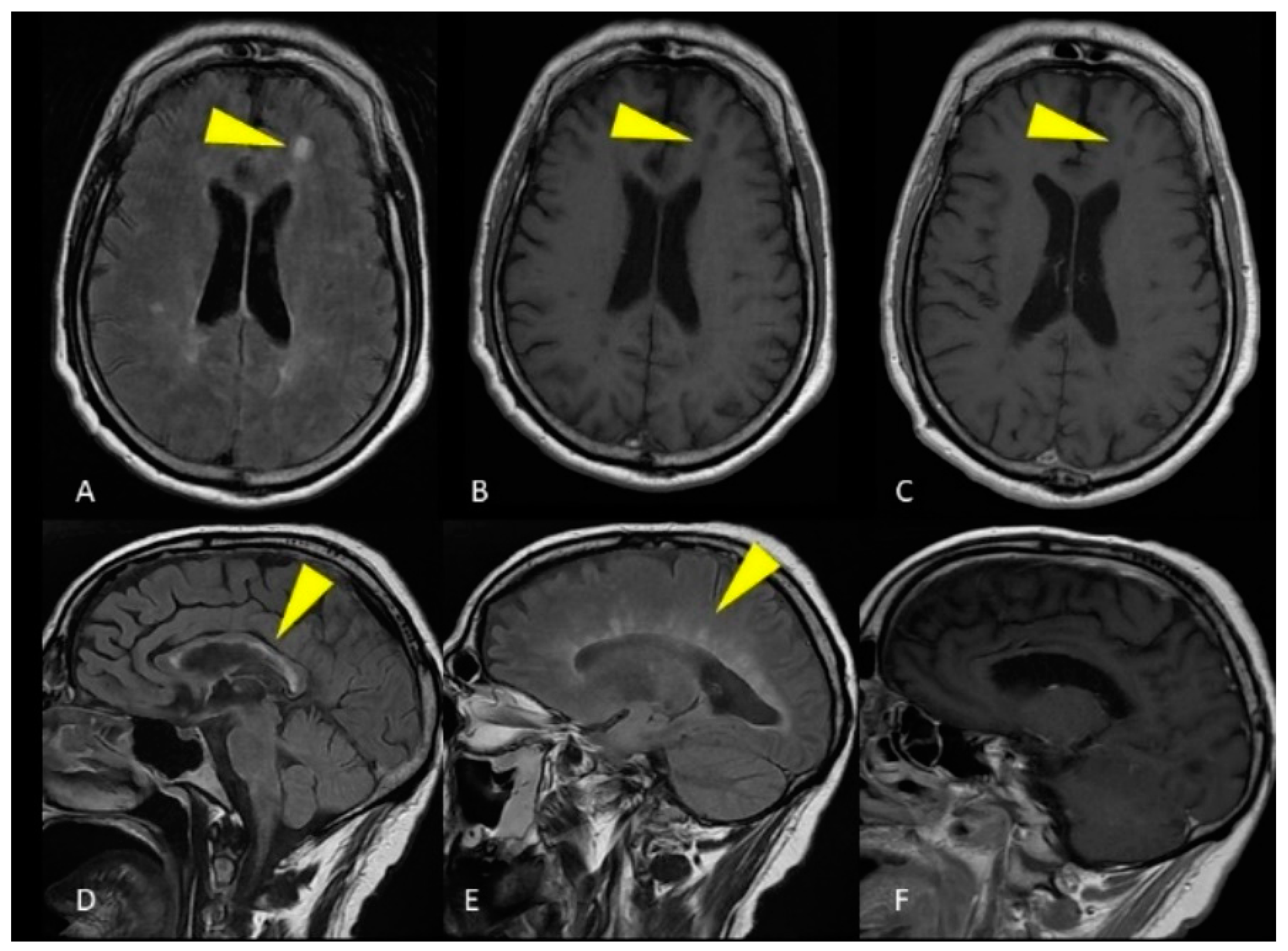
Abstract
:1. Introduction
2. Materials and Methods
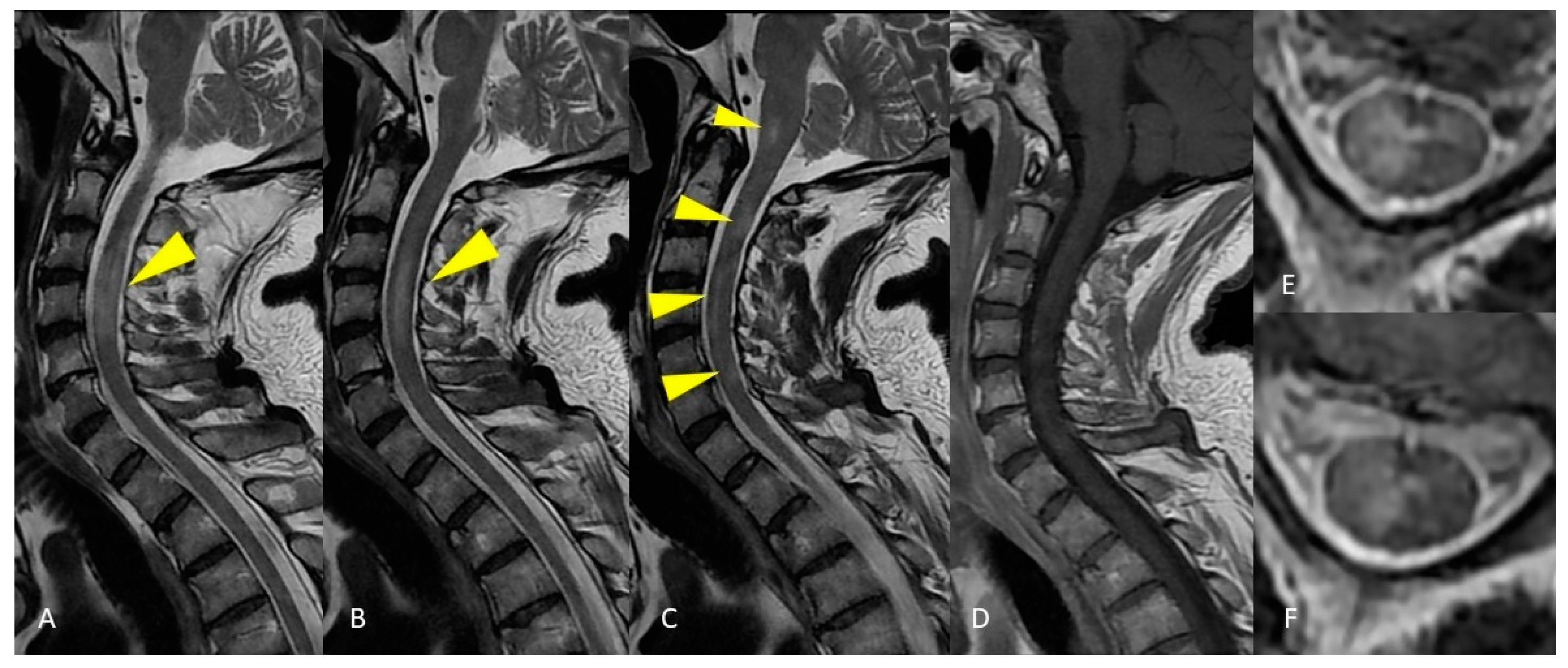
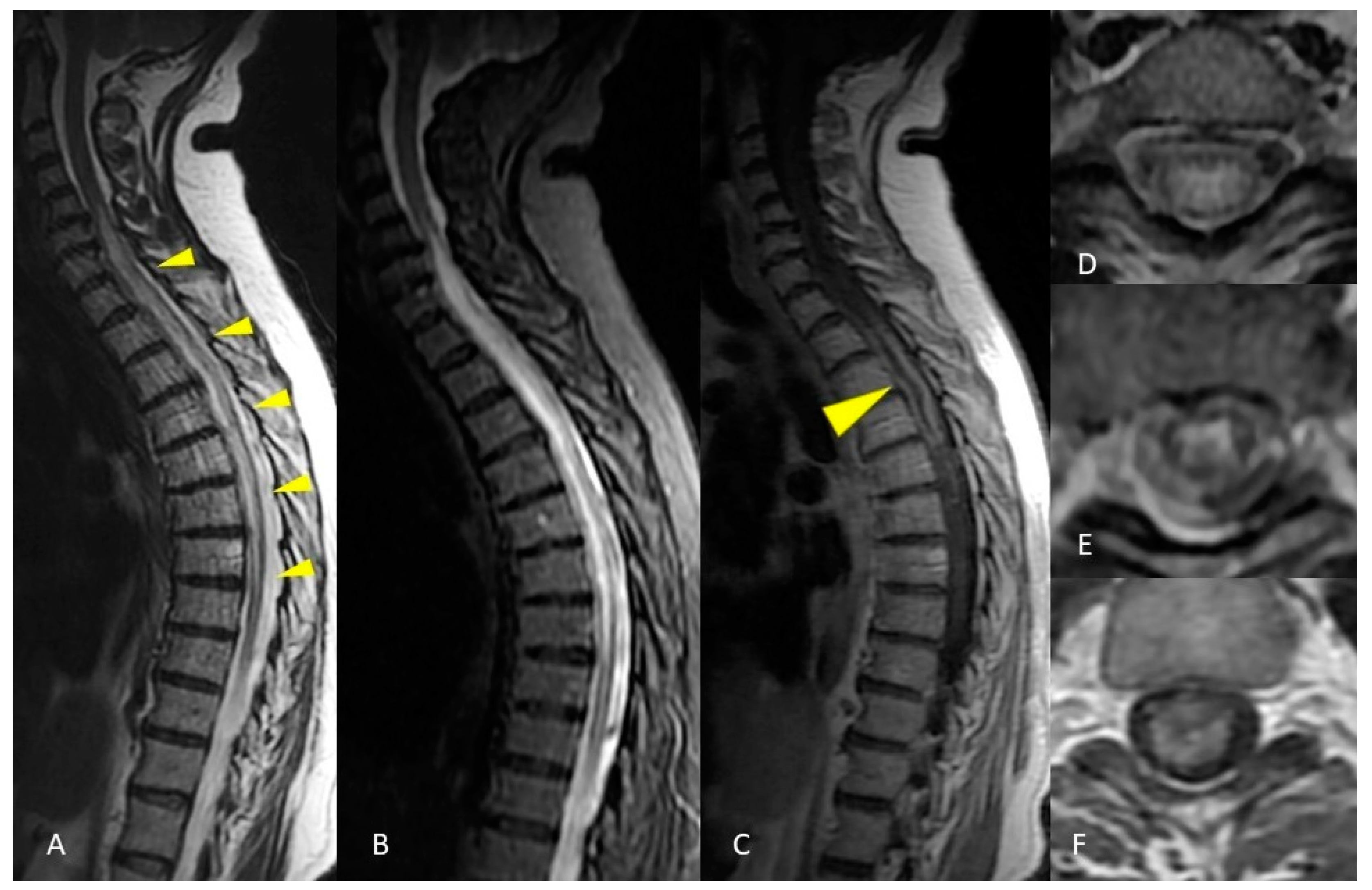
2.1. Case Presentation 1
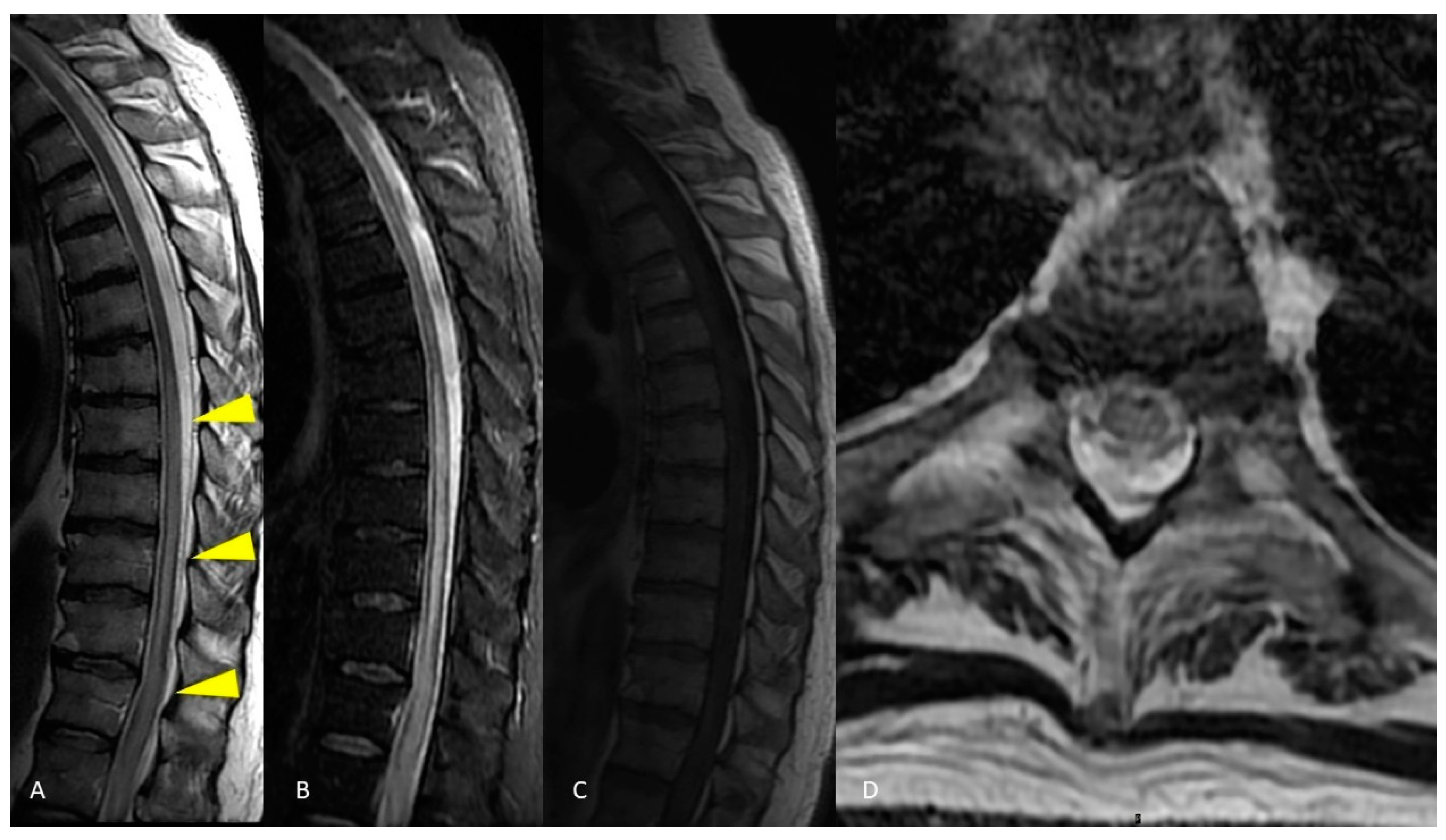
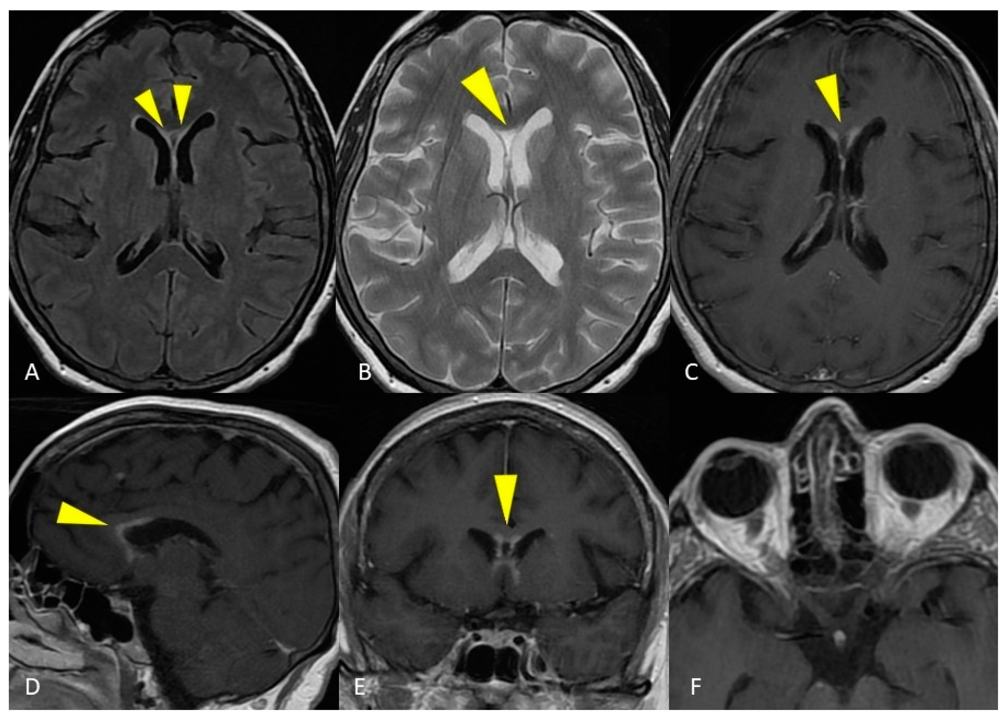
2.2. Case Presentation 2
3. Discussion
3.1. Multiple Sclerosis
3.2. Acute Disseminated Encephalomyelitis—ADEM
3.3. Neuromyelitis Optica—NMO
3.4. Idiopathic Transverse Myelitis
3.5. Myelin Oligodendrocyte Glycoprotein Encephalomyelitis
3.6. CLIPPERS
4. Conclusions
Author Contributions
Funding
Institutional Review Board Statement
Informed Consent Statement
Data Availability Statement
Conflicts of Interest
References
- Lee, M.J.; Aronberg, R.; Manganaro, M.S.; Ibrahim, M.; Parmar, H.A. Diagnostic Approach to Intrinsic Abnormality of Spinal Cord Signal Intensity. Radiographics 2019, 39, 1824–1839. [Google Scholar] [CrossRef]
- Miki, Y. Magnetic resonance imaging diagnosis of demyelinating diseases: An update. Clin. Exp. Neuroimmunol. 2019, 10, 32–48. [Google Scholar] [CrossRef] [Green Version]
- Bunyan, R.F.; Tang, J.; Weinshenker, B. Acute demyelinating disorders: Emergencies and management. Neurol. Clin. 2012, 30, 285–307. [Google Scholar] [CrossRef]
- Katsenos, C.; Androulaki, D.; Lyra, S.; Tsoutsouras, T.; Mandragos, C. A 17 year-old girl with a demyelinating disease requiring mechanical ventilation: A case report. BMC Res. Notes 2013, 6, 22. [Google Scholar] [CrossRef] [Green Version]
- Rossi, A. Imaging of acute disseminated encephalomyelitis. Neuroimaging Clin. N. Am. 2008, 18, 149–161. [Google Scholar] [CrossRef] [PubMed]
- Weidauer, S.; Raab, P.; Hattingen, E. Diagnostic approach in multiple sclerosis with MRI: An update. Clin. Imaging 2021, 78, 276–285. [Google Scholar] [CrossRef]
- Sarbu, N.; Lolli, V.; Smirniotopoulos, J.G. Magnetic resonance imaging in myelopathy: A pictorial review. Clin. Imaging 2019, 57, 56–68. [Google Scholar] [CrossRef] [PubMed]
- Brownlee, W.J.; Hardy, T.A.; Fazekas, F.; Miller, D.H. Diagnosis of multiple sclerosis: Progress and challenges. Lancet 2017, 389, 1336–1346. [Google Scholar] [CrossRef]
- Solomon, A.J.; Corboy, J.R. The tension between early diagnosis and misdiagnosis of multiple sclerosis. Nat. Rev. Neurol. 2017, 13, 567–572. [Google Scholar] [CrossRef] [PubMed]
- Eckstein, C.; Saidha, S.; Levy, M. A differential diagnosis of central nervous system demyelination: Beyond multiple sclerosis. J. Neurol. 2012, 259, 801–816. [Google Scholar] [CrossRef] [PubMed]
- Wildner, P.; Stasiołek, M.; Matysiak, M. Differential diagnosis of multiple sclerosis and other inflammatory CNS diseases. Mult. Scler. Relat. Disord. 2020, 37, 101452. [Google Scholar] [CrossRef]
- Koch-Henriksen, N.; Sørensen, P.S. The changing demographic pattern of multiple sclerosis epidemiology. Lancet Neurol. 2010, 9, 520–532. [Google Scholar] [CrossRef]
- Lublin, F.D.; Reingold, S.C.; Cohen, J.A.; Cutter, G.R.; Sørensen, P.S.; Thompson, A.J.; Wolinsky, J.S.; Balcer, L.J.; Banwell, B.; Barkhof, F.; et al. Defining the clinical course of multiple sclerosis: The 2013 revisions. Neurology 2014, 83, 278–286. [Google Scholar] [CrossRef] [Green Version]
- Ömerhoca, S.; Akkaş, S.Y.; İçen, N.K. Multiple Sclerosis: Diagnosis and Differential Diagnosis. Arch. Neuropsychiatry 2018, 55, S1–S9. [Google Scholar] [CrossRef]
- Yamout, B.I.; Alroughani, R. Multiple Sclerosis. Semin. Neurol. 2018, 38, 212–225. [Google Scholar] [CrossRef] [PubMed]
- Miller, D.; Barkhof, F.; Montalban, X.; Thompson, A.; Filippi, M. Clinically isolated syndromes suggestive of multiple sclerosis, part I: Natural history, pathogenesis, diagnosis, and prognosis. Lancet Neurol. 2005, 4, 281–288. [Google Scholar] [CrossRef]
- Yamout, B.; Sahraian, M.; Bohlega, S.; Al-Jumah, M.; Goueider, R.; Dahdaleh, M.; Inshasi, J.; Hashem, S.; Alsharoqi, I.; Khoury, S.; et al. Consensus recommendations for the diagnosis and treatment of multiple sclerosis: 2019 revisions to the MENACTRIMS guidelines. Mult. Scler. Relat. Disord. 2020, 37, 101459. [Google Scholar] [CrossRef] [PubMed]
- Sarbu, N.; Shih, R.Y.; Jones, R.V.; Horkayne-Szakaly, I.; Oleaga, L.; Smirniotopoulos, J.G. White Matter Diseases with Radiologic-Pathologic Correlation. Radiographics 2016, 36, 1426–1447. [Google Scholar] [CrossRef]
- Lycklama, G.; Thompson, A.; Filippi, M.; Miller, D.; Polman, C.; Fazekas, F.; Barkhof, F. Spinal-cord MRI in multiple sclerosis. Lancet Neurol. 2003, 2, 555–562. [Google Scholar] [CrossRef]
- Mohajeri Moghaddam, S.; Bhatt, A.A. Location, length, and enhancement: Systematic approach to differentiating intramedullary spinal cord lesions. Insights Imaging 2018, 9, 511–526. [Google Scholar] [CrossRef] [PubMed] [Green Version]
- Polman, C.H.; Reingold, S.C.; Banwell, B.; Clanet, M.; Cohen, J.A.; Filippi, M.; Fujihara, K.; Havrdova, E.; Hutchinson, M.; Kappos, L.; et al. Diagnostic criteria for multiple sclerosis: 2010 revisions to the McDonald criteria. Ann. Neurol. 2011, 69, 292–302. [Google Scholar] [CrossRef] [PubMed] [Green Version]
- Baumann, M.; Sahin, K.; Lechner, C.; Hennes, E.M.; Schanda, K.; Mader, S.; Karenfort, M.; Selch, C.; Häusler, M.; Eisenkölbl, A.; et al. Clinical and neuroradiological differences of paediatric acute disseminating encephalomyelitis with and without antibodies to the myelin oligodendrocyte glycoprotein. J. Neurol. Neurosurg. Psychiatry 2015, 86, 265–272. [Google Scholar] [CrossRef] [PubMed]
- Pellegrino, P.; Radice, S.; Clementi, E. Geoepidemiology of acute disseminated encephalomyelitis. Epidemiology 2014, 25, 928–929. [Google Scholar] [CrossRef]
- Xiong, C.H.; Yan, Y.; Liao, Z.; Peng, S.H.; Wen, H.R.; Zhang, Y.X.; Chen, S.H.; Li, J.; Chen, H.Y.; Feng, X.W.; et al. Epidemiological characteristics of acute disseminated encephalomyelitis in Nanchang, China: A retrospective study. BMC Public Health 2014, 14, 111. [Google Scholar] [CrossRef] [PubMed] [Green Version]
- Pohl, D.; Alper, G.; Van Haren, K.; Kornberg, A.J.; Lucchinetti, C.F.; Tenembaum, S.; Belman, A.L. Acute disseminated encephalomyelitis: Updates on an inflammatory CNS syndrome. Neurology 2016, 87, S38–S45. [Google Scholar] [CrossRef]
- Krupp, L.B.; Tardieu, M.; Amato, M.P.; Banwell, B.; Chitnis, T.; Dale, R.C.; Ghezzi, A.; Hintzen, R.; Kornberg, A.; Pohl, D.; et al. International Pediatric Multiple Sclerosis Study Group. International Pediatric Multiple Sclerosis Study Group criteria for pediatric multiple sclerosis and immune-mediated central nervous system demyelinating disorders: Revisions to the 2007 definitions. Mult. Scler. J. 2013, 19, 1261–1267. [Google Scholar] [CrossRef]
- Paolilo, R.B.; Deiva, K.; Neuteboom, R.; Rostásy, K.; Lim, M. Acute Disseminated Encephalomyelitis: Current Perspectives. Children 2020, 7, 210. [Google Scholar] [CrossRef]
- Boesen, M.S.; Blinkenberg, M.; Koch-Henriksen, N.; Thygesen, L.C.; Uldall, P.V.; Magyari, M.; Born, A.P. Implications of the International Paediatric Multiple Sclerosis Study Group consensus criteria for paediatric acute disseminated encephalomyelitis: A nationwide validation study. Dev. Med. Child Neurol. 2018, 60, 1123–1131. [Google Scholar] [CrossRef] [PubMed] [Green Version]
- Huppke, P.; Rostasy, K.; Karenfort, M.; Huppke, B.; Seidl, R.; Leiz, S.; Reindl, M.; Gärtner, J. Acute disseminated encephalomyelitis followed by recurrent or monophasic optic neuritis in pediatric patients. Mult. Scler. 2013, 19, 941–946. [Google Scholar] [CrossRef]
- Mader, I.; Wolff, M.; Niemann, G.; Küker, W. Acute haemorrhagic encephalomyelitis (AHEM): MRI findings. Neuropediatrics 2004, 35, 143–146. [Google Scholar] [CrossRef]
- Lu, Z.; Zhang, B.; Qiu, W.; Kang, Z.; Shen, L.; Long, Y.; Huang, J.; Hu, X. Comparative brain stem lesions on MRI of acute disseminated encephalomyelitis, neuromyelitis optica, and multiple sclerosis. PLoS ONE 2011, 6, e22766. [Google Scholar] [CrossRef] [PubMed]
- Cañellas, A.R.; Gols, A.R.; Izquierdo, J.R.; Subirana, M.T.; Gairin, X.M. Idiopathic inflammatory-demyelinating diseases of the central nervous system. Neuroradiology 2007, 49, 393–409. [Google Scholar] [CrossRef] [PubMed]
- Tenembaum, S.; Chamoles, N.; Fejerman, N. Acute disseminated encephalomyelitis: A long-term follow-up study of 84 pediatric patients. Neurology 2002, 59, 1224–1231. [Google Scholar] [CrossRef]
- Alper, G.; Heyman, R.; Wang, L. Multiple sclerosis and acute disseminated encephalomyelitis diagnosed in children after long-term follow-up: Comparison of presenting features. Dev. Med. Child Neurol. 2009, 51, 480–486. [Google Scholar] [CrossRef] [PubMed]
- Verhey, L.H.; Branson, H.M.; Shroff, M.M.; Callen, D.J.; Sled, J.G.; Narayanan, S.; Sadovnick, A.D.; Bar-Or, A.; Arnold, D.L.; Marrie, R.A.; et al. MRI parameters for prediction of multiple sclerosis diagnosis in children with acute CNS demyelination: A prospective national cohort study. Lancet Neurol. 2011, 10, 1065–1073. [Google Scholar] [CrossRef]
- Tenembaum, S.N. Acute disseminated encephalomyelitis. In Handbook of Clinical Neurology; Dulac, O., Lassonde, M., Sarnat, H.B., Eds.; Elsevier: Amsterdam, The Netherlands, 2013; pp. 1253–1262. [Google Scholar] [CrossRef]
- Osborn, A.G. Acute Disseminated Encephalomyelitis (ADEM). In Imaging in Neurology; Osborn, A.G., Digre, K.B., Eds.; Elsevier: Amsterdam, The Netherlands, 2017; p. 193. [Google Scholar] [CrossRef]
- Koelman, D.L.; Chahin, S.; Mar, S.S.; Venkatesan, A.; Hoganson, G.M.; Yeshokumar, A.K.; Barreras, P.; Majmudar, B.; Klein, J.P.; Chitnis, T.; et al. Acute disseminated encephalomyelitis in 228 patients: A retrospective, multicenter US study. Neurology 2016, 86, 2085–2093. [Google Scholar] [CrossRef]
- Wu, Y.; Zhong, L.; Geng, J. Neuromyelitis optica spectrum disorder: Pathogenesis, treatment, and experimental models. Mult. Scler. Relat. Disord. 2019, 27, 412–418. [Google Scholar] [CrossRef]
- Lennon, V.A.; Wingerchuk, D.M.; Kryzer, T.J.; Pittock, S.J.; Lucchinetti, C.F.; Fujihara, K.; Nakashima, I.; Weinshenker, B.G. A serum autoantibody marker of neuromyelitis optica: Distinction from multiple sclerosis. Lancet 2004, 364, 2106–2112. [Google Scholar] [CrossRef]
- Pittock, S.J.; Lucchinetti, C.F. Neuromyelitis optica and the evolving spectrum of autoimmune aquaporin-4 channelopathies: A decade later. Ann. N. Y. Acad. Sci. 2016, 1366, 20–39. [Google Scholar] [CrossRef]
- Mealy, M.A.; Kessler, R.A.; Rimler, Z.; Reid, A.; Totonis, L.; Cutter, G.; Kister, I.; Levy, M. Mortality in neuromyelitis optica is strongly associated with African ancestry. Neurol.-Neuroimmunol. Neuroinflamm. 2018, 5, e468. [Google Scholar] [CrossRef] [Green Version]
- Quek, A.M.; McKeon, A.; Lennon, V.A.; Mandrekar, J.N.; Iorio, R.; Jiao, Y.; Costanzi, C.; Weinshenker, B.G.; Wingerchuk, D.M.; Lucchinetti, C.F.; et al. Effects of age and sex on aquaporin-4 autoimmunity. Arch. Neurol. 2012, 69, 1039–1043. [Google Scholar] [CrossRef] [PubMed] [Green Version]
- Wingerchuk, D.M.; Banwell, B.; Bennett, J.L.; Cabre, P.; Carroll, W.; Chitnis, T.; de Seze, J.; Fujihara, K.; Greenberg, B.; Jacob, A.; et al. International Panel for NMO Diagnosis. International consensus diagnostic criteria for neuromyelitis optica spectrum disorders. Neurology 2015, 85, 177–189. [Google Scholar] [CrossRef] [PubMed]
- Jasiak-Zatonska, M.; Kalinowska-Lyszczarz, A.; Michalak, S.; Kozubski, W. The Immunology of Neuromyelitis Optica-Current Knowledge, Clinical Implications, Controversies and Future Perspectives. Int. J. Mol. Sci. 2016, 17, 273. [Google Scholar] [CrossRef] [Green Version]
- Jarius, S.; Ruprecht, K.; Wildemann, B.; Kuempfel, T.; Ringelstein, M.; Geis, C.; Kleiter, I.; Kleinschnitz, C.; Berthele, A.; Brettschneider, J.; et al. Contrasting disease patterns in seropositive and seronegative neuromyelitis optica: A multicentre study of 175 patients. J. Neuroinflamm. 2012, 9, 14. [Google Scholar] [CrossRef] [PubMed]
- Andersson, M.; Alvarez-Cermeño, J.; Bernardi, G.; Cogato, I.; Fredman, P.; Frederiksen, J.; Fredrikson, S.; Gallo, P.; Grimaldi, L.M.; Grønning, M. Cerebrospinal fluid in the diagnosis of multiple sclerosis: A consensus report. J. Neurol. Neurosurg. Psychiatry 1994, 57, 897–902, Erratum in J. Neurol. Neurosurg. Psychiatry 2011, 82, 826. [Google Scholar] [CrossRef] [Green Version]
- Weinshenker, B.G.; Wingerchuk, D.M. Neuromyelitis Spectrum Disorders. Mayo Clin. Proc. 2017, 92, 663–679. [Google Scholar] [CrossRef] [Green Version]
- Dutra, B.G.; da Rocha, A.J.; Nunes, R.H.; Maia, A.C.M., Jr. Neuromyelitis Optica Spectrum Disorders: Spectrum of MR Imaging Findings and Their Differential Diagnosis. Radiographics 2018, 38, 169–193, Erratum in Radiographics 2018, 38, 662. [Google Scholar] [CrossRef]
- Kim, H.J.; Paul, F.; Lana-Peixoto, M.A.; Tenembaum, S.; Asgari, N.; Palace, J.; Klawiter, E.C.; Sato, D.K.; de Seze, J.; Wuerfel, J.; et al. MRI characteristics of neuromyelitis optica spectrum disorder: An international update. Neurology 2015, 84, 1165–1173. [Google Scholar] [CrossRef] [Green Version]
- Paul, S.; Mondal, G.P.; Bhattacharyya, R.; Ghosh, K.C.; Bhat, I.A. Neuromyelitis optica spectrum disorders. J. Neurol. Sci. 2021, 420, 117225. [Google Scholar] [CrossRef]
- Lana-Peixoto, M.A.; Talim, N. Neuromyelitis Optica Spectrum Disorder and Anti-MOG Syndromes. Biomedicines 2019, 7, 42. [Google Scholar] [CrossRef] [Green Version]
- Khanna, S.; Sharma, A.; Huecker, J.; Gordon, M.; Naismith, R.T.; Van Stavern, G.P. Magnetic resonance imaging of optic neuritis in patients with neuromyelitis optica versus multiple sclerosis. J. Neuro-Ophthalmol. 2012, 32, 216–220. [Google Scholar] [CrossRef] [PubMed] [Green Version]
- Wingerchuk, D.M.; Lennon, V.A.; Pittock, S.J.; Lucchinetti, C.F.; Weinshenker, B.G. Revised diagnostic criteria for neuromyelitis optica. Neurology 2006, 66, 1485–1489. [Google Scholar] [CrossRef] [PubMed] [Green Version]
- Wang, K.Y.; Chetta, J.; Bains, P.; Balzer, A.; Lincoln, J.; Uribe, T.; Lincoln, C.M. Spectrum of MRI brain lesion patterns in neuromyelitis optica spectrum disorder: A pictorial review. Br. J. Radiol. 2018, 91, 20170690. [Google Scholar] [CrossRef] [PubMed]
- Awad, A.; Stüve, O. Idiopathic transverse myelitis and neuromyelitis optica: Clinical profiles, pathophysiology and therapeutic choices. Curr. Neuropharmacol. 2011, 9, 417–428. [Google Scholar] [CrossRef] [Green Version]
- Jeffery, D.R.; Mandler, R.N.; Davis, L.E. Transverse myelitis. Retrospective analysis of 33 cases, with differentiation of cases associated with multiple sclerosis and parainfectious events. Arch. Neurol. 1993, 50, 532–535. [Google Scholar] [CrossRef]
- Young, J.; Quinn, S.; Hurrell, M.; Taylor, B. Clinically isolated acute transverse myelitis: Prognostic features and incidence. Mult. Scler. 2009, 15, 1295–1302. [Google Scholar] [CrossRef]
- Christensen, P.B.; Wermuth, L.; Hinge, H.H.; Bømers, K. Clinical course and long-term prognosis of acute transverse myelopathy. Acta Neurol. Scand. 1990, 81, 431–435. [Google Scholar] [CrossRef]
- Krishnan, C.; Kaplin, A.I.; Deshpande, D.M.; Pardo, C.A.; Kerr, D.A. Transverse Myelitis: Pathogenesis, diagnosis and treatment. Front. Biosci. 2004, 9, 1483–1499. [Google Scholar] [CrossRef]
- Knebusch, M.; Strassburg, H.M.; Reiners, K. Acute transverse myelitis in childhood: Nine cases and review of the literature. Dev. Med. Child Neurol. 1998, 40, 631–639. [Google Scholar] [CrossRef]
- Kalita, J.; Misra, U.K.; Mandal, S.K. Prognostic predictors of acute transverse myelitis. Acta Neurol. Scand. 1998, 98, 60–63. [Google Scholar] [CrossRef]
- Miller, D.H.; McDonald, W.I.; Blumhardt, L.D.; du Boulay, G.H.; Halliday, A.M.; Johnson, G.; Kendall, B.E.; Kingsley, D.P.; MacManus, D.G.; Moseley, I.F. Magnetic resonance imaging in isolated noncompressive spinal cord syndromes. Ann. Neurol. 1987, 22, 714–723. [Google Scholar] [CrossRef] [PubMed]
- Austin, S.G.; Zee, C.S.; Waters, C. The role of magnetic resonance imaging in acute transverse myelitis. Can. J. Neurol. Sci. 1992, 19, 508–511. [Google Scholar] [CrossRef] [PubMed] [Green Version]
- Lipton, H.L.; Teasdall, R.D. Acute transverse myelopathy in adults. A follow-up study. Arch. Neurol. 1973, 28, 252–257. [Google Scholar] [CrossRef] [PubMed]
- Bakshi, R.; Kinkel, P.R.; Mechtler, L.L.; Bates, V.E.; Lindsay, B.D.; Esposito, S.E.; Kinkel, W.R. Magnetic resonance imaging findings in 22 cases of myelitis: Comparison between patients with and without multiple sclerosis. Eur. J. Neurol. 1998, 5, 35–48. [Google Scholar] [CrossRef]
- Bashir, K.; Whitaker, J.N. Importance of paraclinical and CSF studies in the diagnosis of MS in patients presenting with partial cervical transverse myelopathy and negative cranial MRI. Mult. Scler. 2000, 6, 312–316. [Google Scholar] [CrossRef]
- Ford, B.; Tampieri, D.; Francis, G. Long-term follow-up of acute partial transverse myelopathy. Neurology 1992, 42, 250–252. [Google Scholar] [CrossRef]
- Miller, D.H.; Ormerod, I.E.; Rudge, P.; Kendall, B.E.; Moseley, I.F.; McDonald, W.I. The early risk of multiple sclerosis following isolated acute syndromes of the brainstem and spinal cord. Ann. Neurol. 1989, 26, 635–639. [Google Scholar] [CrossRef]
- Morrissey, S.P.; Miller, D.H.; Kendall, B.E.; Kingsley, D.P.; Kelly, M.A.; Francis, D.A.; MacManus, D.G.; McDonald, W.I. The significance of brain magnetic resonance imaging abnormalities at presentation with clinically isolated syndromes suggestive of multiple sclerosis. A 5-year follow-up study. Brain 1993, 116, 135–146. [Google Scholar] [CrossRef]
- Van Bogaert, L. Post-infectious encephalomyelitis and multiple sclerosis; the significance of perivenous encephalomyelitis. J. Neuropathol. Exp. Neurol. 1950, 9, 219–249. [Google Scholar] [CrossRef]
- Tippett, D.S.; Fishman, P.S.; Panitch, H.S. Relapsing transverse myelitis. Neurology 1991, 41, 703–706. [Google Scholar] [CrossRef]
- Pandit, L.; Rao, S. Recurrent myelitis. J. Neurol. Neurosurg. Psychiatry 1996, 60, 336–338. [Google Scholar] [CrossRef] [PubMed] [Green Version]
- Garcia-Merino, A.; Blasco, M.R. Recurrent transverse myelitis with unusual long-standing Gd-DTPA enhancement. J. Neurol. 2000, 247, 550–551. [Google Scholar] [CrossRef] [PubMed]
- Kaplin, A.I.; Deshpande, D.M.; Scott, E.; Krishnan, C.; Carmen, J.S.; Shats, I.; Martinez, T.; Drummond, J.; Dike, S.; Pletnikov, M.; et al. IL-6 induces regionally selective spinal cord injury in patients with the neuroinflammatory disorder transverse myelitis. J. Clin. Investig. 2005, 115, 2731–2741. [Google Scholar] [CrossRef] [PubMed] [Green Version]
- Byskosh, P.V.; Reder, A.T. Interferon beta-1b effects on cytokine mRNA in peripheral mononuclear cells in multiple sclerosis. Mult. Scler. 1996, 1, 262–269. [Google Scholar] [CrossRef]
- Ransohoff, R.M.; Howe, C.L.; Rodriguez, M. Growth factor treatment of demyelinating disease: At last, a leap into the light. Trends Immunol. 2002, 23, 512–516. [Google Scholar] [CrossRef]
- Gadient, R.A.; Otten, U.H. Interleukin-6 (IL-6)--a molecule with both beneficial and destructive potentials. Prog. Neurobiol. 1997, 52, 379–390. [Google Scholar] [CrossRef]
- Kira, J.; Kawano, Y.; Yamasaki, K.; Tobimatsu, S. Acute myelitis with hyperIgEaemia and mite antigen specific IgE: Atopic myelitis. J. Neurol. Neurosurg. Psychiatry 1998, 64, 676–679. [Google Scholar] [CrossRef]
- Kikuchi, H.; Osoegawa, M.; Ochi, H.; Murai, H.; Horiuchi, I.; Takahashi, H.; Yamabe, K.; Iwaki, T.; Mizutani, T.; Oda, M.; et al. Spinal cord lesions of myelitis with hyperIgEemia and mite antigen specific IgE (atopic myelitis) manifest eosinophilic inflammation. J. Neurol. Sci. 2001, 183, 73–78. [Google Scholar] [CrossRef]
- Oh, J.; Levy, M. Neuromyelitis optica: An antibody-mediated disorder of the central nervous system. Neurol. Res. Int. 2012, 2012, 460825. [Google Scholar] [CrossRef] [Green Version]
- Narayan, R.; Simpson, A.; Fritsche, K.; Salama, S.; Pardo, S.; Mealy, M.; Paul, F.; Levy, M. MOG antibody disease: A review of MOG antibody seropositive neuromyelitis optica spectrum disorder. Mult. Scler. Relat. Disord. 2018, 25, 66–72. [Google Scholar] [CrossRef]
- Hyun, J.W.; Woodhall, M.R.; Kim, S.H.; Jeong, I.H.; Kong, B.; Kim, G.; Kim, Y.; Park, M.S.; Irani, S.R.; Waters, P.; et al. Longitudinal analysis of myelin oligodendrocyte glycoprotein antibodies in CNS inflammatory diseases. J. Neurol. Neurosurg. Psychiatry 2017, 88, 811–817. [Google Scholar] [CrossRef] [PubMed]
- Ramanathan, S.; Mohammad, S.; Tantsis, E.; Nguyen, T.K.; Merheb, V.; Fung, V.S.C.; White, O.B.; Broadley, S.; Lechner-Scott, J.; Vucic, S.; et al. Clinical course, therapeutic responses and outcomes in relapsing MOG antibody-associated demyelination. J. Neurol. Neurosurg. Psychiatry 2018, 89, 127–137. [Google Scholar] [CrossRef] [PubMed] [Green Version]
- Pohl, D.; Hennemuth, I.; von Kries, R.; Hanefeld, F. Paediatric multiple sclerosis and acute disseminated encephalomyelitis in Germany: Results of a nationwide survey. Eur. J. Pediatr. 2007, 166, 405–412. [Google Scholar] [CrossRef] [PubMed]
- Kaneko, K.; Sato, D.K.; Nakashima, I.; Nishiyama, S.; Tanaka, S.; Marignier, R.; Hyun, J.W.; Oliveira, L.M.; Reindl, M.; Seifert-Held, T.; et al. Myelin injury without astrocytopathy in neuroinflammatory disorders with MOG antibodies. J. Neurol. Neurosurg. Psychiatry 2016, 87, 1257–1259. [Google Scholar] [CrossRef]
- Salama, S.; Khan, M.; Pardo, S.; Izbudak, I.; Levy, M. MOG antibody-associated encephalomyelitis/encephalitis. Mult. Scler. 2019, 25, 1427–1433. [Google Scholar] [CrossRef]
- Brunner, C.; Lassmann, H.; Waehneldt, T.V.; Matthieu, J.M.; Linington, C. Differential ultrastructural localization of myelin basic protein, myelin/oligodendroglial glycoprotein, and 2′,3′-cyclic nucleotide 3′-phosphodiesterase in the CNS of adult rats. J. Neurochem. 1989, 52, 296–304. [Google Scholar] [CrossRef]
- Hamid, S.H.M.; Whittam, D.; Saviour, M.; Alorainy, A.; Mutch, K.; Linaker, S.; Solomon, T.; Bhojak, M.; Woodhall, M.; Waters, P.; et al. Seizures and Encephalitis in Myelin Oligodendrocyte Glycoprotein IgG Disease vs Aquaporin 4 IgG Disease. JAMA Neurol. 2018, 75, 65–71. [Google Scholar] [CrossRef] [Green Version]
- Wang, L.; ZhangBao, J.; Zhou, L.; Zhang, Y.; Li, H.; Li, Y.; Huang, Y.; Wang, M.; Lu, C.; Lu, J.; et al. Encephalitis is an important clinical component of myelin oligodendrocyte glycoprotein antibody associated demyelination: A single-center cohort study in Shanghai, China. Eur. J. Neurol. 2019, 26, 168–174. [Google Scholar] [CrossRef] [Green Version]
- Van Pelt, E.D.; Wong, Y.Y.; Ketelslegers, I.A.; Hamann, D.; Hintzen, R.Q. Neuromyelitis optica spectrum disorders: Comparison of clinical and magnetic resonance imaging characteristics of AQP4-IgG versus MOG-IgG seropositive cases in the Netherlands. Eur. J. Neurol. 2016, 23, 580–587. [Google Scholar] [CrossRef]
- Höftberger, R.; Sepulveda, M.; Armangue, T.; Blanco, Y.; Rostásy, K.; Calvo, A.C.; Olascoaga, J.; Ramió-Torrentà, L.; Reindl, M.; Benito-León, J.; et al. Antibodies to MOG and AQP4 in adults with neuromyelitis optica and suspected limited forms of the disease. Mult. Scler. 2015, 21, 866–874. [Google Scholar] [CrossRef]
- López-Chiriboga, A.S.; Majed, M.; Fryer, J.; Dubey, D.; McKeon, A.; Flanagan, E.P.; Jitprapaikulsan, J.; Kothapalli, N.; Tillema, J.M.; Chen, J.; et al. Association of MOG-IgG Serostatus With Relapse After Acute Disseminated Encephalomyelitis and Proposed Diagnostic Criteria for MOG-IgG-Associated Disorders. JAMA Neurol. 2018, 75, 1355–1363. [Google Scholar] [CrossRef] [PubMed]
- Pittock, S.J.; Debruyne, J.; Krecke, K.N.; Giannini, C.; van den Ameele, J.; De Herdt, V.; McKeon, A.; Fealey, R.D.; Weinshenker, B.G.; Aksamit, A.J.; et al. Chronic lymphocytic inflammation with pontine perivascular enhancement responsive to steroids (CLIPPERS). Brain 2010, 133, 2626–2634. [Google Scholar] [CrossRef] [PubMed] [Green Version]
- Dudesek, A.; Rimmele, F.; Tesar, S.; Kolbaske, S.; Rommer, P.S.; Benecke, R.; Zettl, U.K. CLIPPERS: Chronic lymphocytic inflammation with pontine perivascular enhancement responsive to steroids. Review of an increasingly recognized entity within the spectrum of inflammatory central nervous system disorders. Clin. Exp. Immunol. 2014, 175, 385–396. [Google Scholar] [CrossRef] [PubMed]
- Simon, N.G.; Parratt, J.D.; Barnett, M.H.; Buckland, M.E.; Gupta, R.; Hayes, M.W.; Masters, L.T.; Reddel, S.W. Expanding the clinical, radiological and neuropathological phenotype of chronic lymphocytic inflammation with pontine perivascular enhancement responsive to steroids (CLIPPERS). J. Neurol. Neurosurg. Psychiatry 2012, 83, 15–22. [Google Scholar] [CrossRef] [PubMed]
- Tobin, W.O.; Guo, Y.; Krecke, K.N.; Parisi, J.E.; Lucchinetti, C.F.; Pittock, S.J.; Mandrekar, J.; Dubey, D.; Debruyne, J.; Keegan, B.M. Diagnostic criteria for chronic lymphocytic inflammation with pontine perivascular enhancement responsive to steroids (CLIPPERS). Brain 2017, 140, 2415–2425. [Google Scholar] [CrossRef] [Green Version]
- Fang, B.; McKeon, A.; Hinson, S.R.; Kryzer, T.J.; Pittock, S.J.; Aksamit, A.J.; Lennon, V.A. Autoimmune Glial Fibrillary Acidic Protein Astrocytopathy: A Novel Meningoencephalomyelitis. JAMA Neurol. 2016, 73, 1297–1307. [Google Scholar] [CrossRef]
- Oergel, A.; Horger, M.; Bornemann, A.; Hauser, T.K.; Bender, B. MR image findings in CLIPPERS. Rofo 2017, 189, 7–10. [Google Scholar] [CrossRef]
- Kastrup, O.; van de Nes, J.; Gasser, T.; Keyvani, K. Three cases of CLIPPERS: A serial clinical, laboratory and MRI follow-up study. J. Neurol. 2011, 258, 2140–2146. [Google Scholar] [CrossRef]


Publisher’s Note: MDPI stays neutral with regard to jurisdictional claims in published maps and institutional affiliations. |
© 2022 by the authors. Licensee MDPI, Basel, Switzerland. This article is an open access article distributed under the terms and conditions of the Creative Commons Attribution (CC BY) license (https://creativecommons.org/licenses/by/4.0/).
Share and Cite
Pierro, A.; Posa, A.; Addona, T.; Petrosino, A.; Galasso, V.; Tanzilli, A.; Niro, S.; Simone, F.A.; Cilla, S.; Iezzi, R. Magnetic Resonance Imaging of Autoimmune Demyelinating Diseases as a Diagnostic Challenge for Radiologists: Report of Two Cases and Literature Review. Life 2022, 12, 488. https://doi.org/10.3390/life12040488
Pierro A, Posa A, Addona T, Petrosino A, Galasso V, Tanzilli A, Niro S, Simone FA, Cilla S, Iezzi R. Magnetic Resonance Imaging of Autoimmune Demyelinating Diseases as a Diagnostic Challenge for Radiologists: Report of Two Cases and Literature Review. Life. 2022; 12(4):488. https://doi.org/10.3390/life12040488
Chicago/Turabian StylePierro, Antonio, Alessandro Posa, Tiziana Addona, Antonella Petrosino, Vittorio Galasso, Alessandro Tanzilli, Sara Niro, Fernando Antonio Simone, Savino Cilla, and Roberto Iezzi. 2022. "Magnetic Resonance Imaging of Autoimmune Demyelinating Diseases as a Diagnostic Challenge for Radiologists: Report of Two Cases and Literature Review" Life 12, no. 4: 488. https://doi.org/10.3390/life12040488