
Do Seminal Isoprostanes Have a Role in Assisted Reproduction Outcome?

 , , , , and
Abstract
:1. Introduction
2. Materials and Methods
2.1. Patients
2.2. Semen Analysis
2.3. Sperm Preparation
2.4. Ovarian Stimulation
2.5. Acridine Orange (AO) Assay to Evaluate Sperm DNA Susceptibility to Damage
2.6. Aniline Blue (AB) Test
2.7. Determination of Total F2-IsoPs
2.8. Oocyte Preparation and ICSI and IVF Procedure
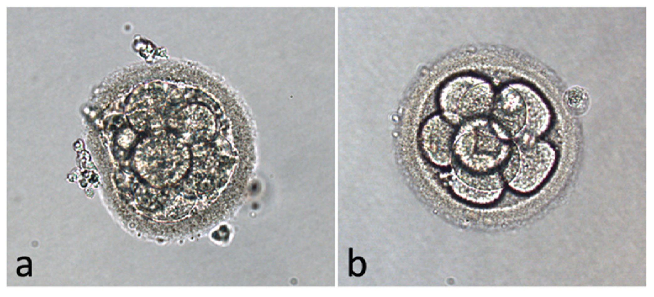
2.9. Embryo Score
2.10. Statistical Analysis
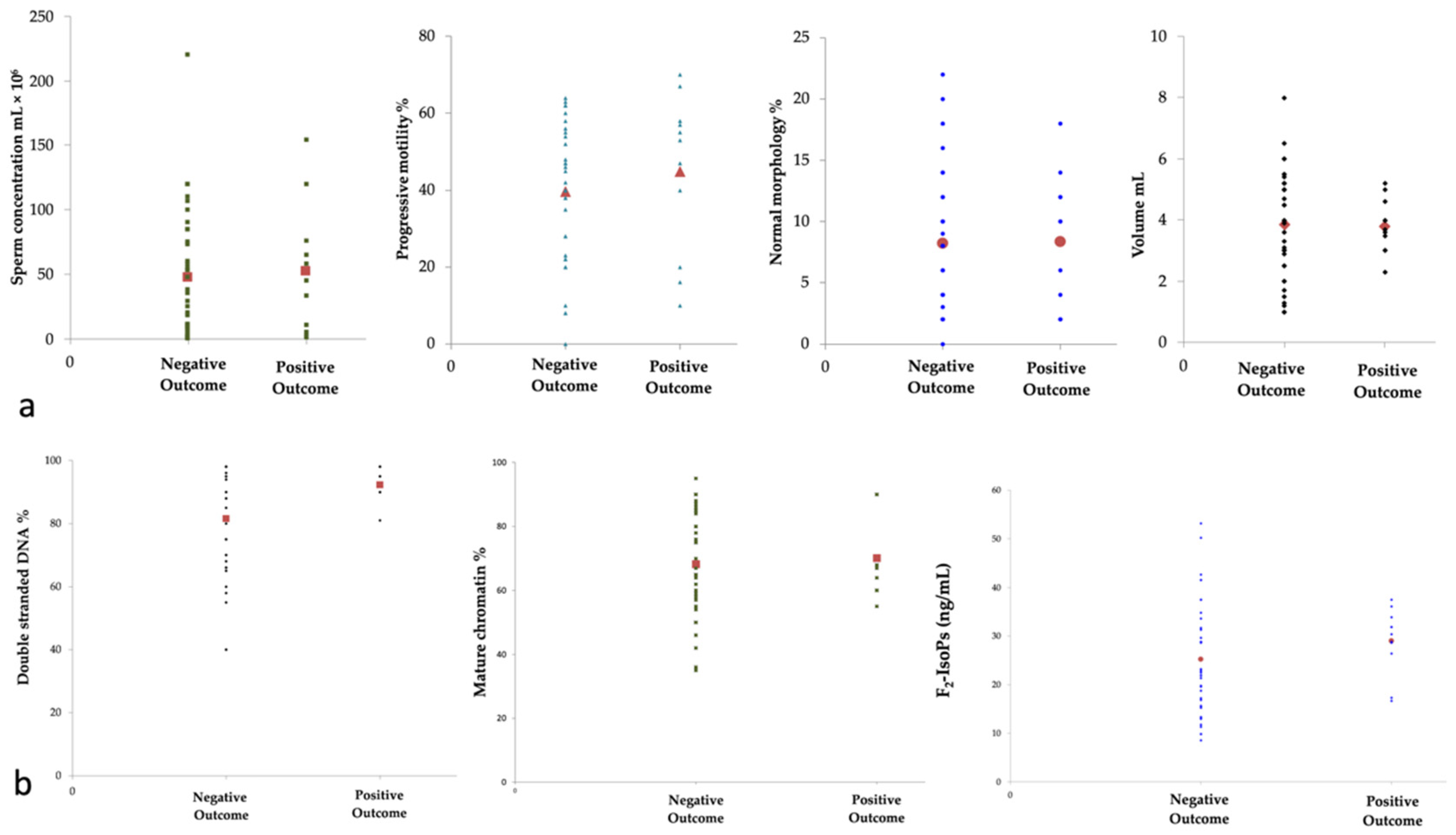
3. Results
4. Discussion
5. Conclusions
Author Contributions
Funding
Institutional Review Board Statement
Informed Consent Statement
Data Availability Statement
Conflicts of Interest
References
- Aitken, R.J. Reactive oxygen species as mediators of sperm capacitation and pathological damage. Mol. Reprod. Dev. 2017, 84, 1039–1052. [Google Scholar] [CrossRef] [PubMed]
- Aitken, R.J.; Baker, M.A.; De Iuliis, G.N.; Nixon, B. New insights into sperm physiology and pathology. In Handbook of Experimental Pharmacology; Springer: Berlin/Heidelberg, Germany, 2010; Volume 198, pp. 99–115. [Google Scholar]
- Bisht, S.; Faiq, M.; Tolahunase, M.; Dada, R. Oxidative stress and male infertility. Nat. Rev. Urol. 2017, 14, 470–485. [Google Scholar] [CrossRef]
- Dandekar, S.P.; Nadkarni, G.D.; Kulkarni, V.S.; Punekar, S. Lipid peroxidation and antioxidant enzymes in male infertility. J. Postgrad Med. 2002, 48, 186–189. [Google Scholar] [PubMed]
- Collodel, G.; Moretti, E.; Micheli, L.; Menchiari, A.; Moltoni, L.; Cerretani, D. Semen characteristics and malondialdehyde levels in men with different reproductive problems. Andrology 2015, 3, 280–286. [Google Scholar] [CrossRef] [PubMed]
- Moazamian, R.; Polhemus, A.; Connaughton, H.; Fraser, B.; Whiting, S.; Gharagozloo, P.; Aitken, R.J. Oxidative stress and human spermatozoa: Diagnostic and functional significance of aldehydes generated as a result of lipid peroxidation. Mol. Hum. Reprod. 2015, 21, 502–515. [Google Scholar] [CrossRef] [PubMed] [Green Version]
- Signorini, C.; Moretti, E.; Collodel, G. Role of isoprostanes in human male infertility. Syst. Biol. Reprod. Med. 2020, 66, 291–299. [Google Scholar] [CrossRef] [PubMed]
- Collodel, G.; Signorini, C.; Nerucci, F.; Gambera, L.; Iacoponi, F.; Moretti, E. Semen Biochemical Components in Varicocele, Leukocytospermia, and Idiopathic Infertility. Reprod. Sci. 2021, 28, 91–101. [Google Scholar] [CrossRef]
- Longini, M.; Moretti, E.; Signorini, C.; Noto, D.; Iacoponi, F.; Collodel, G. Relevance of seminal F2-dihomo-IsoPs, F2-IsoPs and F4-NeuroPs in idiopathic infertility and varicocele. Prostaglandins Other Lipid Mediat 2020, 149, 106448. [Google Scholar] [CrossRef]
- Khosrowbeygi, A.; Zarghami, N. Levels of oxidative stress biomarkers in seminal plasma and their relationship with seminal parameters. BMC Clin. Pathol. 2007, 7, 6. [Google Scholar] [CrossRef] [Green Version]
- Nadjarzadeh, A.; Shidfar, F.; Amirjannati, N.; Vafa, M.R.; Motevalian, S.A.; Gohari, M.R.; Nazeri Kakhki, S.A.; Akhondi, M.M.; Sadeghi, M.R. Effect of Coenzyme Q10 supplementation on antioxidant enzymes activity and oxidative stress of seminal plasma: A double-blind randomised clinical trial. Andrologia 2014, 46, 177–183. [Google Scholar] [CrossRef]
- Collodel, G.; Moretti, E.; Longini, M.; Pascarelli, N.A.; Signorini, C. Increased F2-Isoprostane Levels in Semen and Immunolocalization of the 8-Iso Prostaglandin F2α in Spermatozoa from Infertile Patients with Varicocele. Oxid. Med. Cell. Longev. 2018, 2018, 7508014. [Google Scholar] [CrossRef] [Green Version]
- Moretti, E.; Collodel, G.; Salvatici, M.C.; Belmonte, G.; Signorini, C. New insights into sperm with total globozoospermia: Increased fatty acid oxidation and centrin1 alteration. Syst. Biol. Reprod. Med. 2019, 65, 390–399. [Google Scholar] [CrossRef] [PubMed]
- Colaco, S.; Sakkas, D. Paternal factors contributing to embryo quality. J. Assist. Reprod. Genet. 2018, 35, 1953–1968. [Google Scholar] [CrossRef] [PubMed]
- Kini, S.; Morrell, D.; Thong, K.J.; Kopakaki, A.; Hillier, S.; Irvine, D.S. Lack of impact of semen quality on fertilization in assisted conception. Scott. Med. J. 2010, 55, 20–23. [Google Scholar] [CrossRef] [PubMed]
- Zeyad, A.; Hamad, M.; Amor, H.; Hammadeh, M.E. Relationships between bacteriospermia, DNA integrity, nuclear protamine alteration, sperm quality and ICSI outcome. Reprod. Biol. 2018, 18, 115–121. [Google Scholar] [CrossRef] [PubMed]
- Bounartzi, T.; Dafopoulos, K.; Anifandis, G.; Messini, C.I.; Koutsonikou, C.; Kouris, S.; Satra, M.; Sotiriou, S.; Vamvakopoulos, N.; Messinis, I.E. Pregnancy prediction by free sperm DNA and sperm DNA fragmentation in semen specimens of IVF/ICSI-ET patients. Hum. Fertil. 2016, 19, 56–62. [Google Scholar] [CrossRef]
- World Health Organization. WHO Laboratory Manual for the Examination and Processing of Human Semen, 5th ed.; WHO Press: Geneva, Switzerland, 2010. [Google Scholar]
- Moretti, E.; Pascarelli, N.A.; Belmonte, G.; Renieri, T.; Collodel, G. Sperm with fibrous sheath dysplasia and anomalies in head-neck junction: Focus on centriole and centrin 1. Andrologia 2017, 49, 7. [Google Scholar] [CrossRef] [PubMed]
- Signorini, C.; Comporti, M.; Giorgi, G. Ion trap tandem mass spectrometric determination of F2-isoprostanes. J. Mass Spectrom 2003, 38, 1067–1074. [Google Scholar] [CrossRef]
- Palermo, G.D.; Cohen, J.; Alikani, M.; Adler, A.; Rosenwaks, Z. Development and implementation of intracytoplasmic sperm injection (ICSI). Reprod. Fertil. Dev. 1995, 7, 211–217; discussion 217–218. [Google Scholar] [CrossRef]
- Veeck, L. Abnormal morphology of the human oocyte and conceptus. In An Atlas of Human Gametes and Conceptuses; The Parthenon Publishing Group Ltd.: Lancashire, UK, 1999. [Google Scholar]
- Milne, G.L.; Dai, Q.; Roberts, L.J., 2nd. The isoprostanes—25 years later. Biochim. Biophys. Acta 2015, 1851, 433–445. [Google Scholar] [CrossRef] [Green Version]
- Mishra, S.; Kumar, R.; Malhotra, N.; Singh, N.; Dada, R. Mild oxidative stress is beneficial for sperm telomere length maintenance. World J. Methodol. 2016, 6, 163–170. [Google Scholar] [CrossRef] [PubMed]
- Collodel, G.; Castellini, C.; Iacoponi, F.; Noto, D.; Signorini, C. Cytosolic phospholipase A2 and F2 isoprostanes are involved in semen quality and human infertility-A study on leucocytospermia, varicocele and idiopathic infertility. Andrologia 2020, 52, e13465. [Google Scholar] [CrossRef]
- Carrell, D.T.; Hammoud, S.S. The human sperm epigenome and its potential role in embryonic development. Mol. Hum. Reprod. 2010, 16, 37–47. [Google Scholar] [CrossRef] [Green Version]
- Simon, L.; Castillo, J.; Oliva, R.; Lewis, S.E. Relationships between human sperm protamines, DNA damage and assisted reproduction outcomes. Reprod. Biomed. Online 2011, 23, 724–734. [Google Scholar] [CrossRef] [PubMed] [Green Version]
- Rosen, E.M.; Mínguez-Alarcón, L.; Meeker, J.D.; Williams, P.L.; Milne, G.L.; Hauser, R.; Ferguson, K.K.; EARTH Study Team. Urinary oxidative stress biomarker levels and reproductive outcomes among couples undergoing fertility treatments. Hum. Reprod. 2019, 34, 2399–2409. [Google Scholar] [CrossRef] [Green Version]
- Agarwal, A.; Majzoub, A.; Baskaran, S.; Panner Selvam, M.K.; Cho, C.L.; Henkel, R.; Finelli, R.; Leisegang, K.; Sengupta, P.; Barbarosie, C.; et al. Sperm DNA Fragmentation: A New Guideline for Clinicians. World J. Mens Health 2020, 38, 412–471. [Google Scholar] [CrossRef]
- Basu, S. Review isoprostanes: Novel bioactive products of lipid peroxidation. Free Radic. Res. 2009, 38, 105–122. [Google Scholar] [CrossRef]
- Montero, A.; Munger, K.A.; Khan, R.Z.; Valdivielso, J.M.; Morrow, J.D.; Guasch, A.; Ziyadeh, F.N.; Badr, K.F. F(2)-isoprostanes mediate high glucose-induced TGF-beta synthesis and glomerular proteinuria in experimental type I diabetes. Kidney Int. 2000, 58, 1963–1972. [Google Scholar] [CrossRef]
- Proudfoot, J.M.; Murrey, M.W.; McLean, S.; Greenland, E.L.; Barden, A.E.; Croft, K.D.; Galano, J.M.; Durand, T.; Mori, T.A.; Pixley, F.J. F2-isoprostanes affect macrophage migration and CSF-1 signalling. Free Radic. Biol. Med. 2018, 126, 142–152. [Google Scholar] [CrossRef]
- Arezzini, B.; Vecchio, D.; Signorini, C.; Stringa, B.; Gardi, C. F2-isoprostanes can mediate bleomycin-induced lung fibrosis. Free Radic Biol. Med. 2018, 115, 1–9. [Google Scholar] [CrossRef] [PubMed] [Green Version]
- Takahashi, N.; Takeuchi, K.; Abe, T.; Murakami, K.; Yamaguchi, M.; Abe, K. Immunohistochemical localization of thromboxane receptor and thromboxane synthase in rat testis. Endocrinology 1995, 136, 4143–4146. [Google Scholar] [CrossRef]
- Pandey, A.K.; Yin, X.; Schiffer, R.B.; Hutson, J.C.; Stocco, D.M.; Grammas, P.; Wang, X. Involvement of the thromboxane A2 receptor in the regulation of steroidogenic acute regulatory gene expression in murine Leydig cells. Endocrinology 2009, 150, 3267–3273. [Google Scholar] [CrossRef] [Green Version]
- Pasqualotto, E.B.; Agarwal, A.; Sharma, R.K.; Izzo, V.M.; Pinotti, J.A.; Joshi, N.J.; Rose, B.I. Effect of oxidative stress in follicular fluid on the outcome of assisted reproductive procedures. Fertil. Steril. 2004, 81, 973–976. [Google Scholar] [CrossRef]
- Lishko, P.V.; Botchkina, I.L.; Kirichok, Y. Progesterone activates the principal Ca2+ channel of human sperm. Nature 2011, 471, 387–391. [Google Scholar] [CrossRef] [PubMed]
- Brenker, C.; Goodwin, N.; Weyand, I.; Kashikar, N.D.; Naruse, M.; Krähling, M.; Müller, A.; Kaupp, U.B.; Strünker, T. The CatSper channel: A polymodal chemosensor in human sperm. EMBO J. 2012, 31, 1654–1665. [Google Scholar] [CrossRef] [PubMed] [Green Version]
- Singh, A.P.; Rajender, S. CatSper channel, sperm function and male fertility. Reprod. Biomed. Online 2015, 1, 28–38. [Google Scholar] [CrossRef] [PubMed] [Green Version]
- Lishko, P.V.; Mannowetz, N. CatSper: A Unique Calcium Channel of the Sperm Flagellum. Curr. Opin. Physiol. 2018, 109–113. [Google Scholar] [CrossRef] [PubMed]
- Zerbinati, C.; Caponecchia, L.; Rago, R.; Leoncini, E.; Bottaccioli, A.G.; Ciacciarelli, M.; Pacelli, A.; Salacone, P.; Sebastianelli, A.; Pastore, A.; et al. Fatty acids profiling reveals potential candidate markers of semen quality. Andrology 2016, 4, 1094–1101. [Google Scholar] [CrossRef]
- Zhang, G.; Yang, W.; Zou, P.; Jiang, F.; Zeng, Y.; Chen, Q.; Sun, L.; Yang, H.; Zhou, N.; Wang, X.; et al. Mitochondrial functionality modifies human sperm acrosin activity, acrosome reaction capability and chromatin integrity. Hum. Reprod. 2019, 34, 3–11. [Google Scholar] [CrossRef]
- Spanò, M.; Bonde, J.P.; Hjøllund, H.I.; Kolstad, H.A.; Cordelli, E.; Leter, G. Sperm chromatin damage impairs human fertility. The Danish First Pregnancy Planner Study Team. Fertil. Steril. 2000, 73, 43–50. [Google Scholar] [CrossRef]
- Collins, J.A.; Barnhart, K.T.; Schlegel, P.N. Do sperm DNA integrity tests predict pregnancy with in vitro fertilization? Fertil. Steril. 2008, 89, 823–831. [Google Scholar] [CrossRef] [PubMed]
- Zini, A.; Boman, J.M.; Belzile, E.; Ciampi, A. Sperm DNA damage is associated with an increased risk of pregnancy loss after IVF and ICSI: Systematic review and meta-analysis. Hum. Reprod. 2008, 23, 2663–2668. [Google Scholar] [CrossRef] [PubMed] [Green Version]


| Sperm Concentration mL × 106 | Progressive Motility % | Normal Morphology % | Volume mL | Double-Stranded DNA% | Mature Chromatin % | F2-IsoPs ng/mL | |
|---|---|---|---|---|---|---|---|
| Sperm Concentration mL × 106 | 1 | ||||||
| Progressive Motility % | 0.716 (p = 0.000) | 1 | |||||
| Normal Morphology % | 0.745 (p = 0.000) | 0.826 (p = 0.001) | 1 | ||||
| Volume mL | 0.05 ns | 0.022 ns | 0.158 ns | 1 | |||
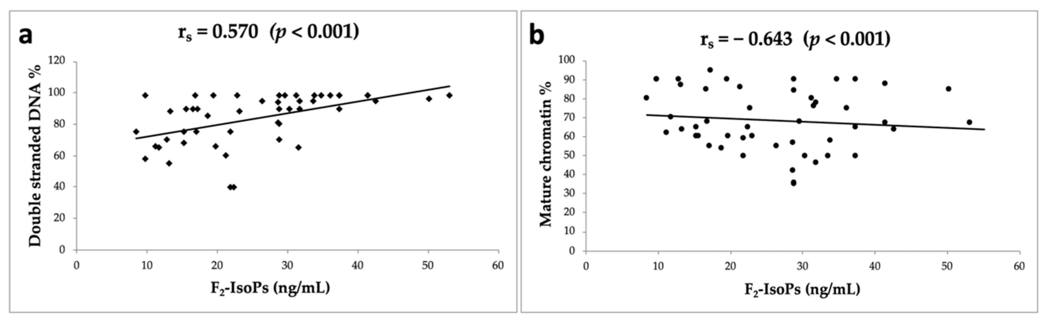
| Double-Stranded DNA% | −0.063 ns | −0.093 ns | 0.213 ns | 0.059 ns | 1 | ||
| Mature Chromatin % | 0.356 ns | 0.391 ns | 0.461 (p = 0.001) | 0.194 ns | −0.016 ns | 1 | |
| F2-IsoPs ng/mL | −0.156 ns | −0.151 ns | −0.084 ns | −0.284 Ns | 0.570 (p = 0.000) | −0.643 (p = 0.000) | 1 |
| Variables | Exp(B)/OR | Significance (p) | 95% CI for Exp(B)/OR | |
|---|---|---|---|---|
| Lower | Upper | |||
| Sperm Concentration × 106 | 0.999 | 0.962 | 0.979 | 1.020 |
| Volume mL | 1.018 | 0.950 | 0.588 | 1.762 |
| F2-IsoPs ng/mL | 1.020 | 0.825 | 0.856 | 1.215 |
| Mature Chromatin % | 1.028 | 0.594 | 0.930 | 1.136 |
| Normal Morphology % | 0.710 | 0.058 | 0.498 | 1.012 |
| Variable(s) entered in Step 1: Sperm Concentration mL × 106; Volume (mL); F2-Isoprostanes (ng/mL); Mature Chromatin; Normal Morphology | ||||
| Parameters tested for positive outcome | ||||
| Progressive Motility % | 1.111 | 0.031 | 1.010 | 1.222 |
| Double-Stranded DNA % | 1.127 | 0.026 | 1.014 | 1.253 |
| Variables | Exp(B)/OR | Significance (p) | 95% CI for Exp(B)/OR | |
|---|---|---|---|---|
| Lower | Upper | |||
| Sperm Concentration × 106 | 0.994 | 0.473 | 0.977 | 1.011 |
| Volume mL | 1.386 | 0.208 | 0.834 | 2.303 |
| Progressive Motility % | 1.026 | 0.426 | 0.963 | 1.094 |
| Mature Chromatin % | 0.988 | 0.757 | 0.916 | 1.066 |
| Normal Morphology % | 0.904 | 0.341 | 0.735 | 1.113 |
| Double-Stranded DNA % | 1.029 | 0.397 | 0.964 | 1.098 |
| Variable(s) entered in Step 1: Sperm Concentration mL × 106; Volume (mL); Mature Chromatin; Normal Morphology; Progressive Motility; Double-Stranded DNA % | ||||
| Parameters tested for embryo quality | ||||
| F2-IsoPs ng/mL | 1.082 | 0.019 | 1.013 | 1.155 |
Publisher’s Note: MDPI stays neutral with regard to jurisdictional claims in published maps and institutional affiliations. |
© 2021 by the authors. Licensee MDPI, Basel, Switzerland. This article is an open access article distributed under the terms and conditions of the Creative Commons Attribution (CC BY) license (https://creativecommons.org/licenses/by/4.0/).
Share and Cite
Collodel, G.; Noto, D.; Signorini, C.; Gambera, L.; Stendardi, A.; Mahmutbegovic, A.; Micheli, L.; Menchiari, A.; Moretti, E. Do Seminal Isoprostanes Have a Role in Assisted Reproduction Outcome? Life 2021, 11, 675. https://doi.org/10.3390/life11070675
Collodel G, Noto D, Signorini C, Gambera L, Stendardi A, Mahmutbegovic A, Micheli L, Menchiari A, Moretti E. Do Seminal Isoprostanes Have a Role in Assisted Reproduction Outcome? Life. 2021; 11(7):675. https://doi.org/10.3390/life11070675
Chicago/Turabian StyleCollodel, Giulia, Daria Noto, Cinzia Signorini, Laura Gambera, Anita Stendardi, Amra Mahmutbegovic, Lucia Micheli, Andrea Menchiari, and Elena Moretti. 2021. "Do Seminal Isoprostanes Have a Role in Assisted Reproduction Outcome?" Life 11, no. 7: 675. https://doi.org/10.3390/life11070675