Article

Overexpression of p75NTR in Human Seminoma: A New Biomarker?

1
Kidney and Transplantation Research Center, Annunziata Hospital, 89100 Cosenza, Italy
2
Department of Pharmacy, Health and Nutritional Sciences, University of Calabria, 87036 Rende, Italy
3
Department of Pathological Anatomy, “Pugliese-Ciaccio” Hospital, 88100 Catanzaro, Italy
4
deHealth Lab—DIMEG, UNICAL, Arcavacata di Rende (C.S.), 87036 Cosenza, Italy
5
Department of Health Sciences, University “Magna Græcia”, 88100 Catanzaro, Italy
6
Department of Experimental and Clinical Medicine, University “Magna Græcia”, 88100 Catanzaro, Italy
7
Department of Clinical and Experimental Medicine, University of Catania, 95100 Catania, Italy
*
Author to whom correspondence should be addressed.
These authors contributed equally to the work.
Life 2021, 11(7), 629; https://doi.org/10.3390/life11070629
Submission received: 26 May 2021 / Revised: 15 June 2021 / Accepted: 24 June 2021 / Published: 29 June 2021
(This article belongs to the Section Reproductive and Developmental Biology)

Abstract

:
Several studies have demonstrated that the p75NTR low-affinity receptor of Nerve Growth Factor (NGF), is produced in abnormally large amounts in several human cancer types. However, the role of p75NTR varies substantially depending on cell context, so that a dual role of this receptor protein in tumor cell survival and invasion, as well as cell death, has been supported in recent studies. Herein we explored for the first time the expression of p75NTR in human specimens (nr = 40) from testicular germ cell tumors (TGCTs), mostly seminomas. Nuclear overexpression of p75NTR was detected by immunohistochemistry in seminoma tissue as compared to normal tissue, whereas neither NGF nor its high-affinity TrkA receptor was detected. An increased nuclear staining of phospho-JNK, belonging to the p75NTR signaling pathway and its pro-apoptotic target gene, p53, was concomitantly observed. Interestingly, our analysis revealed that decreased expression frequency of p75NTR, p-JNK and p53 was related to staging progression, thus suggesting that p75NTR may represent a specific marker for seminoma and staging in TGCTs.

1. Introduction

Testicular germ cell tumors (TGCTs) are the most common tumors in adolescents and young men, accounting for almost all testicular cancers and their incidence is rising especially among Caucasians [1]. TGCTs are divided into seminoma germ cell tumors (SGCT) and non-seminoma germ cell tumors (NSGCT), the latter including undifferentiated (embryonal carcinoma) or differentiated (teratoma, yolk sac tumor and choriocarcinoma) tumors [2]. TGCTs have exceptional cure rates compared to other tumors, as 95% of affected men survive over 5 years [3]. Therefore, accurate staging and correct histopathological recognition of these tumors are critical for targeted therapy and optimal outcomes. Recent proteomic analyses of TGCTs identified novel proteins that might be used to better understand the molecular mechanism(s) involved in TGCTs [4]. As reported elsewhere [5], the histogenesis of TGCTs is complex and it is thought that they might develop from premalignant intratubular germ cell neoplasia (IGCN) that progresses toward invasive seminoma and/or non-seminoma after puberty when cells start to proliferate under the influence of hormones [6]. It is believed that TGCTs may arise from the failure of normal maturation of gonocytes, although, to date, the exact molecular derangements underlying this transformation are not clearly understood, even if the most common genetic finding is the gain of genetic material from chromosome 12p [7]. Since mammalian spermatogenesis is an intricate sequential process of germ cell differentiation from primordial germ cells or spermatogonial stem cells to functional haploid sperm that occurs via a complex interaction between germ and somatic cells, a better knowledge of the regulatory control of spermatogenesis should provide crucial insights into the occurrence and features of TGCTs [8,9,10,11]. Studies in human and animal models have demonstrated that during testicular development, the neurotrophins, a family of polypeptide growth factors and their receptors, are expressed in germ cells throughout their development and also in somatic cells, suggesting that the activation of their receptors may be important in testicular development and spermatogenesis [12,13,14,15].
Among neurotrophins, Nerve Growth Factor (NGF) and its high-affinity receptor, TrkA, exerts before birth an important role in seminiferous cord formation, germ cell differentiation and Sertoli cell viability; after birth, they are involved in sperm motility and acrosome reaction. Fewer data are available on the potential function of the low-affinity NGF receptor p75NTR, a member of the tumor necrosis receptor superfamily, which seems to have important implications in early testicular development [16]. Interestingly, immunohistochemistry studies have demonstrated that NGF/TrkA/p75NTR are expressed in human breast, ovarian and prostatic cancers, suggesting that they may represent new diagnostic markers. Concomitantly, in vitro and in vivo cancer models showed that NGF is involved in cancer cell progression, invasion and chemoresistance, underlying that the NGF axis may represent a new therapeutic target in these tumors [17].
To our knowledge, there are no studies that have investigated the expression pattern of NGF/TrkA and p75NTR in testicular cancer. To better understand the potential implications relevant for the NGF signaling pathway in TGCTs, the objective of the current study was to explore the expression of NGF and its receptors particularly in human testicular seminoma, the most common histological type of TGCTs.

2. Materials and Methods

2.1. Antibodies

The following primary antibodies were used: anti-p75NTR, anti-NGF, anti-TrkA, anti-p53 (Santa Cruz Biotechnology, Santa Cruz, CA, USA), anti-phospho-JNK; (Cell Signaling Technology, Milan, Italy).
Biotinylated goat-anti-rabbit, anti-mouse, anti-donkey IgGs were used as secondary antibodies (Santa Cruz Biotechnology, Santa Cruz, CA, USA).

2.2. Human Tissues

Forty formalin-fixed and paraffin-embedded samples of TGCTs, stored from the 1 January 2015 to 31 December 2019 in the archives of the Division of Pathology, Hospital “A. Pugliese”, Catanzaro (Italy), were collected. Only samples from primary tumors before chemotherapy were selected. The clinicopathological data collected included age, date of diagnosis, histological type, tumor grade (when applicable) and the presence of vascular invasion, according to testicular cancer guidelines of Associazione Italiana di Oncologia Medica (AIOM) 2018 [18]. The median age of the 40 patients was 38 years (ranging from 25 to 51 years); all patients were Caucasian. More than half of the collected specimens presented with stage I neoplasms (67.5%) and the remaining were among stages II (17.5%), III (12.5%) and IV (2.5%), respectively. Seminoma was the most frequent histological type (65%), while embryonal carcinoma and mixed GCT were less represented (27.5% and 7.5%, respectively). Control testicular tissues were obtained from 4 male patients (aged 31 and 44 years), showing testes with a like-sarcoidosis granulomatous lesion. At the time of orchidectomy, all patients gave their informed consent to use the remaining portions of tissue specimens for research purposes after their primary use for routine histologic staining. Therefore, for this study, no formal ethical approval was required for processing archival testicular tissue.

2.3. Tissue Microarray (TMA) Construction and Validation

All samples were analyzed, independently, by two pathological experts to confirm the diagnosis and delimitation of tumor areas for TMA cores. For each sample, both the tumor area and the corresponding normal tissues were selected (when available and sufficient) for triplicate cores of 1.0 mm. For TMA validation, 10 samples were randomly selected. For these 10 samples, the immunohistochemical analysis was performed on both TMA and whole sections and the results were compared.

2.4. Histopathological Analysis

A complete morphological analysis of control and seminoma samples was performed at the Division of Pathology, Hospital “A. Pugliese”, Catanzaro (Italy), upon Hematoxylin & Eosin staining. The histology slides were independently examined by the pathologists, blinded to the clinical diagnosis and the observations made by the other pathologists, careful description of tissue structural features and cellular component was performed on each sample analyzed.

2.5. Immunohistochemistry

The immunohistochemical experiments were carried out on paraffin-embedded sections from all samples. Sections of 5 μm thick, after heat-mediated antigen retrieval, were obtained. Immunodetection was performed at 4 °C overnight, using the specific primary antibodies anti-NGF (1:100), anti-TrkA (1:100), anti-p75NTR (1:100), anti-p53 (1:100), anti-posho-JNK (1:100). Then, biotinylated IgG (1:600) was applied for 1 h at room temperature, followed by avidin-biotin complex (ABC)/horseradish peroxidase (HRP). Immunoreactivity was visualized by using diaminobenzidine chromogen (DAB). Sections were also counterstained with hematoxylin. The specificity of the Abs was verified by using normal rabbit serum and normal mouse serum, respectively, instead of the primary Abs. After immunohistochemistry analysis, slides of tumor samples were visualized using an Olympus BX41 microscope and the images were taken with CSV1.14 software, using a CAM XC-30 for image acquisition.

2.6. Scoring System

Immunoreactivity for human neoplastic tissues was scored using the “Allred Score” [19], which combines a proportion and an intensity score. A proportion score was assigned representing the estimated proportion of positively stained tumor cells on a scale from 0 to 5. An intensity score was assigned by the average estimated intensity of staining in positive cells on a scale from 0 to 3. Proportion score and intensity score were added to obtain a total score that ranged from 0 to 8. A minimum of 100 cells were evaluated in each slide. Six serial sections were scored for each sample.

2.7. Statistical Analysis

The results obtained with the human samples were analyzed using Prism GraphPad (version 9.0). ROC curve was used to define the final score cutoff for positivity, based on the area under the curve. The frequency of protein expression and comparison with clinicopathological data as well as the differences in the scores between seminoma and control samples were analyzed using the one-way ANOVA. The Wilcoxon test was used after ANOVA as post hoc test. The agreement between TMA and the whole section was evaluated by the accuracy of the method.

3. Results

3.1. Morphology Analysis in Control Testis and Seminoma

The morphological analysis of control testes displayed seminiferous tubules with active spermatogenesis and Sertoli cells in the basal compartment, Leydig cells were observed in the interstitial tissue (Figure 1A). The same analysis in Seminoma samples showed large cells with prominent cell membranes containing clear cytoplasm and a hyperchromatic nucleus with a prominent nucleolus. The Seminoma cells are arranged in small clusters separated by connective tissue septae into sheets or cords. Frequently, there is a lymphocytic infiltrate (Figure 1B).

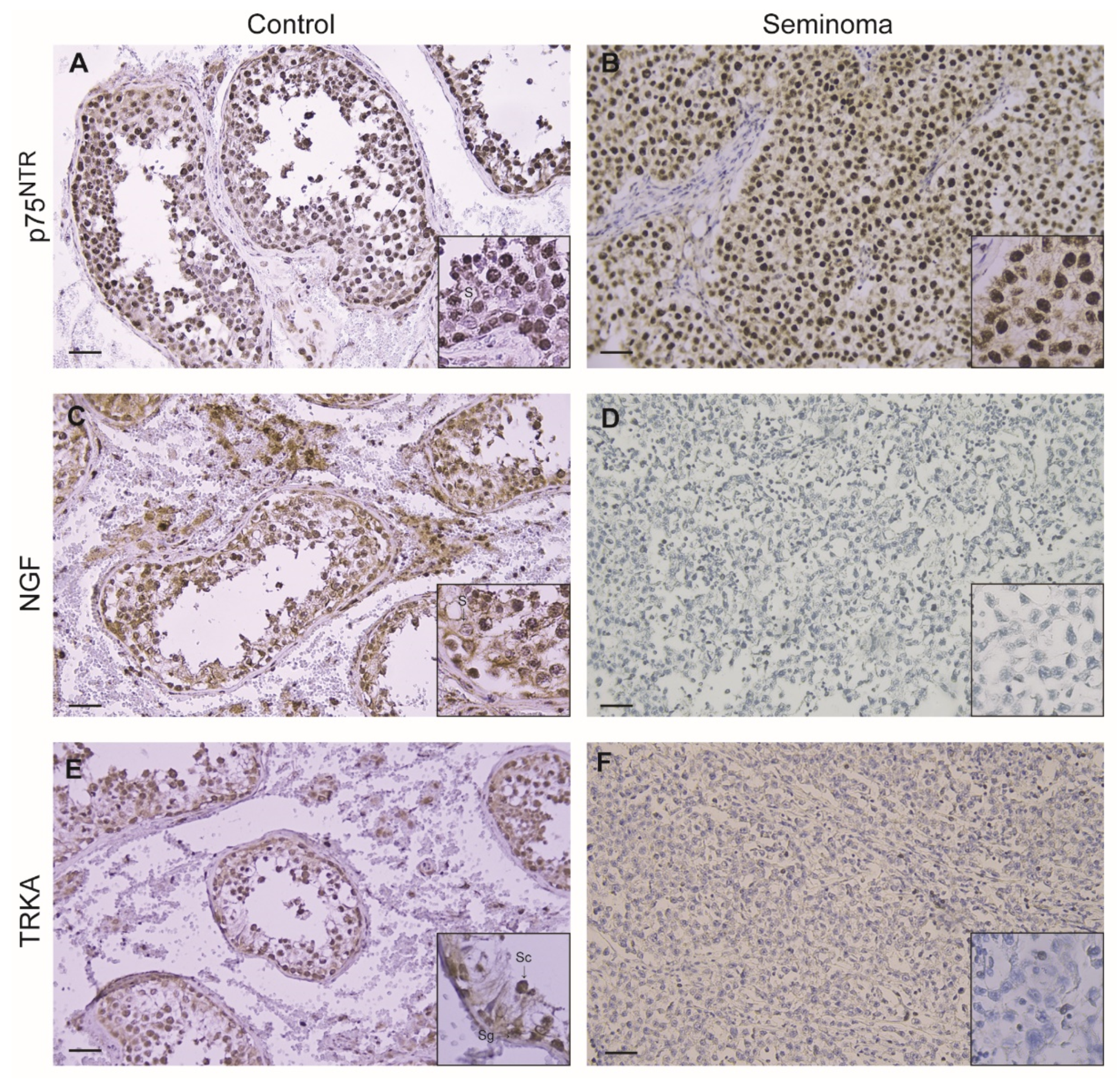
3.2. Immunohistochemical Localization of p75NTR, NGF and TrkA in Control Testis and TGCC

Control testis revealed an intense p75NTR immunoreactivity in tubular compartments, particularly in spermatogonia, spermatocytes and spermatids, while Sertoli cells were immunonegative in the interstitium; the Leydig cells showed the same strong immunopositivity. In tumoral samples, a very strong nuclear immunoreactivity was shown in big neoplastic cells while the leukocytes were immunonegative, (Figure 2A,B; Table 1). A moderate NGF immunoreactivity was evidenced only in Sertoli cells cytoplasm and in spermatogonia as well as in Leydig cell of control testes, while tumoral samples were immunonegative (Figure 2C,D; Table 1). Furthermore, although less intense, the TrkA immunopositivity in control testes was evidenced in the same germinal cells exhibiting p75NTR immunoreactivity, while tumoral samples resulted immunonegative (Figure 2E,F; Table 1). The same immunopositivity pattern for p75NTR was observed only in 4/14 of non-seminoma samples; all samples were immunonegative for NGF and TrkA (data not shown).
Cutoffs for positivity of the p75NTR marker were defined based on the area under the ROC curve, considering the sum of the score and the occurrence of clinical events (Table 2).

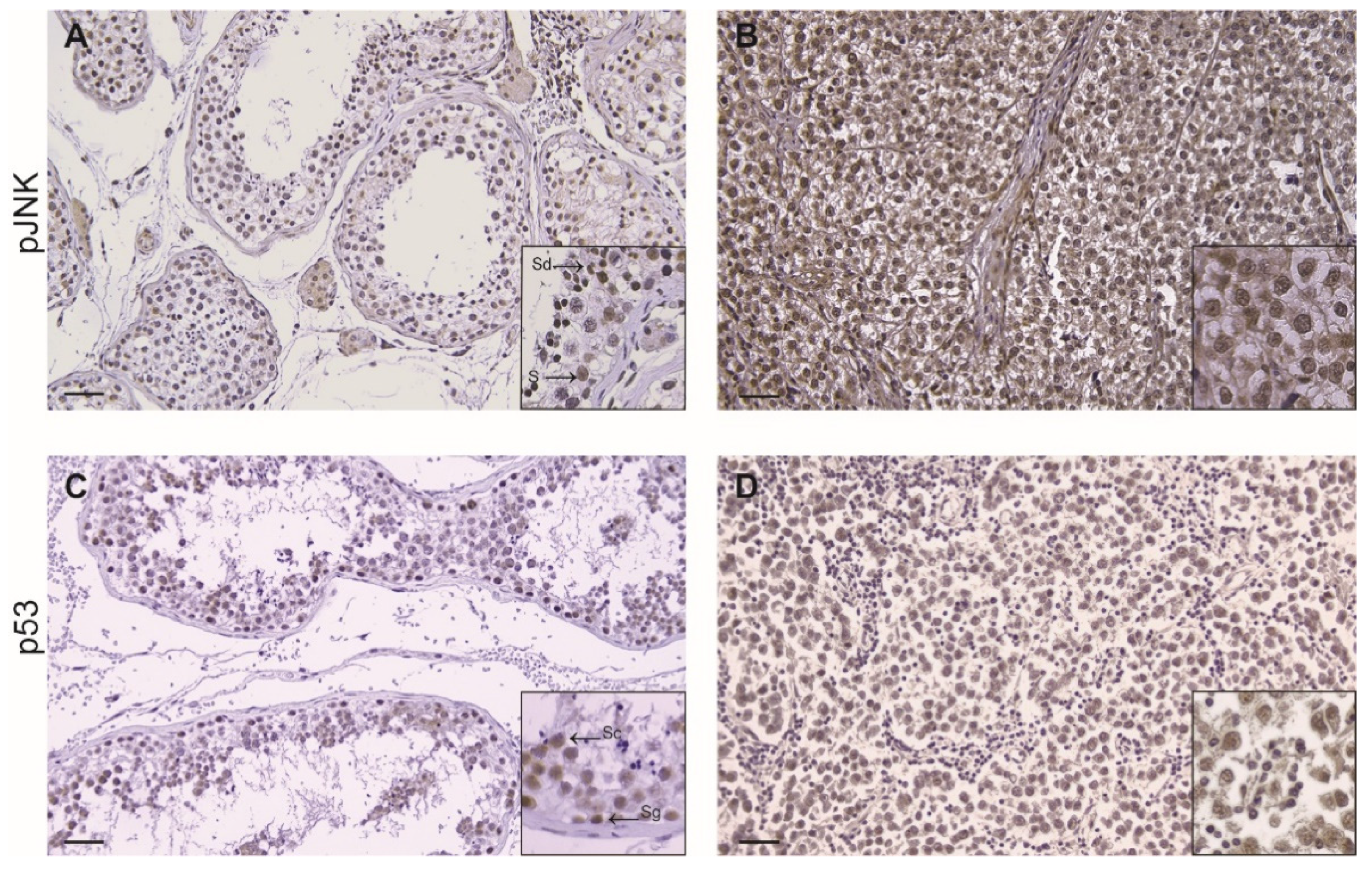
3.3. Immunohistochemistry of p53 in TGCC

Interestingly, in seminoma tissue, we evidenced an increased expression of p-JNK as compared to controls; the tumoral cells showed a strong p-JNK immunopositivity while the lymphocytes were un-stained. In the control samples the positivity was confined in Sertoli cells, in spermatids and Leydig cells, respectively (Figure 3A,B; Table 3). Moreover, immunohistochemical analysis revealed a strong nuclear immunoreactivity of p53 in seminoma samples compared to control tissue sections, where p53 immunolocalization was stronger in spermatogonia and moderate in spermatocytes (Figure 3C,D, Table 3).

3.4. Validation of TMA Method

Analysis of agreement between TMA and whole tissue sections showed an accuracy for the TMA method of 80% for NGF, 98% for TrkA and 78% for p75NTR.

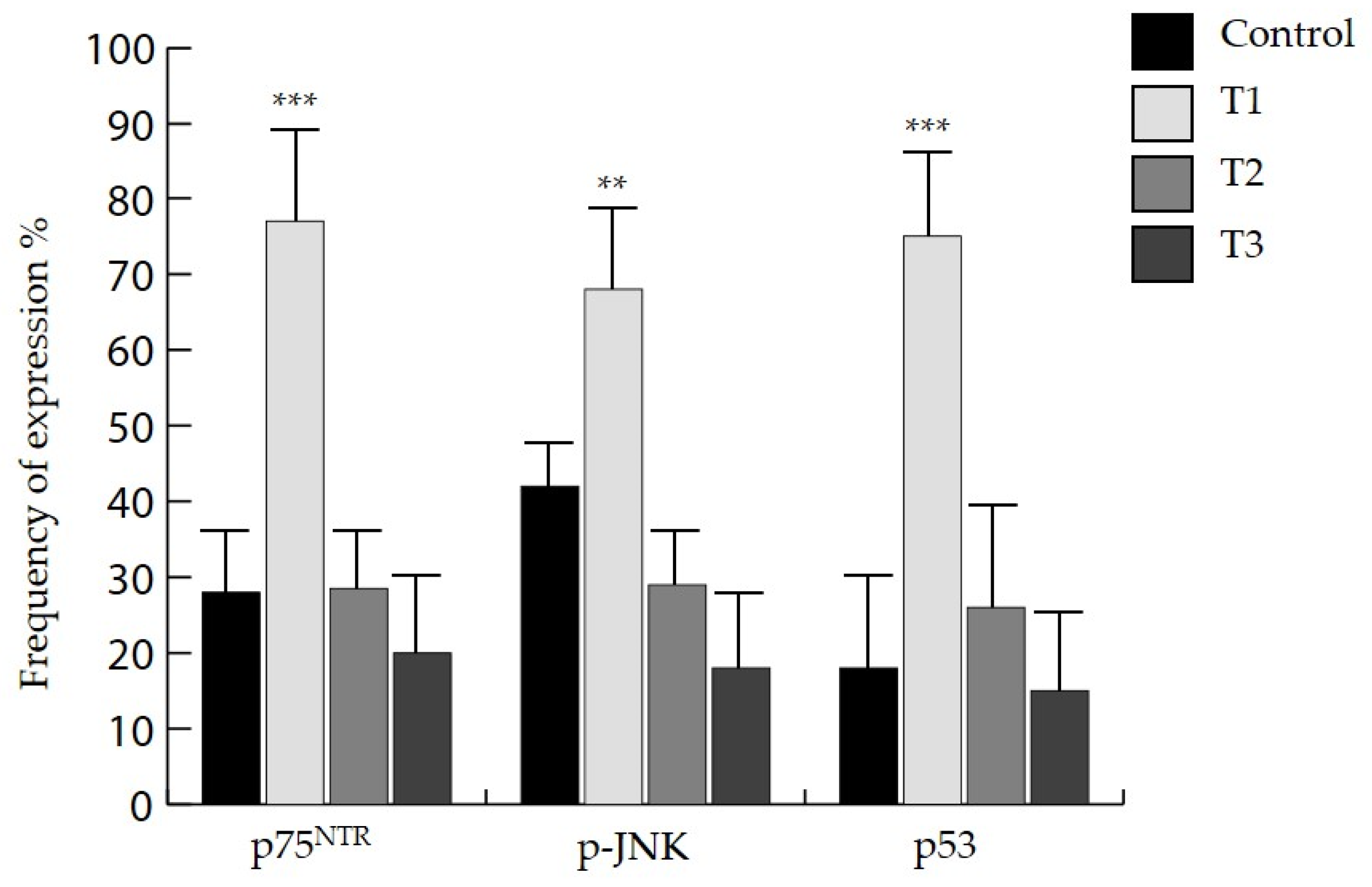
3.5. Clinico-Pathological Significance of p75NTR, p-JNK and p53

To explore the potential clinico-pathological significance of NGF, TrkA and p75NTR, we investigated the frequency of expression of the above-reported markers according to the staging (Table 4). Moreover, our analysis revealed that when compared to control tissue samples, p75NTR, p-JNK and p53 showed higher expression in samples with T1 tumor stage (p < 0.001) (Figure 4).

4. Discussion

As far as we are aware, this study represents the first description of the overexpression of p75NTR in TGCTs, particularly in testicular seminoma and of its downstream signaling molecules, phospho-JNK and p53, whose expression decreases as tumor staging worsens. Different studies have proven that p75NTR plays opposite roles in the context of different cancers, as it acts as a tumor suppressor in carcinomas of prostate, bladder, stomach and liver [20,21,22], whereas, in melanoma, pancreatic carcinoma, glioma and breast cancer, p75NTR acts as a tumor-promoting function facilitating survival and invasion of cancer cells [23,24,25].
Although the involvement of p75NTR has been investigated in tumors affecting the female reproductive system, to our knowledge there are no studies that have explored the expression of p75NTR in TGCTs, in particular in testicular seminoma, which represents the most frequent testicular neoplasm in young men [1,26].
Previous studies have been carried out to identify new molecular markers for TGCTs. Preliminary reports demonstrated that most cells in seminoma express Pituitary-tumor-transforming-gene 1 (PTTG1), as well as Octamer-binding transcription factor 4 (OCT-4) and Krüppel-like factor 4 (KLF-4) [27]. The authors firstly demonstrated that PTTG1 marks some specific OCT4- and KLF4-positive tumor cells, mainly localized at the periphery of the neoplasm. In the intratubular infiltration areas, nests of cells expressing both OCT4/KLF4 and PTTG1 presence were consistent for a sub-population of tumor stem cells OCT4- and KLF4- positive [28]. In the present study, we aimed to investigate a new molecular pathway and found that the overexpression of p75NTR may have a pathogenetic role in testicular germ cell cancer development, which may occur in an NGF-independent manner [29,30], as confirmed by immunohistochemical Allred-score of negative NGF staining and its pro-survival receptor, TrkA, in tumoral tissue. Interestingly, Micera et al. reported that the biological effect mediated by the NGF axis in both normal and cancer cells is related to the TrkA/p75NTR ratio and that the p75NTR expression might facilitate cell proliferation in the absence of TrkA [31].
We can speculate that p75NTR may be a biomarker of the transformed primordial germ cells, which represent the characteristic cellular pattern of testicular seminoma, in particular its pure forms [32]. The pathogenesis of overall TGTCs remains unexplored, although it is known that they originate from transformed gonocytes or undifferentiated spermatogonia [33].
Different observations using several malignancies refer to cells with a remarkable self-renewal potential and extensive proliferation capacity [34,35,36], expressing markers that characterize the stem cells of the original normal tissue [37] and that are strongly involved in growth and tumor propagation. A growing body of evidence has identified p75NTR as a robust cell surface biomarker for neural cancer-initiating or stem-like cells [38]. Interestingly, Okumura et al. demonstrated that in the stem/progenitor cell fraction of normal esophageal epithelial cells, p75NTR is necessary for tumor survival and maintenance [39]. Moreover, the in vivo study of Boiko et al. reported that melanoma tumor stem cells p75NTR-positive, but not p75NTR-negative, were remarkably capable of generating tumors, promoting metastasis and maintaining self-renewal [40].
Furthermore, as expected, we observed that control testicular tissue expressed phospho-JNK; as it has been reported that JNK is involved in regulating various testicular functions, like germ cell development and acrosome reactions, by controlling the expression of genes involved in the apoptosis or survival signaling pathways [41,42]. Concomitantly, we found that the overexpression of p75NTR in tumor tissue was accompanied by an increased nuclear expression of its downstream signaling, phospho-JNK. This result requires further investigation, as it has been extensively demonstrated that both p75NTR and JNK pathway activation can promote different biological effects, depending on cancer type [43]. Expression of p75NTR has been associated with apoptosis, through activation of JNK, which controls the expression of p53 tumor suppressor and with cell survival, through the activation of nuclear factor-kB and AKT [44]. In addition to p75NTR and phospho-JNK overexpression, our analysis revealed a strong nuclear immunoreactivity of p53 in the tumor sections compared to control, highlighting that hyperactivation of p75NTR signaling in seminoma cells may promote cancer cell apoptosis. Furthermore, we hypothesized that p75NTR overexpression may represent a positive prognostic factor in seminoma, as we observed that p75NTR, phospho-JNK and p53 expression frequency decreases with staging progression. These results are in agreement with previous reports, demonstrating an inverse association of p75NTR expression with the neoplastic progression of prostate cancer [45,46]. Interestingly, we observed a strong p75NTR immunolocalization in four non-seminoma samples (two embryonal carcinoma and two mixed TGCT); however, the small sample is not sufficient to assume a potential role of p75NTR in non-seminoma cancer.
Our study has some limitations. First, this is a retrospective study based on an archive of collected samples coming from testicular neoplasms and, therefore, the lack of access to patients’ clinical information limited the evaluation of any relationship between immunohistochemistry findings and tumor aggressiveness. In addition, we used the area under the ROC curve to assess the cutoffs for p75NTR positivity and to investigate the definition of scores better associated with clinical events. Although it may vary depending on tumor type, we used this approach to estimate the possible clinical significance of p75NTR as a biomarker. TMA method demonstrated accuracy for all proteins analyzed. The tumor heterogeneity, quite common in TGCTs, could be a limitation of this method. However, in this study, scores in triplicate for each histological subtype were used, in the attempt to minimize this limitation. Lastly, the present study was not aimed to clarify any molecular pathway that could yield to any relevant translational mechanism(s) linked to tumor progression.

5. Conclusions

In conclusion, our results suggest that p75NTR may exert some role in TGCTs since the loss of its expression may represent a specific hallmark of seminoma and possible staging in TGCTs. However, the mechanism(s) underlying the controversial and paradoxical functions of p75NTR in different cancer cells are not entirely explained at present. Larger translational studies are warranted to better clarify the underlying molecular mechanism by which P75NTR signaling may be involved in testicular germ cell tumorigenesis and represent a new marker of better staging. Therefore, further clinical studies are needed to confirm the relevance of our findings.

Author Contributions

Conceptualization, A.P. and V.R.; Methodology, V.R., R.M., L.M., L.T.; Software and data analysis, D.L. and S.L.V.; Validation V.R., A.A.; Data curation E.A.G., M.V.; Writing, Review & Editing V.R., A.P., A.A., A.B.; Supervision, R.B. and E.C. All authors have read and agreed to the published version of the manuscript.

Funding

This research received no external funding.

Institutional Review Board Statement

Not applicable.

Informed Consent Statement

The informed consent sent to the journal is this signed by patients before surgery, this informed consent allow us use of clinical data.

Data Availability Statement

Not applicable.

Conflicts of Interest

The authors declare no conflict of interest.

References

  1. Ghazarian, A.A.; Kelly, S.P.; Altekruse, S.F.; Rosenberg, P.S.; McGlynn, K.A. Future of Testicular Germ Cell Tumor Incidence in the United States: Forecast through 2026. Cancer 2017, 123, 2320–2328. [Google Scholar] [CrossRef] [Green Version]
  2. Bode, P.K.; Thielken, A.; Brandt, S.; Barghorn, A.; Lohe, B.; Knuth, A.; Moch, H. Cancer Testis Antigen Expression in Testicular Germ Cell Tumorigenesis. Mod. Pathol. 2014, 27, 899–905. [Google Scholar] [CrossRef] [PubMed] [Green Version]
  3. Le Cornet, C.; Lortet-Tieulent, J.; Forman, D.; Béranger, R.; Flechon, A.; Fervers, B.; Schüz, J.; Bray, F. Testicular Cancer Incidence to Rise by 25% by 2025 in Europe? Model-Based Predictions in 40 Countries Using Population-Based Registry Data. Eur. J. Cancer 2014, 50, 831–839. [Google Scholar] [CrossRef]
  4. Milardi, D.; Grande, G.; Vincenzoni, F.; Pierconti, F.; Pontecorvi, A. Proteomics for the Identification of Biomarkers in Testicular Cancer-Review. Front. Endocrinol. 2019, 10, 462. [Google Scholar] [CrossRef] [Green Version]
  5. Batool, A.; Karimi, N.; Wu, X.N.; Chen, S.R.; Liu, Y.X. Testicular Germ Cell Tumor: A Comprehensive Review. Cell. Mol. Life Sci. 2019, 76, 1713–1727. [Google Scholar] [CrossRef]
  6. Skakkebaek, N.E. Possible Carcinoma-in-situ of the Testis. Lancet Lond. Engl. 1972, 2, 516–517. [Google Scholar] [CrossRef]
  7. Sheikine, Y.; Genega, E.; Melamed, J.; Lee, P.; Reuter, V.E.; Ye, H. Molecular Genetics of Testicular Germ Cell Tumors. Am. J. Cancer Res. 2012, 2, 153–167. [Google Scholar] [PubMed]
  8. Chen, S.R.; Liu, Y.X. Regulation of Spermatogonial Stem Cell Self-Renewal and Spermatocyte Meiosis by Sertoli Cell Signaling. Reprod. Camb. Engl. 2015, 149, R159–R167. [Google Scholar] [CrossRef] [PubMed] [Green Version]
  9. Chen, S.R.; Zheng, Q.S.; Zhang, Y.; Gao, F.; Liu, Y.X. Disruption of Genital Ridge Development Causes Aberrant Primordial Germ Cell Proliferation but Does Not Affect Their Directional Migration. BMC Biol. 2013, 11, 22. [Google Scholar] [CrossRef] [Green Version]
  10. Rebourcet, D.; O’Shaughnessy, P.J.; Pitetti, J.L.; Monteiro, A.; O’Hara, L.; Milne, L.; Tsai, Y.T.; Cruickshanks, L.; Riethmacher, D.; Guillou, F.; et al. Sertoli Cells Control Peritubular Myoid Cell Fate and Support Adult Leydig Cell Development in the Prepubertal Testis. Dev. Camb. Engl. 2014, 141, 2139–2149. [Google Scholar] [CrossRef] [Green Version]
  11. Wang, Y.Q.; Batool, A.; Chen, S.R.; Liu, Y.X. GATA4 Is a Negative Regulator of Contractility in Mouse Testicular Peritubular Myoid Cells. Reprod. Camb. Engl. 2018, 156, 343–351. [Google Scholar] [CrossRef] [Green Version]
  12. Persson, H.; Ayer-Le Lievre, C.; Söder, O.; Villar, M.J.; Metsis, M.; Olson, L.; Ritzen, M.; Hökfelt, T. Expression of Beta-Nerve Growth Factor Receptor MRNA in Sertoli Cells Downregulated by Testosterone. Science 1990, 247, 704–707. [Google Scholar] [CrossRef]
  13. Müller, D.; Davidoff, M.S.; Bargheer, O.; Paust, H.J.; Pusch, W.; Koeva, Y.; Jezek, D.; Holstein, A.F.; Middendorff, R. The Expression of Neurotrophins and Their Receptors in the Prenatal and Adult Human Testis: Evidence for Functions in Leydig Cells. Histochem. Cell Biol. 2006, 126, 199–211. [Google Scholar] [CrossRef] [PubMed]
  14. Russo, M.A.; Odorisio, T.; Fradeani, A.; Rienzi, L.; De Felici, M.; Cattaneo, A.; Siracusa, G. Low-Affinity Nerve Growth Factor Receptor Is Expressed during Testicular Morphogenesis and in Germ Cells at Specific Stages of Spermatogenesis. Mol. Reprod. Dev. 1994, 37, 157–166. [Google Scholar] [CrossRef]
  15. Djakiew, D.; Pflug, B.; Dionne, C.; Onoda, M. Postnatal Expression of Nerve Growth Factor Receptors in the Rat Testis. Biol. Reprod. 1994, 51, 214–221. [Google Scholar] [CrossRef] [PubMed] [Green Version]
  16. Li, C.; Zhou, X. The Potential Roles of Neurotrophins in Male Reproduction. Reprod. Camb. Engl. 2013, 145, R89–R95. [Google Scholar] [CrossRef] [Green Version]
  17. Griffin, N.; Faulkner, S.; Jobling, P.; Hondermarck, H. Targeting Neurotrophin Signaling in Cancer: The Renaissance. Pharmacol. Res. 2018, 135, 12–17. [Google Scholar] [CrossRef] [PubMed]
  18. AIOM—Associazione Italiana di Oncologia Medica. Tumore del Testicolo: Linee Guida; AIOM: Milan, Italy, 2018. [Google Scholar]
  19. Allred, D.C.; Harvey, J.M.; Berardo, M.; Clark, G.M. Prognostic and Predictive Factors in Breast Cancer by Immunohistochemical Analysis. Mod. Pathol. 1998, 11, 155–168. [Google Scholar]
  20. Jin, H.; Pan, Y.; He, L.; Zhai, H.; Li, X.; Zhao, L.; Sun, L.; Liu, J.; Hong, L.; Song, J.; et al. P75 Neurotrophin Receptor Inhibits Invasion and Metastasis of Gastric Cancer. Mol. Cancer Res. 2007, 5, 423–433. [Google Scholar] [CrossRef] [Green Version]
  21. Khwaja, F.S.; Quann, E.J.; Pattabiraman, N.; Wynne, S.; Djakiew, D. Carprofen Induction of P75NTR-Dependent Apoptosis via the P38 Mitogen-Activated Protein Kinase Pathway in Prostate Cancer Cells. Mol. Cancer Ther. 2008, 7, 3539–3545. [Google Scholar] [CrossRef] [Green Version]
  22. Gravina, G.L.; Marampon, F.; Sanità, P.; Mancini, A.; Colapietro, A.; Scarsella, L.; Jitariuc, A.; Biordi, L.; Ficorella, C.; Festuccia, C. Increased Expression and Activity of P75NTR Are Crucial Events in Azacitidine-Induced Cell Death in Prostate Cancer. Oncol. Rep. 2016, 36, 125–130. [Google Scholar] [CrossRef]
  23. Marchetti, D.; Aucoin, R.; Blust, J.; Murry, B.; Greiter-Wilke, A. P75 Neurotrophin Receptor Functions as a Survival Receptor in Brain-Metastatic Melanoma Cells. J. Cell. Biochem. 2004, 91, 206–215. [Google Scholar] [CrossRef]
  24. Wang, L.; Rahn, J.J.; Lun, X.; Sun, B.; Kelly, J.J.; Weiss, S.; Robbins, S.M.; Forsyth, P.A.; Senger, D.L. Gamma-Secretase Represents a Therapeutic Target for the Treatment of Invasive Glioma Mediated by the P75 Neurotrophin Receptor. PLoS Biol. 2008, 6, e289. [Google Scholar] [CrossRef] [PubMed]
  25. Verbeke, S.; Tomellini, E.; Dhamani, F.; Meignan, S.; Adriaenssens, E.; Xuefen, L.B. Extracellular Cleavage of the P75 Neurotrophin Receptor Is Implicated in Its Pro-Survival Effect in Breast Cancer Cells. FEBS Lett. 2013, 587, 2591–2596. [Google Scholar] [CrossRef] [PubMed] [Green Version]
  26. Dieckmann, K.P.; Richter-Simonsen, H.; Kulejewski, M.; Ikogho, R.; Zecha, H.; Anheuser, P.; Pichlmeier, U.; Isbarn, H. Testicular Germ-Cell Tumours: A Descriptive Analysis of Clinical Characteristics at First Presentation. Urol. Int. 2018, 100, 409–419. [Google Scholar] [CrossRef]
  27. Pierconti, F.; Milardi, D.; Martini, M.; Grande, G.; Cenci, T.; Gulino, G.; Larocca, L.M.; Rindi, G.; Pontecorvi, A.; De Marinis, L. Pituitary-Tumour-Transforming-Gene 1 Expression in Testicular Cancer. Andrologia 2015, 47, 427–432. [Google Scholar] [CrossRef] [PubMed]
  28. Grande, G.; Milardi, D.; Martini, M.; Cenci, T.; Gulino, G.; Mancini, F.; Bianchi, A.; Pontecorvi, A.; Pierconti, F. Protein Expression of PTTG-1, OCT-4, and KLF-4 in Seminoma: A Pilot Study. Front. Endocrinol. 2019, 10, 619. [Google Scholar] [CrossRef] [PubMed]
  29. Rabizadeh, S.; Oh, J.; Zhong, L.T.; Yang, J.; Bitler, C.M.; Butcher, L.L.; Bredesen, D.E. Induction of Apoptosis by the Low-Affinity NGF Receptor. Science 1993, 261, 345–348. [Google Scholar] [CrossRef]
  30. Gentry, J.J.; Casaccia-Bonnefil, P.; Carter, B.D. Nerve Growth Factor Activation of Nuclear Factor KappaB through Its P75 Receptor Is an Anti-Apoptotic Signal in RN22 Schwannoma Cells. J. Biol. Chem. 2000, 275, 7558–7565. [Google Scholar] [CrossRef] [Green Version]
  31. Micera, A.; Lambiase, A.; Stampachiacchiere, B.; Bonini, S.; Bonini, S.; Levi-Schaffer, F. Nerve Growth Factor and Tissue Repair Remodeling: TrkA(NGFR) and P75(NTR), Two Receptors One Fate. Cytokine Growth Factor Rev. 2007, 18, 245–256. [Google Scholar] [CrossRef]
  32. Sperger, J.M.; Chen, X.; Draper, J.S.; Antosiewicz, J.E.; Chon, C.H.; Jones, S.B.; Brooks, J.D.; Andrews, P.W.; Brown, P.O.; Thomson, J.A. Gene Expression Patterns in Human Embryonic Stem Cells and Human Pluripotent Germ Cell Tumors. Proc. Natl. Acad. Sci. USA 2003, 100, 13350–13355. [Google Scholar] [CrossRef] [PubMed] [Green Version]
  33. Rajpert-De Meyts, E. Developmental Model for the Pathogenesis of Testicular Carcinoma in Situ: Genetic and Environmental Aspects. Hum. Reprod. Update 2006, 12, 303–323. [Google Scholar] [CrossRef] [Green Version]
  34. Al-Hajj, M.; Wicha, M.S.; Benito-Hernandez, A.; Morrison, S.J.; Clarke, M.F. Prospective Identification of Tumorigenic Breast Cancer Cells. Proc. Natl. Acad. Sci. USA 2003, 100, 3983–3988. [Google Scholar] [CrossRef] [Green Version]
  35. Singh, S.K.; Hawkins, C.; Clarke, I.D.; Squire, J.A.; Bayani, J.; Hide, T.; Henkelman, R.M.; Cusimano, M.D.; Dirks, P.B. Identification of Human Brain Tumour Initiating Cells. Nature 2004, 432, 396–401. [Google Scholar] [CrossRef]
  36. Hemmati, H.D.; Nakano, I.; Lazareff, J.A.; Masterman-Smith, M.; Geschwind, D.H.; Bronner-Fraser, M.; Kornblum, H.I. Cancerous Stem Cells Can Arise from Pediatric Brain Tumors. Proc. Natl. Acad. Sci. USA 2003, 100, 15178–15183. [Google Scholar] [CrossRef] [Green Version]
  37. Dean, M.; Fojo, T.; Bates, S. Tumour Stem Cells and Drug Resistance. Nat. Rev. Cancer 2005, 5, 275–284. [Google Scholar] [CrossRef]
  38. Tomellini, E.; Lagadec, C.; Polakowska, R.; Le Bourhis, X. Role of P75 Neurotrophin Receptor in Stem Cell Biology: More than Just a Marker. Cell. Mol. Life Sci. 2014, 71, 2467–2481. [Google Scholar] [CrossRef]
  39. Okumura, T.; Tsunoda, S.; Mori, Y.; Ito, T.; Kikuchi, K.; Wang, T.C.; Yasumoto, S.; Shimada, Y. The Biological Role of the Low-Affinity P75 Neurotrophin Receptor in Esophageal Squamous Cell Carcinoma. Clin. Cancer Res. 2006, 12, 5096–5103. [Google Scholar] [CrossRef] [Green Version]
  40. Boiko, A.D.; Razorenova, O.V.; van de Rijn, M.; Swetter, S.M.; Johnson, D.L.; Ly, D.P.; Butler, P.D.; Yang, G.P.; Joshua, B.; Kaplan, M.J.; et al. Human Melanoma-Initiating Cells Express Neural Crest Nerve Growth Factor Receptor CD271. Nature 2010, 466, 133–137. [Google Scholar] [CrossRef] [PubMed]
  41. Herrera, S.C.; Bach, E.A. JNK Signaling Triggers Spermatogonial Dedifferentiation during Chronic Stress to Maintain the Germline Stem Cell Pool in the Drosophila Testis. eLife 2018, 7, e36095. [Google Scholar] [CrossRef] [PubMed]
  42. Kyriakis, J.M.; Banerjee, P.; Nikolakaki, E.; Dai, T.; Rubie, E.A.; Ahmad, M.F.; Avruch, J.; Woodgett, J.R. The Stress-Activated Protein Kinase Subfamily of c-Jun Kinases. Nature 1994, 369, 156–160. [Google Scholar] [CrossRef] [PubMed]
  43. Bubici, C.; Papa, S. JNK Signalling in Cancer: In Need of New, Smarter Therapeutic Targets. Br. J. Pharmacol. 2014, 171, 24–37. [Google Scholar] [CrossRef] [PubMed]
  44. Roux, P.P.; Bhakar, A.L.; Kennedy, T.E.; Barker, P.A. The P75 Neurotrophin Receptor Activates Akt (Protein Kinase B) through a Phosphatidylinositol 3-Kinase-Dependent Pathway. J. Biol. Chem. 2001, 276, 23097–23104. [Google Scholar] [CrossRef] [PubMed] [Green Version]
  45. Pflug, B.R.; Onoda, M.; Lynch, J.H.; Djakiew, D. Reduced Expression of the Low Affinity Nerve Growth Factor Receptor in Benign and Malignant Human Prostate Tissue and Loss of Expression in Four Human Metastatic Prostate Tumor Cell Lines. Cancer Res. 1992, 52, 5403–5406. [Google Scholar] [PubMed]
  46. Krygier, S.; Djakiew, D. Neurotrophin Receptor P75(NTR) Suppresses Growth and Nerve Growth Factor-Mediated Metastasis of Human Prostate Cancer Cells. Int. J. Cancer 2002, 98, 1–7. [Google Scholar] [CrossRef]
Figure 1. Hematoxylin and Eosin staining in control testis (A) and seminomas (B). Scale bars: 25 μm.
Life 11 00629 g001
Figure 2. Immunohistochemical expression of p75NTR (A,B), NGF (C,D) and TrkA (E,F), in control testis and seminomas. (S) Sertoli cells; (Sg) Spermatogonia; (Sc) Spermatocytes; (Sd) Spermatides. Scale bars: 25 μm. Inserts: higher magnifications of the images.
Life 11 00629 g002
Figure 3. Immunohistochemical expression of pJNK (A,B) and p53 (C,D) in control testis and seminomas. (S) Sertoli cells; (Sg) Spermatogonia; (Sc) Spermatocytes; (Sd) Spermatides. Scale bars: 25 μm. Inserts: higher magnifications of the images.
Life 11 00629 g003
Figure 4. Frequency of expression of p75NTR, p-JNK and p53 in different tumor T stages versus control (** p < 0.005, *** p < 0.001).
Life 11 00629 g004
Table 1. Immunostaining scores (Allred score median) of p75NTR NGF and TrkA in human control testes and seminoma samples.
Control (n = 4)Seminoma (n = 26)
p75NTR4 (3–4)8 (7–8) ***
NGF2 (2–3)0 (0–0)
TrkA3 (2–4)0 (0–0)
Immunostained scores: Total score = Proposition score + Intensity score (range 0–8). *** Seminoma versus Control = p < 0.001 (one-way Anova test).
Table 2. p75NTR cutoff based on the area under the ROC curve.
MarkerCutoffSensitivity (%)Specificity (%)PPVNPVAUC (95% CI)
p75NTR≥767.8%59.0%21.4%76.0%0.80 (0.75–0.98)
AUC—Area under ROC curve; CI—Confidence interval; PPV—Positive predictive value; NPV—Negative predictive value.
Table 3. Immunostaining scores (Allred score median) p-JNK and p53 in human control and seminoma samples.
MarkerControl (n = 4)Seminoma (n = 26)
p-JNK3 (2–3)7 (6–8) ***
P533 (2–3)7 (6–8) ***
Immunostained scores: Total score = Proposition score+ Intensity score (range 0–8). *** Seminoma versus Control = p < 0.001 (one-way ANOVA test).
Table 4. Association of NGF, TrkA and p75NTR expression with clinico-pathological parameters.
nrNGFTrkAp75NTR
Positive (%)pPositive (%)pPositive (%)p
T stage 0.91 0.40 0.001
T1271 (0.03) 5 (5.9) 21 (77.7)
T270 (0.0) 0 (0.0) 2 (28.5)
T350 (0.0) 0 (0.0) 1 (20.0)
T410 (0.0) 0 (0.0) 0 (0.0)
Vascular invasion ns ns 0.59
No340 (0.0) 0 (0.0) 13 (38.2)
Yes60 (0.0) 0 (0.0) 3 (64.3)
Histology ns ns 0.0001
Seminoma260 (0.0) 0 (0.0) 25 (96.1)
Non-seminomatous140 (0.0) 0 (0.0) 4 (28.5)
Publisher’s Note: MDPI stays neutral with regard to jurisdictional claims in published maps and institutional affiliations.

