Case Report

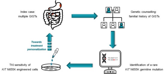
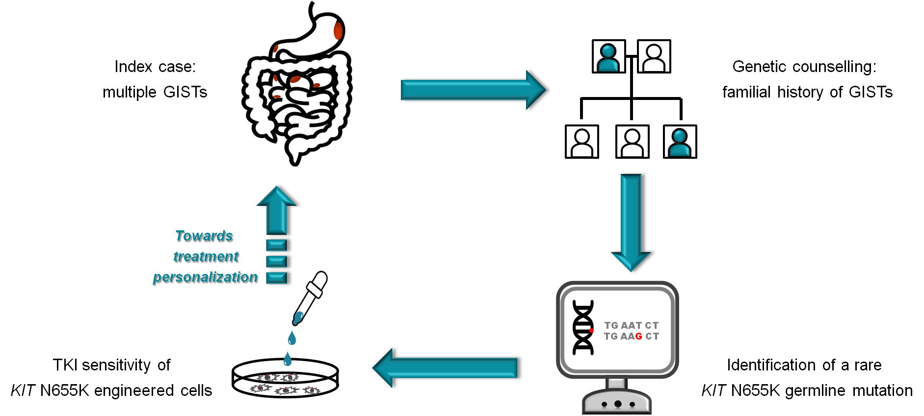
A Novel Kindred with Familial Gastrointestinal Stromal Tumors Caused by a Rare KIT Germline Mutation (N655K): Clinico-Pathological Presentation and TKI Sensitivity

1
Unit of Oncological Gastroenterology, Centro di Riferimento Oncologico di Aviano (CRO Aviano), IRCCS, 33081 Aviano, Italy
2
Unit of Oncogenetics and Functional Oncogenomics, Centro di Riferimento Oncologico di Aviano (CRO Aviano), IRCCS, 33081 Aviano, Italy
3
Unit of Medical Oncology and Cancer Prevention, Centro di Riferimento Oncologico di Aviano (CRO Aviano), IRCCS, 33081 Aviano, Italy
4
Department of Medical, Surgical and Health Sciences, University of Trieste, 34127 Trieste, Italy
5
Department of Medicine, University of Udine, 3310 Udine, Italy
6
Unit of Pathology, Santa Maria Degli Angeli General Hospital, 33170 Pordenone, Italy
7
Unit of Pathology, Centro di Riferimento Oncologico di Aviano (CRO Aviano), IRCCS, 33081 Aviano, Italy
*
Author to whom correspondence should be addressed.
These authors contributed equally to this work.
J. Pers. Med. 2020, 10(4), 234; https://doi.org/10.3390/jpm10040234
Submission received: 16 October 2020 / Revised: 9 November 2020 / Accepted: 14 November 2020 / Published: 17 November 2020
(This article belongs to the Special Issue Molecular Pathology of Cancer: The Past, the Present, and the Future)

Abstract

:
Gastrointestinal stromal tumors (GISTs), the most common mesenchymal tumors of the gastrointestinal tract, are characterized by activating mutations in KIT or PDGFRA genes. The vast majority of GISTs are sporadic, but rare hereditary forms have been reported, often featuring multifocality and younger age of onset. We here report the identification of a novel kindred affected by familial GIST caused by a KIT germline mutation in exon 13 (N655K). No family affected by hereditary GIST due to this KIT variant has been reported in literature so far. We were able to track the mutation in three members of the family (proband, mother, and second-degree cousin), all affected by multiple GISTs. Due to its rarity, the N655K variant is poorly characterized. We conducted in vitro drug sensitivity assays that indicated that most tyrosine kinase inhibitors (TKIs) currently included in the therapeutic armamentarium for GISTs have a limited inhibitory activity toward this mutation. However, when compared to a classical imatinib-resistant KIT mutation (T670I), N655K was slightly more sensitive to imatinib, and encouraging responses were observed with last-generation TKIs.

Graphical Abstract

1. Introduction

Gastrointestinal stromal tumors (GISTs) are the most common mesenchymal tumors of the gastrointestinal tract and are thought to arise from the interstitial cells of Cajal (ICCs) [1,2]. The majority of GISTs (about 75%) harbor activating mutations in the KIT gene, most commonly in exon 11 and 9. Mutations in PDGFRA are detected in about 10–15% of GISTs. The fraction of GISTs devoid of KIT or PDGFRA mutations (~15%) is driven by rarer genetic alterations [1,2,3,4].
GISTs are essentially sporadic tumors, but rare familial forms associated with germline GIST-predisposing mutations have been reported. Among these are inactivating alterations of the NF1 gene, responsible for type I neurofibromatosis, or defects in the genes encoding the succinate dehydrogenase complex (SDH), typically associated with the Carney–Stratakis GIST/paraganglioma syndrome [2,4,5,6].
Hereditary GISTs due to KIT or PDGFRA germline mutations are extremely rare, with very few cases reported so far. Tumor location and histology are similar to sporadic forms, but hereditary GISTs are often of early onset, multifocal, and associated with ICC hyperplasia. Other clinical manifestations include dysphagia, cutaneous hyperpigmentation, increased numbers of nevi, and mast-cell disorders [2,5,6].
We here report a case of a familial GIST syndrome due to a KIT exon 13 N655K germline mutation. The sensitivity of this rare KIT mutation to tyrosine kinase inhibitors (TKIs) currently approved or under trial for GISTs treatment was evaluated in vitro.

2. Materials and Methods

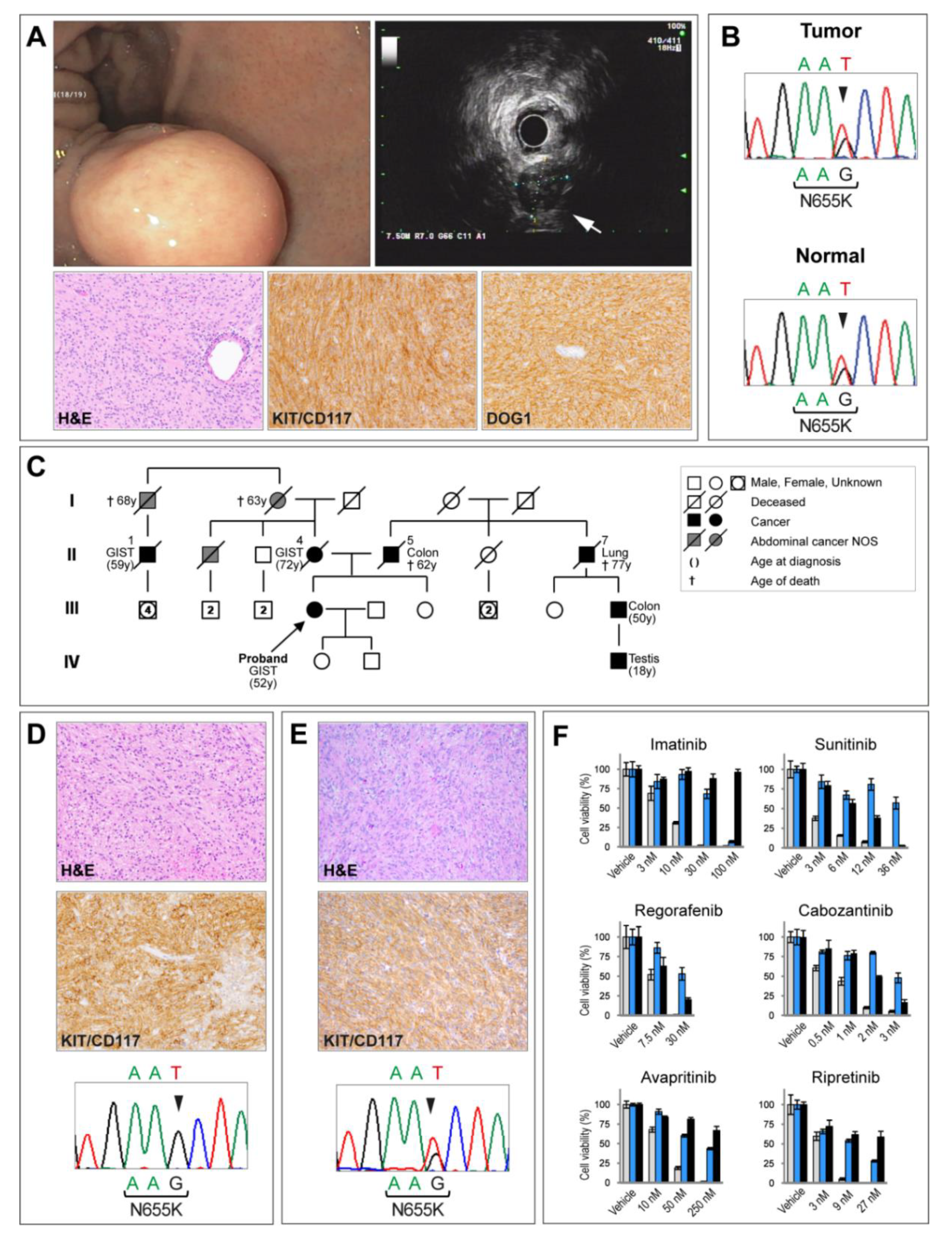
2.1. Histology and Immunohistochemistry

Formalin-fixed paraffin embedded (FFPE) surgical specimens were stained with hematoxylin and eosin (H&E). Immunohistochemical staining was performed by an automated immunostainer (Ventana, Roche, Basel, Switzerland) using the following antibodies: KIT/CD117 (polyclonal; 1:200; DAKO, Agilent, Santa Clara, CA, USA), CD34 (QBEnd 10, 1:1; DAKO), S100 protein (polyclonal; 1:2000; DAKO), alpha-smooth muscle actin (SMA) (1A4; 1:400; DAKO), Ki-67 (MIB-1; 1:200; DAKO), DOG1 (SP31; 1:100; Cell Marque, Merck, Darmstadt, Germany), and desmin (DE-R-11, 1:100; DAKO).

2.2. Molecular Analysis

DNA was extracted from FFPE tumor specimens by QIAamp DNA FFPE Tissue Kit (QIAGEN, Hilden, Germany). DNA from peripheral blood mononuclear cells was extracted with an EZ1 biorobot (QIAGEN).
Next generation sequencing (NGS) libraries were prepared with a TruSeq Custom Amplicon Low-Input kit (Illumina, San Diego, CA, USA) targeting the coding sequence of the KIT, PDGFRA, BRAF, SDHA, SDHB, SDHC, SDHD, HRAS, KRAS, NRAS, SPRED1, NF1, NF2, and TP53, as described [4]. Libraries were sequenced on an MiSeq platform (Illumina) using a v3 kit, 2 × 150 cycles. Data were analyzed with the Miseq Reporter software (v2.6.2), using the custom amplicon workflow and somatic variant caller. Mean amplicon coverage was 3200. Variants were analyzed with the VariantStudio 3.0 software (Illumina) and filtered by coverage > 50 and frequency ≥ 20%. Mutations detected by NGS were validated by Sanger sequencing on an ABI PRISM 3100 Genetic Analyzer (Thermo Fisher Scientific, Waltham, MA, USA).

2.3. Engineering of Ba/F3 Cells for the Expression of KIT Mutants

The murine interleukin-3 (IL-3) dependent Ba/F3 cell line is a cell model widely used in kinase studies. In fact, when ectopically expressed, certain tyrosine kinases can relieve Ba/F3 cells from IL-3 dependency, while tyrosine kinase inhibitors antagonize this effect [7,8].
Ba/F3 cells (ATCC, Manassas, VA, USA) were cultured in RPMI medium supplemented with murine interleukin-3 (IL-3; 10 ng/mL; PeproTech, Rocky Hill, IL, USA), 10% fetal bovine serum (FBS), and gentamicin (8 µg/mL).
Human KIT wild-type cDNA was cloned into the pLVX IRES ZsGreen viral vector (Clontech, Mountain View, CA, USA) and KIT exon 11 (W557_K558del), exon 13 (N655K), and exon 14 (T670I) mutant alleles were generated by PCR. Correct mutation introduction was checked by sequencing. To generate KIT lentiviral particles, human embryonic kidney HEK 293T cells (ATCC) were transfected with vectors encoding KIT mutants. HEK 293T cells were cultured in DMEM medium supplemented with 10% FBS and gentamicin (8 µg/mL). Ba/F3 cells were infected with HEK 293T-derived lentiviral supernatants in the presence of polybrene (8 µg/mL). Ectopically KIT expressing cells were selected by IL-3 withdrawal. Lentiviral infection efficiency was monitored in situ by green fluorescence. Expression of the ectopic KIT alleles was checked by Western blot. Briefly, cells were lysed in RIPA buffer (Santa Cruz Biotechnology, Dallas, TX, USA). Protein lysates were separated by SDS-PAGE and transferred onto a nitrocellulose membrane (Protran Whatman, Merck, Darmstadt, Germany). Expression analyses were performed with the following antibodies: anti c-KIT (H300; Santa Cruz Biotechnology) and anti-GAPDH (MAB374, Chemicon International, Temecula, CA, USA), for total protein loading normalization. A ChemiDoc imaging system (Bio-Rad, Hercules, CA, USA) was used for visualization. Ba/F3 cells expressing comparable levels of ectopic KIT mutants were used in TKI cell viability assays.

2.4. TKI Cell Viability Assay

Ba/F3 cells engineered to express the test mutation (KIT exon 13 N655K), an imatinib-sensitive mutation (KIT exon 11 W557_K558del), or an imatinib-resistant mutation (KIT exon 14 T670I) were seeded on a 48-well plate at a density of 7500 cells/mL. The following day, cells were exposed to the indicated doses of TKIs for 72 h, as previously described [7]. TKIs (Selleckchem, Munich, Germany) were dissolved in DMSO (dimethyl sulfoxide) as a vehicle. Drug dosage ranges were selected based on literature data and were as follows: imatinib (0–1000 nM), sunitinb (0–36 nM), regorafenib (0–480 nM), cabozantinib (0–24 nM), avapritinib (0–250 nM), and ripretinib (0–80 nM). Cell viability was evaluated by Trypan Blue staining and expressed as percentage of cells surviving the treatment, normalized to vehicle (DMSO)-treated cells. Four replicates per dose were evaluated. Results were confirmed on at least two independent Ba/F3 cell infections.

2.5. Ethical Approval

All procedures followed were in accordance with the Helsinki Declaration and with institutional and national ethical standards. The study was approved by the CRO Aviano institutional review board (IRB-04-2017). Written informed consent to be included in the study was obtained from patients.

3. Results

In March 2019, a 52-year-old woman was referred for genetic counseling after a diagnosis of multifocal GIST. She displayed diffuse freckles on her arms, legs, and trunk, and an atypical junctional nevus was surgically removed in February 2019. She had a history of estrogen-receptor-positive intraductal breast carcinoma diagnosed at the age of 43. She was also under surveillance for a thyroid nodule.
GIST was an incidental finding during bilateral salpingo-oophorectomy for uterine myomas and ovarian cyst (November 2018). Multiple nodules, the largest of 6 cm, were detected at the small bowel. At histopathological examination, these nodules showed spindle cells arranged in fascicles, and interlacing bundles with eosinophilic cytoplasm and elongated nuclei. Immunohistochemistry showed positivity for KIT/CD117 and DOG1, weak/focal reactivity for SMA and CD34, negativity for desmin and S100 (Figure 1A). Mitotic index was low (1 per 50 high-power fields). Diagnosis was of GIST of intermediate risk of progression according to Miettinen [9]. The diagnostic workup included a 18FDG-PET (fluorine-18-fluorodeoxyglucose positron emission tomography) that showed two lesions at the small bowel and one at the gastric fundus. Upper gastrointestinal endoscopy and ultrasound endoscopy showed a 2 cm submucosal nodule at the gastric fundus (Figure 1A). A second surgery was performed in January 2019 for resection of the gastric tumor and multiple small bowel nodules (size range 0.5–2 cm). Pathological examination corroborated the diagnosis of multinodular GIST. Molecular analysis, performed using a comprehensive GIST-specific NGS panel, revealed in both 2018 and 2019 tumor specimens a heterozygous T-to-G transversion at codon 655 of KIT exon 13 (c.1965T > G) resulting in an N655K amino acid change (Figure 1B). Besides the N655K KIT mutation, no other pathogenic mutation in GIST driver genes (PDGFRA, BRAF, SDHA, SDHB, SDHC, SDHD, HRAS, KRAS, NRAS, SPRED1, NF1, NF2, and TP53) was found.
Regarding family history (Figure 1C), the index case has a healthy sister (47 years old) with freckles on the trunk. Their mother died in 2012 at the age of 74 of metastatic GIST. She presented with multifocal intestinal GIST (positive for KIT/CD117, DOG1, and CD34; negative for S100, SMA, and desmin) and hepatic metastasis at diagnosis. Sequencing of tumor DNA revealed the same KIT N655K mutation detected in the proband (Figure 1D).
In 2000, the maternal second-degree cousin of the index case (II-1 at pedigree) was diagnosed at the age of 59 with multiple gastric and small-bowel mesenchymal malignant neoplasms compatible with GIST. The patient received two lines of combination chemotherapy for sarcomas (epidoxorubicine plus ifosfamide first, followed by CYVADIC, a combination of cyclophosphamide, vincristine, doxorubicin, and dacarbazine). Five years later (January 2006), he was referred to our center where PET/CT scan showed multifocal abdominal and hepatic increased uptake. Histological revision of the primary tumor supported the diagnosis of KIT/CD117-positive, DOG1-positive, GIST (immunohistochemistry was negative for CD34, S100, SMA, and desmin). Numerous microscopic GIST-like lesions (microGISTs) were also identified alongside the intestinal wall. Molecular analysis revealed a KIT exon 13 N655K mutation (Figure 1E). He started imatinib (400 mg daily). The disease progressed slowly until January 2007 when he underwent surgery for multiple abdominal masses. The patient was shifted to sunitinib for 15 months and, on progression, a rechallenge with imatinib at 800 mg allowed a progression-free interval of 9 months. A last line of treatment with nilotinib did not provide benefit and was stopped after 2 months.
The index case’s uncle (mother’s brother) and the maternal grandmother died of abdominal malignancy of unknown site and pathology (NOS). Colon cancer appeared as a recurrent event in the proband’s paternal branch.
In light of multifocal presentation, family history, and the recurrence of the same rare KIT exon 13 mutation in the neoplasms of the family, genetic testing was proposed to the proband. Analysis of peripheral blood DNA highlighted the presence of the same KIT N655K mutation detected in the tumors thus supporting its germline origin (Figure 1B).
Due to the intermediate risk of recurrence and the fact that KIT exon 13-mutated sporadic GISTs are in general scarcely responsive to imatinib, after curative surgery, the index case was put on close surveillance with alternating CT scan and 18FDG-PET every six months. A papillary thyroid carcinoma, follicular variant, was diagnosed in January 2020.
Genetic counseling was considered for the proband’s first-degree relatives. The sister refused genetic testing. Thus, annual abdominal ultrasound and upper-gastrointestinal endoscopy were suggested. The proband’s children were under age 18 and were therefore considered not eligible for genetic testing.
In sporadic GISTs, the KIT exon 13 N655K detected in this family is very uncommon. Thus, it is poorly characterized [7,8]. To address its sensitivity to TKIs approved or under trial for GIST treatment, we performed in vitro cytotoxicity assays. Specifically, the response to imatinib, sunitinib, regorafenib, avapritinib, cabozantinib, and ripretinib of Ba/F3 cells engineered to express the N655K mutation was compared with that of cells expressing a prototypical imatinib-sensitive (KIT exon 11 W557_K558del) and an imatinib-resistant (KIT exon 14 T670I) mutation (Figure 1F). Compared to the canonical KIT exon 11 sensitizing mutation (W557_K558del), the N655K mutation displayed a lower sensitivity to all TKIs tested. Compared to the reference resistant mutation (KIT exon 14 T670I), N655K appeared less responsive to sunitinib, regorafenib, and cabozantinib; it was instead slightly more sensitive to imatinib, especially at higher dosages, in keeping with previous results [7]. In addition, last-generation TKIs such as avapritinib and ripretinib [10] demonstrated a certain degree of activity toward this mutation (Figure 1F).

4. Discussion

Very few cases of KIT/PDGFRA-associated familial GIST syndromes have been reported so far. A PubMed search retrieved 51 reports, 45 describing hereditary GISTs due to germline KIT mutations, most of which involving exon 11, and 6 reports of familial GIST associated with germline PDGFRA mutations (Table 1) [11,12,13,14,15,16,17,18,19,20,21,22,23,24,25,26,27,28,29,30,31,32,33,34,35,36,37,38,39,40,41,42,43,44,45,46,47,48,49,50,51,52,53,54,55,56,57,58].
Here, we report a new kindred with familial GISTs caused by a KIT exon 13 germline mutation (N655K). In sporadic GISTs, KIT exon 13 mutations, which affect the ATP-binding pocket of the kinase, are uncommon and usually arise as secondary/imatinib-resistance events [1,2]. Among KIT exon 13 mutations, N655K is extremely rare, accounting for less than 0.1% of all GIST-associated KIT variants recorded in the Catalogue of Somatic Mutations in Cancer (COSMIC) database. With the only exception of a single, undetailed entry in the ClinVar database, to the best of our knowledge, there are no literature reports implicating this mutation in hereditary GISTs. Owing to its rarity, the N655K variant is poorly characterized and scanty literature data exist about its sensitivity to TKIs [7,8].
Our in vitro assay results indicate that N655K conveys a limited sensitivity to most TKIs approved for the treatment of GISTs. However, compared to a canonical resistance mutation (T670I), N655K was slightly more sensitive to imatinib, especially at higher dosages, a finding that is somehow in line with the response observed in the proband’s second-degree cousin. Always with reference to the T670I resistance mutation, N655K appeared more sensitive also to avapritinib and ripretinib. These latter are new-generation TKIs that have been proven to be effective in controlling a wide range of KIT and PDGFRA mutations, including classical resistant mutations [10].
The limited number of kindreds reported so far prevents genotype–phenotype correlations and the presentation pattern of these tumor forms and the management of patients carrying GIST-predisposing gene mutations still need to be defined [2,5].
As far as the treatment is concerned, there is currently no evidence supporting a differential therapeutic approach for GISTs developing in a background of germline KIT/PDGFRA mutations compared to sporadic GISTs, although tumor multiplicity and, hence, surgery-requiring complications (e.g., intestinal hemorrhages and occlusions) are more likely to occur in the former.
Definitively, when a familial GIST syndrome is suspected, based on early age of onset, tumor multifocality, and family history, genotyping is highly recommended and, if positive, counseling and predictive genetic testing should be offered to all first-degree relatives. Moreover, it is worth bearing in mind that individuals affected by hereditary GISTs seem to have an increased risk for other tumor types [2,5]. Indeed, our index case also developed breast and papillary thyroid cancers.
In our opinion, GIST-predisposing-mutation carriers should undergo close surveillance for early detection of cancer. This is even more important when the genetic setting makes the tumor poorly amenable to pharmacological inhibition, as in the family presented here, and the earliest diagnosis of neoplastic growth would increase the chance of curative surgery. Unfortunately, no specific guidelines exist on how to manage these subjects. In particular, consensus recommendations for clinical surveillance are lacking and whether regular esophagogastroduodenoscopy (EGD) or 18FDG-PET examinations are useful options remain open questions.
In this context, single case reports such as the one described here are fundamental to build up a body of information that may lay down the ground for the development of evidence-based guidelines.

5. Conclusions

In this paper we report the identification and characterization of a new kindred affected by multiple GISTs due to a rare KIT germline N655K mutation. Very few cases of syndromic KIT/PDGFRA-associated familial GISTs have been described so far. Case studies such as ours may help in defining the presentation pattern of these tumor forms and contribute to the formulation of clinical practice guidelines. In addition, in vitro evaluation of drug sensitivity may provide a basis for treatment personalization.

Author Contributions

Conceptualization and drafting of the manuscript, M.F., D.G., L.F., and R.M.; patient treatment and genetic counselling: M.F., L.F., M.C., A.B., F.P., and R.C.; pathological analyses: S.S. and V.C.; molecular analyses: D.G., S.L., E.D.S., and R.M.; study supervision and funding acquisition, R.M. All authors have read and agreed to the published version of the manuscript.

Funding

This study was supported by the Italian Ministry of Health and by the Italian Association for Cancer Research (AIRC).

Conflicts of Interest

The authors declare no conflict of interest.

References

  1. Corless, C.L. Gastrointestinal stromal tumors: What do we know now? Mod. Pathol. 2014, 27, S1–S16. [Google Scholar] [CrossRef] [Green Version]
  2. Mei, L.; Smith, S.C.; Faber, A.C.; Trent, J.; Grossman, S.R.; Stratakis, C.A.; Boikos, S.A. Gastrointestinal Stromal Tumors: The GIST of Precision Medicine. Trends Cancer 2018, 4, 74–91. [Google Scholar] [CrossRef]
  3. Brenca, M.; Rossi, S.; Polano, M.; Gasparotto, D.; Zanatta, L.; Racanelli, D.; Valori, L.; Lamon, S.; Dei Tos, A.P.; Maestro, R. Transcriptome sequencing identifies ETV6-NTRK3 as a gene fusion involved in GIST. J. Pathol. 2016, 238, 543–549. [Google Scholar] [CrossRef]
  4. Gasparotto, D.; Rossi, S.; Polano, M.; Tamborini, E.; Lorenzetto, E.; Sbaraglia, M.; Mondello, A.; Massani, M.; Lamon, S.; Bracci, R.; et al. Quadruple-Negative GIST Is a Sentinel for Unrecognized Neurofibromatosis Type 1 Syndrome. Clin. Cancer Res. 2017, 23, 273–282. [Google Scholar] [CrossRef] [Green Version]
  5. Ricci, R. Syndromic gastrointestinal stromal tumors. Hered. Cancer Clin. Pract. 2016, 14, 15. [Google Scholar] [CrossRef]
  6. Agarwal, R.; Robson, M. Inherited predisposition to gastrointestinal stromal tumor. Hematol. Oncol. Clin. N. Am. 2009, 23, 1–13. [Google Scholar] [CrossRef]
  7. Garner, A.P.; Gozgit, J.M.; Anjum, R.; Vodala, S.; Schrock, A.; Zhou, T.; Serrano, C.; Eilers, G.; Zhu, M.; Ketzer, J.; et al. Ponatinib inhibits polyclonal drug-resistant KIT oncoproteins and shows therapeutic potential in heavily pretreated gastrointestinal stromal tumor (GIST) patients. Clin. Cancer Res. 2014, 20, 5745–5755. [Google Scholar] [CrossRef] [Green Version]
  8. Kinoshita, K.; Hirota, S.; Isozaki, K.; Nishitani, A.; Tsutsui, S.; Watabe, K.; Tamura, S.; Ishikawa, T.; Kanda, T.; Nishida, T.; et al. Characterization of tyrosine kinase I domain c-kit gene mutation Asn655Lys newly found in primary jejunal gastrointestinal stromal tumor. Am. J. Gastroenterol. 2007, 102, 1134–1136. [Google Scholar] [CrossRef]
  9. Miettinen, M.; Lasota, J. Gastrointestinal stromal tumors: Pathology and prognosis at different sites. Semin. Diagn. Pathol. 2006, 23, 70–83. [Google Scholar] [CrossRef]
  10. Farag, S.; Smith, M.J.; Fotiadis, N.; Constantinidou, A.; Jones, R.L. Revolutions in treatment options in gastrointestinal stromal tumours (GISTs): The latest updates. Curr. Treat. Options Oncol. 2020, 21, 55. [Google Scholar] [CrossRef]
  11. Hartmann, K.; Wardelmann, E.; Ma, Y.; Merkelbach-Bruse, S.; Preussner, L.M.; Woolery, C.; Baldus, S.E.; Heinicke, T.; Thiele, J.; Buettner, R.; et al. Novel germline mutation of KIT associated with familial gastrointestinal stromal tumors and mastocytosis. Gastroenterology 2005, 129, 1042–1046. [Google Scholar] [CrossRef]
  12. Speight, R.A.; Nicolle, A.; Needham, S.J.; Verrill, M.W.; Bryon, J.; Panter, S. Rare, germline mutation of KIT with imatinib-resistant multiple GI stromal tumors and mastocytosis. J. Clin. Oncol. 2013, 31, e245–e247. [Google Scholar] [CrossRef]
  13. Halpern, A.L.; Torphy, R.J.; McCarter, M.D.; Sciotto, C.G.; Glode, L.M.; Robinson, W.A. A familial germline mutation in KIT associated with achalasia, mastocytosis and gastrointestinal stromal tumors shows response to kinase inhibitors. Cancer Genet. 2019, 233–234, 1–6. [Google Scholar] [CrossRef]
  14. Nakai, M.; Hashikura, Y.; Ohkouchi, M.; Yamamura, M.; Akiyama, T.; Shiba, K.; Kajimoto, N.; Tsukamoto, Y.; Hao, H.; Isozaki, K.; et al. Characterization of novel germline c-kit gene mutation, KIT-Tyr553Cys, observed in a family with multiple gastrointestinal stromal tumors. Lab. Investig. 2012, 92, 451–457. [Google Scholar] [CrossRef]
  15. Robson, M.E.; Glogowski, E.; Sommer, G.; Antonescu, C.R.; Nafa, K.; Maki, R.G.; Ellis, N.; Besmer, P.; Brennan, M.; Offit, K. Pleomorphic characteristics of a germ-line KIT mutation in a large kindred with gastrointestinal stromal tumors, hyperpigmentation, and dysphagia. Clin. Cancer Res. 2004, 10, 1250–1254. [Google Scholar] [CrossRef] [Green Version]
  16. Hirota, S.; Okazaki, T.; Kitamura, Y.; O’Brien, P.; Kapusta, L.; Dardick, I. Cause of familial and multiple gastrointestinal autonomic nerve tumors with hyperplasia of interstitial cells of Cajal is germline mutation of the c-kit gene. Am. J. Surg. Pathol. 2000, 24, 326–327. [Google Scholar] [CrossRef]
  17. Farag, S.; van der Kolk, L.E.; van Boven, H.H.; van Akkooi, A.C.J.; Beets, G.L.; Wilmink, J.W.; Steeghs, N. Remarkable effects of imatinib in a family with young onset gastrointestinal stromal tumors and cutaneous hyperpigmentation associated with a germline KIT-Trp557Arg mutation: Case report and literature overview. Fam. Cancer 2018, 17, 247–253. [Google Scholar] [CrossRef]
  18. Antonescu, C.R.; Viale, A.; Sarran, L.; Tschernyavsky, S.J.; Gonen, M.; Segal, N.H.; Maki, R.G.; Socci, N.D.; DeMatteo, R.P.; Besmer, P. Gene expression in gastrointestinal stromal tumors is distinguished by KIT genotype and anatomic site. Clin. Cancer Res. 2004, 10, 3282–3290. [Google Scholar] [CrossRef] [Green Version]
  19. Hasegawa, M.; Shimizu, A.; Ieta, K.; Shibusawa, K.; Ishikawa, O.; Ishida-Yamamoto, A.; Tamura, A. Generalized lentigines associated with familial gastrointestinal stromal tumors dramatically improved by imatinib treatment. J. Dermatol. 2020, 47, e241–e242. [Google Scholar] [CrossRef]
  20. Sekido, Y.; Ohigashi, S.; Takahashi, T.; Hayashi, N.; Suzuki, K.; Hirota, S. Familial Gastrointestinal Stromal Tumor with Germline KIT Mutations Accompanying Hereditary Breast and Ovarian Cancer Syndrome. Anticancer Res. 2017, 37, 1425–1431. [Google Scholar] [CrossRef] [Green Version]
  21. Gupta, D.; Chandrashekar, L.; Larizza, L.; Colombo, E.A.; Fontana, L.; Gervasini, C.; Thappa, D.M.; Rajappa, M.; Rajendiran, K.S.; Sreenath, G.S.; et al. Familial gastrointestinal stromal tumors, lentigines, and café-au-lait macules associated with germline c-kit mutation treated with imatinib. Int. J. Dermatol. 2017, 56, 195–201. [Google Scholar] [CrossRef] [PubMed]
  22. Maeyama, H.; Hidaka, E.; Ota, H.; Minami, S.; Kajiyama, M.; Kuraishi, A.; Mori, H.; Matsuda, Y.; Wada, S.; Sodeyama, H.; et al. Familial gastrointestinal stromal tumor with hyperpigmentation: Association with a germline mutation of the c-kit gene. Gastroenterology 2001, 120, 210–215. [Google Scholar] [CrossRef] [PubMed]
  23. Adela Avila, S.; Peñaloza, J.; González, F.; Abdo, I.; Rainville, I.; Root, E.; Carrero Valenzuela, R.D.; Garber, J. Dysphagia, melanosis, gastrointestinal stromal tumors and a germinal mutation of the KIT gene in an Argentine family. Acta Gastroenterol. Latinoam. 2014, 44, 9–15. [Google Scholar] [PubMed]
  24. Beghini, A.; Tibiletti, M.G.; Roversi, G.; Chiaravalli, A.M.; Serio, G.; Capella, C.; Larizza, L. Germline mutation in the juxtamembrane domain of the kit gene in a family with gastrointestinal stromal tumors and urticaria pigmentosa. Cancer 2001, 92, 657–662. [Google Scholar] [CrossRef]
  25. Kuroda, N.; Tanida, N.; Hirota, S.; Daum, O.; Hes, O.; Michal, M.; Lee, G.H. Familial gastrointestinal stromal tumor with germ line mutation of the juxtamembrane domain of the KIT gene observed in relatively young women. Ann. Diagn. Pathol. 2011, 15, 358–361. [Google Scholar] [CrossRef]
  26. Kim, H.J.; Lim, S.J.; Park, K.; Yuh, Y.J.; Jang, S.J.; Choi, J. Multiple gastrointestinal stromal tumors with a germline c-kit mutation. Pathol. Int. 2005, 55, 655–659. [Google Scholar] [CrossRef] [PubMed]
  27. Nishida, T.; Hirota, S.; Taniguchi, M.; Hashimoto, K.; Isozaki, K.; Nakamura, H.; Kanakura, Y.; Tanaka, T.; Takabayashi, A.; Matsuda, H.; et al. Familial gastrointestinal stromal tumours with germline mutation of the KIT gene. Nat. Genet. 1998, 19, 323–324. [Google Scholar] [CrossRef]
  28. Bamba, S.; Hirota, S.; Inatomi, O.; Ban, H.; Nishimura, T.; Shioya, M.; Imaeda, H.; Nishida, A.; Sasaki, M.; Murata, S.; et al. Familial and multiple gastrointestinal stromal tumors with fair response to a half-dose of imatinib. Intern. Med. 2015, 54, 759–764. [Google Scholar] [CrossRef] [Green Version]
  29. Kang, D.Y.; Park, C.K.; Choi, J.S.; Jin, S.Y.; Kim, H.J.; Joo, M.; Kang, M.S.; Moon, W.S.; Yun, K.J.; Yu, E.S.; et al. Multiple gastrointestinal stromal tumors: Clinicopathologic and genetic analysis of 12 patients. Am. J. Surg. Pathol. 2007, 31, 224–232. [Google Scholar] [CrossRef]
  30. Woźniak, A.; Rutkowski, P.; Sciot, R.; Ruka, W.; Michej, W.; Debiec-Rychter, M. Rectal gastrointestinal stromal tumors associated with a novel germline KIT mutation. Int. J. Cancer 2008, 122, 2160–2164. [Google Scholar] [CrossRef]
  31. Neuhann, T.M.; Mansmann, V.; Merkelbach-Bruse, S.; Klink, B.; Hellinger, A.; Höffkes, H.G.; Wardelmann, E.; Schildhaus, H.U.; Tinschert, S. A novel germline KIT mutation (p.L576P) in a family presenting with juvenile onset of multiple gastrointestinal stromal tumors, skin hyperpigmentations, and esophageal stenosis. Am. J. Surg. Pathol. 2013, 37, 898–905. [Google Scholar] [CrossRef] [PubMed]
  32. Vale Rodrigues, R.; Santos, F.; Pereira da Silva, J.; Francisco, I.; Claro, I.; Albuquerque, C.; Lemos, M.M.; Limbert, M.; Dias Pereira, A. A case of multiple gastrointestinal stromal tumors caused by a germline KIT gene mutation (p.Leu576Pro). Fam. Cancer 2017, 16, 267–270. [Google Scholar] [CrossRef] [PubMed]
  33. Carballo, M.; Roig, I.; Aguilar, F.; Pol, M.A.; Gamundi, M.J.; Hernan, I.; Martinez-Gimeno, M. Novel c-KIT germline mutation in a family with gastrointestinal stromal tumors and cutaneous hyperpigmentation. Am. J. Med. Genet. A 2005, 132, 361–364. [Google Scholar] [CrossRef] [PubMed]
  34. Forde, P.M.; Cochran, R.L.; Boikos, S.A.; Zabransky, D.J.; Beaver, J.A.; Meyer, C.F.; Thornton, K.A.; Montgomery, E.A.; Lidor, A.O.; Donehower, R.C.; et al. Familial GI Stromal Tumor With Loss of Heterozygosity and Amplification of Mutant KIT. J. Clin. Oncol. 2016, 34, e13–e16. [Google Scholar] [CrossRef]
  35. Tarn, C.; Merkel, E.; Canutescu, A.A.; Shen, W.; Skorobogatko, Y.; Heslin, M.J.; Eisenberg, B.; Birbe, R.; Patchefsky, A.; Dunbrack, R.; et al. Analysis of KIT mutations in sporadic and familial gastrointestinal stromal tumors: Therapeutic implications through protein modeling. Clin. Cancer Res. 2005, 11, 3668–3677. [Google Scholar] [CrossRef] [Green Version]
  36. Wali, G.N.; Halliday, D.; Dua, J.; Ieremia, E.; McPherson, T.; Matin, R.N. Cutaneous hyperpigmentation and familial gastrointestinal stromal tumour associated with KIT mutation. Clin. Exp. Dermatol. 2019, 44, 418–421. [Google Scholar] [CrossRef]
  37. Jones, D.H.; Caracciolo, J.T.; Hodul, P.J.; Strosberg, J.R.; Coppola, D.; Bui, M.M. Familial gastrointestinal stromal tumor syndrome: Report of 2 cases with KIT exon 11 mutation. Cancer Control 2015, 22, 102–108. [Google Scholar] [CrossRef] [Green Version]
  38. Lasota, J.; Miettinen, M. A new familial GIST identified. Am. J. Surg. Pathol. 2006, 30, 1342. [Google Scholar] [CrossRef]
  39. Kleinbaum, E.P.; Lazar, A.J.F.; Tamborini, E.; Mcauliffe, J.C.; Sylvestre, P.B.; Sunnenberg, T.D.; Strong, L.; Chen, L.L.; Choi, H.; Benjamin, R.S.; et al. Clinical, histopathologic, molecular and therapeutic findings in a large kindred with gastrointestinal stromal tumor. Int. J. Cancer 2008, 122, 711–718. [Google Scholar] [CrossRef]
  40. Yamanoi, K.; Higuchi, K.; Kishimoto, H.; Nishida, Y.; Nakamura, M.; Sudoh, M.; Hirota, S. Multiple gastrointestinal stromal tumors with novel germline c-kit gene mutation, K642T, at exon 13. Hum. Pathol. 2014, 45, 884–888. [Google Scholar] [CrossRef]
  41. Wadt, K.; Andersen, M.K.; Hansen, T.V.O.; Gerdes, A.M. A new genetic diagnosis of familiar gastrointestinal stromal tumour. Ugeskr. Laeg. 2012, 174, 1462–1464. [Google Scholar] [PubMed]
  42. Bachet, J.B.; Landi, B.; Laurent-Puig, P.; Italiano, A.; Le Cesne, A.; Lévy, P.; Safar, V.; Duffaud, F.; Blay, J.Y.; Emile, J.F. Diagnosis, prognosis and treatment of patients with gastrointestinal stromal tumour (GIST) and germline mutation of KIT exon 13. Eur. J. Cancer 2013, 49, 2531–2541. [Google Scholar] [CrossRef] [PubMed]
  43. Isozaki, K.; Terris, B.; Belghiti, J.; Schiffmann, S.; Hirota, S.; Vanderwinden, J.M. Germline-activating mutation in the kinase domain of KIT gene in familial gastrointestinal stromal tumors. Am. J. Pathol. 2000, 157, 1581–1585. [Google Scholar] [CrossRef] [Green Version]
  44. Graham, J.; Debiec-Rychter, M.; Corless, C.L.; Reid, R.; Davidson, R.; White, J.D. Imatinib in the management of multiple gastrointestinal stromal tumors associated with a germline KIT K642E mutation. Arch. Pathol. Lab. Med. 2007, 131, 1393–1396. [Google Scholar] [CrossRef]
  45. Vilain, R.E.; Dudding, T.; Braye, S.G.; Groombridge, C.; Meldrum, C.; Spigelman, A.D.; Ackland, S.; Ashman, L.; Scott, R.J. Can a familial gastrointestinal tumour syndrome be allelic with Waardenburg syndrome? Clin. Genet. 2011, 79, 554–560. [Google Scholar] [CrossRef]
  46. Engin, G.; Eraslan, S.; Kayserili, H.; Kapran, Y.; Akman, H.; Akyuz, A.; Aykan, N.F. Imatinib response of gastrointestinal stromal tumor patients with germline mutation on KIT exon 13: A family report. World J. Radiol. 2017, 9, 365–370. [Google Scholar] [CrossRef]
  47. Hirota, S.; Nishida, T.; Isozaki, K.; Taniguchi, M.; Nishikawa, K.; Ohashi, A.; Takabayashi, A.; Obayashi, T.; Okuno, T.; Kinoshita, K.; et al. Familial gastrointestinal stromal tumors associated with dysphagia and novel type germline mutation of KIT gene. Gastroenterology 2002, 122, 1493–1499. [Google Scholar] [CrossRef]
  48. O’Riain, C.; Corless, C.L.; Heinrich, M.C.; Keegan, D.; Vioreanu, M.; Maguire, D.; Sheahan, K. Gastrointestinal stromal tumors: Insights from a new familial GIST kindred with unusual genetic and pathologic features. Am. J. Surg. Pathol. 2005, 29, 1680–1683. [Google Scholar] [CrossRef]
  49. Veiga, I.; Silva, M.; Vieira, J.; Pinto, C.; Pinheiro, M.; Torres, L.; Soares, M.; Santos, L.; Duarte, H.; Bastos, A.L.; et al. Hereditary gastrointestinal stromal tumors sharing the KIT Exon 17 germline mutation p.Asp820Tyr develop through different cytogenetic progression pathways. Genes Chromosomes Cancer 2010, 49, 91–98. [Google Scholar] [CrossRef]
  50. Arima, J.; Hiramatsu, M.; Taniguchi, K.; Kobayashi, T.; Tsunematsu, I.; Kagota, S.; Sakane, J.; Suzuki, Y.; Hirota, S. Multiple gastrointestinal stromal tumors caused by a novel germline KIT gene mutation (Asp820Gly): A case report and literature review. Gastric Cancer 2020, 23, 760–764. [Google Scholar] [CrossRef]
  51. Thalheimer, A.; Schlemmer, M.; Bueter, M.; Merkelbach-Bruse, S.; Schildhaus, H.U.; Buettner, R.; Hartung, E.; Thiede, A.; Meyer, D.; Fein, M.; et al. Familial gastrointestinal stromal tumors caused by the novel KIT exon 17 germline mutation N822Y. Am. J. Surg. Pathol. 2008, 32, 1560–1565. [Google Scholar] [CrossRef] [PubMed]
  52. de Raedt, T.; Cools, J.; Debiec-Rychter, M.; Brems, H.; Mentens, N.; Sciot, R.; Himpens, J.; de Wever, I.; Schöffski, P.; Marynen, P.; et al. Intestinal neurofibromatosis is a subtype of familial GIST and results from a dominant activating mutation in PDGFRA. Gastroenterology 2006, 131, 1907–1912. [Google Scholar] [CrossRef] [Green Version]
  53. Pasini, B.; Matyakhina, L.; Bei, T.; Muchow, M.; Boikos, S.; Ferrando, B.; Carney, J.A.; Stratakis, C.A. Multiple gastrointestinal stromal and other tumors caused by platelet-derived growth factor receptor alpha gene mutations: A case associated with a germline V561D defect. J. Clin. Endocrinol. Metab. 2007, 92, 3728–3732. [Google Scholar] [CrossRef] [PubMed] [Green Version]
  54. Carney, J.A.; Stratakis, C.A. Stromal, fibrous, and fatty gastrointestinal tumors in a patient with a PDGFRA gene mutation. Am. J. Surg. Pathol. 2008, 32, 1412–1420. [Google Scholar] [CrossRef] [PubMed] [Green Version]
  55. Ricci, R.; Martini, M.; Cenci, T.; Carbone, A.; Lanza, P.; Biondi, A.; Rindi, G.; Cassano, A.; Larghi, A.; Persiani, R.; et al. PDGFRA-mutant syndrome. Mod. Pathol. 2015, 28, 954–964. [Google Scholar] [CrossRef] [Green Version]
  56. Ricci, R.; Martini, M.; Cenci, T.; Riccioni, M.E.; Maria, G.; Cassano, A.; Larocca, L.M. Divergent gastrointestinal stromal tumors in syndromic settings. Cancer Genet. 2016, 209, 354–358. [Google Scholar] [CrossRef]
  57. Chompret, A.; Kannengiesser, C.; Barrois, M.; Terrier, P.; Dahan, P.; Tursz, T.; Lenoir, G.M.; Bressac-De Paillerets, B. PDGFRA germline mutation in a family with multiple cases of gastrointestinal stromal tumor. Gastroenterology 2004, 126, 318–321. [Google Scholar] [CrossRef]
  58. Manley, P.N.; Abu-Abed, S.; Kirsch, R.; Hawrysh, A.; Perrier, N.; Feilotter, H.; Pollett, A.; Riddell, R.H.; Hookey, L.; Walia, J.S. Familial PDGFRA-mutation syndrome: Somatic and gastrointestinal phenotype. Hum. Pathol. 2018, 76, 52–57. [Google Scholar] [CrossRef]
Figure 1. (A) Index case: endoscopic and histopathologic characteristics. Endoscopic (upper panel, left) and endoscopic ultrasound (upper panel, right) appearance of the submucosal gastric tumor of the index case (arrow). Lower panels: hematoxylin and eosin (H&E) staining of the lesion revealed bundles of spindle cells arranged in fascicles. Tumor cells displayed positive immunoreactivity for KIT/CD117 and DOG1 (original magnification: ×20). (B) Index case: molecular analysis. Sanger sequencing of tumor DNA revealed a T-to-G transversion at codon 655 of KIT exon 13 resulting in an N655K amino acid change (upper panel). The same KIT mutation was subsequently detected in the peripheral blood DNA (normal), indicating a germline origin (lower panel). (C) Family pedigree. (D) Index case’s mother: pathological and molecular characterization of the gastrointestinal stromal tumor (GIST). H&E staining of the tumor showing a proliferation of spindle cells. Tumor cells were positive for KIT/CD117 expression (original magnification: ×20). Molecular analysis of tumor DNA revealed a KIT exon 13 N655K mutation. No significant signal for the wild-type nucleotide (T) was detected, indicating that the mutation in the tumor of the index case’s mother was homozygous/hemizygous. (E) Index case’s second-degree cousin. Pathological and molecular characterization of the GIST. H&E staining showing spindle-shaped tumor cells disposed in fascicles. KIT/CD117 immunostaining demonstrating diffuse and strong membranous and cytoplasmic reactivity (original magnification: ×20). Sanger sequencing of tumor DNA revealing a KIT exon 13 N655K mutation. (F) In vitro tyrosine kinase inhibitor (TKI) sensitivity assay. Ba/F3 cells, engineered to express KIT mutant alleles, were treated with different TKIs at the indicated dosages. Cell viability is expressed as percentage of cells surviving the treatment, normalized to vehicle (DMSO)-treated cells. Color coding for KIT mutants: Light grey: imatinib-sensitive KIT exon 11 W557_K558del; Blue: the KIT exon 13 N655K mutation detected in the familial GIST syndrome described here; Black: imatinib-resistant KIT exon 14 T670I. Error bars represent standard error of the mean.
Jpm 10 00234 g001
Table 1. Currently reported cases of KIT- or PDGFRA-associated familial Gastrointestinal Stromal Tumors (GISTs).
GeneExonMutationNo. of KindredsMain Clinical Features [Reference]
KIT8D419del1systemic mastocytosis, multiple GISTs, dysphagia [11]
9K509I2systemic mastocytosis, multiple GISTs [12]; achalasia; mastocytosis, multiple GISTs [13]
11Y533C1multiple GISTs [14]
11W557R4multiple GISTs, skin hyperpigmentation, dysphagia [15]; multiple gastrointestinal autonomic nerve tumor [16]; multiple GISTs; skin hyperpigmentation [17]; multiple GISTs [18]
11W557S1multiple GISTs; lentigines [19]
11W557L K558E1multiple GISTs, hereditary breast cancer [20]
11V559A7multiple GISTs; lentigines, cafe-au-lait macules [21]; multiple GIST, cutaneous hyperpigmentation [22]; multiple GISTs, melanosis, lentiginosis, hyperpigmentation, dysphagia [23]; multiple GISTs, hyperpigmentation, urticaria pigmentosa [24]; multiple GISTs, cutaneous hyperpigmentation [25]; multiple GISTs [26]
11V559_V560del1multiple GISTs, cutaneous hyperpigmentation [27]
11V560del1multiple GISTs [28]
11V560G1multiple GISTs, cutaneous hyperpigmentation [29]
11V560A1multiple GISTs [29]
11Q575_P577delinsH1rectal GIST [30]
11L576P2multiple GISTs; skin hyperpigmentation, achalasia-like stenosis [31]; multiple GISTs [32]
11L576_P577insQL1multiple GISTs, cutaneous hyperpigmentation [33]
11D579del7multiple GISTs, cutaneous hyperpigmentation, dysphagia [34]; GIST [35]; GIST, cutaneous hyperpigmentation [36]; multiple GISTs [37]; multiple GISTs [38]; multiple GIST, nevi, hyperpigmentation [39]
13K642T1multiple GISTs, dysphagia [40];
13K642E7multiple GISTs, breast cancer [41]; multiple GISTs, dysphagia, multiple nevi and lentigines [42]; multiple GISTs [43]; multiple GISTs including rectal GIST [44]; multiple GISTs, dysphagia, pigmentary defects (hyper- and hypopigmentation) [45]; multiple GISTs including rectal GIST, pigmentary defects (hyper- and hypopigmentation) [46]
13N655K1multiple GISTs, lentigines, atypical junctional nevus, breast and thyroid cancer [present report]
17D820Y3multiple GISTs, dysphagia [47]; multiple GISTs [48]; multiple GISTs including rectal GIST [49]
17D820G1multiple GISTs [50]
17N822Y1multiple GISTs [51]
PDGFRA12Y555C1multiple GISTs, intestinal neurofibromatosis, glaucoma, coarse facies, broad hands [52]
12V561D1multiple GISTs, fibrous tumors, lipoma [53,54]
14P653L2multiple GISTs, fibrous tumors, inflammatory fibroid polyps [55,56]
18D846Y1multiple GISTs, broad hands [57]
18D846V1multiple GISTs, coarse facies/skin, broad extremities [58]
Publisher’s Note: MDPI stays neutral with regard to jurisdictional claims in published maps and institutional affiliations.

